RESEARCH
Genotypic and phenotypic
heterogeneity in Streptococcus mutans isolated
from diabetic patients in Rome, Italy
Arpan De
1, Guido Pasquantonio
2, Loredana Cerroni
2, Dezemona Petrelli
3, Davide Lauro
4, Marta Longhi
2and Luca A. Vitali
1*Abstract
Our study focuses on the antimicrobial susceptibility, genotypic and phenotypic heterogeneity, and serotype clas-sification of the Streptococcus mutans isolated from type II diabetic patients (n = 25; age 42–68). Eighty-two percent of isolates were classified as serotype c. No serotype k was present. Macrorestriction analysis of genomic DNA of the isolates exhibited a clonal diversity that paralleled the phenotypic heterogeneity, which was also assessed in terms of biofilm forming ability. Isolates were susceptible to all the classes of antibiotics. In conclusion a great heterogeneity and no antimicrobial resistance were apparent in the considered S. mutans strains from diabetic patients.
Keywords: Caries, Plaque, PFGE, Antimicrobial susceptibility
© 2016 The Author(s). This article is distributed under the terms of the Creative Commons Attribution 4.0 International License
(http://creativecommons.org/licenses/by/4.0/), which permits unrestricted use, distribution, and reproduction in any medium,
provided you give appropriate credit to the original author(s) and the source, provide a link to the Creative Commons license, and indicate if changes were made.
Background
Globally, dental caries is a major public health concern imposing an economic impact among developed coun-tries. Italy was estimated with the highest percentage of Gross National Product spent on oral health in 2010 (Patel Reena 2012). Predisposition of diabetic patients to dental caries may result as sequelae of poor glycemic control, due to high levels of glucose in saliva in con-junction with xerostomia, although a strong association has not been found hitherto (King et al. 1998; Ship 2003; Taylor 2004). Streptococcus mutans, one of the main causative agents of caries (Loesche 1986), is a commen-sal inhabitant of the tooth surface, participating in the formation of a complex multispecies microbial commu-nity (i.e. a biofilm) called dental plaque (Kuramitsu et al. 2007). It adheres to the pellicle of tooth enamel surface through adhesins (AgI/II) or extracellular polysaccha-ride (EPS) formed by glucosyltransferases (GtfB, GtfC, and GtfD) in presence of sucrose (Krzyściak et al. 2014). Increased intake of dietary carbohydrates acts as source
of sugar increasing the production of metabolic acid end-products, and thus demineralization of teeth enamel and dentine at low pH (Loesche 1986). The EPS also serves as a reservoir of sugar and maintains acidic pH in the depths of plaque (Dibdin and Shellis 1988), and a con-tinuous flush of salivary sugar in the EPS may pose a greater caries threat in diabetics. In a biofilm, S. mutans expresses competence and bacteriocin genes that can facilitate integration of various dispensable genes (cnm, gbpA), endowing a competitive advantage and a wide array of genome heterogeneity (Waterhouse and Rus-sell 2006; Waterhouse et al. 2007). In general, Horizon-tal Gene Transfer (HGT) is known to be strongly favored in biofilm increasing the acquisition rate of exogenous genetic material. Among this material are the determi-nants of antibiotic resistance (Davies and Davies 2010), posing a continuous threat to the antimicrobial suscepti-bility of S. mutans (Baddour et al. 2005). In this context, diabetic individuals are subjected to various long term medications escalating the chances of acquiring resist-ance to these medicines by the indigenous flora of diabet-ics. Frequent use of antibiotics against infections in such immune-compromised patients has shown increased risk
Open Access
*Correspondence: luca.vitali@unicam.it
1 Microbiology Unit, School of Pharmacy, University of Camerino, Via
Gentile III da Varano, 62032 Camerino, MC, Italy
of antibiotic resistance among pathogens (Boyanova and Mitov 2013).
Based on the rhamnose glucose polysaccharide surface antigen (RGP) S. mutans is classified into four serotypes (c, e, f, k), among which k exhibits reduced cariogenecity and immunogenicity (Hamada and Slade 1980; Nakano et al. 2010). In addition, infective endocarditis (IE) is reportedly caused at higher frequency by non-c serotype
S. mutans (Nakano et al. 2010). Also DNA
fingerprint-ing methods such as RFLP, AP-PCR, PFGE have demon-strated significant genotypic diversity among S. mutans isolates (Caufield and Walker 1989; Brady et al. 1991; Napimoga 2004; Lembo et al. 2007; Moser et al. 2010).
In the present study, we have focused on the antimi-crobial susceptibility as well as genotypic and phenotypic heterogeneity of S. mutans isolates from type II diabetic patients aged >40 and bring the importance of such studies from patients with metabolic disorders to the forefront.
Results and discussion Dental health analysis of diabetes
The DMF index is a key measure of the caries experi-ence in dental epidemiology. The mean DMFT values among the Italian middle aged diabetic patients was 16.3 (SD = 9.0) while the median plaque index was 0.544. Pre-vious oral health studies among Type II diabetic popula-tion in Sudan also found a significant higher plaque index (Mohamed et al. 2013). Moreover, Taylor et al. (2004) found six large cohort studies reporting an association of poor glycemic control with high caries experience. Diabetic subjects are usually restricted to refined carbo-hydrates and proteinaceous diet. Nevertheless, poor gly-cemic control, xerostomia and repeated food intake may disrupt the microbial homeostasis of plaque and increase the incidence of caries due to acidogenic bacteria (Taylor 2004; Marsh 2009).
Screening of S. mutans isolates and serotyping
500 bp amplicons obtained using gtfB primers con-firmed the putative S. mutans clinical isolates from diabetics. Only 68 % (n = 17) isolates from the serially diluted plaque or saliva samples showed positive ampli-fication. Selection of adult patients in a particular age group with no other medical complications except dia-betes might have restricted to end up with low number of patients and consequently isolating few clinical strains. Even though possessing a higher plaque index, the viable count of S. mutans was low presumably due to proper maintenance of their oral health by 2 times brushing and usage of mouth washes among 48 % respondents (data not shown). Screening of the serotype by a molecular approach classified 14 isolates as c type, while 2 and 1 as
type e and f, respectively. We did not record any k sero-type among our isolates. This result is in accordance with the global prevalence of serotype c S. mutans in the oral cavity (Nakano et al. 2006). Serotype e, f, k are commonly found in atheromatous plaque (Abranches et al. 2011). People with diabetes are usually more prone to heart disease and atherosclerosis (Chait and Bornfeldt 2009), which may follow dental surgical procedures favor-ing the access of S. mutans in sufficient numbers in the blood-stream. Hence, a thorough stomatological study of the diabetic subjects can serve as a prophylaxis to such health disorder.
Antimicrobial susceptibility testing
Diabetic foot infection, urinary tract and lower respira-tory tract infections are a constant threat to poor gly-cemic control, which has demonstrated an increase in antibiotic prescription rates in Netherlands (Venmans et al. 2009). Notwithstanding this fact prompts a warn-ing situation, pointwarn-ing towards increased antimicrobial resistance among indigenous flora, all the bacterial strains isolated from diabetics were susceptible to all antibiotics tested (Table 1). According to EUCAST clinical break-point, the average zone diameter against penicillin, clin-damycin and vancomycin were far from the breakpoints (12, <19 and 15 mm, respectively). Concomitantly, the susceptibility against other antibiotics (erythromycin, oxa-cillin, rifampicin, tetracycline, gentamicin, cefoxitin, lin-ezolid, norfloxacin and levofloxacin) was compared with the performance standard values published by CLSI for streptococci other than S. pneumoniae (CLSI 2011). Clin-damycin, erythromycin, rifampicin and cefoxitin exhibited higher inhibition compared to others with a zone diame-ter ranging between 35–41, 30–38, 29–36 and 28–34 mm respectively. Penicillin resistance was not observed, which countertrends the previously recorded 14 % penicillin-resistant S. mutans isolates from dental patients in Rome (Pasquantonio et al. 2012). Hence, with scarce avail-able information on antimicrobial disc susceptibility tests based on EUCAST and CLSI guidelines, our work may aid as a reference scale for determining reduced susceptibility to various class of antibiotics for future studies.
Macrorestriction analysis
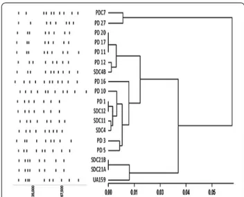
Macrorestriction analyses using SmaI and BssHII of S. mutans isolates were studied previously to identify maternal transmission as well as genotypic uniform-ity in an individual (Mineyama et al. 2007; Mitchell et al. 2011). This work illustrates genotypic heteroge-neity of S. mutans from caries of diabetic patients of a specific region. The two clustered dendogram along with the schematic representation of the digested bands in Fig. 1 illustrates the clonal diversity of the S. mutans
Table 1 D istribution of S. m ut ans isola tes fr om diab etic pa tien ts ac cor ding t o the inhibition z one diamet ers (mm) e xhibit ed b y various an tibiotics (EUC AST f or -ma t) * Z one diamet er e xhibit ed b y the c on tr ol str ain U A159 A ntibiotic D isk C on ten t (μg) Inhibition z one diamet er (mm) 12 13 14 15 16 17 18 19 20 21 22 23 24 25 26 27 28 29 30 31 32 33 34 35 36 37 38 39 40 41 42 Vancom ycin 5 0 0 0 0 0 1 6 5 1 4 1* 0 0 0 0 0 0 0 0 0 0 0 0 0 0 0 0 0 0 0 0 Er ythr om ycin 30 0 0 0 0 0 0 0 0 0 0 0 0 0 0 0 0 0 0 1 2 2 1 5 2 2 1 2* 0 0 0 0 Oxacillin 1 0 0 0 0 0 0 0 0 0 0 0 0 3 3 2 5* 3 2 0 0 0 0 0 0 0 0 0 0 0 0 0 Penicillin 1 0 0 0 0 0 0 0 0 0 0 0 0 0 0 0 1 2 3 4 4* 2 1 1 0 0 0 0 0 0 0 0 Rifampicin 5 0 0 0 0 0 0 0 0 0 0 0 0 0 0 0 0 0 3 2 3 3 0 3 3* 1 0 0 0 0 0 0 Tetrac ycline 30 0 0 0 0 0 0 0 0 0 0 0 0 1 1 0 1 4 4 1 2 2* 1 1 0 0 0 0 0 0 0 0 G entamicin 10 0 0 0 2 1 2 3 1 3 4 0 0 2* 0 0 0 0 0 0 0 0 0 0 0 0 0 0 0 0 0 0 Cef oxitin 30 0 0 0 0 0 0 0 0 0 0 0 0 0 0 0 0 1 2 2 0 6 4 3* 0 0 0 0 0 0 0 0 Linez olid 10 0 0 0 0 0 0 0 0 0 2 0 1 1 0 4 5* 2 1 2 0 0 0 0 0 0 0 0 0 0 0 0 Nor flo xacin 10 1 1 3 1 1 3 2 3 2 1* 0 0 0 0 0 0 0 0 0 0 0 0 0 0 0 0 0 0 0 0 0 Clindam ycin 10 0 0 0 0 0 0 0 0 0 0 0 0 0 0 0 0 0 0 0 0 0 0 0 2 1 6 5 0 0 3* 1 Le voflo xacin 5 0 0 0 0 0 0 0 1 1 0 4 4 2 3 2* 1 0 0 0 0 0 0 0 0 0 0 0 0 0 0 0
clinical strains. One major cluster accounts for 88 % of the clinical strains along with reference strain UA159 in it. PD27 and PDC7 formed the other cluster. PFGE of PD11, PD17 and PD20 projected similarity, but PD17 and PD20 showed one less band in the lower part of the gel. Conversely, SDC12 and PD1 exhibited complete over-lapping of the digested bands. SDC21A–SDC21B and SDC4–SDC4B were isolated from the oral cavity of the same patient and were included in the study due to their differences in growth behavior in broth. As SDC21A and SDC21B belonged to the same serotype and showed the
same macrorestriction pattern, they may be considered as variants of the same strain. SDC4 and SDC4B were instead dissimilar in both the characteristics and may be, therefore, considered as different strains. Genotypic vari-ability is well known in S. mutans and records suggests 23 % gene content divergence through whole genome sequence comparison, which validates the diversity found in our study (Cornejo et al. 2013). Although PFGE is considered as the gold standard for discerning clonal relatedness (Birren and Lai 2012), a robust conclusion about the phylogenetic relationship between the strains can be reached only through Whole Genome Sequenc-ing (WGS) studies. WGS of our set of clinical strains are underway, among which the genome sequence of SDC21A has been deposited in GenBank as S. mutans AD01 (Accession number: LGAC00000000).
Phenotypic features of isolates
Biofilm formation, aciduricity and acidogenesis are the key virulence factors which have been extensively com-pared among large number of clinical isolates with geno-typic differences, demonstrating phenogeno-typic variability at the same time (Palmer et al. 2013). Wide genetic variabil-ity as observed by PFGE urged us to compare few viru-lence characteristics among isolates. The strains showed high variability in the capacity to develop mature biofilm. While SDC21A, SDC21B, PD20 and PD5 exhibited high amount of biomass, PD27 and PD11 were poor biofilm formers in presence of glucose Fig. 2. A sucrose inde-pendent biofilm formation is governed by various surface antigens among which WapA, P1 and competence factors are known to aid in initial adherence of S. mutans to the Fig. 1 Dendogram and schematic representation of PFGE pattern.
Scale represents Pearson distance
Fig. 2 Percentage of biofilm formation (bars) and cell aggregation (squares and dotted lines) among isolates. Error bar represents %Relative Standard deviation (RSD)
tooth surface, and a potential variability in these genes among the strains may be associated with such pheno-typic heterogeneity (Bowen et al. 1991; Zhu et al. 2006; Senadheera and Cvitkovitch 2008). Gene sequence com-parative studies of these gene loci upon WGS may reveal the underlying cause for such phenotypic variation.
Co-aggregation is an important event in the initial stages of biofilm development (Kolenbrander 2000). Apparently there was merely any correlation between amount of biofilm biomass and aggregation (Pearson Correlation r = −0.1), as exemplified by SDC21A and SDC21B, which were high biofilm formers but had a con-trasting low aggregation capability Fig. 2. PD27 and PD11 showed an opposite behavior. Hence, co-aggregation is not a major factor determining biofilm formation in S. mutans.
Furthermore, the strains were equally acidogenic with a mean final pH of 4.4 ± 0.08 after 72 h (pH range of 4.3–4.6). This may reflect the acidogenic potential of the caries causing bacteria in various patients as well as significant criteria in the multifactorial nature of dental caries. With a mean pH far below the pH of saliva, these bacteria in co-operation with other acidogenic bacteria may pose a serious threat.
Conclusion
Numerous studies have been conducted on etiology of dental caries in children and young adults (Lembo et al. 2007; Sgolastra et al. 2010). But with an increasing aver-age aver-age of individuals and a globally ascending level of diabetes, it is very essential to make cohort studies spe-cifically focused on oral epidemiology of adult diabet-ics. Future studies will aim at WGS and comparative sequence analysis of the virulence genes of the clinical strains. Our study also encourages and acts as a reference for future epidemiological studies of S. mutans isolates from Italian diabetic patients.
Methods
Sampling and growth conditions
Patients were enrolled during the period October 2012– March 2013. Ethical approval was granted by Comi-tato Etico Indipendente Fondazione PTV Policlinico Tor Vergata (prot. n. 101/12). All the diabetic patients were ≥40 years of age, had ≥10 natural teeth and had no history of long-term antibiotic use (≥14 days) in the 6 months preceding the study. The considered diabetic hosts (n = 25) where diagnosed with a Type II diabe-tes and had an average age of 58 (SD = 7.2 years). All patients older than 70 (n = 6) were excluded from the study due to various associated combordities. The tooth examination was performed by the same clinician and the dichotomous plaque index (PI) was assessed at four sites
per tooth (Löe 1967). For the evaluation of caries expe-rience, the DMFT index (number of decayed, missing and filled teeth) was used. Supragingival plaque on buc-cal and lingual surface was collected in 10 mL of distilled water. About 5 mL of unstimulated saliva was obtained from all subjects 2 h after the breakfast and 10 min after mouthwash with 10 mL of deionized water. The plaque and saliva samples were homogenized by ultrasonic dis-persion for 20 s at 0 °C. 100 µL of these dispersed sam-ples were spread plate on selective agar medium TSY20B (Trypticase Soy agar with 1 % yeast extract, 20 % sucrose and 200 IE of bacitracin) or MSB (Mitis-Salivarius agar containing 15 % sucrose, 1 % tellurite and 200 IE bacitra-cin) at appropriate dilutions (Gold et al. 1973; Schaeken et al. 1986). All plates were incubated at 37 °C, in an atmosphere of 91 % N2, 5 % CO2, and 4 % H2 for 5 days. PCR screening and serotyping
Colonies obtained on selective media were transferred on BHI agar and subsequently screened by colony PCR using S. mutans specific primers for gtfB (Oho et al. 2000). Similarly, PCR based serotypic classification of the clinical isolates were determined using rhamnose–glu-cose polysaccharide specific primers (Shibata et al. 2003; Nakano et al. 2004). S. mutans UA159 was always used as a positive or negative control as appropriate.
Susceptibility tests
The antimicrobial susceptibility of the clinical isolates was determined by disc diffusion method based on the protocol prescribed by European Committee on Anti-microbial Susceptibility Testing (EUCAST) on viridans group streptococci (Matuschek et al. 2014). Filter discs (Oxoid Ltd., Hants, United Kingdom and Liofilchem, Italy) containing the antimicrobial agents penicillin (1 U), rifampicin (5 µg), oxacillin (1 µg), clindamycin (10 µg), cefoxitin (30 µg), erythromycin (30 µg), levofloxacin (5 µg), linezolid (10 µg), gentamicin (10 µg), norfloxacin (10 µg), tetracycline (30 µg) and vancomycin (5 µg) were used for the study. The zone diameters were measured at three positions for each disc using a caliper. The antimi-crobial susceptibility of S. mutans UA159 was evaluated as a reference strain.
Macrorestriction and PFGE
Genomic DNA extraction from clinical isolates was performed by modification of a protocol described previously (Ripa et al. 2001). A high resolution low background gel picture was used to analyze the DNA fin-gerprint of clinical isolates. PFGE patterns were analyzed and compared using Gel Analyzer. The molecular weight and retention (Rf) values of the digested bands were cal-culated based on the molecular weight of λ phage ladder.
The band size values of the isolates were compared to create a similarity matrix using Pearson correlation coef-ficient and subsequently cluster analysis using UPGMA in Infostat Professional version 2014 (Di Rienzo et al. 2014).
Cell aggregation assay
Clinical isolates were grown in BHI broth till the mid exponential phase (OD600 nm = 0.7) from an overnight culture, harvested, washed twice in PBS and re-sus-pended in the same buffer to obtain an OD600 = 0.6 U. One mL of suspension was added with 5 µL of 0.1 M CaCl2, vortexed and transferred to a cuvette. After equili-brating at room temperature for 1 min, the decrease in OD600 of the samples was recorded for 120 min in a spectrophotometer (Cary 100) at 37° C. The percent of aggregation was calculated as [(OD600 at 0 min–OD600 at 120 min)/(OD600 at 0 min)] × 100 (Ahn et al. 2008). Biofilm Assay
Biofilm formation was assessed in polystyrene 96-well (flat bottom) cell culture clusters (Costar 3595; Corning Inc., NY). An overnight culture of each isolate was trans-ferred in pre-warmed BHI and grown at 37 °C in micro-aerophilic condition till the mid-exponential phase and then diluted 100 fold in Semi Defined Minimal medium containing 0.8 % glucose (Ahn et al. 2008). An aliquot of culture was dispensed in a microtiter plate and incubated at 37 °C, in 5 % CO2. After 20 h incubation, the culture medium was decanted and the wells were washed thrice with saline. The adhered cells were stained for 15 min using 200 µL of 0.1 % crystal violet at room temperature. Wells were then rinsed twice with saline (0.9 % NaCl). The bound dye was extracted from the adherent cells using 200 µL of 99 % ethanol and quantified at 495 nm. The assay was performed in triplicates.
Final pH analysis
Final pH analysis measures pH at which the growth of each isolate is completely inhibited (van Houte et al. 1996). 50 µL of an overnight culture was inoculated in 5 mL of Phenol Red Dextrose broth (Difco) supple-mented with 1 % glucose and incubated at 37 °C, 10 % CO2 for 3 days. The final pH was determined using a pH meter. Non-inoculated medium was used as a control. The tests were done in triplicates.
Statistical analysis
Statistical data analysis was done in Statgraphics Centu-rion ver. XIV and Infostat Professional version 2014 (Di Rienzo et al. 2014). All the data set were analyzed by non parametric Kruskal–Wallis one way ANOVA at 95 % confidence interval.
Authors’ contributions
AD performed the microbiology experiments and wrote the manuscript, LC and ML performed isolation and first identification of bacteria and contributed to the editing of the manuscript, GP supervised the study and contributed to manuscript editing, DP supervised the molecular microbiology section and contributed to the editing of the manuscript, DL designed the study and enrolled patients, LAV designed and supervised the study, wrote and edited the manuscript. All authors read and approved the final manuscript.
Author details
1 Microbiology Unit, School of Pharmacy, University of Camerino, Via Gentile
III da Varano, 62032 Camerino, MC, Italy. 2 Department of Clinical Science
and Translational Medicine, University of Rome Tor Vergata, Rome, Italy.
3 School of Biosciences and Veterinary Medicine, University of Camerino,
Camerino, Italy. 4 Department of Systems Medicine, University of Rome Tor
Vergata, Rome, Italy.
Acknowledgements
We acknowledge sincere thanks to Daniela Bencardino for her assistance in PFGE. This work was supported by a Grant from Ministry of University and Research MIUR-ITALY-PRIN Project 2009 No. 2009P5EKH4_004 (to G.P.) and by the University of Camerino Grant No. FPA00057 (to V.L.A.).
Competing interests
The authors declare that they have no competing interests.
Ethical approval
All procedures performed in studies involving human participants were in accordance with the ethical standards of the institutional research committee (Ethical approval was granted by Comitato Etico Indipendente Fondazione PTV Policlinico Tor Vergata—prot. n. 101/12) and with the 1964 Helsinki decla-ration and its later amendments or comparable ethical standards.
Informed consent
Informed consent was obtained from all individual participants included in the study.
Received: 7 December 2015 Accepted: 4 October 2016
References
Abranches J, Miller JH, Martinez AR et al (2011) The collagen-binding protein Cnm is required for Streptococcus mutans adherence to and intracel-lular invasion of human coronary artery endothelial cells. Infect Immun 79:2277–2284. doi:10.1128/IAI.00767-10
Ahn SJ, Ahn SJ, Wen ZT et al (2008) Characteristics of biofilm formation by
Streptococcus mutans in the presence of saliva. Infect Immun 76:4259–
4268. doi:10.1128/IAI.00422-08
Baddour LM, Wilson WR, Bayer AS et al (2005) Infective endocarditis: diagnosis, antimicrobial therapy, and management of complications: a state-ment for healthcare professionals from the Committee on Rheumatic Fever, Endocarditis, and Kawasaki Disease, Council on Cardiovascu-lar Disease in the Young. Circulation 111:e394–e434. doi:10.1161/ CIRCULATIONAHA.105.165564
Birren B, Lai E (2012) Pulsed field gel electrophoresis: a practical guide. Aca-demic Press, Cambridge
Bowen WH, Schilling K, Giertsen E et al (1991) Role of a cell surface-associated protein in adherence and dental caries. Infect Immun 59:4606–4609 Boyanova L, Mitov I (2013) Antibiotic resistance rates in causative agents of
infections in diabetic patients: rising concerns. Expert Rev Anti Infect Ther 11:411–420. doi:10.1586/eri.13.19
Brady LJ, Crowley PJ, Ma JK et al (1991) Restriction fragment length polymor-phisms and sequence variation within the spaP gene of Streptococcus
mutans serotype c isolates. Infect Immun 59:1803–1810
Caufield PW, Walker TM (1989) Genetic diversity within Streptococcus mutans evident from chromosomal DNA restriction fragment polymorphisms. J Clin Microbiol 27:274–278
Chait A, Bornfeldt KE (2009) Diabetes and atherosclerosis: is there a role for hyperglycemia? J Lipid Res 50(Suppl):S335–S339. doi:10.1194/jlr. R800059-JLR200
CLSI (2011) Performance standards for antimicrobial susceptibility testing; Twenty First Informational Supplement. Clinical and Laboratory Stand-ards Institute, Wayne
Cornejo OE, Lefébure T, Bitar PDP et al (2013) Evolutionary and population genomics of the cavity causing bacteria Streptococcus mutans. Mol Biol Evol 30:881–893. doi:10.1093/molbev/mss278
Davies J, Davies D (2010) Origins and evolution of antibiotic resistance. Micro-biol Mol Biol Rev 74:417–433. doi:10.1128/MMBR.00016-10
Di Rienzo JA, Casanoves F, Balzarini MG et al (2014) InfoStat versión 2014. InfoStat Group, Facultad de Ciencias Agropecuarias, Universidad Nacional de Córdoba, Argentina. http://www.infostat.com.ar
Dibdin GH, Shellis RP (1988) Physical and biochemical studies of
Streptococ-cus mutans sediments suggest new factors linking the cariogenecity
of plaque with its extracellular polysaccharide content. J Dent Res 67:890–895
Gold OG, Jordan HV, van Houte J (1973) A selective medium for Streptococcus
mutans. Archs oral Biol 18:1357–1364
Hamada S, Slade HD (1980) Biology, immunology, and cariogenicity of
Strepto-coccus mutans. Microbiol Rev 44:331–384
King H, Aubert R, Herman W (1998) Global burden of diabetes 1995–2025. Diabetes Care 21:1414–1431
Kolenbrander PE (2000) Oral microbial communities: biofilms, interactions, and genetic systems. Annu Rev Microbiol 54:413–437. doi:10.1146/annurev. micro.54.1.413
Krzyściak W, Jurczak A, Kościelniak D et al (2014) The virulence of Streptococcus
mutans and the ability to form biofilms. Eur J Clin Microbiol Infect Dis
33:499–515. doi:10.1007/s10096-013-1993-7
Kuramitsu HK, He X, Lux R et al (2007) Interspecies interactions within oral microbial communities. Microbiol Mol Biol Rev 71:653–670. doi:10.1128/ MMBR.00024-07
Lembo FL, Longo PL, Ota-Tsuzuki C et al (2007) Genotypic and phenotypic analysis of Streptococcus mutans from different oral cavity sites of caries-free and caries-active children. Oral Microbiol Immunol 22:313–319. doi:10.1111/j.1399-302X.2007.00361.x
Löe H (1967) The gingival index, the plaque index and the retention index systems. J Periodontol 38:610–616. doi:10.1902/jop.1967.38.6.610 Loesche WJ (1986) Role of Streptococcus mutans in human dental decay.
Microbiol Rev 50:353–380
Marsh PD (2009) Dental plaque as a biofilm: the significance of pH in health and caries. Compend Contin Educ Dent 30:76–78, 80, 83–87; quiz 88, 90 Matuschek E, Brown DFJ, Kahlmeter G (2014) Development of the EUCAST disk
diffusion antimicrobial susceptibility testing method and its implementa-tion in routine microbiology laboratories. Clin Microbiol Infect 20:O255– O266. doi:10.1111/1469-0691.12373
Mineyama R, Yoshino S, Maeda N (2007) DNA fingerprinting of isolates of
Streptococcus mutans by pulsed-field gel electrophoresis. Microbiol Res
162:244–249. doi:10.1016/j.micres.2006.06.014
Mitchell SC, Ruby JD, Moser S et al (2011) Maternal transmission of mutans Streptococci in severe-early childhood caries. Pediatr Dent 31:193–201 Mohamed HG, Idris SB, Ahmed MF et al (2013) Association between oral
health status and type 2 diabetes mellitus among sudanese adults: a matched case-control study. PLoS One 8:e82158. doi:10.1371/journal. pone.0082158
Moser S, Mitchell SC, Ruby JD et al (2010) Repetitive extragenic palindro-mic PCR for study of Streptococcus mutans diversity and transmission in human populations. J Clin Microbiol 48:599–602. doi:10.1128/ JCM.01828-09
Nakano K, Nomura R, Shimizu N et al (2004) Development of a PCR method for rapid identification of new Streptococcus mutans serotype k strains. J Clin Microbiol 42:4925–4930. doi:10.1128/JCM.42.11.4925-4930.2004 Nakano K, Inaba H, Nomura R et al (2006) Detection of cariogenic
Streptococ-cus mutans in extirpated heart valve and atheromatous plaque
speci-mens. J Clin Microbiol 44:3313–3317. doi:10.1128/JCM.00377-06 Nakano K, Nomura R, Matsumoto M, Ooshima T (2010) Roles of oral bacteria
in cardiovascular diseases—from molecular mechanisms to clinical cases: cell-surface structures of novel serotype k Streptococcus mutans strains and their correlation to virulence. J Pharmacol Sci 113:120–125. doi:10.1254/jphs.09R24FM
Napimoga MH (2004) Genotypic diversity and virulence traits of
Streptococ-cus mutans in caries-free and caries-active individuals. J Med Microbiol
53:697–703. doi:10.1099/jmm.0.05512-0
Oho T, Yamashita Y, Shimazaki Y et al (2000) Simple and rapid detection of
Streptococcus mutans and Streptococcus sobrinus in human saliva by
polymerase chain reaction. Oral Microbiol Immunol 15:258–262 Palmer SR, Miller JH, Abranches J et al (2013) Phenotypic heterogeneity of
genomically-diverse isolates of Streptococcus mutans. PLoS One 8:e61358. doi:10.1371/journal.pone.0061358
Pasquantonio G, Condò S, Cerroni L et al (2012) Antibacterial activity of various antibiotics against oral streptococci isolated in the oral cavity. Int J Immu-nopathol Pharmacol 25:805–809
Patel Reena (2012) The State of Oral Health in Europe
Ripa S, Zampaloni C, Vitali LA et al (2001) SmaI macrorestriction analysis of Italian isolates of erythromycin-resistant Streptococcus pyogenes and correlations with macrolide-resistance phenotypes. Microb Drug Resist 7:65–71. doi:10.1089/107662901750152828
Schaeken MJ, van der Hoeven JS, Franken HC (1986) Comparative recovery of Streptococcus mutans on five isolation media, including a new simple selective medium. J Dent Res 65:906–908
Senadheera D, Cvitkovitch DG (2008) Quorum sensing and biofilm formation by Streptococcus mutans. In: Bacterial signal transduction: networks and drug targets. Springer, New York, pp 178–188
Sgolastra F, Fidanza F, Carosi D et al (2010) An interdisciplinary approach to a survey on dental caries in a group of 3-year-olds in Ascoli Piceno (Italy). Eur J Paediatr Dent 11:137–140
Shibata Y, Ozaki K, Seki M et al (2003) Analysis of loci required for determina-tion of serotype antigenicity in Streptococcus mutans and its clinical utilization. J Clin Microbiol 41:4107–4112. doi:10.1128/JCM.41.9.4107 Ship JA (2003) Diabetes and oral Health. J Am Diet Assoc 134:54–55.
doi:10.1024/0301-1526.32.1.54
Taylor GW (2004) Diabetes, periodontal diseases, dental caries and tooth loss: a review of the literature. Compend Contin Educ Dent 25:179–192 Van Houte J, Lopman J, Kent R (1996) The final pH of bacteria comprising
the predominant flora on sound and carious human root and enamel surfaces. J Dent Res 75:1008–1014
Venmans LMAJ, Hak E, Gorter KJ, Rutten GEHM (2009) Incidence and antibiotic prescription rates for common infections in patients with diabetes in primary care over the years 1995 to 2003. Int J Infect Dis 13:e344–e351. doi:10.1016/j.ijid.2008.12.003
Waterhouse JC, Russell RB (2006) Dispensable genes and foreign DNA in
Streptococcus mutans. Microbiology 152:1777–1788. doi:10.1099/ mic.0.28647-0
Waterhouse JC, Swan DC, Russell RRB (2007) Comparative genome hybridiza-tion of Streptococcus mutans strains. Oral Microbiol Immunol 22:103–110. doi:10.1111/j.1399-302X.2007.00330.x
Zhu L, Kreth J, Cross SE et al (2006) Functional characterization of cell-wall-associated protein WapA in Streptococcus mutans. Microbiology 152:2395–2404. doi:10.1099/mic.0.28883-0