1
School of Medicine
Department of Health Sciences
PhD Program in Medical Sciences and Biotechnology
RNA-mediated Correction of Aberrantly Methylated Genes
Supervisor:
Prof. Claudio Santoro
PhD candidate:
Marta Borchiellini
Cycle XXXII
2 Index Summary/Sommario ... 4 Introduction ... 9 1 Epigenetics ... 10 2 DNA methylation ... 11
3 Non coding RNAs ... 14
3.1 Long noncoding RNAs (lncRNAs) ... 14
3.2.1 DNMT1-interacting RNAs (DiRs) ... 18
3.3 Small noncoding RNAs (sncRNAs) ... 19
3.3.1 MicroRNAs (miRNAs) ... 20
3.3.2 Short interfering RNAs (siRNAs) ... 22
3.3.3 PIWI-interacting RNAs (piRNAs) ... 23
3.3.4 Short activating RNAs (saRNAs) ... 24
4 CEBPA ... 26
4.1 CEBPA in normal hematopoiesis ... 27
4.2 CEBPA in lung development ... 29
5 P15 ... 30
5.1 P15: a cell-cycle regulator ... 30
5.2 P15: a tumor suppressor gene ... 31
Aims of the thesis ... 35
Material and methods ... 37
1. Cell culture ... 38
2. RNA isolation ... 39
3. DNA isolation ... 41
4. Sodium bisulfite conversion ... 41
5. Combined Bisulfite Restriction Analysis (COBRA) ... 42
6. Bisulfite Sequencing PCR (BSP) ... 43
7. DNA Fluorescence in Situ Hybridization (DNA-FISH) ... 44
8. RNA Fluorescence In Situ Hybridization (RNA-FISH) ... 45
9. Cell synchronization... 46
10. Cell cycle analysis ... 47
11. Short-activating RNAs (saRNAs) ... 48
12. saRNAs delivery ... 49
3
12.2 Lipid-based transfection ... 50
13. saRNAs transfection efficiency... 51
14. Fluorescent microscopy ... 51
15. Western blotting ... 52
Results ... 53
CEBPA-saRNA AW1-51 induced re-expression of CEBPA and ecCEBPA mRNA in myeloid and lung cancer cell lines ... 54
The CEBPA-saRNA induced, to different extent, demethylation of the CEBPA upstream promoter in A549 and K562 cell lines ... 58
Characterization of the P15 locus in various cell lines ... 61
The P15-saRNAs induced upregulation of the aberrantly methylated P15 locus in KG1a and Raji cell lines ... 63
P15-saRNAs induce demethylation, to different extent, in KG1a and Raji cell lines, respectively... 67
Discussion... 70
References ... 76
4
5
Promoter DNA methylation, which is carried by members of the DNA methyltransferase (DNMT) family, negatively regulates genes transcription. In most cancers, silencing of tumor suppressor genes by aberrant promoter DNA methylation is considered the initiating event to uncontrolled cell growth and tumorigenesis. Currently, the approved hypomethylating protocols are based on two drugs: Azacytidine (AC) and Decitabine (DAC). Unfortunately, cytotoxic and global non-specific demethylation effects limit their clinical application.
The identification of thousands of noncoding RNAs (ncRNAs), for long time deemed as merely “transcriptional noise”, has provided an additional layer of control to many biological processes. The ncRNA revolution has not only impacted our understanding on gene regulation but has laid the foundations for a new “RNA-based” therapeutics era.
Short activating RNAs (saRNAs) are small noncoding RNAs able to trigger an RNA activation mechanism. They can be endogenously expressed or artificially designed to recognize targeted sequences and promote gene-specific activation upon ectopic delivery. Thus, saRNAs might represent a novel strategy to selectively induce gene expression with profound implications for basic research and clinical applications. Yet, their mechanism of action remains elusive. Our group has previously identified a novel class of functional RNAs, named DNMT1-interacting RNAs (DiRs), able to inhibit the DNMT1 activity and to prevent DNA methylation in a locus specific manner. On the example of the CEBPA locus, we showed that a specific DiR, the extra-coding CEBPA (ecCEBPA), prevented downstream CEBPA methylation by forming complexes with DNMT1. Expression of ecCEBPA in cells in which CEBPA is methylated resulted in promoter demethylation and gene activation. Collectively, our data suggested that RNAs similar to ecCEBPA could protect their related gene loci from DNA methylation.
6
Provided their gene-specific mode of action, we investigated whether saRNAs could function as DiR-mimicking molecules, thereby modifying DNA methylation and restoring expression of silenced gene loci. We modeled our study on two genes, frequently undergoing aberrant DNA methylation in lung cancer and/or hematopoietic malignancies: CEBPA and P15, respectively. The former is a critical transcription factor that controls tissue-specific gene expression and proliferation arrest, the latter is a cell cycle regulator and tumor suppressor gene.
By using number of cell lines displaying varied CEBPA or P15 expression levels, from undetectable to low, paralleled by similar promoter DNA methylation profiles, from fully- to hemi-methylated, we showed that transcriptional activation by saRNAs promotes, to different extent, DNA methylation changes of the targeted loci. Thus, saRNAs may represent the first demethylating tool to correct DNA methylation in a gene-specific fashion.
In conclusion, this study delineates a new mechanistic action of saRNAs and suggest a novel RNA-based therapeutic strategy, aiming at reestablishing proper expression of genes aberrantly silenced by DNA methylation.
7
La metilazione del promotore, meccanismo catalizzato dalla famiglia di enzimi chiamati DNA metiltrasferasi (DNMT), è associata ad una ridotta trascrizione genica. Nella maggior parte dei tumori, il silenziamento di soppressori tumorali a motivo di una metilazione aberrante del promotore è considerato il meccanismo iniziale che porta a crescita cellulare incontrollata ed a tumorigenesi. Attualmente, i protocolli ipometilanti si basano sull’utilizzo di due farmaci: Azacitidina (AC) e Decitabina (DAC). Purtroppo, la loro citotossicità e l’effetto ipometilante non specifico limitano il loro uso clinico.
Il riconoscimento di migliaia di RNA non codificanti, per molto tempo considerati semplicemente come “rumore trascrizionale di fondo”, ha offerto un ulteriore livello di controllo in molti processi biologici. La rivoluzione degli RNA non codificanti non solo ha cambiato la nostra comprensione della regolazione genica ma ha anche posto le fondamenta per un’era terapeutica basata sull’RNA. Gli short activating RNA (saRNAs) sono piccoli RNA non codificanti capaci di attivare meccanismi trascrizionali. Possono essere endogeni oppure disegnati artificialmente al fine di riconoscere sequenze target e favorire l’attivazione specifica di geni in seguito alla loro trasfezione. Tuttavia, il loro meccanismo di azione rimane non del tutto compreso. Il nostro gruppo ha identificato una nuova classe di RNA funzionali, definiti DNMT1-interacting RNAs (DiRs), in grado di inibire l’attività di DNMT1 e prevenire la metilazione del DNA tramite un meccanismo locus specifico. Usando il locus di CEBPA come esempio, abbiamo dimostrato che uno specifico DiR, l’extra-coding CEBPA (ecCEBPA), è in grado di prevenire la metilazione del gene CEBPA grazie alla formazione di un complesso con DNMT1. L’espressione di ecCEBPA nelle cellule in cui CEBPA è metilato risulta in una demetilazione del promotore ed attivazione genica. Globalmente, i nostri dati suggeriscono che RNA simili ad ecCEBPA potrebbero proteggere i rispettivi loci genici dalla metilazione del DNA.
8
Considerando il loro meccanismo di azione gene-specifico, abbiamo valutato se gli saRNAs potessero agire in maniera simile ai DiRs, modificando la metilazione del DNA e ristabilendo l’espressione di geni non trascritti. Abbiamo condotto la nostra ricerca su due geni che spesso sono caratterizzati da metilazione aberrante nel tumore al polmone e/o in malattie ematopoietiche: CEBPA e P15, rispettivamente. CEBPA è un fattore di trascrizione chiave che controlla l’espressione genica tessuto-specifica ed arresta la proliferazione cellulare, P15 invece è un regolatore del ciclo cellulare ed anche un soppressore tumorale.
Usando linee cellulari con diversa espressione di CEBPA e P15, da non rilevabile a bassa, insieme anche ai profili di metilazione, da completamente a parzialmente metilato, abbiamo dimostrato che l’attivazione trascrizionale tramite saRNAs favorisce, in misura diversa, cambiamenti nel profilo di metilazione dei geni target. Di conseguenza, gli saRNAs possono rappresentare il primo approccio demetilante per correggere difetti di metilazione in maniera gene-specifica.
In conclusione, tale studio identifica un nuovo meccanismo di azione degli saRNAs e suggerisce una nuova strategia terapeutica basata sull’RNA, al fine di ristabilire una corretta espressione di geni la cui trascrizione è soppressa a motivo di una metilazione difettiva.
9
10 1 Epigenetics
The term epigenetics was originally coined in the 17th century by the physician and physiologist William Harvey to indicate the gradual development of the embryo from a homogeneous to a heterogeneous material, referred to as “epigenesis” [1]. Later on, in the 1940s, Conrad Waddington used the term “epigenetics” to explain the relationship between the genotype, defined as the whole genetic system of an organism, and the phenotype, indicating the entire set of characteristics that an organism develops over time [2]. Waddington established the first causal relationship between genes and their outcomes by introducing the concept of the “epigenetic landscape” as “the various developmental pathways that undifferentiated cells (sharing identical genotype) might take toward differentiation” (Figure 1) [3]. In other words, he described how the static information written in the form of nucleotide sequences is dynamically translated into tissues and organs, thus driving cell fate decisions [4]. In the last two decades, the definition of epigenetics has evolved from “the study of mitotically and/or meiotically heritable changes in gene function that cannot be explained by changes in DNA sequence” [5] to “the structural adaptation of chromosomal regions so as to register, signal or perpetuate altered activity states”, which is inclusive of all the stable or transient chromosomal markers arising in response to different stimuli [6].
Figure 1. An outline depicting cell-fate plasticity according to the Waddington’s epigenetic landscape (inspired by the model proposed by Waddington [3]).
11 2 DNA methylation
DNA methylation is a key epigenetic signature implicated in regulation of gene expression that occurs predominantly within CpG dinucleotides. DNA methylation can also regulate other mechanisms, such as DNA binding of transcription factors, nucleosome positioning, i.e. limiting the access of protein complexes to DNA regulatory regions, and gene splicing [7]. CpG dinucleotides are under-represented in the mammalian genome (1%), but tend to cluster in CpG-rich regions called CpG islands (CGIs), located in the proximity of the transcription start sites (TSSs) of the majority (70%) of human protein-coding genes [8, 9]. CGIs are stretches of DNA sequences of 200 nucleotides or greater [10], with the GC ratio observed/expected to be greater than 0.6. Although the bulk of genome is methylated at 70– 80% of its CpGs, CGIs are mostly unmethylated in somatic cells [10, 11].
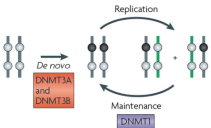
DNA methylation is mediated by members of the DNA methyltransferase (DNMT) family that can covalently transfer a methyl group (CH3) from the universal donor S-adenosyl-L-methionine (SAM) to the carbon 5-position of the cytosine ring. Conventionally, DNMTs are classified as de novo (DNMT3a and DNMT3b) or maintenance (DNMT1) enzymes (Figure 2) [12, 13].
Figure 2. De novo and maintenance DNA methylation schema. De novo DNA methyltransferases (DNMTs) DNMT3A and DNMT3B establish DNA methylation in germ line and developing embryos, (the black circles represent methylated CpG dinucleotides and the grey circles represent unmethylated CpG dinucleotides), whereas DNMT1, which has a preference for hemimethylated DNA, maintains the methylation pattern throughout replication (Jones and Liang, 2009) [13].
12
Of note, DNMT2, a member of the methyltransferase family, catalyzes methylation of RNA at position 38 in tRNAAsp GUC [14-16] (Figure 3). like (DNMT3L) is a DNMT3-associated protein lacking an enzymatic domain and interacting with DNMT3a/3b to modulate its activity [17, 18]. In mice, Dnmt1 and Dnmt3b are essential for embryonic development, as DNA methylation changes dramatically. An overall demethylation after fertilization is followed by de novo methylation of discrete regions upon implantation [19]. Dnmt1 knockout
Figure 3. Schematic representation of the methylation reaction catalyzed by the DNA methyltransferases (DNMTs) (adapted from [14]). Shown are the mechanisms proposed for methylation of cytosine by DNMT1, 3a and 3b on DNA (upper left panel) or by DNMT2 on RNA (lower left panel). Briefly, a thiol group (SH) from the binding site of the enzyme provides the nucleophilic attack to position 6 of the cytosine heterocycle, to activate position 5 towards one-carbon transfer (I). The methyl group on position 5 is donated by the coenzyme AdoMet (II). A proton in position 5 of the 5,6-dihydropyrimidine is then removed (II–III), and a consequent β-elimination generate 5-methylcitosyne and free enzyme (IV).
mice show early lethality at embryonic day (E) 9.5, whereas Dnmt3b depletion induces death at E 14.5–18.5, due to developmental impairment. On the contrary, Dnmt3a knockout mice do not display defects in embryonic development, but they do die at 4 weeks of age. Although this binary classification is convenient, the function of the de novo and maintenance DNMTs overlaps in many instances [13, 20, 21].
A number of studies have shown that DNA methylation is not randomly distributed across the genome, but displays regional specificity [22]. Methyl groups promote conformational changes in the major groove of DNA, thus altering protein-DNA binding [23] and, as a result, gene
13
expression. Most studies have initially focused on the effect of CGI methylation within the promoter and TSS of protein-coding genes. Recently [24], however, more comprehensive genome-wide methylation analyses have started elucidating the role of DNA methylation at CpG clusters within exons, introns, and intergenic sequences, expanding on the previous knowledge of CGIs and leading to the identification of CG shores (regions up to 2 kb from CGI), shelves (regions from 2 to 4 kb from CGI), and open sea (the rest of the genome) regions [25-27].
Hence, DNA methylation needs to be framed in the context of the genomic location. As a proof of concept, methylation of TSS-associated CGIs negatively correlates with gene expression, leading to long-term gene silencing [8], whilst gene-body methylation positively correlates to gene expression [28-31]. Another interesting finding emerging from bulk methylome studies is that non-CGI-CpGs are mostly methylated and therefore less stable than CGIs, due to the tendency of 5-methylcytosine (5mC) to undergo spontaneous or enzymatic deamination to T [32]. The C-to-T transition causes germ-line or somatic mutations, resulting in the depletion of CpGs dinucleotides in the human genome. Methylation at other genomic regions, such as enhancers and insulators, does not follow a specific pattern and may vary in different settings. Enhancers and insulators are long-range regulatory elements able to alter gene expression or protect gene promoters from inappropriate signals, respectively [33, 34]. Aran et al. have shown that distal methylation sites in estrogen receptor (ER)-positive breast tumors associates with breast cancer-related gene expression better than promoter methylation [35]. Moreover, Tatetsu et al. demonstrated that aberrant methylation of the 17-kb 5′ upstream enhancer of PU.1 is required for myeloma cell growth [36]. Insulators are bifunctional instead, acting either as a blocking enhancer, by preventing enhancer-mediated transcription, or as barriers, by limiting the advance of nearby heterochromatin that would otherwise silence expression [37]. CCCTC-binding factor (CTCF), an enhancer-blocking protein, does not bind to its DNA
14
consensus sequence if methylated, as demonstrated for the imprinted IGF2-H19 locus and CD45 gene [38]. It follows that DNA methylation can regulate gene expression indirectly by controlling access of enhancers to gene promoters [39]. Finally, CGI shores, with a lower CpG density, have recently emerged as critical regulatory elements affecting gene expression depending on their DNA methylation profile [40].
3 Non coding RNAs
The genome sequencing project revealed that complex organisms have a lower number of protein coding genes than expected. Differences between individuals and species cannot account for variation in protein-coding sequences, but rather for variation in gene expression regulated by a control architecture [41]. In this regard, perhaps one of the biggest surprise of the post-genome era is indeed the great number and diversity of transcripts arising from the previously assumed wastelands of non-coding regions. The first efforts in mapping transcriptional events to the genome led to the identification of thousands of long noncoding RNA (lncRNA) transcripts resulting in potential novel RNA regulatory elements [42]. Later on, other classes of regulatory noncoding RNAs were discovered, whose biological functions are still largely unknown. Thus, the growing number and increasing pace of discovery of new noncoding RNAs are followed by the challenge of their definition and annotation [43]. Noncoding RNAs (ncRNAs) can be classified either by function, as housekeeping (ribosomal RNA and transfer RNA) or regulatory RNAs, either by length, as lncRNAs or small RNAs (snRNAs).
3.1 Long noncoding RNAs (lncRNAs)
The general term long noncoding RNAs (lncRNA) defines a highly heterogeneous and poorly conserved group of transcripts >200 nucleotides (nt) in length that do not contain protein-coding sequences. The size threshold represents an arbitrary but convenient cutoff that excludes other classes of small infrastructural and regulatory RNAs [43, 44]. Some annotated lncRNAs
15
are not functional and do not confer any fitness advantage to the cell, e.g., spurious RNAs produced during transcription and showing tissue-specificity due to variations in chromatin states within different cellular contexts [45]. Despite the idea that the majority of lncRNAs are not functional, there exists a percentage of functional polyadenylated and non-polyadenylated lncRNAs, including sense or antisense, intronic, intergenic or enhancer, and overlapping transcripts with respect to protein-coding loci. They have been proposed to accomplish a variety of functions, such as gene regulation. lncRNAs indeed can be categorized into transcripts that regulate chromatin structure and gene expression in cis versus those that, by leaving the site of transcription, act in trans [46]. Cis-acting lncRNAs can regulate gene expression through three potential different mechanisms: (a) the lncRNA itself regulates the expression of neighboring genes by recruiting activating or repressing factors, (b) transcriptional or splicing events of the lncRNA acquire a gene-regulation functionality that is independent of the sequence of the transcripts, or (c) the recruitment of DNA elements within
16
the lncRNA’s promoter is eventually responsible for the neighbor gene’s regulation and is lncRNA’s sequence independent (Figure 4).
Figure 4. LncRNA’s function in local gene regulation . lncRNAs can control the expression of nearby genes in cis (A) by sequence-specific functions of the mature lncRNA transcript, (B) by mechanisms of transcription or splicing of an RNA, but the lncRNA itself is not functional or (C) by DNA elements within the lncRNA promoter or gene body that function independently of the transcribed RNA . Pol II, RNA polymerase II; TF, transcription factor (Kopp and Mendell, 2018) [46].
(a) The most known and well-studied example of cis-acting lncRNA is the X-inactive-specific-transcript (Xist) that orchestrates X chromosome inactivation. In female mammals, during early embryonic development, one of the two X chromosomes is transcriptionally silenced for dosage compensation. The silencing process depends upon transcription of Xist, that is transcribed from only one X chromosome, which will be later inactivated. Despite aspects of the Xist-silencing mechanism are still elusive, recent development of focused and innovative
17
methods able to capture RNA-binding proteins (RBPs) has allowed a step forward in understanding Xist’s interactors as well as in re-evaluating previously proposed mechanisms [47]. Therefore, studies have demonstrated that Xist directly recruits SMART/histone deacetylase 1 (HDAC1)-associated repressor protein (SHARP or SPEN). This further leads to the recruitment of the SHARP/SPEN-interacting protein silencing mediator for retinoid and thyroid hormone receptors (SMRT) and its interactor histone deacetylase 3 (HDAC3), ultimately inducing histone deacetylation, one of the earliest event in X inactivation [48]. Chen et colleagues also demonstrated that Xist entails remodeling of the three-dimensional (3D) structure of the X chromosome by recruiting it to the nuclear lamina and, by doing so, Xist is able to spread to actively transcribed genes across the X [49].
(b) In contrast with the activity of Xist, where sequence recognition plays a key role in recruiting protein interactors, Engreitz et colleagues demonstrated that other cis-acting lncRNAs exert local effects on neighboring genes without sequence-specific function [50]. By analyzing 12 lncRNA loci in mouse embryonic stem cells (mES cells), 5 of these 12 lncRNA loci influence the expression of adjacent genes, by ultimately acting in cis. As one example, the linc1536, also called Bendr (Bend4-regulating effects not dependent on the RNA), regulates expression of the adjacent Bend4 gene by DNA regulatory elements within the 750 bp promoter-proximal region. Thus, knocking-out the Bendr promoter reduces the expression of the neighboring gene by 57%, whereas introducing polyadenylating signals (pAS) into the first intron of the locus does not exert any effect.
The process of transcription and the 5’ splice in the Blustr locus (bivalent locus (Sfmbt2) is upregulated by the splicing and transcription of an RNA, also known as linc1319) are both necessary for regulating the neighboring gene Sfmbt2, located 5 kb upstream. Hence, Blustr is essential for Sfmb2 activation, but the entire process does not require precise RNA sequence beyond the initial splice signals.
18
(c) As for the Bendr locus, in most of lncRNA loci, effects on adjacent genes are mediated by enhancer-like function of DNA elements located within promoters and functioning independently of the production or sequence of the transcript [50]. lincRNA-p21 is a nuclear intergenic noncoding RNA that neighbors the CDKN1A in human and mouse. An initial study defines lincRNA-p21 as a p53-dependent trans-acting lncRNA, able to mediates apoptosis upon p53 signal. However, more recent studies suggest a cis-acting regulatory function. Indeed, even in tissues with no detectable lincRNA-p21 transcript, deletion of the locus dramatically affects the expression of neighboring genes, as Cdkn1a. Further analysis has defined DNA enhancer elements within promoter of the locus, which are responsible of its regulatory activity. Therefore, the lincRNA-p21 locus can exert an RNA-independent function on gene expression [51].
lncRNAs can also act in trans therefore regulating the expression of genes located at distant regions from their transcriptional start site (TSS), influencing chromatin structure and, interacting with proteins and other RNAs. Cis and trans lncRNA’s activity are not mutually exclusive. The Xist has indeed the ability to shape X chromosome structure during X chromosome inactivation process, in addition to cis-acting gene silencing mechanism [49, 52]. Firre, a lncRNA transcribed from the active X chromosome and escaping X chromosome inactivation, has been detected, in addition to its transcriptional site, at other autosomal loci using RNA antisense purification (RAP) and RNA fluorescent in situ hybridization (FISH) in murine embryonic stem cells. Moreover, Firre expression is essential to maintaining the Xi structure and its epigenetic features [53].
3.2.1 DNMT1-interacting RNAs (DiRs)
Previous work has shown the existence of noncoding RNAs able to regulate methylation patterns of their respective coding transcripts [54]. These novel class of noncoding RNAs, namely DNMT1-interacting RNAs (DiRs), interact with and inhibit DNMT1, hence preventing
19
DNA methylation. Using as example the CEBPA locus, Di Ruscio et al. demonstrated the existence of an overlapping, sense-orientated transcript, the extra coding CEBPA (ecCEBPA), whose transcription regulated the expression of the corresponding coding CEBPA mRNA (Figure 5).
Figure 5. Schematic of DNMT1 sequestration. Top panel, DNMT1 can access transcriptionally inactive methylated sequences. Bottom panel, DNMT1 cannot access transcriptionally active hemi-methylated sequences. (Di Ruscio et al, 2013) [54].
Characterization of ecCEBPA demonstrated that DiRs were non-polyadenylated, enriched in the nuclear fraction, and overlapped the gene in the sense orientation. From genome-wide analysis, they found that, besides CEBPA, DiRs regulated other loci and were associated with hypomethylated regions. The discovery of DiRs provided a new mechanism showing how genes can be regulated by noncoding transcripts arising around a gene body..
3.3 Small noncoding RNAs (sncRNAs)
All noncoding RNA species smaller than 200 nt can be classified as small noncoding RNAs. However, what defines eukaryotic small noncoding RNAs in the RNA silencing pathway is their limited size (20-30 nt) and their association with Ago-family proteins [55]. At least three classes of snRNAs can be identified, based on their mechanism of biogenesis and the type of Ago-protein they are associated with: microRNAs (miRNAs), endogenous short interfering
20
RNAs (endo-siRNA) and piwi-interacting RNAs (piRNAs). An additional subgroup, the short activating RNAs (saRNAs), that positively regulate gene expression, will be also described.
3.3.1 MicroRNAs (miRNAs)
MicroRNAs (miRNAs or miRs) are short noncoding RNAs able to control expression by targeting specific messenger RNAs (mRNAs). Canonically, miRNAs are encoded by intronic regions of noncoding and coding transcripts, however, some miRNAs can also be encoded by exons. miRNAs are transcribed by RNA Pol II as poly- or mono-cistronic primary miRNAs, also named pri-miRNAs, and containing an m7G cap at 5’ end in addition to a poly(A) tail at 3’ untranslated region (3’UTR) [56]; alternatively, some miRNAs requires Pol III, e.g. in the case of the human chromosome 19 miRNA cluster (C19MC) [57]. The miRNA precursors can be several thousand base pairs in length and are processed in the nucleus by the Drosha/DGCR8 complex to produce 70 bp stem-loops (pre-miRNAs). The small hairpin-shape is then exported to the cytoplasm by means of the nuclear export receptor exportin5 and cleaved by Dicer. Dicer’s cleavage results in an RNA duplex of about 21 nt in length. Once the RNA duplex is unwound, it is loaded onto an Argonaute protein complex, named RNA-induced silencing complex (miRNP/RISC complex), which identifies the sense and passenger strands in the RNA duplex and guides mature miRNA (single-stranded) to its target mRNA (Figure 6) [58, 59]. miRNAs recognize their target mRNAs by Watson-Crick base pairing: they contain a seed region, centered on
21
Figure 6. miRNAs’ mediated expression regulation. Primary miRNA transcripts are processed to miRNA precursors in the nucleus by Drosha. The pre-miRNA is then exported to the cytoplasm by means of the nuclear export receptor exportin5 and cleaved by Dicer in 21-nt RNA duplex. The RNA duplex is resolved and loaded onto miRNP/RISC complex. Mature miRNAs bind to Ago proteins, which mediate translational repression or cleavage of target mRNAs. Other sources of long dsRNA in the cytoplasm of a cell are processed by Dicer into 21–23 nucleotide dsRNA intermediates. Following unwinding of the dsRNA (mediated by Armitage and R2D2) the single-stranded siRNA-containing RISC is formed. ADARs and exonuclease ERI-1 can regulate dsRNA stability during processing (Meister and Tuschl, 2004) [58].
nucleotides 2-7, that binds within the 3’ UTR of the targeted RNA. However, miRNAs responsive elements (MREs) can be found
within 5’ UTR sequences as well as coding regions of the target mRNAs. In addition, non-canonical seed pairing can also drive miRNA-mRNA recognition and annealing [60]. The degree of miRNA-mRNA complementarity has been considered a pivotal element of the regulatory mechanism. While perfect complementarity is responsible for Ago-catalyzed cleavage of the targeted mRNA, central mismatches prevent cleavage of the mRNA and promote translational repression of the transcript [61]. The canonical function of miRNAs is that these small RNA molecules repress gene expression by targeting the 3’UTR of targeted mRNAs, thus inducing mRNA’s cleavage and/or transcriptional gene silencing in the cytoplasm. However, since the discovery of the first miRNA, lin-4, in the 1993 [62], more recent studies have demonstrated that miRNAs can localize into the nucleus and are able to regulate transcription [63-65]. MiRNAs nuclear localization is strictly dependent on tissue, cell line and condition analyzed. The regulation can be either repression or activation and the miRNA’s modus operandi can be affected by the presence of TATA box motif, CpG island region and the epigenetic status of the promoter [66]. However, whether miRNAs function depends on DNA methylation status of the target sequence or whether miRNAs can determine changes in DNA methylation profile is still a matter of debate [67, 68].
22 3.3.2 Short interfering RNAs (siRNAs)
Short interfering RNAs partake, together with miRNAs, in the RNA interference mechanism [69]. Initially, siRNAs were thought to be primarily exogenous, that is directly derived from the virus, transposon or transgene trigger. Subsequently, they were identified upon transgene- and virus-induced silencing in plants, persistent with a natural role in genome defense. Further studies of small-RNA profiling in mice reveal the presence of various types of endogenous siRNAs (endo-siRNAs) in oocytes and, to a lesser extent, in embryonic stem (ES) cells [70]. Endo-siRNAs are slightly shorter than miRNAs: they are 21 nt in length and derive from different sources of double stranded RNAs (dsRNAs), such as transposable elements (TEs), cis-natural antisense transcripts (cis-NATs), transNATs and hairpin RNA transcripts [55]. Some siRNAs originate from expressed pseudogenes, suggesting that pseudogenes can regulate levels of the founding mRNA by means of the RNAi machinery [71]. Long dsRNA precursors are processed by Dicer into discrete siRNAs duplex whose guide strand will direct the RNA interference (RNAi) complex at the targeted mRNA. During the canonical RNAi, a perfectly complementary mRNA to the siRNA’s guide strand is recognized and the targeted mRNA is cleaved at a single site within the duplex siRNA-mRNA. After cleavage, the mRNA fragments are further degraded. In some cases, siRNAs can also recognize targets with imperfect complementarity therefore acting as miRNA-like molecules. Finally, in plants, siRNAs can activate or engage DNA methyltransferases (DNMTs), leading to an increase in DNA methylation and heterochromatin formation [61, 72, 73]. Therefore, siRNAs mediate gene specific silencing resulting in an exquisitely specific mRNA suppression. As a consequence, synthetic siRNAs can be used as a powerful tool for regulating exogenous and endogenous gene expression, as well as innovative therapeutic approach [74]. Conventional synthetic siRNAs consists of 19-21 nt with two nucleotide overhangs at 3’ end, usually TT or UU [75]. siRNAs longer than 21 nt require Dicer cleavage to obtain shorter and active
23
noncoding transcripts. Increasing the length of siRNAs has been demonstrated to potentiate siRNAs’ silencing effect. However, dsRNAs longer than 30 nt can activate the interferon (IFN) response, and should be avoided for therapeutic applications [76, 77].
3.3.3 PIWI-interacting RNAs (piRNAs)
PIWI-interacting RNAs (piRNAs) are a class of animal-specific small single-stranded RNAs that associate with PIWI proteins. Piwi proteins are a clade within the larger family of Argonaute proteins and are mainly expressed in the germline. In general, Argonaute proteins silence their target RNAs through RNA degradation, inhibition of translation or chromatin modification [78]. Biogenesis, as well as function, differentiate piRNAs from miRNAs and siRNAs. PiRNAs originate from long single-stranded RNAs independently from RNase III enzymes, whose role is essential in miRNAs and siRNAs expression. Single-stranded precursors are then processed into discrete piRNAs’ units. PiRNAs precursors arise from genomic loci known as piRNAs clusters, from which long and single-stranded RNAs are produced. They usually consist of more than 100.000 bases and include transposable DNA elements. The majority of them have an antisense orientation to transposons, thus inducing their silencing. Loss-of-function mutations in piRNAs and PIWI proteins activate transposons that are able to randomly relocate or insert copies within the genome: these events can often impair gonadal development and lead to infertility [79, 80]. Indeed, the ancestral function attributed to piRNAs relates to transposon-induced genomic instability in the germline, where piRNAs repress transposable elements thus preventing transposons mobilization. However, some piRNAs correspond to unique genomic sequences unrelated to transposons. Therefore, there exist evidence suggesting that these piRNAs might regulate expression of host mRNAs [81, 82]. In addition, it has been demonstrated that piRNAs are not only expressed in the germline but, to a lesser extent, can also be detected in somatic tissues where regulate gene expression [83]. Indeed, disruption of piRNAs involved in transposable elements silencing
24
might results in genomic instability, transposons mobilization and hence, contribute to tumorigenesis. Besides being involved in silencing of transposable elements, piRNAs can also modulate DNA methylation. In this regard, genome-wide methylation profile revealed changes in DNA methylation in Farage and MCF7 cell lines upon transfection with single copy piRNAs. The study showed that genomic regions close to differentially methylated CpG sequences were enriched for sequences recognized by the transfected synthetic piRNA. In conclusion, piRNAs could induce DNA methylation of non-transposable elements loci by means of directly binding to genomic DNA or nascent mRNA [84].
3.3.4 Short activating RNAs (saRNAs)
Small double-stranded RNAs (dsRNAs) molecules, such as miRNAs, siRNAs and piRNAs, have been originally identified as the trigger for RNA silencing, where dsRNAs inhibit translation or degrade complementary target mRNA sequences. However, if RNAi is considered a mechanism to regulate gene expression, it is reasonable to suggest that RNA-mediated gene regulation can also positively control target sequences [85]. RNA activation (RNAa) is indeed a mechanism of enhancing gene expression at transcriptional level by means of short dsRNAs and is triggered by both endogenous and artificially designed small RNAs. dsRNAs usually target gene promoter [86, 87], however, there are examples of short activating RNAs (saRNAs) recognizing sequences within coding regions of target genes, e.g. the intronless gene CEBPA [88]. Therefore, saRNAs offer a new method to promote gene overexpression and represent a powerful laboratory tool to enhance transcription by a more natural approach. Moreover, RNAa can also be a new option for gene therapies: the ability to specifically upregulate transcription in the absence of exogenous DNA can have profound impacts either in basic research and therapeutics [87]. RNAa has been recently identified in mammals as endogenous mechanism for upregulating transcription. For example, Kuwabara et al identified a small double-stranded noncoding RNA able to induce transcription of genes
25
containing NRSE/RE1 sequences and to promote neuronal differentiation in adult stem cells [89]. Moreover, in the liver, mir-122 facilitates replication of hepatitis C viral RNAs by interacting with 5’ noncoding region of the viral genome; hence, it may represent a new target for antiviral therapies [90]. To further study the role of dsRNA in RNAa, synthetic dsRNAs can be designed and tested in vitro. Based on empirical observations, a set of rules for saRNAs design has been generated in order to improve the chance to identify functional saRNAs targets, including (i) RNA duplex size of about 19 nt, (ii) 2-nt overhangs on the 3’ ends of both RNA strands, (iii) GC content between 40-60 % and (iv) thermodynamic stability in base pairing within the 5’ end, whose modification can interfere with Ago2 processing [91]. In regards to target location, saRNAs are usually designed between -100 to -1000 bp relative to the TSS, with the most responsive site usually occurring within -200 and -500 bp region of the promoter. Moreover, avoidance of GC islands, lack of repeat elements (Alu sequences) or inverted sequences are also considered during the design [87]. Due to their nuclear nature, high concentrations of saRNAs are required in order to compensate for the potential nuclear exclusion of duplex RNAs [92]. Therefore, concentrations between 10 to 100 nM can be tested for initial studies to identify optimal conditions for gene activation.
As RNai, RNAa depends on argonaute proteins but with a different kinetics. During RNAa, gene activation is delayed by about 48 hours and, even though the underlying reason is not entirely known yet, it might depend on cell division and accessibility of the nuclear membrane. Moreover, since duplex RNAs are loaded by Ago2 in the cytoplasm before entering the nucleus, it is possible that this process is a passive mechanism that occurs during cell division. Therefore, gene expression analysis should be performed between 48 and 96 hours post transfection in order to validate saRNAs activity. However, saRNA-mediated gene activation is a stable mechanism whose effects can be detected up to 13 days after a single transfection [86].
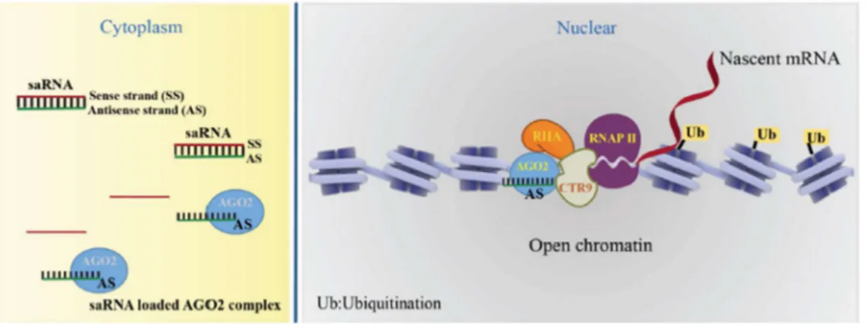
26
Thus far, the mechanism responsible for RNAa describes Ago2 protein loading the guide strand (antisense to the sense target gene) of the duplex saRNAs and forming an active saRNA-Ago2 complex. The complex binds to promoter of target genes and induces open chromatin structure and transcription by recruiting CTR9 and RHA proteins. Activation of transcription is also associated with phosphorylation at serine 2 of RNA polymerase II (RNAP II) and monoubiquitination of H2B (H2Bub1) (Figure 7) [93].
Figure 7. Accepted working model of the molecular mechanisms of saRNA-induced gene activation. The saRNA:AGO2 complex enters the nucleus, binds at promoter of target genes and induces open chromatin structure. AGO2 recruits CTR9, RHA and promotes transcript initiation associating with phosphorylation of RNAP II on Ser2 and H2Bub1 (Yoon and Rossi, 2018) [93].
Even though there is a sound understanding of the saRNAs mechanism, there exist some unresolved areas regarding RNAa effect on DNA methylation and whether both saRNAs strands might function in transcriptional activation.
4 CEBPA
The CCAAT/enhancer-binding protein alfa (CEBPA) is a decisive transcription factor that regulates tissue-specific gene expression by coupling lineage commitment to terminal differentiation and cell cycle arrest [94]. CEBPA is a member of the bZIP family and consists of N-terminal transactivating domains, a basic region necessary for specific DNA sequence binding, and a leucine-zipper region necessary for dimerization at the C-terminal end [95]. Moreover, evidences demonstrated that it acts as tumor suppressor, supporting the
27
general view that disruption of normal differentiation and its uncoupling with cell cycle arrest are key features for tumor development [96]. The intronless CEBPA gene localizes on chromosome 19q13.11 and its mRNA is transcribed into different protein isoforms generated from two different but consecutive in-frame AUGs within the CEBPA mRNA. The shorter isoform, namely p30, lacks the amino-terminal 117 amino acids whereas retains the same carboxyl terminus as the full-length form, known as p42. Differences in the N-terminal sequences confer distinct functions to the two isoform in the regulation of differentiation and proliferation. The p30 isoform maintains the DNA-binding domain but miss the N-terminal transactivation domain and is a dominant-negative regulator of the full-length p42 isoform. Since p30 fails to induce differentiation and promote proliferation of myeloid progenitors, the ratio p30/p40 is critical for granulopoiesis [97]. An additional CEBPA protein isoform, named extended-CEBPA, have been identified. This isoform, translated from an alternative non-AUG initiation codon and having an extended N-terminal sequence, occupies the ribosomal DNA promoter in the nucleoli and stimulates rRNA synthesis [98].
4.1 CEBPA in normal hematopoiesis
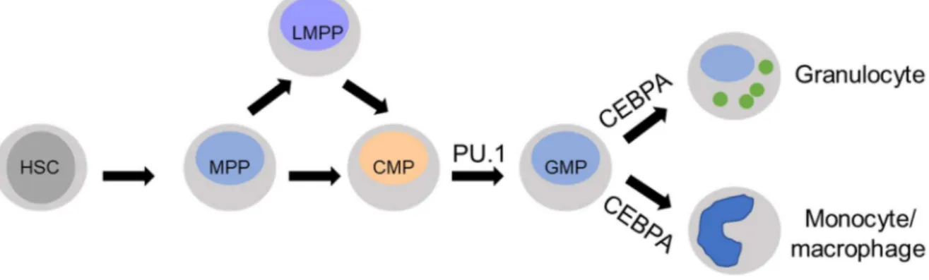
Fine mechanisms of differentiation regulate formation of terminally differentiated hematopoietic cells from hematopoietic stem cells (HSCs). HSCs maintain the ability to self-renew throughout the entire life of an organism and also to generate all types of mature blood cells [99, 100]. The interplay between tissue-specific transcription factors is a decisive element driving the expression of genes defining a precise cell type. As examples, CEBPA synergistically cooperates with PU.1, c-Myb, and RUNX1 to regulate transcription of genes such as myeloperoxidase, neutrophil elastase, lysozyme, lactoferrin, granulocyte colony
28
stimulating factor receptor (G-CSFr), macrophage colony stimulating factor receptor (M-CSFr), and granulocyte-macrophage colony stimulating factor receptor (GM-CSFr) (Figure 8).
Figure 8. Development of granulocytes and monocytes/macrophages cells from hematopoietic stem cell (HSC). CEBPA drives differentiation towards granulocytic and mononcytic/macrophagic population. MPP, multipotent progenitor; LMPP, lymphoid-primed multipotent progenitor; CMP, common myeloid progenitor; GMP, granulocyte–macrophage progenitor (adapted from Imperato et al, 2015) [101].
CEBPA is largely expressed in several cell lineages, including adipocytes, hepatocytes, and type II pneumocytes. Within hematopoiesis, CEBPA is primarily expressed in granulocyte, monocyte, and eosinophil as compared to the lymphoid or megakaryocyte/erythroid lineages [102]. Reducing CEBPA expression promotes monopoiesis by forming heterodimers with AP-1 proteins, as c-Jun or c-Fos. On the contrary, formation of active CEBPA homodimers is essential for granulopoiesis. Indeed CEBPA induces transcription of various proteins indispensable for subsequent lineage maturation, as CEBPE, Gfi-1 and KLF5 [103]. Moreover, CEBPA induces mir-223 transcription that leads to NFI-A mRNA degradation thus enhancing granulopoiesis. CEBPA-deficient mice have normal numbers of common myeloid progenitors (CMPs) but lack granulocyte-monocyte progenitors (GMPs) and all subsequent granulocytic stages [104]. Conditional CEBPA knockout in GMPs allows for normal granulopoiesis in vitro, indicating that CEBPA is not necessary for differentiation towards granulocytes beyond the
GMP stage [96].
29
hypermethylation. Methylation of the core promoter, located -600 bp from the ATG, was found to be an infrequent event in AML [105], whereas methylation of the distal promoter (-1600 to -600 from ATG), also known as upstream promoter, occurs with higher frequency in AML patients [106, 107]. Surprisingly, Hackanson et al demonstrated that, while treatment with DAC of AML cell lines induced upregulation of CEBPA mRNA in vitro, the CEBPA protein levels decreased. Further investigation proved that mir124a, often silenced by epigenetic mechanism and targeting CEBPA transcript, was upregulated upon epigenetic treatment and responsible for CEBPA degradation [106]
4.2 CEBPA in lung development
CEBPA is expressed in a number of epithelial tissues, including the respiratory epithelium, breast, colon, and prostate [108]. In the respiratory epithelium, CEBPA is involved in lung development and airway epithelial cells differentiation. CEBPA, indeed, regulates the expression of several genes during lung differentiation, as surfactant B and uteroglobin [109]. Since CEBPA abnormalities were originally observed in CEBPA (-/-) knockout mice, where aberrant proliferation of type II pneumocytes was detected, it has been demonstrated that CEBPA plays also a critical role in lung cancer development. CEBPA has growth inhibition properties, therefore its downregulation in lung cancer leads to uncontrolled cell proliferation, whereas its induction promotes growth arrest, morphological changes characteristic of differentiation, and apoptosis [110]. Reduced expression of CEBPA in lung cancer might occur because of promoter hypermethylation. As in AML, methylation of the upstream promoter is a critical event regulating the CEBPA gene in lung cancer [111].
30 5 P15
5.1 P15: a cell-cycle regulator
P15INK4b, also known as P15, CDKN2B or MTS2, and its functional homologue P16INK4a (P16 and CDKN2A) are genes located in close proximity within a small 25 kb fragment on chromosome 9p21 and they are both transcribed in the centromere to telomere direction. The P16 locus, in addition to P16, encodes for the alternative transcript P14ARF (p19ARF in mice). P16 and P14ARF share common exons 2 and 3, but have different first exons, 1a for P16 and 1b for P14 [112]. The three genes are tandemly linked to each other within 42 Kb of genomic locus on the 9p21 chromosome. The 9p21 locus also contains the Antisense RNA in the INK4 Locus (ANRIL), a long-noncoding antisense transcript that overlaps the entire P15 gene body and shares a bidirectional promoter with P16 (Figure 9 ) [113, 114].
Figure 9. Schematic of the 9p21 locus. Red rectangles indicate P15, striped-pattern rectangles indicate P14 and squared-pattern rectangles indicate P16 transcripts. P15, P14 and P16 mRNAs are transcribed in the direction telomere to centromere. Black rectangles depict ANRIL transcript, whose transcription follows centromere to telomere direction.
P15 and P16 encode for cyclin-dependent kinase inhibitors (CDKIs) that negatively regulate the cell cycle by inhibition of cyclin-dependent kinases (CDKs) 4 and 6, which control G1 to S phase progression through regulation of the Rb pathway [115, 116]. Once the cell pass the G1/S phase checkpoint, it is irreversibly committed to the next cell division. CDKs are a family of multifunctional enzymes able to phosphorylate target proteins involved in the cell cycle and
31
require the presence of cyclins to be activated. There exist multiple cyclins acting during specific stage of the cell cycle; in this respect, CDK4 and 6 only partner D-type cyclins which regulate the early G1 phase. P15 and P16, acting as negative regulators of cell-cycle progression, directly bind CDKs thus inducing allosteric changes that propagate to the cyclin binding site. Since interaction with cyclins is essential for CDK’s activation, conformational changes within cyclins’ binding site prevent cyclins from binding hence causing CDK’s inhibition [117]. P15 and P16 genes share 90% homology at coding exon 2, and isoform 1 of either proteins show 83 % identity. Therefore, the high degree of structural identity explains the high degree of functional homology. However, despite their similarity, in a variety of tumors, genetic alterations of sole P16 expression due to deletions and/or mutations are observed. On the contrary, deletions of P15 in lymphomas, carcinomas and sarcomas, occurred in conjunction with P16 and P14ARF loci [118].
5.2 P15: a tumor suppressor gene
P15 is important during differentiation of early hematopoietic progenitors: it indeed drives normal CD34+ towards granulocytic and megakaryocytic commitment. P15 mediates antimitotic signals and it is the effector of transforming growth factor β1 (TGF-β1), which induces arrest at G1 phase [119]. Aberrant P15 methylation is a major gene silencing mechanism in hematological malignancies, whereas P16 and P14 methylation more often occurs in solid tumors as well as in leukemias and lymphomas [120]. P15 promoter hypermethylation occurs at high frequency in acute myeloid leukemia (AML) and myelodysplastic syndrome (MDS), 80 and 50 % respectively [121, 122]. Aberrant methylation of the P15 locus is associated with poor prognosis in AML and with increased risk of progression to AML in MDS patients [118]. P15 aberrant methylation has also been reported in up to 60 % of chronic myelomonocytic leukemia (CMML) cases and is associated with a higher degree of disease’s severity [123]. Due to its important role in hematopoietic
32
differentiation, P15 has been suggested to be a tumor suppressor. To assess whether inactivation of P15 is a critical event for premalignant myeloid disorder and development of leukemia, a p15 (-/-) mouse model, in which the second coding exon of the gene was eliminated by homologous recombination, was developed [124]. The p15 knockout mouse did not show a correlation between loss of p15 and AML outcome, even though the p15 (-/-) mice have a competitive advantage over wild-type cells in myeloid cell formation, therefore suggesting an important role in common myeloid progenitors’ (CMPs) differentiation [125]. A more informative mouse model generated to define the role of p15 as tumor suppressor is a conditional one, the p15Ink4bfl/flLysMcre mouse, where P15 was silenced specifically in myeloid cells. Even though the p15Ink4bfl/flLysMcre mice did not develop acute myeloid leukemia, inactivation of the p15 locus increased susceptibility when additional oncogenic hits were provided. Moreover, the mice developed non-reactive monocytosis in the peripheral blood and expansion of myeloid blast progenitors in the bone marrow (BM), either symptoms resembling CMML in human patients [123]. These data demonstrate that loss of P15 contribute to preleukemic conditions and can be defined as tumor suppressor gene for AML [118, 126]. As in AML, P15 is not expressed in acute promyelocytic leukemias (APL), hence favoring blasts expansion, but it is progressively upregulated by all-trans retinoic acid (ATRA) treatment, able to drive differentiation [121]. Restoring normal methylation is therefore a rational therapeutic approach when aberrant methylation occurs.
33
5-azaytidine (5AC, trade name; Vidaza®, Azadine) and 5-aza-2’-deoxycytidine (5-aza-CdR; DAC; also known as Decitabine, trade name; Dacogen®) are nucleoside analogs of cytidine used as demethylating agents (Figure 10) [127].
Figure 10. Chemical structures of cytidine nucleoside (a) and azanucleoside (b, c). Sugar moieties are indicated in grey whereas chemical changes between cytidine nucleoside and azanucleosides are highlighted in red (Diesch et al, 2016) [128]
Later, they both were approved for the treatment of MDS and AML. 5AC and DAC act differently within the cell: 5AC is mainly incorporated into the RNA (80-90%) and a small percentage into DNA; DAC, instead, incorporate into DNA only. 5AC and DAC are cell-cycle dependent drugs able to exert their activity during S phase, when irreversibly and covalently trap DNMTs at C-6 position [129]. DNMTs’ inhibition has disparate consequences: it can induce mutagenesis within CpG dinucleotides, inhibit methylation during the following DNA replication, and cause cytotoxic effects when additional drug molecules do not bind any further DNMTs. Moreover, 5AC and DAC can trigger genomic instability resulting from demethylation of repetitive sequences. It has long been debated whether 5AC and DAC’s anticancer effect depends on their cytotoxicity or on the analog itself once incorporated into nucleic acids [129]. However, it is emerging that low doses of analogs inhibit DNMTs, whereas high doses induce cell-cycle arrest because of cytotoxic effects. Besides their efficacy, epigenetic therapies induce off-target effects that limit their clinical use. Moreover, little
34
success has been achieved thus far in treatment of solid tumors [130]. Therefore, these evidences suggest the need for more specific therapies to treat hematological malignancies, where aberrant methylation occurs.
35
36
CEBPA and P15 are genes frequently silenced by aberrant promoter DNA methylation in hematological malignancies and/or lung cancer.
These past years, our laboratory has been testing a double stranded RNA platform, the short activating RNAs (saRNAs) to reactivate expression of aberrantly methylated genes. SaRNAs are double stranded RNAs shown to upregulate transcription in a gene-specific manner, yet the mechanism behind this activation remains elusive.
This study is built on the hypothesis that saRNAs may promote gene expression by acting as DiR mimicking molecules and changing DNA promoter methylation.
In collaboration with the U.K. biotech firm MiNA Therapeutics [131, 132], saRNAs targeting CEBPA and P15 gene loci were tested.
The CEBPA saRNA AW1-51, developed by Voutila et al. [88] and currently under evaluation in patients with advanced liver cancer in a phase 1/2a trial (ClinicalTrials.gov: NCT02716012), was delivered by nucleofection in the AML cell line K562 and by lipid-based transfection in the lung cancer cell line A549, which have low-to-medium CEBPA expression and fully- to- hemi methylated promoter profile, respectively.
For the P15 locus three saRNAs targeting: the promoter PR313); the first exon (saP15-PR11) or the intron (saP15-PR56) were designed. All three saRNAs were delivered by nucleofection into the AML cell lines KG1a and Raji, which display low-to- medium P15 expression and fully- to-hemi-methylated promoter profile, respectively.
The effects of RNA-mediated activation was assayed by PCR and strand-specific qRT-PCR analyses. RNA sequencing was performed to account for potential off-target effects and activation of specific pathways correlated to P15 and CEBPA re-expression. DNA methylation changes, upon gene reactivation, were assessed for the respective loci by Bisulfite sequencing PCR and comprehensive genome-scale (in progress), to exclude additional off-target effects, by the Infinium EPIC arrays platform which covers 850k CpG sites across the genome.
37
38 1. Cell culture
KG1a, K562, Raji, U937 and A549 cell lines were growth in RPMI medium supplemented with 10% fetal bovine serum (FBS). HEK293 cell line was cultured in DMEM High glucose medium supplemented with 10% FBS. All cell lines were purchased from ATCC and grown at 37°C, 5% CO2 in the absence of antibiotics. For all cell lines, media was replaced every 2/3 days; KG1a, K562, Raji and U937 were passaged 1:3/1:4, whereas A549 and HEK293 were passaged 1:5/1:7 every 48/72 hours.
In contrast to the parental KG1, the KG1a population, a male-derived acute myelogenous leukemia cell line, is mainly composed of undifferentiated promyeloblasts, thus being morphologically, cytochemically and functionally less mature. The KG-1a cell is insensitive to colony stimulating factor (CSF) and does not spontaneously differentiate to granulocyte and macrophage like cells.
K562 is a highly undifferentiated population and has been classified as human erythroleukemia line. K562 blasts are able to spontaneously differentiate into progenitors of the erythrocytic, granulocytic and monocytic series. K562 cells are positive for BCR-ABL fusion gene whose oncoprotein leads to transcriptional inhibition of the granulocyte colony–stimulating factor receptor (G-CSF-R), through down-regulation of CEBPA [133].
U937 was derived from histiocytic lymphoma and can be differentiated into macrophages or myeloid lineages dendritic cells (DC) by supernatants from human mixed lymphocyte cultures. The Raji cell line was derived from B-lymphocytes of a Burkitt’s lymphoma and holds the t(8;14) translocation, which results in the juxtaposition of the MYC gene to the IGH enhancers, which leads to its activation and oncogenic transformation [134, 135]. KG1a, K562 and Raji are suspension cell lines. A549 is an epithelial-like cell line derived from a lung carcinomatous tissue from a Caucasian male. HEK293 is a human embryonic kidney cell line. Either A549 and HEK293 are adherent cell lines.
39 2. RNA isolation
Total RNA was isolated by phenol chloroform purification using the commercially available Trizol (Invitrogen) according to manufacturer’s instructions. Briefly, cells are harvested by centrifugation and supernatant is carefully removed, leaving just a drop of liquid. Cells are briefly vortexed and Trizol is added to the cells solution. Chloroform is added to Trizol (1:5 v:v, respectively) and tube is inverted several times. Follows centrifugation at 13200 rpm, 15 minutes at 4°C. The suspension is now a lower red phenol-chloroform phase (organic), an interphase and a colorless upper phase (aqueous). RNA is exclusively in the aqueous phase. The aqueous solution is pipetted out and isopropyl alcohol is added (ratio 1:1 v:v). Sample is incubated at -20°C O/N (low amount) or 2 hours (high amount). The sample is centrifuged at 13200 rpm, 4°C and pellet is resuspended in 178 μl of RNase free water. DNase I (Roche) is added to sample in the presence of RNase inhibitors (Promega) and solution is incubated 1 hour at 37°C. EDTA 0.5 M and NaCl 5 M are added to sample at the end of incubation to stop DNase I reaction and precipitate RNA, respectively. Cold phenol solution saturated with 0.1M citrate buffer, pH 4.3 (Sigma) is added to sample and tube is centrifuged at 13200 rpm 15 min at 4°C. The supernatant is moved to a new tube and 2.5 volumes of cold 100% ethanol is added. Sample is incubated at -20°C, O/N (low amount) or 2-3hrs (high amount). Pellet is recovered by centrifugation, 13200 rpm at 4°C for >20 minutes. Supernatant is carefully removed and pellet is resuspended in 30-40 μl of RNase free water.
For one-step quantitative reverse transcription PCR (qRT-PCR) using Taqman probes, 50 ng of total RNA was used per 14 μl reaction (Affymetrix USB; HotStart-IT Probe One-Step qRT-PCR Master Mix Kit). GAPDH was used as normalization control (Applied Biosystems). Taqman qRT-PCR conditions were: 50°C for 10 minutes (to generate cDNA), 95°C for 2 minutes (to inactivate the reverse transcriptase and activate the Hot Start DNA polymerase), followed by 40 cycles at 95°C (denaturation) for 15 seconds and 60°C (annealing and
40
elongation) for 1 minute with fluorescence acquisition during the final step. qRT-PCR was performed using a StepOnePlus Real-Time PCR System (Applied Biosystems) using the standard run protocol. For relative expression by qRT-PCR, target gene amplification was calculated using the formula 2^-∆∆DDCt as described by Livak et al [136].
To accurately assess expression and re-expression of low level transcripts, we verified transcript levels using strand specific qRT-PCR (ssqRT-PCR). For ssqRT-PCR 250 or 1000 ng of total RNA were retrotranscribed using SuperScript IV RT, according to the manufactuer’s instructions (Invitrogen). In place of random hexamers, specific primers were used for P15 (KG1a and Raji) and ecCEBPA (K562) detection. cDNA was purified using the High Pure PCR Product Purification Kit (Roche) and 60 ng of purified cDNA was used for 14 μl reaction. For P15, iTaq Universal SYBR Green Supermix (BioRad) was used; for ecCEBPA, One-Step qRT-PCR Master Mix Kit was used and conditions of reaction were modified (95°C for 2 minutes, followed by 40 cycles at 95°C for 15 seconds and 60°C for 1 minute). SYBR Green conditions were: 95°C for 5 minutes (to activate Taq polymerase), followed by 40 cycles at 95 °C for 15 seconds (denaturation) and 60 °C for 1 minutes (elongation and annealing). To measure number of copy of P15 transcript per each sample, we used a reference standard with known number of copies. The standard curve was generated by serial dilutions of a reference plasmid, pGEM-T easy vector (Promega), containing the amplified PCR product. SsqRpGEM-T-PCR was performed using a StepOnePlus Real-Time PCR System (Applied Biosystems) using the standard curve protocol. qRT-PCR primer set for the CEBPA mRNA is located in the coding region and after the poly(A) signal for ecCEBPA (Fig1f and g). qRT-PCR primer set for P15 is located across exon-exon junctions, as depicted in Fig3d and 4d.
Primer sequences are as the following: Human CEBPA:
41
Reverse 5’-GCA GGC GGT CAT TG -3’;
TaqMan Probe 5’-ACA AGG CCA AGC AGC GC-3’ Human ecCEBPA:
Forward 5’-GGT TGT CTG TGG GCC AGG TCA-3’; Reverse 5’-AGA GCT CAT GAA AGT CAG GAT TG-3’;
TaqMan Probe 5’-AAT AAT ACA GCA TTT TCC CTG GCG G-3’ Human P15:
Forward 5’-CGG GGA CTA GTG GAG AAG G-3’ Reverse 5’-GTG AGA GTG GCA GGG TCT G-3’ P15 specific primer for RT reaction:
Reverse 5’-CCT GTG AAC CTT TAA CAT TTC TCA-3’ ecCEBPA specific primer for RT reaction:
Reverse 5’-GGT AGG GTG TAG CCA CAT GGT CTA-3’
3. DNA isolation
DNA was isolated by phenol:chloroform:isoamyl alcohol (25:24:1) purification. Briefly, cells were resuspended in lysis buffer (0.5% SDS, 25 mM EDTA pH 8, 10 mM TRIS pH 8, 200 mM NaCl) and treated with RNAse A (Roche) for 20 minutes at 37°C. The cell lysate was digested with proteinase K (Roche) at 65˚C overnight. Phenol solution (pH 8) was added to cell lysate (v:v 1:1) and sample centrifuged 15 minutes at 13200 rpm. The aqueous phase was recovered with a cut tip and transferred to a new tube. Isopropyl alcohol 1:1 v:v was added and sample incubated at -20°C for 20 minutes (high amount) or O/N (low amount). Pellet was recovered by centrifugation and resuspended in TE (pH8).
4. Sodium bisulfite conversion
Bisulfite genomic sequencing is defined as the gold-standard for detection of DNA methylation at single base-pair resolution. This method was first developed by Frommer et al and it is based
42
on the finding that the amination reactions of cytosine and 5-methylcytosine (5mC) proceeds with very different consequences after the treatment of sodium bisulfite [137]. Therefore, cytosines will be converted into uracil residues and recognized as thymine in subsequent PCR amplification and sequencing, whereas 5mCs are immune to this conversion and remain as cytosines allowing 5mCs to be distinguished from unmethylated cytosines. A subsequent PCR process is performed to define the methylation status of the loci of interest [138]. From 200 to 1000 ng of genomic DNA were bisulfite converted according to manufacturer’s instructions (EZ DNA Methylation kit, Zymo Research) and eluted in 20 µl of warm RNase free water. The conversion was performed as follows: 98˚C for 8 minutes, 64˚C for 7.5 hour, 4˚C storage.
5. Combined Bisulfite Restriction Analysis (COBRA)
Combine Bisulfite Restriction Analysis (COBRA) is a qualitative approach to detect the methylation status of CpG-containing regions. Sodium bisulfite treatment introduces methylation-dependent sequence modifications and new restriction enzyme (RE) sites can be generated or pre-existing ones can be lost or retained depending on the methylation status [139]. As an example, the BstUI enzyme recognizes the CGCG sequence, which can be altered upon bisulfite conversion depending on the methylation status of the Cs. This property can be exploited to study DNA methylation in a qualitative way, by digesting the PCR amplicon with different REs and looking at the resulting digestion patterns, that is the basic principle of Combined Bisulfite Restriction Analysis (COBRA).
4 µl of bisulfite converted DNA were used in 50 µl of PCR reaction (FastStart Taq DNA Polymerase, Roche). The PCR reaction was performed as follows: 95°C for 6 minutes, followed by 35 cycles at 95°C for 30 seconds, 53-57°C for 1 minute, 72°C for 1 minute, and a final step at 72°C for 7 minutes. PCR product was gel-purified (QIAEXII Gel Extraction Kit, Qiagen) in a 2% TAE agarose gel. After gel purification, about 400 ng of gel-purified product
43
were digested at 60°C for 2 hours with 10 units of BstUI enzyme (NEB). Digested and undigested product were run on a 3% TAE agarose gel. Primer sequences are as the following: CEBPA upstream promoter, -1.4 kb from the TSS:
Forward 5’-GGT GTT TTT AGT TGT GTT TTT TT- 3’ Reverse 5’-AAA CCC TAA AAC CCC TTA-3’
CEBPA upstream promoter, -1.1 kb from the TSS: Forward 5’-TAT TTA AGG GGT TTT AGG- 3’ Reverse 5’-AAA AAC AAA CTT AAC TCT AA- 3’ CEBPA upstream promoter, -0.8 kb from the TSS:
Forward 5’-TAG TTT YGT TAG TTT GGG GGG TTT- 3’ Reverse 5’-AGG TTA AGG YGG TTG TGG GTT TTA- 3’ P15 promoter:
Forward 5’-GAT ATT TAG YGA GTA GTG TAG TTA GTA TTT TTG G- 3’ Reverse 5’-CCT YGC TCT AAC AAA ATA AAA AAC CAA- 3’
6. Bisulfite Sequencing PCR (BSP)
After bisulfite treatment of genomic DNA and PCR amplification of the CEBPA upstream promoter or P15 promoter, the PCR product was gel purified as previously described. The amplicon was cloned into the pGEM-T Easy Vector (Promega) as manufacturer’s instructions. E.coli competent cells (bacterial strain JM109, Promega) were transformed with 3 µl of ligase and plated on LB agar plates containing ampicillin (Sigma Aldrich), IPTG (Sigma Aldrich) and X-Gal (Sigma Aldrich) for blue/white screening and standard selection. White positive colonies were amplified and extracted using Pure yield plasmid Miniprep system Kit (Promega). Plasmid DNA was sent for sequencing (Quintara Biosciences, Boston, MA) and results analyzed by QUMA software (http://quma.cdb.riken.jp/). Clones with percentage of unconverted CpGs higher than 95 and with percentage of identity lower than 90 were excluded
44
from the final analysis. For DNA methylation analysis of the CEBPA upstream promoter, 42 nt, preceding the reverse primer 5’-AAAAACAAACTTAACTCTAA-3’ and containing 6 CpGs dinucleotides, were excluded from the analysis. The 6 CpGs were unmethylated both in the CEBPA-saRNA treated samples and in the NC-treated control. Moreover, the region where the primer was designed is the only one without CpGs, whose presence within primer sequence can affect primers’ efficiency.
7. DNA Fluorescence in Situ Hybridization (DNA-FISH)
DNA Fluorescence In Situ Hybridization (DNA-FISH) takes advantage of fluorophore labeled Bacterial Artificial Chromosome (BAC) probes to visualize the presence or absence of genomic DNA fragments on interphase DNA. BAC probes (Empire Genomics) covering selected areas of the P16/P15 locus were used to assess the presence of the locus. The manufacturer’s recommended hybridization protocol was followed with minor modifications. Briefly, cells were fixed with methanol acetic acid (3:1 ratio) for more than 2 hours at -20°C, dropped onto uncharged glass slides, and then left for at least 20 minutes at 45°C. To increase accessibility of the chromatin to the BAC probe, dried slides were then washed for 1 minute in 2X SSC (Cold Spring Harbor Protocols) and digested with 1 μg/ml proteinase K (Roche) in proteinase K buffer (Cold Spring Harbor Protocols) for 3 minutes at room temperature. Slides were then washed twice in 70% ethanol, 85% ethanol, 100% ethanol, and then allowed to dry. Hybridization was carried out with 1 uL of probe and 9 μL of hybridization buffer for at least 16 hours in a humidified Hybrite chamber: 85°C for 3 minutes, followed by 37°C for at least 16 hours. The following day, excess probe was removed in Wash 1 (0.4X SSC/ 0.3% NP-40 warmed to 73°C), 2 minutes; Wash 2 (2X SSC/ 0.1% NP-40 at room temperature), 1 minute; and dried in the dark. Prolong Gold antifade with DAPI (Thermo Fisher Scientific) was used to counterstain and cover the slide. A chromosome 9 control probe (9q21) was used as hybridization control. BAC probe coverage is depicted in Figure 3b.
![Figure 1. An outline depicting cell-fate plasticity according to the Waddington’s epigenetic landscape (inspired by the model proposed by Waddington [3])](https://thumb-eu.123doks.com/thumbv2/123dokorg/4816255.50141/10.892.330.591.789.1000/depicting-plasticity-according-waddington-epigenetic-landscape-inspired-waddington.webp)
![Figure 3. Schematic representation of the methylation reaction catalyzed by the DNA methyltransferases (DNMTs) (adapted from [14])](https://thumb-eu.123doks.com/thumbv2/123dokorg/4816255.50141/12.892.162.725.363.646/figure-schematic-representation-methylation-reaction-catalyzed-methyltransferases-adapted.webp)


![Figure 10. Chemical structures of cytidine nucleoside (a) and azanucleoside (b, c). Sugar moieties are indicated in grey whereas chemical changes between cytidine nucleoside and azanucleosides are highlighted in red (Diesch et al, 2016) [128]](https://thumb-eu.123doks.com/thumbv2/123dokorg/4816255.50141/33.892.153.744.238.520/chemical-structures-nucleoside-azanucleoside-indicated-nucleoside-azanucleosides-highlighted.webp)

