Volume 2011, Article ID 262674,6pages doi:10.1155/2011/262674
Case Report
Primary Pulmonary Epithelioid Hemangioendothelioma:
A Rare Cause of PET-Negative Pulmonary Nodules
Riccardo Cazzuffi,
1Nunzio Calia,
1Franco Ravenna,
1Claudio Pasquini,
1Sara Saturni,
1Giorgio Narciso Cavallesco,
2Francesco Quarantotto,
2Rosa Rinaldi,
3Annaluisa Cogo,
1Gaetano Caramori,
1, 4and Alberto Papi
11Sezione di Malattie dell’Apparato Respiratorio, Dipartimento di Medicina Clinica e Sperimentale, Universit`a di Ferrara, Italy
2Dipartimento di Scienze Chirurgiche, Anestesiologiche e Radiologiche, Modulo di Chirurgia Toracica, Universit`a di Ferrara, Italy
3Sezione di Anatomia, Istologia e Citologia Patologica, Dipartimento di Medicina Sperimentale e Diagnostica,
Universit`a di Ferrara, Italy
4Centro per lo Studio delle Malattie Infiammatorie Croniche delle Vie Aeree e Patologie Fumo Correlate dell’Apparato Respiratorio
(CEMICEF; formerly Centro di Ricerca su Asma e BPCO), Universit`a di Ferrara, Via Savonarola 9, 44121 Ferrara, Italy
Correspondence should be addressed to Gaetano Caramori,[email protected] Received 11 February 2011; Accepted 16 May 2011
Academic Editor: A. Curt
Copyright © 2011 Riccardo Cazzuffi et al. This is an open access article distributed under the Creative Commons Attribution License, which permits unrestricted use, distribution, and reproduction in any medium, provided the original work is properly cited.
We report here a case of primary pulmonary epithelioid hemangioendothelioma diagnosed in a 67-year-old Caucasian man, presenting with exertion dyspnoea, dry cough, and multiple bilateral pulmonary nodules revealed by computed tomography. At the 18F-fluorodeoxyglucose positron emission tomography, these nodules were negative. The histopathological diagnosis was made on a pulmonary wedge resection (performed during video-thoracoscopic surgery).
1. Introduction
Pulmonary epithelioid hemangioendothelioma (PEH) is a rare, low- to intermediate-grade tumor of endothelial origin [1] with around 120 cases reported in the literature [2]. At the onset, the patients are usually asymptomatic or present with nonspecific symptoms (such as weight loss, fatigue, dyspnoea, cough, and chest pain). Chest imaging usually shows the presence of multiple, bilateral small pulmonary lesions. The diagnosis usually requires a lung biopsy. The treatment is still not standardized. Surgery is suggested in presence of a solitary nodule or few unilateral nodules. In-stead if the lesions are unresectable or multiple and bilateral several chemotherapy protocols have been used. The prog-nosis is variable with a median survival of 4-5 years.
2. Case Report
A 67-year-old Caucasian man, lifelong nonsmoker, with no prior history of lung diseases, was referred to our university
hospital for the presence, in the last 3 months, of exertion dyspnoea (grade 1 according to the Medical Research Coun-cil scale [3]) and dry cough. He had worked as an employee without occupational exposures of clinical relevance. His past medical history was characterized by an herniated lum-bar disc, systemic arterial hypertension (treated with calcium blockers), diabetes mellitus (treated with oral hypoglycaemic agents), and polycythemia vera (diagnosed in 2007 and treat-ed from 2008 with hydroxyurea). Physical examination was unremarkable. Arterial blood gases analysis performed with the patient breathing room air demonstrated normal val-ues (pH 7.40, PaCO243 mmHg, PaO291 mmHg, and
bicar-bonate level 26 mmoL/L). Routine laboratory tests were within the normal range except for those listed inTable 1. Serum levels of neoplastic markers were within normal range, including carcinoembryonic antigen (2.7 ng/mL), prostate-specific antigen (3.75 ng/mL), and CA19.9 (15 U/ mL). Also the serum levels of angiotensin-converting en-zyme (11 IU/mL) andβ2microglobulin (2.50 mg/mL) were
2 Case Reports in Medicine
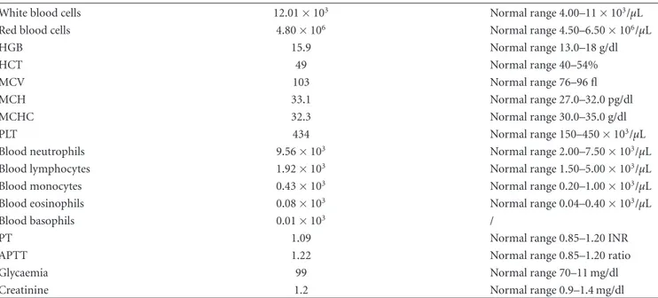
Table 1: Laboratory tests concerning diabetes mellitus and polycythemia vera.
White blood cells 12.01×10³ Normal range 4.00–11×103/μL
Red blood cells 4.80×10⁶ Normal range 4.50–6.50×10⁶/μL
HGB 15.9 Normal range 13.0–18 g/dl HCT 49 Normal range 40–54% MCV 103 Normal range 76–96 fl MCH 33.1 Normal range 27.0–32.0 pg/dl MCHC 32.3 Normal range 30.0–35.0 g/dl PLT 434 Normal range 150–450×103/μL
Blood neutrophils 9.56×10³ Normal range 2.00–7.50×10³/μL
Blood lymphocytes 1.92×10³ Normal range 1.50–5.00×10³/μL
Blood monocytes 0.43×10³ Normal range 0.20–1.00×10³/μL
Blood eosinophils 0.08×10³ Normal range 0.04–0.40×10³/μL
Blood basophils 0.01×10³ /
PT 1.09 Normal range 0.85–1.20 INR
APTT 1.22 Normal range 0.85–1.20 ratio
Glycaemia 99 Normal range 70–11 mg/dl
Creatinine 1.2 Normal range 0.9–1.4 mg/dl
APTT: Activated thromboplastin time. HCT: Hematocrit. HGB: Haemoglobin. LDH: Lactate dehydrogenase. MCH: Mean corpuscular Haemoglobin. MCV: Mean blood cell volume. MCHC: Mean cell haemoglobin concentration. PLT: Platelets. PT: Prothrombin time.
The pulmonary function tests showed the presence of a restrictive pattern with a vital capacity (VC) of 3.14 litres (70% of the predicted value), a forced vital capacity (FVC) of 3.11 litres (72% of the predicted value), forced expir-atory volume in one second (FEV1) of 2.55 litres (76.5% of
predicted value), a FEV1/VC ratio of 81% (108% of the
pre-dicted value) and a total lung capacity (TLC), 5.35 litres (73.3% of the predicted value). DLCO was in the normal range (Figure 1).
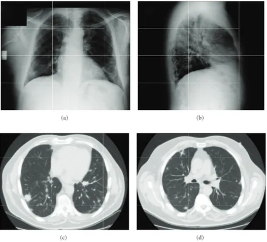
The chest radiography and a computed tomography of the chest (performed with iodine intravenous contrast me-dium) showed the presence of multiple bilateral pulmonary nodules (with a diameter variable between 3 mm and 3 cm) without contrast enhancement and the presence of one nod-ule (with the diameter of 2 cm) in the fourth hepatic segment showing late-phase contrast enhancement and another in the spleen (with the diameter of 1 cm) with cystic features (Figure 2).
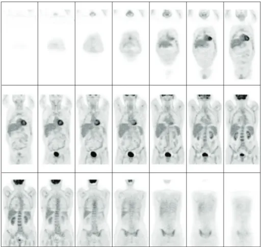
Interestingly, at the 18F-fluorodeoxyglucose positron emission tomography (PET), all these nodules were not showing any uptake of the tracer (Figure 3).
The fiberoptic bronchoscopy was normal. A transbron-chial lung biopsy and an ultrasound-guided transthoracic lung biopsy were both nondiagnostic.
The final diagnosis of pulmonary epithelioid heman-gioendothelioma was made by pulmonary wedge resection of a nodule located in the left lower lobe by video-thoracoscopic surgery. Histopathological examination of the resected tissue revealed round- to oval-shaped nodules, with a central sclerotic, hypocellular zone, and peripheral zone rich of cells. There was tumour diffusion into the adjacent bronchioles and the alveolar spaces with micropolypoid aspects. The
ex-F/V ex 1 Vol (L) 10 5 5 0 10 1 2 3 4 5 6 7 8 F/V in Flo w (L/s)
Figure 1: Flow volume loop.
tracellular stroma consisted of an abundant matrix of chon-droid, hyaline, mucinous, or myxomatous appearance.
The neoplastic cells were of polygonal shape and eosin-ophilic with round nuclei and uniform small to moderately sized nucleoli. Some cells had cytoplasmic vacuoles.
Immunohistochemical studies demonstrated that the neoplastic cells are endothelial cells immunoreactive for Von Willebrand factor, CD31, CD34, and vimentin (Figure 4).
Immunostaining for other markers (including caudal type homeobox transcription factor 2 (CDX2), cytokeratin (CK) 20, CK7, prostate-specific antigen (PSA), pan-cytok-eratin, chromogranin, epithelial membrane antigen (EMA), HMB45, progesterone receptor, and thyroid transcription factor (TTF)-1) was negative.
(a) (b)
(c) (d)
Figure 2: The chest radiography (a, b) and a computed tomography of the chest (performed with iodine intravenous contrast medium) (c, d) showed the presence of multiple bilateral pulmonary nodules.
3. Discussion
Pulmonary epithelioid hemangioendothelioma (PEH) is a rare, low- to intermediate-grade tumor of endothelial origin [1]. Epithelioid hemangioendothelioma can arise from many organs, including lungs, liver, bone, and soft tissue, simulta-neously or sequentially. When this occurs, it may be difficult to determinate if the tumor is multicentric from the begin-ning or there is a primary lesion with metastases to the other tissues [4]. Around 120 cases have been reported in literature [2,Table 2], only five of them were in Italian patients [5,15–
18]. Mean (SD) age of patients is 40.1 (17.5) years, and 73% are females [6]. Interestingly, our patient is instead a 67-year-old male. Most patients are asymptomatic at presentation, and some are complaining of weight loss, fatigue, dyspnoea, fever, pleuritic chest pain, mild nonproductive cough, and haemoptysis [19]. In most cases, the physical examination is normal, but few cases of digital clubbing and pleural effusions have been reported [1,6].
The most characteristic feature of PEH on chest radio-graphs or CT is the presence of multiple perivascular nodules with well- or ill-defined margins in both lungs. The nodules may range in size up to 2 cm, but most of them are≤1 cm in diameter; they are usually found near medium-sized vessels and bronchi [1]. This radiological presentation is suggestive of many lung diseases (Table 3).
Some authors consider 18F-fluorodeoxyglucose positron emission tomography an important tool for PEH diagnosis [7]; however, in our patient, the PET was negative, suggesting that a negative PET cannot exclude PEH. Unfortunately, we do not have a molecular explanation for this finding but may be related to a low proliferation rate of the neoplastic cells.
The diagnosis of PEH is made on the basis of the histo-pathological features and confirmed using immunohisto-chemical staining for endothelial cell markers, such as against factor VIII-related von Willebrand antigen, CD31 or CD34 [2,4,13,20,21].
The prognosis is variable. The 5-year survival is around 60% (range 47–71%). In fact, there are two groups of PEH at the clinical presentation: (1) one asymptomatic, with a solitary pulmonary nodule or unilateral multiple nodules. Usually they can be managed with surgery alone, and lym-phatic invasion is unlikely. Their prognosis is good with a median survival of more than 10 years; (2) another symp-tomatic group presents with multiple bilateral pulmonary nodules or pleural effusion with scarce response to chemo-therapy. The prognosis of this group is poor [11]. Most patients die from pulmonary insufficiency as a result of an in-creasing number of tumor nodules [9]. Hematogeneous metastases are rare and have been described especially in the liver but also in other sites [2,4].
4 Case Reports in Medicine
Figure 3: 18F-fluorodeoxyglucose positron emission tomography, all these nodules were not showing any uptake of the tracer.
Table 2: Review of the clinical, radiological and pathological features of the published cases of primary pulmonary epithelioid heman-gioendothelioma.
Review of the literature Our case report
Female/male ratio 3 : 1 female Male
Mean (SD) age 40.1 (17.5) years 67 years
Symptoms
Weight loss, fatigue, and respiratory symptoms (dyspnoea, chest pain, mild nonproductive cough, and mild haemoptysis)
Dyspnoea and dry cough
Chest radiography and
computed tomography Usually multiple bilateral pulmonary nodules Multiple bilateral pulmonary nodules
PET Positive Negative
Metastatic sites
Lymph nodes, liver, bone, skin, serosal membranes, spleen, tonsils, retroperitoneum, kidney and central nervous system
Single spleen and liver metastatic nodules
Immunohistochemical features
Factor VIII von Willebrand factor+, CD31+, or CD34+
Factor VIII von Willebrand factor+, CD31+, CD34+
5-year survival (%) 60% Alive for eight months then lost at the follow up Obtained with the data from [4–18].
(a) (b)
(c) (d)
Figure 4: (a) Neoplastic nodule showing increased number of cells at the periphery with an eosinophilic stroma (H/E, 100x magnification). (b) The neoplastic cells are of polygonal shape and eosinophilic with round nuclei and uniform small to moderately sized nucleoli. (H/E, 200x magnification). (c) Immunoperoxidase staining for CD34 of the neoplastic cells (brown colour) (200x magnification). (d) Immunoperoxidase staining for CD31 of the neoplastic cells (brown colour) (200x magnification).
Table 3: Differential diagnosis of multiple pulmonary bilateral nodules.
Metastases
Primary lung cancer (particularly bronchioloalveolar carcinoma) Lymphoid tumors and myeloma
Leukaemic infiltrates
Benign vascular tumors (hemangioma and lymphangioma) Malignant vascular tumors (angiosarcoma and Kaposi’s sarcoma) Neuroendocrine tumourlets
Nodular lesions in pulmonary fibrosis Pneumoconiosis
Infections (tuberculosis, nocardiosis, aspergillosis, and histop-lasmosis)
Sarcoidosis
Langerhan’s cell histiocytosis Vasculitis
Connectivitis
Pulmonary arteriovenous malformations Obtained with the data from [4,5,9,12–14].
The treatment of PEH is not standardized. Surgery alone is indicated in the presence of a single pulmonary nodule or unilateral multiple nodules. Lung transplantation should be considered in patients with vascular infiltration [10].
Various chemotherapies have been reported for unre-sectable or metastatic PEH, with variable effectiveness [22]. Other authors have suggested a role for the hormonal therapy (antiestrogens and progesterone) in patients with diffuse dis-ease if the neoplastic cells express estrogen and progesterone receptors [23]. A slight regression of the pulmonary lesions was also obtained with interferon (IFN)-2α, probably for its antiangiogenic activity [24]. Thalidomide and IFN-α have also been proposed for unresectable cases [22]. Some bene-ficial results were also obtained with bevacizumab, a mon-oclonal antibody that blocks human VEGF-A [22,25]. The neoplastic cells express glucocorticoid receptors and the en-zyme 11β-hydroxysteroid dehydrogenase involved in the syn-thesis of the steroids, suggesting a potential role for steroid modulators [26].
In conclusion, we had the opportunity to observe an un-usual primary pulmonary hemangioepithelioma in an old
6 Case Reports in Medicine
male patient, and interestingly, in comparison with the pub-lished literature, the neoplastic lesions were PET negative. Again, as previously suggested [27], we stress the importance of starting an international clinical registry of this unusual neoplasm.
Acknowledgment
Riccardo Cazzuffi and Gaetano Caramori have equally con-tributed to this work.
References
[1] H. Baba, M. Tomiyasu, H. Makino, A. Yamamoto, H. Yokoyama, and Y. Oshiro, “Surgical resection of a primary pulmonary epithelioid hemangioendothelioma in bilateral lungs,” General Thoracic and Cardiovascular Surgery, vol. 58, no. 8, pp. 431–433, 2010.
[2] O. Leleu, F. Lenglet, C. Clarot, P. Kleinmann, and V. Jounieaux, “Pulmonary epithelioid haemangioendothelioma: reports of three cases and a review of the literature,” Revue des Maladies
Respiratoires, vol. 27, no. 7, pp. 778–783, 2010.
[3] J. C. Bestall, E. A. Paul, R. Garrod, R. Garnham, P. W. Jones, and J. A. Wedzicha, “Usefulness of the Medical Research Council (MRC) dyspnoea scale as a measure of disability in patients with chronic obstructive pulmonary disease,” Thorax, vol. 54, no. 7, pp. 581–586, 1999.
[4] B. Ye, W. Li, X. Y. Liu et al., “Multiple organ metastases of pulmonary epithelioid haemangioendothelioma and a review of the literature,” Medical Oncology, vol. 27, no. 1, pp. 49–54, 2010.
[5] G. A. Rossi, P. Tom`a, O. Sacco et al., “A 14-yr-old male with dyspnoea, productive cough and chest pain,” European
Respir-atory Journal, vol. 22, no. 2, pp. 387–391, 2003.
[6] R. M. S. Amin, K. Hiroshima, T. Kokubo et al., “Risk factors and independent predictors of survival in patients with pulmonary epithelioid haemangioendothelioma. Review of the literature and a case report,” Respirology, vol. 11, no. 6, pp. 818–825, 2006.
[7] S. Watanabe, F. Yano, T. Kita et al., “18F-FDG-PET/CT as an indicator for resection of pulmonary epithelioid hemangioen-dothelioma,” Annals of Nuclear Medicine, vol. 22, no. 6, pp. 521–524, 2008.
[8] N. Tochigi, K. Tsuta, A. M. Maeshima et al., “Malignant pul-monary epithelioid hemangioendothelioma with hilar lymph node metastasis,” Annals of Diagnostic Pathology, vol. 15, no. 3, pp. 207–212, 2011.
[9] T. Schattenberg, R. Kam, M. Klopp et al., “Pulmonary epithelioid hemangioendothelioma: report of three cases,”
Surgery Today, vol. 38, no. 9, pp. 844–849, 2008.
[10] P. Bagan, M. Hassan, F. L. P. Barthes et al., “Prognostic factors and surgical indications of pulmonary epithelioid heman-gioendothelioma: a review of the literature,” Annals of Thoracic
Surgery, vol. 82, no. 6, pp. 2010–2013, 2006.
[11] Y. Ouadnouni, M. Bouchikh, A. Achir et al., “Pulmonary epithelioid hemangioendothelioma: a case report,” Cases
Journal, vol. 2, no. 9, article no. 8235, 2009.
[12] L. Azc´arate Perea, E. Oliveros Acebes, N. Moreno Mata, R. Salom ´on P´erez, E. Vilalta Castel, and F. Gonz´alez Aragoneses, “Pulmonary epithelioid hemangioendothelioma,” Archivos de
Bronconeumologia, vol. 45, no. 9, pp. 466–468, 2009.
[13] A. Weissferdt and C. A. Moran, “Primary vascular tumors of the lungs: a review,” Annals of Diagnostic Pathology, vol. 14, no. 4, pp. 296–308, 2010.
[14] N. J. Mayer, W. A. H. Wallace, and H. H. Kamel, “Nodular lesions of the lung: a practical approach to histological diagnosis,” Current Diagnostic Pathology, vol. 9, no. 3, pp. 188– 198, 2003.
[15] V. Poletti, “Epithelioid haemangioendothelioma of the lung imitating clinical features of pulmonary histiocytosis X,”
Mo-naldi Archives for Chest Disease, vol. 52, no. 4, pp. 346–348,
1997.
[16] G. Paciocco, U. Caterino, and D. D’Auria, “Epithelioid hae-mangioendothelioma of the lung: a high malignancy case,”
Monaldi Archives for Chest Disease, vol. 54, no. 3, pp. 231–233,
1999.
[17] A. Sortini, M. Santini, P. Carcoforo, D. Sortini, E. Pozza, and I. Donini, “Primary lung epithelioid hemangio-endothelioma with multiple bilateral metachronous localizations: case report and review,” International Surgery, vol. 85, no. 4, pp. 336–338, 2000.
[18] F. Massera, R. Delfanti, G. Rocco, P. Antonelli, M. Donghi, and M. Robustellini, “Pulmonary epithelioid haemangioen-dothelioma mimicking bronchogenic carcinoma,” Journal of
Cardiovascular Surgery, vol. 45, no. 4, pp. 397–398, 2004.
[19] J. J. Erasmus, H. P. McAdams, and M. S. Carraway, “A 63-year-old woman with weight loss and multiple lung nodules,” Chest, vol. 111, no. 1, pp. 236–238, 1997.
[20] M. Miettinen, A. E. Lindenmayer, and A. Chaubal, “Endothe-lial cell markers CD31, CD34, and BNH9 antibody to H- and Y-antigens—evaluation of their specificity and sensitivity in the diagnosis of vascular tumors and comparison with von Willebrand factor,” Modern Pathology, vol. 7, no. 1, pp. 82–90, 1994.
[21] W. D. Travis, E. Brambilla, H. K. Mueller-Hermelink, and C. C. Harris, Pathology and Genetics of Tumours of the Lung, Pleura,
Thymus and Heart, WHO Classification of Tumours, IARC
Press, Lyon, France, 2004.
[22] L. Belmont, L. Zemoura, and L. J. Couderc, “Pulmonary epi-thelioid haemangioendothelioma and bevacizumab,” Journal
of Thoracic Oncology, vol. 3, no. 5, pp. 557–558, 2008.
[23] N. P. Ohori, S. A. Yousem, E. Sonmez-Alpan, and T. V. Colby, “Estrogen and progesterone receptors in lymphangiol-eiomyomatosis, epithelioid hemangioendothelioma, and scle-rosing hemangioma of the lung,” American Journal of Clinical
Pathology, vol. 96, no. 4, pp. 529–535, 1991.
[24] E. Radzikowska, E. Szczepulska-W ´ojcik, M. Chabowski, K. Oniszh, R. Langfort, and K. Roszkowski, “Pulmonary epithelioid haemangioendothelioma—interferon 2-alpha treatment—case report,” Pneumonologia i Alergologia Polska, vol. 76, no. 4, pp. 281–285, 2008.
[25] Y. H. Kim, M. Mishima, and A. Miyagawa-Hayashino, “Treat-ment of pulmonary epithelioid hemangioendothelioma with bevacizumab,” Journal of Thoracic Oncology, vol. 5, no. 7, pp. 1107–1108, 2010.
[26] Y. Kumazawa, K. Maeda, M. Ito et al., “Expression of glu-cocorticoid receptor and 11β hydroxysteroid dehydrogenase in a case of pulmonary epithelioid haemangioendothelioma,”
Molecular Pathology, vol. 55, no. 1, pp. 61–64, 2002.
[27] J. Kpodonu, C. Tshibaka, and M. G. Massad, “The importance of clinical registries for pulmonary epithelioid hemangioen-dothelioma,” Chest, vol. 127, no. 5, pp. 1870–1871, 2005.
Submit your manuscripts at
http://www.hindawi.com
Stem Cells
International
Hindawi Publishing Corporation
http://www.hindawi.com Volume 2014
Hindawi Publishing Corporation
http://www.hindawi.com Volume 2014
INFLAMMATION
Hindawi Publishing Corporation
http://www.hindawi.com Volume 2014
Behavioural
Neurology
Endocrinology
International Journal ofHindawi Publishing Corporation
http://www.hindawi.com Volume 2014 Hindawi Publishing Corporation
http://www.hindawi.com Volume 2014
Disease Markers
Hindawi Publishing Corporation
http://www.hindawi.com Volume 2014
BioMed
Research International
Oncology
Journal ofHindawi Publishing Corporation
http://www.hindawi.com Volume 2014
Hindawi Publishing Corporation
http://www.hindawi.com Volume 2014
Oxidative Medicine and Cellular Longevity
Hindawi Publishing Corporation
http://www.hindawi.com Volume 2014
PPAR Research
The Scientific
World Journal
Hindawi Publishing Corporation
http://www.hindawi.com Volume 2014
Immunology Research
Hindawi Publishing Corporation
http://www.hindawi.com Volume 2014
Journal of
Obesity
Journal ofHindawi Publishing Corporation
http://www.hindawi.com Volume 2014
Hindawi Publishing Corporation
http://www.hindawi.com Volume 2014
Computational and Mathematical Methods in Medicine
Ophthalmology
Journal ofHindawi Publishing Corporation
http://www.hindawi.com Volume 2014
Diabetes Research
Journal of Hindawi Publishing Corporationhttp://www.hindawi.com Volume 2014
Hindawi Publishing Corporation
http://www.hindawi.com Volume 2014
Research and Treatment
AIDS
Hindawi Publishing Corporation
http://www.hindawi.com Volume 2014
Gastroenterology Research and Practice
Hindawi Publishing Corporation
http://www.hindawi.com Volume 2014