REVIEW ARTICLE
Distinctive features of female reproductive physiology and artificial insemination
in the dromedary camel species
D. Monaco1*, B. Padalino2and G. M. Lacalandra1 1
Department of Emergency and Organ Transplantation (D.E.T.O.), Veterinary Clinics and Animal Production Section, University of Bari Aldo Moro, Valenzano (Bari), Italy
2
Department of Veterinary Medicine, University of Bari, Valenzano (Bari), Italy Abstract
Female dromedary camel are seasonal breeders and induced ovulators therefore the follicular wave pattern differs from that of other domestic animals and notably it is composed of recruitment phase, growth phase, mature phase and regression phase. Ultrasonography allows a more clear determination of the ovarian activity. The optimum time for inducing ovulation, either with GnRH or hCG, is when the dominant follicle diameter measures between 13 and 18 mm. The semen should be deposited into the uterus at least 24 hours after induction of ovulation. Pregnancy rates depend on the number of sperm deposited and on the site of deposition as well as on the semen extender. The corpus luteum start developing two-three days after the ovulating stimulus and, if the maternal recognition of the pregnancy does not occur, it is completely regressed the 13thday after it. The placenta of camelids is epitheliochorial and the pregnancy is located in the left uterine horn in 98% of the cases. Pregnancy could be diagnosed at 18 days, by ultrasonography, or later if diagnosed by rectal palpation of the uterus. The pregnancy lasts about 13 months and the uterine involution is completed within 40 days after parturition. After parturition, the period of resumption of the ovarian activity is variable.
Key words: Dromedary camel, Female reproductive physiology, Artificial insemination 1. Puberty and Seasonality
Female dromedary camels reach puberty at three years of age but first mating is generally delayed at 4 years; first parturition generally occurs at five years (Moslah and Megdiche, 1989). Type/breed, management (traditional, semi-intensive, intensive) and the nutritional status can influence the onset of reproductive activity (Skidmore, 2003).
Camels are considered seasonal breeders; the breeding season is reported to occur from November to March in India (Matharu, 1966), from December to May in Egypt (Shalash, 1965), December to March in Tunisia (Minoia et al., 1992), and October to April in Saudi Arabia
(Abdel-Rahim and El-Nazier, 1990). Proper nutrition and a good management may help to override seasonal effects and allow the breeding to occur throughout the year (Arthur et al., 1985; Arthur, 1992). Ovarian activity could be observed, even during summer, in females with good Body Condition Score, but the responsiveness of the follicle to ovulate after proper stimuli could be impaired. Therefore Nagy and Juhasz (2012a) consider the starting of the breeding season after the occurrence of first induced ovulation.
2. Follicular wave pattern
Camels are ‘induced ovulators’ and normally ovulate only in response to mating; follicles tend to grow, have a period of maturity during which are capable to ovulate, and then regress if ovulation is not induced (Skidmore et al., 2013). The changes in the ovarian follicular dynamics in dromedary camels are described as a ‘follicular wave pattern’. Received 08 May 2014; Revised 27 November 2014; Accepted
10 December 2014; Published Online 01 April 2015 *Corresponding Author
recruited in this phase (Manjunata et al., 2012). The follicular growth follows the recruitment phase; 3-6 follicles grow until they reach approximately 8 mm in diameter (duration: 6.10 ± 0.15 days). The Mature Phase (or dominance) starts when one or two growing follicles become dominant (duration: 10.20 ± 0.47 days) and ends when the dominant follicle (DF) lose its dominance and allows the emergence of the next follicular wave; the diameter of the dominant follicle could continue increasing during this phase (Manjunatha et al., 2012). The uterine tone and edema steadily increase from follicular growth to the dominance phase. The organ become turgid particularly during the follicular dominance and then the tone is slowly lost once the follicle starts its regression phase (Tibary and Anouassi, 1996). The follicular Regression Phase occurs after the mature phase and is due to the absence of mating or ovulation-inducing treatment; it last, in average, 11.9±0.8 days if the mature follicle measures 15-25 mm. During the regression phase the echotexture and tone of the uterus depend on the development reached by the new follicular wave (Skidmore et al., 1996; Tibary and Anouassi, 1996, Manjunatha et al., 2012). In some cases (about 50%) dominant follicles continue to grow, even after losing their dominance, reaching a mean maximum diameter of 42 mm (range 40-64 mm), before start regressing. These large oversize follicles, however, do not inhibit the growth and responsiveness to ovulatory stimulus of other follicles, in the same or contra-lateral ovary. Overlarge follicles sometimes become luteinized and produce levels of progesterone similar to that observed in presence of a Corpus Luteum (Skidmore et al., 1996; Tibary and Anouassi, 1996).
3. Induced ovulation and Corpus luteum development
The follicle is responsive to ovulate during its dominant phase; the natural mating, the intravenous injection of 20 µg of Buserelin, or of 3000 IU of human chorionic gonadotropin (hCG) are equally efficacious in inducing ovulation but the crucial factor for ensuring the occurrence of ovulation is the diameter of the dominant follicle (Skidmore et al., 1996). Ovulation rates of 85% are achieved if it
measures between 10 and 19 mm in diameter, but ovulation rates dramatically drop to 12.5% if the follicle measures between 20-29 mm. Follicles with diameter >30 mm cannot ovulate as well as follicles during the regression phase (Skidmore et al., 1996). Although follicles of only 10 mm in diameter acquire the ability to ovulate, the functionality of the developed Corpus luteum is impaired; consequently it is not advisable to induce ovulation in follicles less than 12 mm (Skidmore et al., 1996, 2000; Nagy and Juhasz, 2008). Skidmore and Billah (2006a), suggested to induce ovulation and to perform timed mating or artificial insemination when the dominant follicle measures between 13 and 18 mm. Ovulation occurs 24-36 hours after the stimuli (Marie and Anouassi 1987); 31 hours in average, according to Manjunata et al. (2011).
The Corpus Luteum (CL) starts developing 3-4 days after the inducing stimulus and reaches its maximum size by days 8-9 (Marie et Anouassi, 1987). In absence of pregnancy the corpus luteum starts regressing 9-10 days post-mating (Skidmore et al., 1996; Tibary and Anouassi 1996). Progesterone concentrations remain low for 3 days then increase to maximum values by day 8-9 and decrease to basal concentrations by day 11-12 (Marie et Anouassi, 1987; Skidmore et al., 1996).
The short luteal lifespan entails that the camel conceptus must send an antiluteolytic signal to the endometrium by day 7 or 8 to maintain CL (Skidmore et al., 1994). In case of maternal recognition of pregnancy the CL continues to grow until day 35 (Musa and Abusineina, 1978b).
A marked increase of PGF2α have been observed 8-10 post mating in female camels when the maternal recognition of pregnancy did not occur; the PGF2α increase corresponds with the progesterone decline, indicating that luteolysis is mediated by the PGF2α action. Both PGF2α and progesterone concentrations return to basal levels by day 12 after mating. The PGF2α involvement in the luteolysis of dromedary camel was demonstrated by the suppressive effect of meclofenamic acid (a prostaglandin synthetase inhibitor), the latter could be used to prevents luteolysis and synchronize recipients for Embryo transfer procedures (Skidmore at al., 1998).
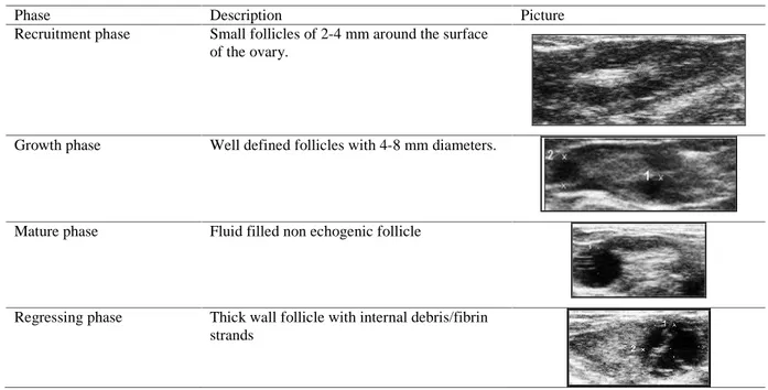
Table 1. Description and images of female dromedary camel ovarian follicular phases.
Phase Description Picture
Recruitment phase Small follicles of 2-4 mm around the surface of the ovary.
Growth phase Well defined follicles with 4-8 mm diameters.
Mature phase Fluid filled non echogenic follicle
Regressing phase Thick wall follicle with internal debris/fibrin strands
4. Pregnancy
The placenta of the dromedary camel is epitheliochorial, similar to that of mare and sow (Hafez, 1955; Van Lennep, 1961) and the maintenance of pregnancy, in camelids, completely depends on Corpus Luteum (Al-Eknah et al., 2001). The length of gestation has been reported to be about 13 months (average 385 days) (Matharu, 1966). The pregnancy is almost 99% located in left uterine horn. Double ovulation is frequent but twin parturitions are rare 0.4%. Longer left horn and development of satisfactory placental attachment, as well as embryo migration were suggested, as reasons for the left horn pregnancy, by Arthur and Al-Rahim (1982). Studies about the migration of the embryonic vesicle are missing in this species. 5. Post Partum period
The involution of the uterus is completed in about 40 days (Ahmed, 1990; Vyas and Sahani, 2000), and, as in other animals is significantly shorter in primiparous females; the lochial discharge ceases after 12.5±3.5 days (Elias and Cohen, 1986). The starting of follicular activity after parturition can vary according with the period of the year (Wilson, 1984), the breeding system and
2000). Calving and mating take place during the breeding season; females having birth are generally mated during the following breeding season thus giving an intercalving period of about two years or more (Minoia et al., 1992). The management of reproduction and adequate nutrition could help in maximizing the reproductive performance of camels (Wilson, 1986). Abdel Rahim and El Nazier (1990) reported intercalving periods lower than two years in camels bred in semi-intensive system. A “modern” approach to the dromedary camel management of reproduction and the application of assisted reproductive technologies could help in reduce the intercalving period and overcome specific breeding problems (i.e. the seasonality of males) (Fatnassi et al., 2014; Padalino et al., 2014). 7. Monitoring of ovarian activity
Behavioural (estrus) signs that made distinguishable females with a dominant follicle on their ovaries from other females, are rarely displayed in camels; consequently the monitoring of ovarian activity became, an essential step for the reproductive management of this species (Skidmore et al., 1996).
the back; through this restraining method the animal cannot rise (Arthur et al., 1985). Occasionally, some females need to be sedated with Xylazine (0.1-0.3 mg/kg b.w. i.v.) (Tibary and Anouassi, 1996).
The technique adopted for rectal palpation of the genital tract is similar to that described in the cow: the uterus should be grasped at the level of the uterine bifurcation and flipped dorsally so that each horn can be palpated from the base to apex in order to assess its width, length and consistency and any physiological or pathological variation noted (Tibary and Anouassi, 1996). Palpation of the ovaries is the most important part of the gynecological examination but sometimes they are very difficult to be found. On many occasions they are hidden beneath the uterus, so unless the uterus is retracted and rolled from side to side, they would be missed. The right ovary is always easier, than the left, to be found.
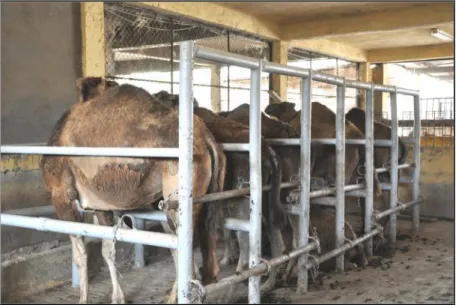
Ultrasonography significantly helps in assessing the phase of the dominant follicle, the Corpus Luteum development, ovarian irregularities and other pathological condition such as intrauterine accumulation of fluid, air or debris (Tinson and McKinnon, 1992; Tibary and Anouassi, 1996; 2000). Adapted stocks have been ideated for serial gynecological examinations of a large number of animals; protected chains are passed above and below the neck and beneath the flanks to stop it from rearing or sitting; the stock have also metal bars for preventing the examiner being kicked from the camel (McKinnon and Tinson, 1992), other kind of strokes have also been conceived considering the particular behavior of the animals to assume the sternal recumbence for avoiding the gynecological examination (Figure 1). Ultrasonographic examination can be performed using a real time, B-mode scanner equipped with a 5 MHz, or with a multifrequency, linear probe. The latter is manually inserted per rectum and the genital tract is scanned over all its parts starting from the cervix, then the uterine horn, followed by
the ipsilateral ovary. Recorded parameters could be the echotexture of the uterus (homogeneus-heterogeneus) and the amount of oedema (0 to 3 score). The ovary can be classified as quiescent (no structures) or active; ovarian structures, if present, can be measured using the electronic caliper (Tibary and Anouassi, 1996).
The follicular recruitment is characterized by the presence of multiple, small follicles (2-3 mm in size) arranged at the periphery of the ovary. Dominant follicles, like other fluid filled structures, are non-echogenic and appear as black, circular images protruding on ovarian surface. Regressing follicles can be identified for the increased echogenicity of their wall and some echogenic debris could be found within their cavity (Tibary and Anouassi, 2000).
During regression, the follicular fluid of overlarge follicles becomes more echogenic and free floating echogenic strands could be observed. Those echogenic strands becomes then organized into transecting strands of fibrin.
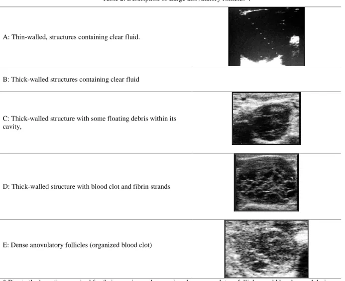
According to their ultrasonographical appearance, large oversized follicles have been divided into 5 categories: 1) thin-walled, structures containing clear fluid. 2) thick-walled structures containing clear fluid, 3) thick-walled structures with some floating debris within their cavity, 4) thick-walled structures with blood clot and fibrin strands within the cavity (hemorrhagic follicles) and 5) dense anovulatory follicles corresponding to organized blood clots (Table 2) (Skidmore et al., 1996; Tibary and Anouassi, 1996; 1997; 2000).
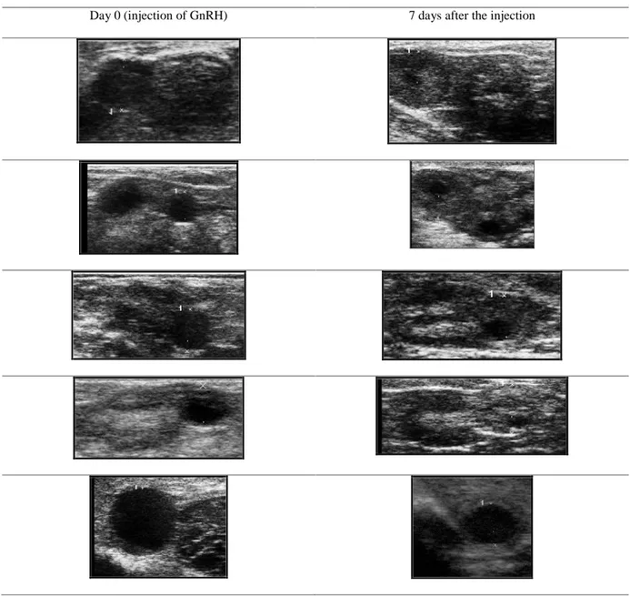
Ovulation could be diagnosed if the dominant follicle disappears 36-48 hours, following mating or injection of ovulation inducing treatments (GnRH o hCG); Corpora haemorragica, are usually very difficult to be seen (Tibary and Anouassi, 2000). The Corpus luteum appears as an uniformly echogenic round body that frequently exhibit a slight less echogenic center (Table 3) (Tinson and McKinnon, 1992).
Figure 1. Multiple strokes for serial gynecological examination of female dromedary camels (Desert Research Center, Alexandria, Egypt). As camels are gregarious animals this kind of stroke allow an easy training and management of
animals for the gynecological examination. Metal bars could be inserted below the female if the animal attempts to assume the sternal recumbence.
8. Artificial Insemination
Dromedary camels are induced ovulators therefore the synchronization protocols used for other domestic species are not effective in this species. This aspect could limit the application of AI, particularly if the latter has to be performed in large camel herds bred in semi intensive system. Recently, a new synchronization protocol has been proposed by Skidmore et al. (2009) and later implemented by Nagy and Juhasz (2012b). The method consists in performing a double injection of GnRH 14 days apart. In general, a single ovarian examination in a group of camels reveals the following animal categories: 10% of animals are found with the largest follicle being < 10 mm, 65% of the animals with largest follicle between 10-19 mm and 25% with the largest follicle measuring more than 20 mm (Nagy and Juhasz, 2012b). The percentage of females with the largest follicle of 10-19 mm would increase to 90%, 14 days after the 2nd injection of GnRH (Nagy and Juhasz, 2012b). This method would be a very useful tool for
synchronizing ovarian follicular wave and performing fixed time mating or AI on a large group of animals but providing that a sufficient number of bulls or semen doses (either fresh, cooled or frozen) are available for the timed matings or the inseminations.
For performing the AI, the semen should be deposited, through bovine or equine insemination catheters, just cranial to the cervix, into the uterus. Indeed, during mating, the semen is deposited partly into the uterus and partly intracervically, due to the particular twisting movement of the glans penis (Tibary and Anouassi, 1997). The cervix could be located by transvaginal approach (as AI performed in the mare) or by transrectal guiding of the insemination catheter. Skidmore and Billah (2006b) verified that a lower dose of sperm could be equally effective for obtaining pregnancies if the sperms are released near the Utero-Tubarian Junction, left or right, according to the position of the preovulatory follicle.
Table 2. Description of Large anovulatory follicles*.
A: Thin-walled, structures containing clear fluid.
B: Thick-walled structures containing clear fluid
C: Thick-walled structure with some floating debris within its cavity,
D: Thick-walled structure with blood clot and fibrin strands
E: Dense anovulatory follicles (organized blood clot)
* Due to the long time required for their growing and regression, large anovulatory follicles could be observed during any of the above described follicular phases
9. Use of extended semen
Pregnancy rates of 50% (5/10) have been achieved after diluting semen with an extender containing 11% lactose and 20% egg yolk. Similar pregnancy rates were achieved with Laciphos + 20% egg yolk (53%; 7/13), and Green Buffer + 20% egg yolk (47%; 10/21) (Skidmore et al., 2013). Recent studies have proved that it is possible to obtain 72 % pregnancy rates by using 1.5–3.5 ml of semen (150 x 106 spz/ml) extended with green
buffer plus + 20% egg yolk but providing that the Green Buffer extender have not been previously frozen, as the freezing process is deleterious for the preservation properties of this extender (Morton et al., 2011). Pregnancy rates could be possibly improved through the use of a higher concentration of sperms (300 x106 spz/ml). The semen (either fresh or chilled) should be deposited into the uterus at least 24 hours after induction of ovulation.
Table 3. Control of ovulation: Corpora lutea, developed 7 days after the injection of GnRH. Day 0 (injection of GnRH) 7 days after the injection
10. Use of chilled and frozen semen
Pregnancy rates (PR) of 17.6% have been obtained by depositing 150 x 106 motile sperms chilled in the INRA 96 extender. No pregnancies were obtained by using same sperm concentration but diluted with Green Buffer extender. Such results have been improved to 10.5% by using 300 x 106 motile sperms and to 25% with 600 x 106
but although 50% of spermatozoa were motile after cooling, this percentage was reduced to 35% immediately post-thaw and to 0% 3 h post thaw. El-Bahrawy, (2010) reported that tris-lactose containing a final concentration of 3% glycerol recorded the highest post-thaw motility (45.8%) with the highest survival rate (73.3%) but did not report pregnancy rates using such freezing-thawing protocol.
spontaneous ovulation or persistent Corpus Luteum or progesterone secreting cysts as well as also in young animals alarmed by the male. For the same above mentioned reason the assessment of pregnancies through the serum progesterone examination could provide erroneous conclusions (Skidmore et al., 2011). Positive pregnancy diagnosis can be achieved only if the CL and fetus are palpated (Skidmore, 2000). Uterine changes due to pregnancy can be detected by rectal palpation 45 day after mating and the first sign is an increase in the diameter of the left horn. At about three month of pregnancy, as the amount of fetal fluid increases, the gravid horn feels bigger and softer than the non-gravid horn and the uterus becomes more abdominal (Musa and Abusineina, 1978).
The pregnancy could be diagnosed as early as 17 days of gestation by ultrasonography. The diagnosis is based on two main criteria, the visualization of the embryonic vesicle and the presence of a Corpus Luteum (Tibary and Anouassi, 1997). On day 17 the embryonic vesicle appears, in the uterine lumen, as a star-shaped accumulation of fluid. Then after it increases in size and becomes more visible and elongated, in longitudinal view of the uterus, or more round in cross section. The embryo becomes visible, at about 20-22 days, as a small, echogenic speck within the fluid fixed at one pole of the vesicle; the heartbeat becomes discernible between days 23-25 as a small fluttering within the echogenic speck of the fetus (Vyas et al., 2002).
Acknowledgement
This document has been produced with the financial assistance of the European Union through the “PROCAMED” Project: Promotion des systèmes camelins innovants et des filières locales pour une gestion durable des territoires saharienne: reference number: I.B/1.1/493. The contents of this document are the sole responsibility of Veterinary Clinics and Animal Productions Unit D.E.T.O. Bari, Italy, and can under no circumstances be regarded as reflecting the position of the European Union.
Author Contributions
D. M., B. P. and G. M. L. equally contributed in the manuscript.
References
Abdel Rahim, S. E. A. and A. E. El Nazier. 1990.
Factors affecting camel reproductive
performance in the tropics. Proceeding of the Workshop: Is it possible to improve the reproductive performance of the camel?. ed.
Saint Martin. Paris 10-12 September.
Ahmed, M. S. H. 1990. Some studies on the postpartum period in she camels. Camel News Lett. 7:27.
Al-Eknah, M. M. and A. M. A. Ali. 2001. Infundibular cyst jeopardize reproduction in the camel (Camelus dromedarius). Emir. J. Agric. Sci. 13:52-56.
Arthur, G. H. 1992. An overview of reproduction in the camelids. In: Proceedings of 1st International Camel Conference. R & W Publications (Newmarket) Ltd., UK, pp. 109-115.
Arthur, G. H. and A. T. Al-Rahim. 1982. Aspects of reproduction in the female camel in Saudi Arabia. Vet. Med. Rev. 1:83-88.
Arthur, G. H., A. T. Rahim and A. S. Al-Hindi. 1985. Reproduction and genital desease of camel. Br. Vet. J. 141:650-659.
El-Bahrawy, K. A. 2010. Cryopreservation of dromedary camel semen supplemented with α-amylase enzyme. J. Cam. Pract. Res. 17:211-216.
Elias, E. and D. Cohen. 1996. Parturition in the camel and some behavioral aspects of the newborn. Comp. Biochem. Physiol. 84A:413-419.
Fatnassi M., B. Padalino, D. Monaco, T.
Khorchani, G. M. Lacalandra and M.
Hammadi. 2014. Evaluation of sexual
behavior of housed male camels (Camelus
dromedarius) through female parades:
correlation with climatic parameters. Trop. Anim. Health Prod. 46:313-321.
Hafez, A. S. A. 1955. Fetal maternal attachment in the buffalo and camel. Indian. J. Vet. Sci. 25:109-115.
Manjunatha, B. M., N. Pratap, S. Al-Bulushi, and B. E. Hago. 2012. Characterization of ovarian follicular dynamics in dromedary camels
(Camelus dromedarius). Theriogenology
78:965-973.
Manjunatha, B. M., N. Pratap and B. E. Hago. Acquisition of ovulatory capacity in camel (Camelus dromedarius) follicles. http://www. camelidconference.com/resources/dyn/files/19 1128/_fn/Manjunatha-Abstract-2011.pdf. Marie, M. and A. Anouassi. 1987. Induction of
the non-pregnant one-humped camel (Camelus dromedarius) J. Reprod. Fertil. 80:183-192. Matharu, B. S. 1966. Camel care. Indian Farming.
16:19-22.
Minoia, P., M. Moslah, G. M. Lacalandra, T. Korchani and A. Zarrilli. 1992. Induction of oestrus and Management of reproduction in the female dromedary camel. In: Proceedings of 1stInternational Camel Conference. R & W Publications (Newmarket) Ltd., UK, pp. 173-174
Morton, K. M., M. Billah and J. A. Skidmore. 2010. Artificial insemination of dromedary camels with fresh and chilled semen: effect of diluent and sperm dose, preliminary results. Proceedings of the Eighth International Symposium on Reproduction in Domestic Ruminants. Nottingham University press United Kingdom. p. 493.
Morton, K. M., M. Billah and J. A. Skidmore. 2011. Effect of green buffer storage on the fertility of fresh camel semen after artificial insemination. Reprod. Domest. Anim. 46:554-7.
Moslah, M. and F. Megdiche. 1989. L’elevage
cameline en Tunisie. Options
méditerranéennes serie A n°. 2:33-36.
Musa, B. E. and M. E. Abusineina. 1978. Clinical Pregnancy in the camel and a comparison with bovine pregnancy. Vet. Rec. 102:7-10.
Nagy, P. and J. Juhasz. 2008. Effect of different GnRH analogue and follicular size on ovulation and CL development in dromedary camels (Camelus dromedarius). Proceedings of 16th International Congress on Animal Reproduction (ICAR). p 95.
Nagy, P. and J. Juhasz. 2012a. Environmental factors affecting reproduction in dromedary camels (Camelus dromedarius). Proceedings of the ICAR satellite meeting on Camelid
reproduction. Publisher International
Veterinary Information Service (IVIS), Ithaca New York, USA. P 3-8.
1:103-109.
Padalino B., L. Aube’, M. Fatnassi, D. Monaco, T. Khorchani, M. Hammadi and G. M. Lacalandra. 2014. Could Dromedary Camels Develop Stereotypy? The First Description of Stereotypical Behaviour in Housed Male Dromedary Camels and How It Is Affected by Different Management Systems. PLOS One 9:1-7.
Skidmore, J. A. 1994. Reproduction in the dromedary camel. Ph.D. Thesis. University of Cambridge, UK.
Skidmore, J. A. 2000. Reproductive Physiology in Male and Female Camels. In: Recent
Advances in Camelid Reproduction.
Publisher: International Veterinary
Information Service (www.ivis.org) Ithaca, New York.
Skidmore, J. A. 2003. The main challenges facing camel reproduction research in the 21st century. Reprod. Suppl. 61:37-47.
Skidmore, J. A. 2011. Reproductive physiology in female Old World Camelids. Anim. Reprod. Sci. 124:148-54.
Skidmore, J. A. and M. Billah. 2006a. Investigation of the most appropriate time for insemination of female camels (Camelus dromedarius) after GnRH injection and comparison of pregnancy rates after deep intra-uterine versus cervical insemination. In: Proceedings of First Conference of International Society of
Camelids Research and Development
(ISOCARD), p. 54.
Skidmore J. A. and M. Billah. 2006b. Comparison of pregnancy rates in dromedary camels (Camelus dromedarius) after deep intra-uterine versus cervical insemination. Theriogenol. 66:292-296
Skidmore, J. A., M. Billah and W. R. Allen. 1996. The ovarian follicular wave pattern and induction of ovulation in the mated and non-mated one humped camel. J. Reprod. Fertil. 106:185-92.
Skidmore J. A., G. R. Starbuck, G. E. Lamming and W. R. Allen. 1998. Control of luteolysis in the one humped camel (Camelus dromedarius). J. Reprod. Fertil. 114:201-209.
Tibary, A. and A. Anouassi. 1997. Reproductive physiology in the female camelidae. In: Theriogenology in camelidae: anatomy, physiology, pathology and artificial breeding. IAVH II. Rabat, Morocco. p. 169-241.
Tibary, A. and A. Anouassi. 1996.
Ultrasonographic changes of the reproductive
tract in the female camel (Camelus
dromedarius) during the follicular cycle and pregnancy. J. Cam. Pract. Res. 3:71-90. Tibary, A. and A. Anouassi. 2000. Ultrasonography
of the genital tract in camels (Camelus dromedarius and Camelus bactrianus), In: T. K. Ghalot (Ed.), pp. 431-465. Selected topics on camelids, Bikaner, India.
Tinson, A. H. and A. O. McKinnon. 1992. Ultrasonography of the reproductive tract of the female dromedary camel. In: Proceedings of 1stInternational Camel Conference. R & W
Publications (Newmarket) Ltd., UK, pp. 129-135.
Van Lennep, E. W. 1961. The histology of the placenta of the one-humped camel (Camelus dromedarius) during the first half of pregnancy. Acta Morphl. Neerl. Sc. 4:180-193.
Vyas, S. and M. S. Sahani. 2000. Real-time ultrasonography of ovaries and breeding of the one-humped camel (Camelus dromedarius) during the early postpartum period. Anim. Reprod. Sci. 59:179-184.
Vyas, S., G. N. Purohit, P. K. Pareek and M. S. Sahani. 2002. Ultrasonographic imaging to monitor early pregnancy in the camel (Camelus dromedarius). Revue Élev. Méd. vét. Pays trop. 55:241-245.
Wilson, R. T. 1984. The Camel. Ed. Longmans, London, p. 83-101.
Wilson, R. T. 1986. Reproductive performance and survival of young one-humped camels on Kenya commercial ranches. Anim. Prod. 42:375-380.