Scuola Normale Superiore
Pisa
Role of Polarity and Polarized Secretion in Dendritic Cells Functions
Thesis submitted for the Degree of Doctor of Philosophiae
Academic year 2010/2011
Candidate: Jelena Petrovic
Contents
Introduction
1.1. Dendritic cells ...1
1.1. 1. Dendritic cells general features ... 1
1.1.2. Dendritic cells heterogeneity ... 3
1.1.3. Immature DCs ... 6
1.1.5. Role of DCs in priming adaptive immunity ... 10
1.2. Cytoskeleton and polarity proteins ...12
1.2.1. Cytoskeleton general features and role ... 13
1.2.2. Polarity proteins general features and role ... 15
1.2.2.1. Three polarity protein complexes - Par, Crumbs and Scrib ...15
1.3. Regulation of polarization ...21
1.4. Cytoskeleton and polarity proteins in immune system ...23
1.4.1. Cell migration ... 24
1.4.1.1. The role of cytoskeleton in T cells migration ...24
1.4.1.2. The role of polarity proteins in T cells migration ...25
1.4.1.3. DCs migration ...27
1.4.2. Immunological synapse (IS) formation ... 28
1.4.2.1. The role of cytoskeleton in T cells during IS ...29
1.4.2.2. The role of polarity proteins in T cell during IS ...31
1.4.2.3. DCs in synapse ...33
1.4.2.4. Polarized secretion ...35
Materials and Methods ...39
Results ...47
3.1. The functional aspect of MTOC polarization in DCs during synapse formation ...49
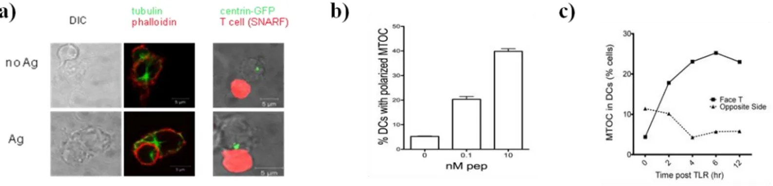
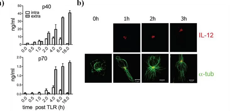
3.1.1. DCs polarize MTOC/IL-12 toward antigen-specific synapse with T cells ... 49
3.1.2. IL-12 signaling at the synapse ... 53
3.1.3. WASp as a putative model to follow MTOC polarization... 55
3.1.5. Assessing the effect of Cdc42-mediate MTOC polarization in DCs on the functionality of T cells ... 61
3.2. Further into polarity-Disc Large protein (Dlg1) ...65
3.2.1. Dlg1 localizes mainly in the membrane area in immature DCs ... 65
3.2.2. Dlg1 undergoes relocalization from membrane toward the protrusions in mature DCs ... 67
3.2.3. Dlg1 is phosphorilated upon TLR stimulation ... 68
3.2.4. Dlg1 polarizes toward the synapse in TLR and antigen dependent manner ... 70
3.3. Function of Dlg1 in DCs ...72
3.3.1. Dlg1 and migration ... 73
3.3.2. Dlg1 in synapse formation and T cell functioning ... 75
3.3.2.1. Assessing the functional consequences of Dlg1 depletion in DCs on T cells functions ... 76
3.3.2.1.1. CD69 expression and IL-2 production in T cells stimulated by Dlg1-depleted DCs ...76
3.3.2.1.2. INF-γ production in T cells stimulated by Dlg1-depleted DCs ...78
Discussion ...83
Conclusions ...99
Abstract
A timely and spatially well-organized utilization of polarity determinants appears to be an absolute functional prerequisite of all cells, including leukocytes. Numerous in vitro and in vivo studies performed in the field of DCs biology have strongly stressed the idea that DCs ascertain a correct immune response by finely tuning signal transmission within IS and by temporally regulating contact duration with T cells. In this study we elucidate another important aspect of DCs biology as we demonstrate that TLR-stimulated DCs preferentially secrete cytokines (IL-12) within the IS. We provide further insights into the mechanism of IL-12 delivery at the synapse area as we show that it strictly relies on microtubule organizing center (MTOC) reorientation toward antigen-specific synapse. On the molecular level, we find that the phenomenon of MTOC polarization in DCs is regulated by Cdc42 (a Rho GTPase) and Wiskott-Aldrich protein (WASp). Interfering with MTOC polarization had drastic impact on IL-12-dependent events occurring very early following synapse formation: pSTAT4 phosphorilation and IFN-γ production. At later time points, the absence of MTOC reorientation reflected in the decreased survival of T cells and strong reduction in their IFN-γ-producing capacity. Polarity proteins belonging to three main families (Par, Crb and Scrib), have been shown essential in ensuring polarized state of various cells. We find that in immature DCs, disc large protein (Dlg) is preferentially clustered underneath the membrane area whereas it undergoes relocation and hyperphosporilation following TLR stimulation. During DCs-T cell contact, we observed that Dlg clusters at DC-T cell interface in antigen-specific manner. Dlg depletion in DCs markedly reduced early, synapse-specific IFN-γ mRNA production by T cells; that reflected in a decreased IFN-γ production by differentiated T cells at alter time-points. Collectively, our results revealed that polarized cytokine secretion and Dlg clustering at IS are features acquired by DCs through their maturation program and provide a proof of principle that T cells fate is highly controlled by DCs.
1 | P a g e
1.1. Dendritic cells
The evolution of the immune system has been advancing toward the expansion in the number of available components and optimization of effector mechanisms, all with a goal of an improved protection against numerous pathogens in the environment. From the “traces” of an immune system in the form of phagocytes in Protozoa, via heightened specialization of the components of this system in Pisces, Amphibia and Reptilia, the evolution reached complex and pathogen specific immune system of Mammalia. Here, on the basis of functional criteria, this complex immune system can be divided into two distinct parts: innate and adaptive immune system. The components of the innate immune system (i.e. macrophages, neutrophilles, and system of complement) represent the first line of defense as they have a capacity to recognize pathogens and inhibit its further spreading by activating inflammatory reactions. However, the activation of a specialized and more efficient adaptive immune response is essential for complete protection from pathogens. Dendritic cells (DCs) represent a link between innate and adaptive immune response owing to their ability to recognize pathogens within the innate immune response and inform the components of adaptive immunity about the presence of infection by presenting pathogen-derived antigens (Ag) to T lymphocytes; a process necessary for adaptive immune response activation. Thus activated, the constituents of adaptive immune response clear the infection by either direct killing of infected cells (cytotoxic T lymphocytes, CTL), by opsonizing pathogens with antibodies (B lymphocytes) thus making them more susceptible to the action of phagocytes and by secreting soluble mediators (i.e. cytokines) that are crucial for spatio-temporal orchestration of immune response (helper T cells, Th) (Steinman and Hemmi, 2006).
1.1. 1. Dendritic cells general features
For proper immune response awakening, the internalization of antigens (Ag) and their presentation to T lymphocytes within immunological synapse (IS) is necessary. The cells bestowed with this function are called antigen presenting cells (APC). By many criteria, DCs represent the most efficient APC of the immune system (Itano and Jenkins, 2003).
DCs are a heterogeneous group of leukocytes originating from bone-marrow precursors. Within the first phase of their life, DCs (so called immature DCs) are strategically situated within
2 | P a g e
epithelial tissues at the organisms’ frontiers, where they constantly patrol the environment for the presence of pathogens, owing to the expression of many receptors (PRRs, pattern recognition receptors) recognizing the evolutionary conserved patterns within pathogens (PAMPs, pathogen-associated molecular patterns). This strategical function, coupled with their high intrinsic capacity to internalize recognized microbes via endocytic/phagocytic pathway, makes them extremely predisposed for fast antigen recognition.
Once the invader is internalized, DCs enter into integrated developmental program called maturation. Within this program, DCs downregulate their endocytotic capacity and upregulate the expression of cytokines, the expression of molecules necessary for antigen presentation (MHC), and co-stimulatory molecules (CD80, CD86 and CD40) necessary to optimally activate T cells. Furthermore, these cells undergo a global redistribution of their actin cytoskeleton resulting in the acquirement of a migration capacity. Further changes in the expression of chemokine receptors and adhesion molecules induce migration of these pathogen-carrying DCs toward proximal lymph nodes (LN) where they present antigens to naïve T cells.
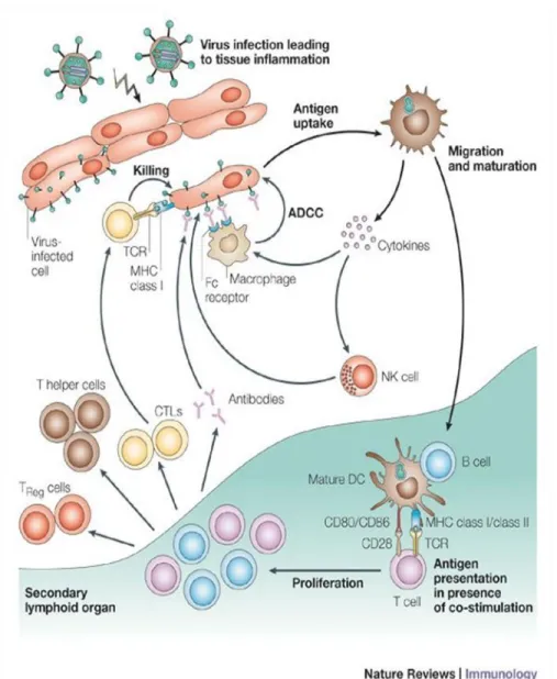
Within IS, DCs (mature DCs) adopt specific morphology and engage the adhesion molecules on T cell side necessary for the synapse preservation. At the same time, they present degraded pathogen sequences to naïve CD4+ and CD8+ T cells, inducing their activation and differentiation toward a specific T helper cell (Th1 or Th2) or cytotoxic T cells (CTL) lineages, respectively (Reis e Sousa, 2004; Steinman and Hemmi, 2006) (Figure 1).
3 | P a g e
Figure 1. Dendritic cells (DCs) lifecycle. DCs are generally situated within the peripheral tissues where they have a pivotal role in recognizing the invading antigens. Once the antigen is recognized and internalized, DCs produce a high level of inflammatory cytokines essential to induce recruitment of cells specialized to induce inflammatory reactions (macrophages and neutrophilles) or direct killing of infected cells (NK cells). Concomitantly, DCs migrate toward a secondary lymph node where they present antigen-derived sequences to naïve T cells. This process initiates a cascade of events leading to activation and differentiation of T cells. Thus activated, T cells clear the infection by direct killing of infected cells (CTLs) and by secreting soluble mediators (cytokines) necessary to recruit other members of adaptive immune system (B cells) and to potentiate anti-pathogen activities of other cells (macrophages, DCs) (Th cells).
1.1.2. Dendritic cells heterogeneity
Currently accepted paradigm regarding DCs life cycle, suggests four stages of maturation
i.e. functional characteristics of DCs: (i) progenitors from bone marrow (ii) DCs precursors (preDCs),
which patrol through blood and lymphaticus and, upon contact with antigen differentiate into corresponding DCs (iii) immature DCs, localized within peripheral tissues, with great endocytotic and phagocytotic capacity that enables them to recognize and ingest antigens (iv) mature DCs, localized in the secondary lymphoid tissues, with a function in antigen presentation (Banchereau et al., 2000; Palucka and Banchereau, 1999)
4 | P a g e
The scheme given above is a representative of a general view of DCs life cycle, mostly known as Langerhans cell paradigm. In the light of this, however, one may think that the pathway from the pluripotent stem cell to the development of mature DC would be a straightforward process. However, a huge functional and phenotypical diversity of DCs found in lymph nodes and spleen challenges this simplistic theory of DCs ontogeny, reporting a vast degree of heterogeneity in all aspects of DCs life cycle: (i) The precursors population. DCs originate from both myeloid and lymphoid lineages. (ii) Function. DCs are responsible both for the activation of the immune response as well as for the induction of tolerance. (iv) Anatomical localization. DCs can be found in the peripheral tissues (Langerhans DCs, interstitial DCs), in spleen, in lymph nodes (interdigitate DCs), in liver and in blood (blood-derived DCs) (Banchereau et al., 2000; Naik et al., 2007; Shortman and Naik, 2007; Villadangos and Heath, 2005). There have been several trials to classify DCs according to various criteria that are still debated. In very general terms, DCs can be divided into two major groups: (i) non-lymphoid migratory DCs and lymphoid tissue resident DCs (ii) plasmacytoid DCs (pDCs) (Merad and Manz, 2009).
Non-lymphoid migratory DCs are mainly strategically localized in those tissues where
they can quickly recognize the invading pathogens. Thus, they are found within tissues on the interface with the environment (lung, gut and skin) and at big filtering sites (liver and kidney). The prototype of this big family of DCs are Langerhans DCs (LC). They are easily distinguished by the surface expression of C-type lectin langerin and by LC-specific intracellular organelles known as Birbeck granules. Phenotypically, they are recognizable by a constitutive expression of major histocompatibility (MHC) class II complex and by low levels of CD11b. Furthermore, these cells express CD103 molecule, a ligand for the adhesion molecule E-cadherine expressed on most epithelial cells, which enables their retention within the tissues. LCs are discovered by Steinman in 1978 and Langerhans cell paradigm was postulated according to their functional characteristic (Gallegos and Bevan, 2006; Ginhoux and Merad, 2010).
Lymphoid tissue resident DCs are the most studied population of DCs. They are a very
heterogeneous population that can be divided, based on the combination of phenotypical markers they express, in three major categories: CD4+CD8-CD11b+, CD4-CD8+CD11b-, and double negative CD4 -CD8-CD11b+cells. CD4+ DCs are mainly localized at the marginal zone and are specialized in MHC II presentation. CD8+ DCs are situated in T cell rich zones and were shown to be able to cross-present cell-associated antigens. The lymphoid resident DCs do not conform to the Langerhans cell paradigm as
5 | P a g e
they develop within the lymphoid organs directly from the precursors, without prior passing through peripheral tissues. In the absence of infection, these cells retain immature phenotype and are most likely involved in the maintenance of central tolerance. However, upon contact with pathogens, lymphoid DCs do acquire mature phenotype, secrete big amounts of inflammatory cytokines and migrate toward T cell rich zones. They have a role in activating adaptive immune response against blood borne antigens arriving at LN and spleen (den Haan et al., 2000; Wilson et al., 2003)
Plasmacytoid DCs were identified for the first time in human blood and lymphoid tissues.
Due to their morphological similarity with plasma cells and expression of some T cell markers, they were initially named plasmacytoid T cells or plasmacytoid monocytes (Wu and Dakic, 2004). Later it was discovered that, following pathogen recognition, these cells obtain the ability to release high amounts of IFN-α and IFN-β; for this characteristic they were also named IFN-producing cells (IPC) (Colonna et al., 2004; Siegal et al., 1999). pDCs are specialized to combat against viral infections due to the expression of specific set of PRRs (TLR7 and TLR9), that recognize patterns within viral nucleic acids and stimulate the cascade of intracellular events resulting in IRF-7 induced production of IFN type I cytokines. In addition, viral recognition induces maturation of pDCs, visible in the upregulated expression of MHC class II molecules, co-stimulatory molecules (CD80, CD86 and cD40) and release of proinflamatory cytokines (IL-6 and TNF-α) (Dai et al., 2004).
Finally, DCs can be differentiated in vitro (in vitro generated DCs) owing to the existence of well-established methods. The first method reports that DCs can be generated from bone-marrow or spleen precursors in medium supplemented with granulocyte-monocyte colony stimulating factor
(GM-CSF) with or without interleukin-4 (IL-4). Thus obtained cells are partially similar to some lymphoid
organ resident DCs, as determined by phenotypical markers examination (Shortman and Naik, 2007). A more precise way to differentiate lymphoid organ-like DCs from bone-marrow precursors is by supplementing the medium with FMS-like tyrosine kinase 3 ligand (Flt3L). These DCs expressed phenotypical and functional characteristics equivalent to CD8+ and CD8- DCs: CD11b, CD24 surface marker expression; expression of mRNA for TLRs and chemokines receptors; the ability to produce cytokine (IL-12 and IFN) following TLR engagement and the ability to cross-present antigens (Naik et al., 2005).
6 | P a g e
1.1.3. Immature DCs
Immature DCs express sets of chemokine receptors (CCR1, CCR2, CCR4, CCR5, CXCR1 and CXCR4), which enable their specific localization within the peripheral epithelial tissues (Banchereau et al., 2000) The term immature is related to the incapacity of these cells to induce T cells proliferation, but they have a great capacity to recognize and engulf pathogens (Reis e Sousa, 2006). Immature DCs employ three major endocytotic mechanisms of pathogens internalization: phagocytosis, macropinocytosis and receptor-mediated endocytosis.
Antigen internalization
Phagocytosis. Phagocytosis represents most prevalent mechanism for pathogen
engulfment. One of the reasons for that probably lies within the fact that this mechanism is used for multitude of processes. For instance, it includes receptor (FcγR, complement and lectin) mediated phagocytosis of extracellular pathogens; it is used as a main mechanism for sampling and presentation of the apoptotic bodies within the steady-state conditions (and thus tolerance maintenance); it is employed in sampling foreign antigens by engulfing the cells died from the infection; it enables inert particles and liposomes uptake (Trombetta and Mellman, 2005).
Once recognized, pathogens are uptaken through a formation of cytoskeleton-based membranous protrusions, wrapping in the pathogen within a vesicle. A special sophistication of this mechanism lies within a fact that different regulatory pathways and different types of membranous protrusions are activated depending on the nature of pathogen being internalized. Thus, phagocytosis through FcγR mediation induces Cdc42-dependent formation of pseudopodes while Rac activation is necessary for phagosome closure. On the other hand, complement mediated phagocytosis is Rho GTPase dependent. It does not include the formation of pseudopodes but rather involves integrin mediated ligation for the formation of phagosomes (Symons and Rusk, 2003).
Macropinocytosis. As its name suggests (from Greek pinein-“to drink”), this type of endocytic process is involved in sampling soluble antigens by uptaking huge amounts of fluidic fractions. The base of this process is Cdc42-regulated formation of huge membranous protrusions, encircling a big proportion of extracellular fluids and their fusion with the membrane. It is a constitutive process within immature DCs and is downregulated upon maturation (Conner and Schmid, 2003; Symons and Rusk, 2003).
7 | P a g e
Receptor mediated endocytosis. Mechanistically, receptor mediated endocytosis could be
seen as the further refinement of general process of macropinocytosis. It is involved in sampling soluble antigens, but the presence of very sensitive pathogen-pattern specific receptors enables the cell to recognize pathogens that are present in extremely low concentrations. For that purpose DCs express the array of receptors able to perform endocytosis of antigens: (i) Receptors that recognize Fc region of immunoglobulins (FcRs) and complement receptors (CRs) are used to internalize immunoglobulin opsonized antigens (Fanger et al., 1996). (ii) C type lectin receptor that recognizes carbohydrate remnants on the surface of many pathogens (Steinman and Hemmi, 2006) (iii) a few of not well characterized receptors (CD36, αβ3 or αvβ5 integrins) for internalization of apoptotic and necrotic cells (iv) Scavenger receptors used to recognize low density lipoproteins (LDLs) (Delneste et al., 2002).
Antigen processing and presentation
Upon internalization, antigens are degraded into small immunogenic epitops that associate with MHC and are transported to the plasma membrane where they trigger the activation of naïve T cells. Generally accepted view is that exogenous antigens are presented within MHC class II (MHC II) complex while endogenous antigens are presented within MHC class I (MHC I), activating CD4+ and CD8+ T cells, respectively.
Specifically, MHC II are constantly being synthesized in immature DCs and accumulate in the form of trimer with the invariant chain (Ii) within specific, multilamelar structures, called MHC II
rich compartments. These structures are involved in the endocytic pathway as they fuse with endocytic
vesicles carrying exogenous antigen. The presence of Ag causes the exchange of the invariant chain with the antigen and its presentation on the DCs surface. Immature DCs express low levels of short-lived MHC II molecules on their surface. However, upon antigen recognition, MHC II-Ag complexes are stabilized and remain on DCs surface for couple of days, thus giving the time to rare Ag-specific CD4+ T lymphocytes to recognize them and initiate a series of proliferation and differentiation processes (Trombetta and Mellman, 2005).
On the other side, intracellular Ags are processed within MHC class I pathway. In short, endogenous antigens are ubiquitinated and, following the transport through proteaosome, degraded to a certain length (6-30 amino acid long), characterized by the presence of basic or hydrophobic amino acids on their carboxyl terminus. Oligonucleotides of this length and specificity are recognized by transporter associated with antigen presentation (TAP), which translocates them inside endoplasmic
8 | P a g e
reticulum (ER) where they associate with de novo synthesized MHC I dimer. Exocytotic vesicles carrying these trimers bud from the ER and fusing with plasma membrane, enable the presentation of antigens within MHC I complexes. Thus expressed antigens are being recognized by CD8+ T lymphocytes (Rock et al., 2004; Trombetta and Mellman, 2005).
However, this general view has been very shaken by the discovery that DCs can present exogenous antigens within MHC class I pathway (a process termed cross-presentation) very efficiently. Cross-presentation has been shown to enable the activation of CD8+T cells in response to either bacterial or viral stimulus. Furthermore, cross-presentation was shown to be important against pathogens which do not infect DCs (den Haan and Bevan, 2001) and in the presentation of self antigens; coming from the cells that died by apoptosis as a part of normal cellular turnover (necessary for peripheral tolerance maintenance) (Heath and Carbone, 2001) . However, mechanisms that DCs employ in order to perform this function is still elusive. It has been suggested to involve exogenous antigen “escape” from the phagosomes and its leakage into the cytosol; where it is recognized as foreign by the components of MHC class I-pathway (Monu and Trombetta, 2007).
1.1.4. Pathogen recognition and TLRs
At the same time that pathogen is being recognized and internalized by endocytotic machinery, another group of molecules expressed by immature DCs recognizes antigens. These molecules belong to pathogen recognition receptors family (PRRs), as they recognize the evolutionary conserved sequences invariably present on the surface of every pathogen (PAMP, pathogen associated
molecular pattern) (Akira et al., 2001).
The best described group of PAMPs is the family of Toll like receptors (TLR). This family consists of about 12 known receptors that are different in their pattern of expression as well as in their specificity for the pathogens. Thus, some of them (TLR1, 2, 4, 5 and 6) are expressed on the cell surface where they mainly recognize bacterial products, while others (TLR3, 7, 8 and 9) are located on the membranes of the endocytic vesicles and are specialized in recognition of viral products. For instance, TLR4 is shown to recognize lipopolysaccharides (LPS), a major component of Gram- bacteria wall; TLR2 recognizes bacterial peptidoglycans and lipoproteins; TLR5 recognizes bacterial flagellin; TLR7 and TLR9 recognize CpG-rich islands of bacterial and viral DNA (Matzinger, 2002).
9 | P a g e
Furthermore, the complexity is increased by the ability of some of these receptors to form heterodimers, thus increasing the sensitivity of the response (Takeda et al., 2003).
Following pathogen recognition, TLRs were shown to be able to activate two independent signaling pathways. The first pathway relies on the association with an adaptor molecule, MyD88, via its C-terminal domain. On the other side, Myd88 engages a kinase IRAK, whose consequent phosphorilation induces further association with TRAF6. The final outcome is the activation of two different signaling cascades: JNK and NF-κB. The second pathway evoked by TLR-mediated antigen recognition also results in the activation of NF-κB signaling pathway; however it involves a different adaptor molecule, namely TRIF (that further activates RIP1 and TRAF6). At the same time, however, TRIF was shown to induce the expression of IFN type I genes as it associates with two kinases, TBK1 and IKKi who further mediate phosphorilation-dependent activation of IFN-regulatory factor 3 (IRF3) ((Janeway and Medzhitov, 2002; Trinchieri and Sher, 2007).
From their side, these transcriptional factors ensure a complete turnover in DCs genetical program. Dendritic cells lose their capacity to internalize antigens due to a decrease in the expression of receptors performing antigen uptake and downregulation of endocytotic activity (Garrett et al., 2000). The expression of genes for cytokines, that indirectly induce inflammatory reactions by recruiting specific cells of the innate immunity (i.e. macrophages, neutrophilles,) is enhanced. These cytokines are also secreted later within immunological synapse where they are crucial for differentiation of T cells toward a specific linage. Furthermore, the expression of genes for co-stimulators (CD40, CD80 and CD86) is augmented, whose function in ensuring signal amplification is essential for the proper presentation of antigens in IS (Janeway and Medzhitov, 2002). Finally, inflammatory stimuli turn off DCs’ response to those chemokines responsible for DCs retention within tissues, by negative autocrine effect or by receptor desensitization (Sallusto et al., 1998). At the same time, the expression of another group of chemokine receptors, namely CCR7, is upregulated, enabling these cells to respond to corresponding CCL19 and CCL21 chemokines. Taking into account that these chemokines are expressed in T cell zones of lymph nodes, the change in chemokine expression profile allows DCs to leave peripheral tissues and migrate in the direction of chemokine gradient- i.e. toward lymph nodes (Alvarez et al., 2008; Dieu et al., 1998).
10 | P a g e
1.1.5. Role of DCs in priming adaptive immunity
At present, the idea that the transmission of the signal between Ag-bearing DCs and T cells occurs during their contact in the lymph node via a specialized structure called immunological synapse (IS), is a well established paradigm. Later in vivo studies have further revealed the complexity of the interactions and demonstrated that a refined and dynamical interplay between DCs and T cells is a prelude for the formation of informational synapse (Bousso, 2008). Once the stable contact is formed, DCs deliver three types of signals that directly influence the outcome of the immune response. The first signal occurs upon interaction between TCR and MHC-bearing antigen which secures Ag-specific response. The second signal consists of the engagement of co-stimulatory molecules on DCs side by either CD28 or CTLA-4, which induces activation or inhibition of T cell activation, respectively. The third signal is mirrored in the combined effect of various cytokines secreted by DCs and integrated by T cell as the signals for proliferation and differentiation toward specific subsets (Kalinski et al., 1999b; Villadangos and Schnorrer, 2007).
The direct cause of T cell fate determination is given by the integral message of cytokines (“third signal”) being secreted toward T in synapse with DCs. However, the decision regarding the nature of immune response being awakened has been decided much earlier, upon first contact of DCs with the pathogen. Specifically, pathogens engage a specific combination of TLRs present on the surface of immature DCs which further signal DCs to enter into a certain program of maturation, each of which results in the specific combination of cytokines being produced. Based on the type of antigen being recognized, DCs may prime the differentiation of T cells toward Th1, Th2, Th17 or Treg lineages (Reiner, 2007).
Th1 subset has specialized for the defense against intracellular bacteria and viruses. The activation of TLR3, TLR4, TLR7 or TLR9 on DCs surface induces the activation of DC maturation program characterized by the production of IL-12 cytokine. This cytokine induces IFN-γ production in T cells through the activation of special combination of signaling molecules (JAK2) and transcriptional factors (STAT4). Once produced, IFN-γ further polarizes the response toward Th1 dominated response by enhancing further IL-12 production in DCs and by sensitizing activated T cells to the effect of IL-12 (by upregulating the expression of IL-12β2R subunit on their surface) (Reiner, 2007; Szabo et al., 2000). Th2 dominated response is specialized for the defense against helminthes and various allergens. TLRs being engaged by these microbes are still unknown, while the outcome of their
11 | P a g e
recognition by DCs is manifested in high production of IL-4. IL-4 induces the polarization of activated T cells toward Th2 dominated response by upregulating the expression of a transcriptional factor GATA3. Targets of this transcriptional factor are genes for IL-4 and IL-5 cytokines. These cytokines cause a complete polarization toward Th2 pathway by suppressing the expression of factors critical for the development of Th1 response.
Th17 are fairly recently discovered subtype of CD4+ T cells that have probably evolved to enhance the clearance of pathogens distinct from those targeted by Th1 and Th2. Their role has been implicated in defense against extracellular bacteria (Klebsie pneumoniae) and fungi (Candida albicans). The receptors being engaged by DCs in the presence of these pathogens is largely unknown, but initial reports by Reis e Sousa implicate the activation of dectin-1-Syk-CARd9 signaling pathway. DCs that recognize these pathogens secret a specific combination of cytokines having a role in inducing inflammatory reaction 6 and TNFα) or polarizing Th response toward a Th17-dominated one (IL-23). The necessity of regulatory T cells (Tregs) and TGFβ in the polarization of the response toward Th17 dominated lineage has also been reported. This is in line with the reports suggesting that IL-6 and TGFβ are necessary to induce the activation of Th17 cells, while the presence of IL-23 secreted by DCs induces complete polarization of this response (Huang et al., 2004; Reiner, 2007; Weaver et al., 2006).
Regulatory T cells (Treg) develop from naïve CD4+T cells through their contact with DCs that have previously been exposed to cytokines IL-10 and TGFβ. The development of Tregs is associated with the maintenance of peripheral tolerance through a suppression of unwanted adaptive immune response and autoimmunity prevention. Regulatory T cells that develop at the periphery through DC-mediated contact (so called adaptive T reg) are different than naturally occurring Tregs developing in thymus. Within the population of adaptive Tregs, two main, functionally different populations can be distinguished. Treg1 develop upon the contact with IL-10-conditioned DCs and are characterized by the lack of transcription factor Foxp3 and production of large amounts of IL-10. Treg2 develop as the result of TGFβ signaling, they express Foxp3 and are functionally indistinguishably from intrathymical Tregs (Hall et al., 2011; Reiner, 2007) (Figure 2).
12 | P a g e
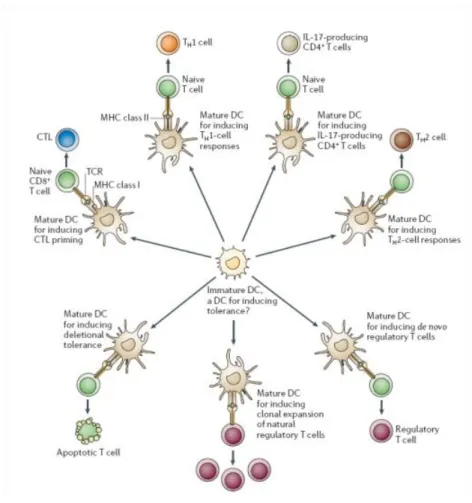
Figure 2. Dendritic cells effector functions. DCs maturation program is a process where DCs acquire phenotypical and functional characteristics necessary to present internalized antigens to naïve T cells. However, the outcome of DCs priming is very heterogeneous as DCs can potentiate the development of various lineages of T cells. This capacity of DCs depends on the type of pathogens being recognized by DCs, the environmental clues or DCs ontogeny (predisposing a certain pool of DCs toward a specific pathway). A direct cause of diverse T cell fate during priming by DCs relies on different sets of cytokines being secreted by DCs in response to various pathogenic circumstances promoting DCs maturation (Reis e Sousa, 2006).
1.2. Cytoskeleton and polarity proteins
At present, an emerging concept indicating that spatio-temporal interplay between numerous polarity players shapes cells’ destinies and ensures their function becomes more evident. Current knowledge regarding polarity determined processes within cells has helped divide polarity determinants into arbitrary five categories actin cytoskeleton 2) microtubule cytoskeleton 3) surface proteins (receptors and lipid rafts) 4) secretion pathways (endo- and exocytosis) 5) polarity proteins complexes. In the following few chapters we will try to briefly communicate how dynamical changes of
13 | P a g e
cytoskeleton and mutual distribution of polarity proteins enable various aspects of cell functioning (Krummel and Macara, 2006).
1.2.1. Cytoskeleton general features and role
Whether stationary cells (e.g. epithelial cells), highly motile cells (e.g. neutrophilles, fibroblasts, astrocytes), cells with high proliferative capacity (e.g. embryonic stem cells) or the cells forming the synapse (e.g. lymphocyte, dendritic cells, neurons); all functions of these cells depend on the presence of stable and dynamic cytoskeleton architecture at the same time. This cytoskeleton networks needs to be firm in order to ensure consistency of the cellular processes in time; and it needs to be dynamic in order to meet cellular morphological prerogatives in response to various clues from the environment.
All cells studied possess three types of cytoskeleton elements: actin, microtubules and intermediate filaments. So far, the role of intermediate filaments has not been implicated in polarity processes regulation, while actin and microtubules have long been known for their role in supporting functional dynamism of cells. Moreover, until recently, it has been thought that actin and microtubules react independently to changes in intracellular and extracellular milieu; however, recent advances have made it clear that adequate cellular functioning is a consequence of integrated action of these two networks (Banerjee et al., 2007; Kodama et al., 2004; Rodriguez et al., 2003).
Actin is usually present within cells in the form of F-actin (filamentous actin), a polymer composed of only one type of monomer, G-actin (globular actin). The process of actin nucleation depends on the presence of Arp2/3 complex that stimulates G-actin incorporation. Cycle of actin polimerization and depolimerization, regulated by ATP hydrolysis and assisted by accessory proteins enable the cells to respond to various signals by obtaining adequate morphology. Microtubule (MT) network is preferentially structured as a polymeric network of α and β-tubulin spanning radially from a core consisting of two centriols and pericentriolar material (PCM; consisting mainly of γ-tubulin monomers). This central structure was termed microtubule organizing center (MTOC) also known as
centrosome. MT polimerization from this center is characterized by growth of one end (plus end) of
MTs, while the other end remains attached to MTOC (minus end) (Gundersen, 2002; Rodriguez et al., 2003).
Actin and MTOC have been implicated, as mentioned, in various cellular functions. For instance, during cell migration, changes in the composition in extracellular environment induce actin
14 | P a g e
recruitment toward cell periphery and MTOC reorientation toward one pole of the cell. Actin subcortical clustering is crucial for cellular migration as it ensures a stability of cells’ migration shape and it represents a base of membrane protrusions e.g. filopodia, lamellipodia (Hall, 1998); while MTOC translocation toward one pole is important to bring the directionality of the movement (Kodama et al., 2004). Moreover, adequate spindle orientation is important for the proper segregation of fate determinants and consequential asymmetrical division during D.melanogaster and mouse neurogenesis (Knoblich, 2008). In mouse embryonic hippocampus neurons, polarization of MT and actin cytoskeleton within one of the numerous membrane protrusions (minor processusses) predestines it to become axon (Arimura and Kaibuchi, 2007). Finally, apico-basal polarity of epithelial cells is a result of junction complexes being held firmly in their places by a meshwork of actin cytoskeleton (Knust and Bossinger, 2002) (Figure 3).
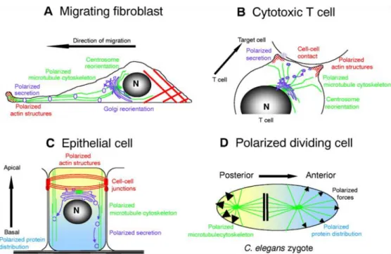
Figure 3. Role of cytoskeleton in various cells. a) In motile cells, actin cytoskeleton polarization toward the leading edge enables formation of membranous protrusions (lamelopodia and filopodia); while centrosome positioning in the rear end probably contributes to the directionality of the movement. b) Polarized secretion of soluble mediators in cytotoxic T cells is ensured by centrosome polarization toward the synapse region. Docking of plus-end MTs at the outskirts of synapse represents a pathway for vesicle transport. c) Functionally important apico-basal polarity of epithelial cells is a result of junction complexes held together by the inner actin cytoskeleton network. MTOC (centrosome) has a function in directing polarized secretion of vesicles toward the specific region of epithelial cells. d) In dividing C.elegans zygote, spindle positioning establishes the axis of polarity, ensures unequal distribution of other fate determinants and enables asymmetrical cell division (Etienne-Manneville, 2004).
15 | P a g e
1.2.2. Polarity proteins general features and role
First results considering phenomenon of polarity came about the end of the 19th century but they were restricted to the mere morphological descriptions of asymmetrically distributed components within cells and tissues. With the first postulated model on the topic of asymmetrical cell divisions in ascidian egg back in 1905 (Conklin, 1905), the idea that the relative distribution of proteins within a cell may play an important role in cell destiny came to light. It was not until the end of 20th century, however, that the first determinant of segregation was molecularly characterized in the sensory organ precursors (SOP) in peripheral nervous system of D.melanogaster (Rhyu et al., 1994). Asymmetrical inheritance of cell fate determinants was then also shown to induce emergence of diverse embryonic tissues in C.elegans (Betschinger and Knoblich, 2004). Studies in mammalian cell systems have demonstrated that asymmetry seems to be a common characteristic of all multicellular organisms, rather than the exception (Horvitz and Herskowitz, 1992). For instance, the presence of asymmetrical division is evident also in mouse zygote (Knoblich, 2008); formation of transient polarity modules represents the base of cellular locomotion (Etienne-Manneville, 2008); polarized clustering of specific proteins in the region of immunological synapse ensures proper signal transmission between the cells (Krummel and Macara, 2006).
Further reports aimed to understand the mechanisms and players involved in establishing cellular polarity were mainly performed on epithelial tissues. Intensive research in this field has revealed the existence of evolutionary conserved group of proteins with a role in ensuring apico-basal organization of cells. Following these observations the proteins were classified within specific polarity complexes and the term polarity protein was created.
1.2.2.1. Three polarity protein complexes - Par, Crumbs and Scrib
PAR complex
The name PAR stands for partition defective, the term that crystallized itself upon early works in C.elegans, where it was shown that mutants lacking par genes experienced defects in division patterns during C.elegans development (Kemphues et al., 1988). In the same work, genetic analysis revealed six par genes (i.e. proteins) responsible for this phenomenon: par1, par2, Par3, par4, par5 and
par6. Through sequence alignment it was observed that C.elegans Par6, D.melanogaster dmPar6 and
16 | P a g e
demonstrated that C.elegans Par3 protein is a D.melanogaster protein Bazook (one of the main players that leads to the proper asymmetrical division in D.melanogaster) homologue (Kuchinke et al., 1998). The addition of another protein, aPKC, shown to be physically and functionally connected with Par proteins in D.melanogaster epithelia, formed the Par trilogy complex known as Par3/Par6/aPKC complex (Suzuki et al., 2001).
General mechanism of action: Although the meaning of polarity diverges with the respect
to cell types and organisms involved, the general mode of Par protein functioning seems to be preserved. This protein complex, in invertebrates as well as in vertebrates, has been shown to contribute to the establishment of epithelial cell polarity, through its role in apical domain and zonula adherens (ZO) formation. In these cells, Par6 seems to have the initial role in scaffolding other proteins (Joberty et al., 2000; Suzuki and Ohno, 2006). Specifically, Par6 receives a message regarding the change in polarity cues through the action of a small RhoGTPase-Cdc42. This signal is then translated into inner polarization signal by Par6 binding to aPKC (Joberty et al., 2000; Lin et al., 2000). Interestingly, inactive aPKC is found associated with a protein from another polarity complex, Lgl (lethal giant
larvae) (Plant et al., 2003). Par6 binding induces aPKC phosphorilation-dependent dissociations from this complex and its adherence to Par3. Initially, this association induce the formation of the final Par6/Par3/aPKC complex (Yamanaka et al., 2006; Yamanaka et al., 2003). However, at the same time, Par3 phosphorilation by aPKC induces the loss of Par3 affinity for the complex and its disassociation (Nagai-Tamai et al., 2002). This is in line with some later reports stating that Par3 and aPKC/Par6 complex are not always found co-localizing in epithelial cells (Harris and Peifer, 2005). Moreover, it appears that exactly this dynamical association between the members of this complex is fundamental for adequate polarity pattern development of epithelial cells. Specifically, Par6/Par3/aPKC association was shown to be important for the formation of apical domain in epithelial cells, whilst its dissociation is important for the tight junction formation (Horikoshi et al., 2009; Nagai-Tamai et al., 2002). Overall, this prototype of temporary connections between Par polarity complex proteins enabling cell morphology has found its proof of principle in various cell types (McCaffrey and Macara, 2011).
Crumbs (Crb) complex
This complex originally got its name from morphological appearance of D.melanogaster cuticle, resulting from the absence of the gene crumbs in the epithelial cells underneath that secrete the cuticle (Tepass et al., 1990). Further studies on the nature of this protein revealed that Crumbs is
17 | P a g e
responsible for apical domain maintenance of epithelial cells in D.melanogaster, as its overexpression in
crb mutants restored the apical phenotype (Wodarz et al., 1995). Afterwards, functional colocalization
of Stardust (Std) and Crumbs in the adequate organization of the apical domain of epithelial cells was demonstrated (Bachmann et al., 2001; Hong et al., 2001). Finally, a third protein whose depletion induced complete perturbation of the epithelial cell polarity, as well as Crb mislocalization, was revealed (Bhat et al., 1999). The name of this protein is Disc Lost (Dlt), and that marked the formation of another polarity complex. All these proteins, through various and multiple studies have found their homologues in vertebrates. Thus, the appropriate counterparts of these three proteins in mammals are: Crb1, Crb2 and Crb3 (homologues of Crb), PALS1 (homologue of Sdt) and PATJ/MUPP1 (homologues of Dlt) (Assemat et al., 2008).
Mechanisms of action: Most of the data regarding the function of this complex comes
from works in mammalian epithelia. As deciphered from these studies Crb, Sdt and Dlt are all physically associated and are responsible for the formation of apical domain and tight junction maintenance in these cells. Specifically, Roh et.al., were able to see that PALS1 was indirectly targeted toward the tight junction through its association with a 200kDa protein which, upon sequencing, turned out to be PATJ (Roh et al., 2002). In the same study, fluorescent tracking of Myc-Crb1 within MDCK cells revealed that this protein localizes at tight junctions where it makes a ternary complex with both PALS1 and PATJ. Moreover, depletion of PALS1 induces perturbation not only of PATJ and Crb3, but also of two other proteins specifically found within tight junctions: occludin and ZO-3 (Michel et al., 2005). Finally, the association between these three proteins was shown to have functional consequences on the apical domain identity as the apical zone overgrowth was noticed in the cells in which Crb was overexpressed (Roh et al., 2003). The role of the ultimate member of this complex, MUPP1, remains still elusive. Its relation to the Crb complex was demonstrated since its L27 domain was shown to bind to PALS1 (Roh et al., 2002). However, its role in the formation/maintenance of the epithelial cell polarity still remains to be explored.
Scrib complex
This third complex consists of three proteins: Scrib, Dlg (Disc Large protein) and Lgl (Lethal Giant Larvae protein). Two of these proteins, Dlg and Lgl, were initially isolated from the tumors in imaginal disk epithelia of D.melanogaster. Specifically, the mutations in these two genes caused the tumoral overgrowth of imaginal epithelia and consequential death of D.melanogaster larvaes
18 | P a g e
(Gateff, 1978); hence, they were categorized as tumor-suppressor proteins. Scrib was initially mapped in a screen for the genes whose mutations affect the adequate morphogenesis of epithelial cells in
D.melanogaster embryos. Specifically, the cuticle (a product of secretion of epithelial cells beneath it)
of scrib mutants was found wrinkled and intensively perforated. On the other side, the cuticle of wild-type embryos had an even and immaculate appearance (Bilder and Perrimon, 2000). Thus, these three proteins got their names based on these initial observations regarding the phenotype they cause when mutated. All of these proteins found their orthologues in mammals (Dow et al., 2003; Lue et al., 1994).
Scrib. Scrib is a cytoplasmic multidomain scaffolding protein belonging to
the LAP protein family (Assemat et al., 2008; Santoni et al., 2002). Even though the role of Scrib in the formation of cell junction complexes has long been established for D.melanogaster epithelial cells, its role in the mammalian counterparts was proven much harder to elucidate. One of the first results regarding its role in the mammalian epithelia came from few contemporaneous studies. Specifically, Navarro C et.al. have shown that hScrib is enriched at the adherens junctions (AJ) in MDCK cells, as demonstrated by fluorescent staining and the loss-of-functions experiments (Navarro et al., 2005). Functional contribution of Scrib to overall epithelial cell polarity was shown by Zeitler J et.al. who demonstrated that cells transfected with scrib mutant for one of the domains (LRR) failed to localize Scrib toward junction complexes and caused actin cytoskeleton displacement, epithelial cell rounding and uncontrolled proliferation (Navarro et al., 2005; Zeitler et al., 2004). Immediately after, Qin Y et.al., further broadened its function by demonstrated that hScrib does not only regulate the adhesion between MDCK cells, but their migration as well. Specifically, MDCK cell in which hScrib has been depleted were shown to express increased motility as well as loss of directionality (Qin et al., 2005). Additionally, hScrib was also shown to negatively regulate cell cycle progression by its localizing in the area of basolateral junction complexes (Nagasaka et al., 2006). For its role in negatively affecting migration and proliferation, Scrib was suggested to belong to a group of tumor suppressors. This was additionally confirmed by many studies that correlated the effect of Scrib absence to the emergence of tumorogenic phenotype (Gardiol et al., 2006; Nakagawa et al., 2004; Nguyen et al., 2003).
Dlg. Dlg is a protein belonging to a membrane associated guanilate kinase
(MAGUK) family, a family that functions as a scaffold for other proteins. Dlg is represented by four homologues in mammals: Dlg1-Dlg4, out of which Dlg1 is the one with most widespread distribution within the tissues as well as closest homolog to D.melanogaster Dlg (Assemat et al., 2008)
19 | P a g e
The initial data suggesting that Dlg might be involved in establishing epithelial cell polarity came from few studies reporting its association with cytoskeleton (protein 4.1.) and structural (E-cadherine and β-catenine) proteins whose role in adhesion junctions formation was well documented (Hanada et al., 2003; Matsumine et al., 1996; Reuver and Garner, 1998). Direct role of Dlg in the adherens junction establishment was then demonstrated as the colonal epithelial cells in which Dlg was absent showed a marked loss of AJ integrity and failed to recruit other proteins necessary for AJ formation (p85/PI3K) (Laprise et al., 2004). This observation was later confirmed as Dlgh1 down-regulation causes junction complexes misdistribution in epithelial Caco-2 cells (Stucke et al., 2007). Dlg was found to be co-localizing with hScrib in subapical region of the mammalian epithelial cells (Dow et al., 2003); however the functional interaction between these two proteins is still lacking. Its role as tumor-suppressor seems to be intimately linked with a role of another tumor-suppressor, Adenomotous Polyposis Coli protein (APC), a protein whose mutations are involved in familial adenomotous polyposis (FAP) colon diseases and sporadical colon tumors (Groden et al., 1991; Kinzler et al., 1991). The association between Dlg and APC was soon demonstrated to negatively regulate epithelial cell proliferation (Makino et al., 1997) most likely by directly blocking G0/G1 to S phase transition (Ishidate et al., 2000). The association between Dlg and tumor development is mostly linked to its feature of being a direct target of oncogenic human papiloma viruses (HPV16 and HPV18) (Thomas et al., 2008); however the mutations in Dlg were also observed to be directly responsible for the development of tumorogenic phenotype in various mammalian cells (Humbert et al., 2008).
Lgl. The “mammalian story” of this last remaining member of Scrib family starts with a discovery of its two mammalian homologues: Lgl and Lgl2 (Wirtz-Peitz and Knoblich, 2006). However, its categorization within Scrib protein family is somewhat obscure as its colocalization with Dlg and Scrib, as well as its function in establishing/maintaining basolateral domain of epithelial cells certainly do categorize Lgl within Scrib family (Bilder et al., 2000; Bilder and Perrimon, 2000; Dow et al., 2003; Kallay et al., 2006). However, no functional link between these proteins has yet been found while, on the other side, its function is intimately related to its association with the members of Par complex, aPKC and Par3 (Yamanaka et al., 2006; Yamanaka et al., 2003). Moreover, Lgl has been shown to influence the formation of the basolateral pattern of epithelial cells by being directly involved in exocytosis via its association with Stx4 (a member of SNARE family) (Musch et al., 2002).
In short, Yamanaka et al., proposed that Lgl is retained within the region of the basolateral membrane through its phosphorilation by aPKC, as Lgl was mislocalized in the cells where
20 | P a g e
aPKC kinase-deficient mutant (aPKC kn) was overexpressed. Since aPKC is a member of another polarity complex (Par), and since Lgl dissociates from Par6/aPKC complex upon its phosphorilation these authors suggested that the proper apico-basal polarity of epithelial cells is the consequence of the interplay between different polarity complexes (Yamanaka et al., 2003). This idea was further strengthen by the observation that apical protein disassembly was either attenuated or enhanced by interchangeable using siRNA for either Lgl or Par3, respectively. With yet another result that depletion of Lgl increases the bond between the Par6/aPKC and Par3, it might be concluded that Lgl inhibits the apical domain formation by inducing the disassembly of Par6/Par3/aPKC complex (Yamanaka et al., 2006).
Moreover, Musch et al. were able to see that Lgl associates with Stx4 in MDCK cells. As Stx4 is a SNARE that regulates fusion of the vesicles with plasma membrane these authors suggested that Lgl ensures apico-basal polarity of the epithelial cells by regulating secretion within basolateral membrane domain (Musch et al., 2002). These observations are not so unexpected as the same kind of functional association between Lgl and SNAREs was found also in mammalian neuronal cells (Fujita et al., 1998) and the involvement of Lgl in exocytosis was later proven in yeast as well (Zhang et al., 2005). The mechanism by which Lgl clusters Stx4-marked vesicles toward basolateral part of the membrane seems to involve cytoskeleton protein myosin, both in yeast (Rossi and Brennwald, 2011) and in mammals (Musch et al., 2002).
General mechanism of action of Scrib complex Even though the functional association
between proteins of other polarity complexes (Crb and Par) has been fairly well elucidated, biochemical and functional connection between the members of Scrib family is still lacking. Their interaction has been genetically mapped (Bilder, 2003; Johnson and Wodarz, 2003; Tanentzapf and Tepass, 2003) and some of them are found physically associated in epithelial tissues (Kallay et al., 2006; Mathew et al., 2002), but no clear mechanism of action has been proposed. The role of these proteins is correlated to establishment of basal polarity probably by counteracting the function of apically situated Par and Crb complexes. Specifically, apical zone overgrowth was noticed in epithelial cells in which Crb3 was overexpressed (Roh et al., 2003) while Par3 and aPKCλ depletion induce faster disassembly of apical protein complexes. Lgl depletion in epithelial cells enhances the formation of the apical membrane domain probably by strengthening the interaction between Par6-aPKC and Par3 complex (Yamanaka et al., 2006) (Figure 4).
21 | P a g e
Figure 4. Polarity proteins shape apico-basal polarity of epithelial cells. Par and Crumb complex proteins are situated in the upper part of the epithelial cells where they ensure apical membrane integrity and contribute to the formation of adherens junction. Localized basally within the epithelial cells are Scrib complex proteins. Even though genetical and, to some extent, physical interaction between Scrib members has been found; biochemical and functional link is still lacking. It is thought they ensure the formation of basal domain indirectly, by counteracting the functions of Par and Crb complexes. Overall, formation of apico-basal polarity of epithelial cells is the outcome of spatio-temporal interplay between three polarity protein complexes (Humbert et al., 2008).
1.3. Regulation of polarization
Cell division cycle protein 42 (Cdc42)
So far, we have been able to see that the process of polarization within various cells involves multiple structures, mechanisms and players. However, in order to ensure proper establishment of polarization all these processes need to be spatially and temporally coordinated. Numerous research performed on this topic in various cell types revealed that cell division cycle 42 (Cdc42 GTPase), a GTP binding protein, is responsible for orchestrating the interplay between various polarity modules (Etienne-Manneville, 2004).
Cdc42 belongs to a family of Rho GTPases and expresses general mode of functioning shared by all GTPases: it has the capacity to bind and hydrolyze GTP, thus oscillating between GTP-bound (active state) and GDP-GTP-bound (inactive state). When in its active state, Cdc42 has the ability to pass the activation signal on its targets. In addition, three types of factors are catalyzing the GTPases activity: i) Guanine nucleotide exchange factors (GEFs) help GDP to GTP nucleotide exchange ii)
22 | P a g e
GTPase activating proteins (GAPs) catalyze the hydrolytic reaction and iii) Guanine nucleotide exchange inhibitors (GDIs) sequester the inactive GTPases from the membrane.
The specific property that makes Cdc42 a target of such intense research is that it directly or indirectly influences multitude of polarity-related processes in various cell types. This small molecule appears to be a main dispatcher in the cell, integrating the incoming polarity cues and translating them into organized reaction of various polarity modules. Cdc42 has been implicated in organization of a polarized phenotype of migrating cells in couple of ways. It indirectly induces actin polimerization toward the leading edge via WASp and p65PAK activation. Furthermore, Cdc42 exhibits a couple of integrated functions that result in MTOC reorientation toward one pole of the migrating cell. Specifically, Cdc42 stabilizes MTs through p65PAK activation and it ensures membrane docking of MTs plus end via IQGAP1-mediated pathway. Microtubule network stabilization and docking at the plasma membrane is thought to induce MTOC reorientation (Etienne-Manneville, 2004). Additionally, in migrating astrocytes it has been recently demonstrated that Cdc42 can directly induce MTOC polarization toward the leading edge through Par6-aPKCδ-mediated pathway. Downstream effects of this pathway induce Dlg and APC clustering at the leading edge that is further responsible for MTOC polarization (Etienne-Manneville, 2005) During epithelial morphogenesis, Cdc42 initiates the processes of polarity proteins recruitment toward junction complexes by directly binding to Par6 and inducing phosphorilation of aPKC (Joberty et al., 2000; Lin et al., 2000). In the cells of the immune system, Cdc42-dependent pathways induce actin clustering underneath the synapse area in T cells (Valitutti et al., 1995; Vicente-Manzanares and Sanchez-Madrid, 2004) and simultaneous MTOC translocation toward cSMAC zone (Banerjee et al., 2007; Stowers et al., 1995). In C.elegans embryogenesis, this Rho GTPase ensures asymmetrical cell division by directly affecting spindle positioning (Gotta et al., 2001). Furthermore, a few studies implicate role of Cdc42 in cell secretion as this Rho GTPase was found to regulate secretory transit between ER and Golgi complex (Wu et al., 2000) and directional secretion in epithelial cells as well (Musch et al., 2002).
Wiskott-Aldrich Syndrome protein (WASp)
Wiskott - Aldrich syndrome (WAS) is an X-linked immunodeficiency manifested in three clinical symptoms: eczema, thrombocytopenia and repeating infections. This immunodeficiency arises
23 | P a g e
as a consequence of one of few hundred possible mutations in Wiskott - Aldrich syndrome protein (WASp) occurring in the cells of hematopoietic lineage (Orange et al., 2004).
WASp contains three domains, most of them responsible for binding with various components of cytoskeleton (actin and tubulin) of with Rho GTPases (Cdc42 and Rac). However, it should be noted that WASp also contains the regions that enable it to bind to specific members of the signaling pathway (e.g. Fyn, Lyn Btk) as well. In homeostasis WASp is usually held in an autoinhibited status: its central (GBD) and carboxyl (VCA) domains physically interact and thus disable binding of other factors. Upon activation signal (phosphorilation), WASp is freed from the inhibited state and able to react with other factor (Cory et al., 2003; Orange et al., 2004).
The role of WASp has been mainly linked to actin cytoskeleton. Indeed, WASp was demonstrated to bind to actin either directly or by inducing the activation of Arp2/3 (Miki and Takenawa, 1998; Thrasher, 2002). WASp-mediated actin polimerization and redistribution toward cellular periphery was demonstrated to be essential for migration of various cells (Machesky and Insall, 1999; Thrasher, 2002). Furthermore, the role of WASp has been implicated in actin cytoskeleton clustering underneath the IS in the cells of the immune system (Zhang et al., 1999). The mechanism of WASp-dependent actin clustering was suggested to occur via activation of Zap70/CrkL/WIP/WASp pathway (Sasahara et al., 2002). Finally, WASp was found to be a link for a physical connection between actin reorganization and MTOC translocation toward the synapse in NK cells. This was verified by the absence of MTOC polarization in WASp-depleted NK cells (Banerjee et al., 2007).
1.4. Cytoskeleton and polarity proteins in immune system
Immune system is specialized to protect the body from the numerous pathogens in the environment. This implies orchestration of processes occurring at distant places and different times. In its most drastic and simplistic sense it involves pathogen recognition at the periphery and the recruitment of the multiple effector mechanisms to the site of infection via activation of a centralized adaptive immune response. The cells of the immune system change their location driven by soluble mediators and cytoskeleton rearrangements and ensure the transmission of the signal by engaging in highly dynamical interactions; thus overcoming this temporal and spatial gap.
24 | P a g e
1.4.1. Cell migration
1.4.1.1. The role of cytoskeleton in T cells migration
The molecular details of cytoskeleton remodeling that occurs in T cells during migration have been quite well characterized. The initial recognition of chemokine gradient at the leading edge of a migrating T cell is indirectly translated into activation of Cdc42 GTPase, via integrin conformational shift. This GTPase further induces actin clustering at the leading edge of lymphocytes, most likely through WASp-dependent activation of Arp2/3 complex. Actin clustering at the leading edge is important as it enables the formation of chemokine receptor-rich membrane protrusions (i.e. lamelopodia and filopodia) that survey the environment through their constant elongation and shrinking Mechanical forces that enable these movements are polimerization of actin on one side (anterograde
movement) balanced by a myosin-induced backward actin movement (retrograde movement). Once the
decision regarding directionality is made, the cell migrates by actin-based pulling onto transient structures forming between the base of the cell and the substratum (focal contacts) (Hall, 1998; Krummel and Macara, 2006; Rodriguez et al., 2003).
At the same time, formation of another functional microdomain occurs at the uropod of the cell. Specifically, Rho activation at the rear end leads to the activation of ezrin-radixin-meosin (ERM) complex, a major scaffolding protein complex that further induces clustering of other proteins Some of these proteins are the transmembrane proteins (CD44, CD44) and polarity proteins of Scrib family (del Pozo et al., 1998; Lue et al., 1996; Serrador et al., 1998). Another structure that is invariably found at the uropod of migrating leukocytes is MTOC (Affolter and Weijer, 2005; Russell, 2008). Although MTOC represents a hallmark of uropod in T cells, the mechanisms that induce its polarization are not well understood. It is, however, likely that members of Rho GTPase family are involved as their role in stabilizing/docking MT plus end and inducing MTOC translocation has been shown in various cell types (Daub et al., 2001; Manneville and Etienne-Manneville, 2006; Palazzo et al., 2004). In migrating astrocytes, MTOC polarization was shown to occur through Dlg mediated pathway (Manneville et al., 2010). Furthermore, it was suggested that MTOC can bind to two proteins localized at the uropod, myosin and meosin, and this association might be a mechanism of MTOC retention (Gundersen et al., 2004).
Functional outcome of MTOC translocation toward the rear end of migrating leukocytes is still elusive. As Golgi is invariably associated to MTOC (Kupfer et al., 1985), MTOC polarization
25 | P a g e
was suggested to have a role in directional secretion of vesicles, most likely along the axis of migration. An indication that preferential secretion is parallel to the direction of migration comes from the observation that t-SNAREs are mainly localized at cell’s front. (Krummel and Macara, 2006) Microtubules have already been shown to be the major pathway for the vesicle transport in epithelial cells (Manneville et al., 2003) and they undergo post-transcriptional modifications that most likely further strengthen the binding of the vesicles (Hammond et al., 2008; Kirschner and Mitchison, 1986). Alternatively, MTOC positioning at one pole might represent a general mechanism that establishes directionality of the movement (Kodama et al., 2004) (Figire 5).
1.4.1.2. The role of polarity proteins in T cells migration
In a study where the distribution of polarity proteins was fluorescently tracked in migrating T cells, Ludford-Menting et al., were able to assess relative position of polarity proteins belonging to different complexes. In these experimental settings, they observed that Dlg and Scrib were clustered at the uropod, Crumbs3 was found distributed throughout the cell but with a slight level of overlapping with Dlg at the uropod, Par3 was localized at cell body while PKCδ did not show a clear pattern of clustering but was rather found within the whole cell. The initial clustering of Scribble at the uropod was further reflected on the assembly of other cell proteins in uropod: Ezrin, Dlg, CD44 and CD46. Finally, the loss-of-function experiments demonstrated that Scrib has a role in enabling adequate T cell locomotion, most likely due to its aggregation at the uropod (Ludford-Menting et al., 2005). Proteins belonging to Par complex have also been implicated in ensuring migration capacity of the cells of the immune system. Par3 and PKCδ enable proper T cell migration by chemokine induced clustering at the leading edge (Gerard et al., 2007). In line with that is the observation that clustering of PAR6 and
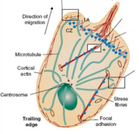
Figure 5. Organization of actin and microtubule cytoskeleton during T cell migration. In motile T lymphocytes, actin cytoskeleton is present subcortically (in order to support motile morphology of cell) and at the leading edge (where it is important for the formation of lemellopodia and filopodia). Invariably found at the uropod of motile T cells is centrosome (MTOC). Although it represents a hallmark of the uropod, its exact function is still elusive. It was suggested to have a role in establishing directionality of the movement and/or polarized vesicle secretion (Rodriguez et al., 2003).