“Amedeo Avogadro”
Department of Pharmaceutical Sciences
Ph.D. in Chemistry & Biology
Drug discovery and development
SSD BIO-14
XXX cycle a.y. 2014-2017
“In vitro” and “in vivo” studies on innate immune cells
Alvaro Brittoli
Supervisor: Prof. Grazia Lombardi
Co-Supervisor: Prof. Giulio Spagnoli
Co-Supervisor: Dr. Emanuele Trella
“Amedeo Avogadro”
Department of Pharmaceutical Sciences
Ph.D. in Chemistry & Biology
XXX cycle a.y. 2014-2017
“In vitro” and “in vivo” studies on innate immune cells
Alvaro Brittoli
Supervisor: Prof. Grazia Lombardi
Co-Supervisor: Prof. Giulio Spagnoli
Co-Supervisor: Dr. Emanuele Trella
secure future. The very basic core of a man's living spirit is his passion for adventure. The joy of life comes from our encounters with new experiences, and hence there is no greater joy than to have an endlessly changing horizon, for each day to have a new and different sun.
Christopher Johnson McCandless
“
List of abbreviations……….. VII
Preface………... 1
Chapter I: “In vitro” studies on galectin-3 in human natural killer cells….. 3
1. Introduction………... 5
1.1 The Sugar Code………... 5
1.2 Lectins……… 5
1.3 Galectins………. 6
1.4 Galectin-3………... 8
1.4.1 The carbohydrate recognition domain……… 9
1.4.2 The N-terminal domain………... 10
1.4.3 Galectin-3 ligand specificity………... 12
1.5 Galectin-3 expression and distribution in human tissues…... 14
1.5.1 Galectin-3 expression in human non-hematopoietic tissues……….. 15
1.5.2 Galectin-3 expression and cellular localization in human hematopoietic cells...………….……... 16
1.5.3 Regulation of galectin-3 gene expression……….. 18
1.6 Cellular localization...………...………… 23
1.7 Function of extracellular and intracellular galectin-3 in regulating cellular homeostasis………... 23
1.7.1 Function of extracellular galectin-3………... 23
1.7.2 Gal-3 function in endocytosis………….………... 23
1.7.3 Cell adhesion……….………. 25
1.7.4 Function of nuclear and cytoplasmic galectin-3……… 26
1.7.5 Regulation of cell cycle………….………. 26
1.7.6 Galectin-3 in gene regulation…….……… 27
1.8.1 The pro/anti-apoptotic effect of galectin-3………. 29
1.8.2 Regulation of cell growth and differentiation by endogenous and exogenous galectin-3……… 33
1.9. Function in immune system..……… 34
1.9.1 T cells and B cells………... 35
1.9.2 Monocytes/macrophages……… 37
1.9.3 Neutrophils………. 38
1.9.4 Mast cells and eosinophils……….. 38
1.9.5 Adhesion and migration of immune cells…... 39
1.9.6 Galectin-3 at the interface between innate and adaptive immunity………. 41
1.9.7 Recognition of foreign pathogens…………... 42
1.9.8 Role in immune system regulation………. 44
1.10 Natural killer cells……… 45
1.10.1 Natural killer cells activation and functions…... 48
2. Outline of the thesis……….. 55
3. Materials and methods………... 57
3.1 Human NK cell isolation and cell cultures……… 57
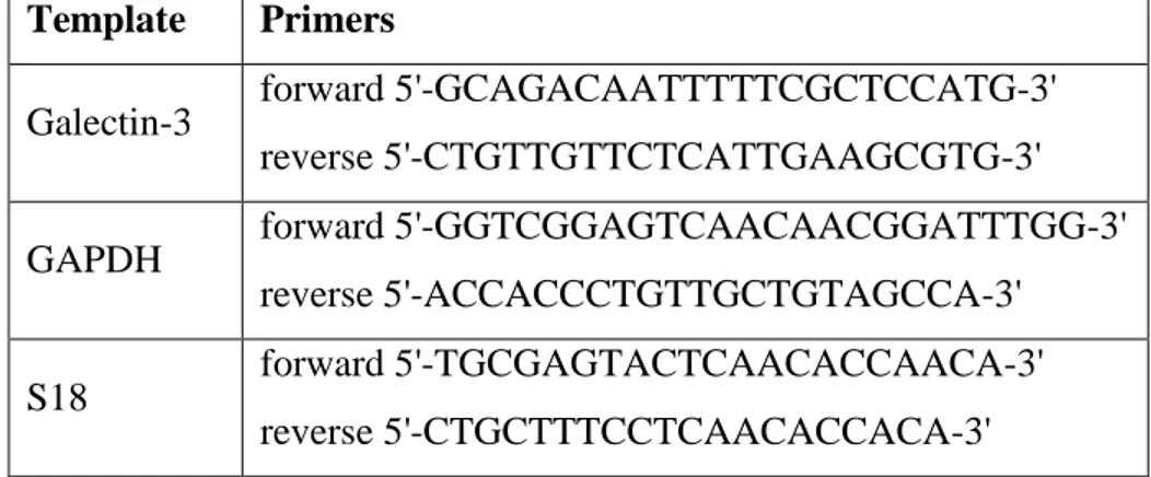
3.2 PCR and real-time PCR………. 57
3.3 Western blot.………...………... 58
3.4 Flow cytometry………... 59
3.5 Confocal microscopy.……… 60
3.6 NK cell degranulation assay……... 60
3.7 Statistical analysis………... 61
4. Results………... 63
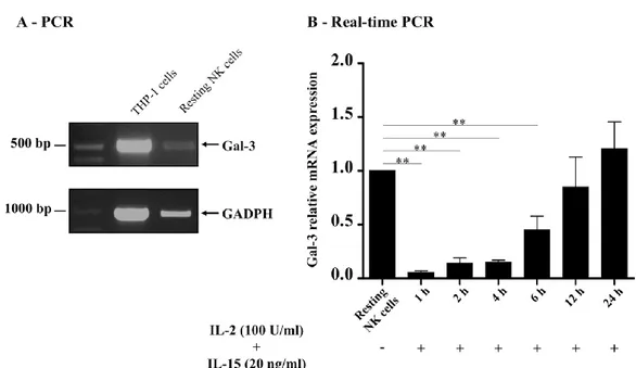
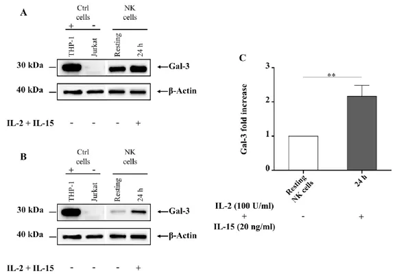
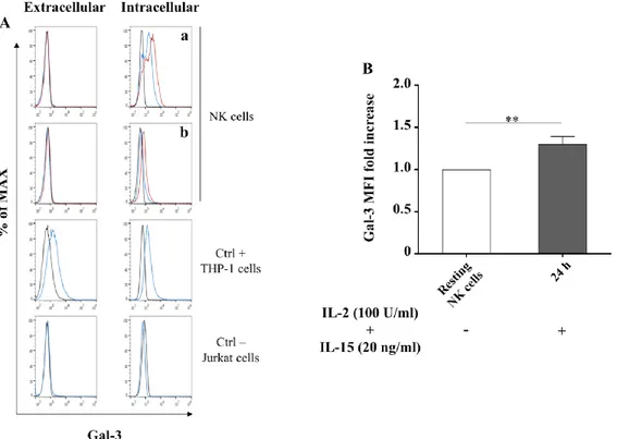
4.1 Galectin-3 gene expression in human resting NK cells …… 63
4.2 Galectin-3 mRNA in human activated NK cells ……... 63
4.5 Galectin-3 functional studies in human NK cells……... 70
5. Discussion………. 73
6. Conclusion and outlook……… 79
References………. 81
Chapter II: CD154/40L recombinant vaccinia virus induces direct and macrophage-mediated in vitro and in vivo antitumor effects………... 95
1. Introduction………... 97
1.1 Cancer and immune therapeutic strategy………... 97
1.2 The CD40 receptor and CD40 ligand ………... 102
1.2.1 The CD40 receptor : CD40 ligand (CD154) pathway… 103 1.2.2 The CD40 receptor : CD40 ligand (CD154) in the immune system……… 106
1.2.3 Targeting CD40 receptor to harness the immune system against cancer………... 110
1.3 Macrophages………... 114
1.3.1 Macrophages and cancer……… 116
1.3.2 Targeting the CD40 pathway to enhance antitumor macrophage activity……… 121
1.4 Vaccinia virus and cancer immunotherapy……… 124
2. Outline of the thesis………... 129
3. Materials and methods………... 131
3.1 Immunohistochemistry………... 131
3.2 CD40 ligand-expressing recombinant Vaccinia Virus (rVV40L)………...………….. 131
3.3 Established tumor cell lines………... 131
3.4 In vitro generation of CD14+ derived macrophages……….. 132
differentiation………... 133
3.7 Co-culture of M1 and M2 macrophages with established tumor cell lines………. 133
3.8 Cross-presentation assays………... 133
3.9 Migration assay……….. 134
3. 10 In vivo experiments………. 134
3.11 Gene expression analysis………. 135
3.12 Flow cytometry……… 135
3.14 ELISA………... 135
3.15 Histological evaluation……… 136
3.16 Statistical analysis……… 136
4. Results………... 137
4.1 CD40 receptor expression in clinical tumor specimens……. 137
4.2 rVV40L infection induces apoptosis of CD40(+) tumor cell lines………... 138
4.3 Impaired CD40 signaling pathway is associated with tumor cell resistance to rVV40L induced apoptosis…………... 142
4.4 Modulation of M1/M2 functional profiles by rVV40L infection………... 144
4.5 CD40 receptor-stimulated M1 and M2 macrophages promote the recruitment of CD8+ T cells………... 150
4.6 rVV40L promotes M1-mediated cross-presentation of tumor-associated antigens (TAAs) to CD8+ T cells…………... 154
4.7 rVV40L promotes in vivo tumor regression through macrophage activation………. 155
5. Discussion………. 159
6. Conclusion and outlook……… 165
aa amino acids
ADCC antibody-dependent cell-mediated cytotoxicity
AdCD40L CD40L-expressing
recombinant adenovirus
AGE advanced glycation end
products
AIP ALG-2 interacting protein Alix ALG-2 linked protein x AP activator protein
APC antigen presenting cell Apo apoptosis antigen
ATF activating transcription factor Bcl B-cell lymphoma
CCL C-C motif chemokine ligand CCR C-C motif chemokine receptor CD40/CD40R/CD40(+) CD40
receptor
CD40L CD40 ligand
CDC complement-dependent cytotoxicity
CEA carcinoembryonic antigen CM complete medium
CRD carbohydrate recognition
domain
CRE cAMP-dependent response
CRM1 chromosome maintenance region 1 CXCL C-X-C motif chemokine ligand CXCR C-X-C motif chemokine receptor DAMPs damage-associated molecular patterns DCs dendritic cells
DISC cell death-inducing signaling
complex
EGF epidermal growth factor ERK extracellular signal-regulated
kinase
ESCRT endosomal sorting complex
required for transport
Fc fragment crystallizable FcεRI high-affinity IgE receptors Fuc fucose Gal galactose Gal Galectin Gal-1 galectin-1 Gal-3 galectin-2 GI gastrointestinal Glc glucose GM-CSF granulocyte-macrophage colony-stimulating factor
GRIFIN galectin-related interfibre
protein
GSK glycogen synthase kinase H&E hematoxylin and eosin HCC hepatocellular carcinoma HIPK homeodomain-interacting
protein kinase
HLA human Leucocyte Antigens HLA-DR human leukocyte
antigen-antigen D Related hnRPN heterogeneous nuclear ribonucleoproteins IFN interferon Ig Immunoglobulin IL interleukin
iNOS inducible nitric oxide synthase IRF interferon regulatory factor IS the immunological synapse JNK c-Jun N-terminal kinase KIRs killer cell Ig-like receptors LacNAc N-acetyllactosamine
LAK lymphokine-activated killer cell LAMPs
lysosomal-membrane-associated glycoproteins
LcK lymphocyte-specific protein
tyrosine kinase
LDL low density lipoproteins
mAb monoclonal antibody Mac macrophage antigen
MAPK mitogen activated protein
kinase
mCD40L transmembrane form of
CD40 ligand
M-CSF macrophage
colony-stimulating factor
MDSC myeloid derived suppressor
cells
MEK mitogen-activated protein MHC major histocompatibility
complex
MIC MHC class I
polypeptide-related sequence
MOI multiplicity of infection NAc N-acetyl
NCRs natural cytotoxicity receptors Neu neuraminic acid
NF-IL6 nuclear factor for
interleukin-6
NF-kB nuclear facto kappa B NK natural killer
NO nitric oxide
NORE1a novel Ras effector 1a NSCLC non-small-cell lung cancer OC ovarian cancer
PB peripheral blood
PBMC peripheral blood mononuclear
cells
PDA pancreatic ductal
adenocarcinoma
PI propidium iodide
PI3K phosphoinositide 3-kinase PLCγ phospholipase Cγ
Ras rat sarcoma
Rassf5 Ras association
domain-containing protein 5
RCC renal cell carcinoma
RNI reactive nitrogen intermediates
ROI reactive oxygen intermediates rVV40L recombinant vaccinia virus
encoding for CD40L
rVVMART-1 FG MelanA/MART-1
full gene
rVVs recombinant vaccinia viruses sCD40L soluble form of CD40
ligand
sCD40L/s40L soluble form of CD40
ligand
SIE sis-inducible element Sp specificity protein
STAT signal transducer and activator
of transcription
Tcf T cell factor
TCM T central memory cell TEM T effector memory cell TEMRA T effector memory RA TGF transforming growth factor Th T helper lymphocyte
TLR toll like receptor
TMA Tissue microarray TNF tumor necrosis factor
TNFR tumor necrosis factor receptor TRAFs TNFR-associated factors TRAIL necrosis factor-related
apoptosis-inducing ligand
Tregs regulatory T cells
TTF thyroid-specific transcription
factor
VCAM vascular cell adhesion
molecule
VEGF vascular endothelial growth
factor
VV Vaccinia Virus WT wild type
M2 alternatively activated
macrophages
Preface
During the three years of my Ph.D. course, I was involved in two different research projects, both concerning cellular components of the innate immune system: natural killer cells and macrophages.
In particular, my principal project (University of Eastern Piedmont, Italy) aims to clarify the expression and role of a protein, galectin-3, on natural killer cells. The other one (University of Basel, Switzerland) deals with the use of a viral vector to boost the antitumor activity of macrophages.
Chapter I
1. Introduction
1.1 The Sugar Code
An enormous amount of events take place among cells, either intra-, inter-, or extra-cellularly and the whole apparatus of proteins and nucleic acids is not sufficient to adequately perform all of them. A third category of bio-molecules with the ability to transmit a multitude of different messages seems to be required for assuring fine cellular processes, such as cell adhesion and communication.
Sugars, in the form of mono-, oligo-, or poly-saccharides, as well as glycoconjugates, are a perfect example of such type of bio-molecules. These compounds are, in fact, endowed with a large capacity in information-storing and a great ability to transfer information through protein bio-recognition.
As reported by Gabius (2000), oligo-saccharides surpass peptides by more than seven orders of magnitude in the theoretical ability to build isomers, when the total of conceivable hexamers is calculated. Most of the carbohydrates, in fact, contain at least one asymmetric carbon atom with a number of possible stereoisomers that grow rapidly with molecular weight. In addition, each carbohydrate has approximately four functional groups that can be used to establish connections with other sugar units in a linear and/or in a nonlinear/branched fashion. All the complex information stored in the glycostructures decorating the surface of a cell, protein, or lipid molecule can be deciphered by a protein having the ability to bind sugar, such as, for example, the lectins (Gabius2000).
1.2 Lectins
The name lectin derives from the latin word legere, whose meaning is to pick/choose or select. Lectins represent a group of structurally diverse proteins able to bind and recognize simple/complex sugar moieties protruding from glycolipids or glycoproteins (Ghazarian et al. 2011). Carbohydrate-binding proteins can be roughly
classified into two main groups: lectins and sulfated glycosaminoglycan-binding proteins. The lectin superfamily phylogenetically comprises ancient proteins, defined by their ability to bind either soluble carbohydrates or glycoconjugates, mediating a large array of biological processes, including cell to cell interactions, communications at both inner and outer side of the cellular membrane and modulation of immune responses.
The broad range of functions carried out by this heterogeneous group of proteins are possible thanks to their ability to bind many different types of carbohydrates, which are present in the extracellular matrix, at the cell surfaces, or into secreted glycoproteins. Lectins can read the sugar code, recognizing even slight difference in the complex structure of oligosaccharides and/or in simple monosaccharides, binding only to specific sugars.
Originally isolated from plants, they are widely distributed in nature, ranging from bacteria and virus to animals. Today, based on the structures of animal lectins at least 15 structural families of mammalian lectins exist, where galectins and C-type lectins are the largest families (Gupta 2012).
1.3 Galectins
Galectins are a family of highly conserved proteins whose members are characterized by a highly conserved carbohydrate recognition domain (CRD), consisting of approximately 130 amino acids (aa) (Barondes et al. 1994) which bind small β-galactosides/poly-N-acetyllactosamine (LacNAc)-enriched glycoconjugates (Gupta 2012). These proteins were initially classified as S-type lectins, where the S (sulphydryl or thiol) was used to indicate the dependence on reducing conditions for activity, a specific property of galectin-1 (Gal-1), the first studied galectin (Gupta 2012).
To date, up to 15 galectins have been identified in mammals and, based on their molecular architecture and structural differences in CRD presentation, they can be
classified into 3 main groups, as proposed by Hirabayashi and Kasai in 1993 (Fig. 1A).
The prototype group are galectins containing one CRD, that can exist as monomer (galectins -5, -7, -10) or homodimers with two polypeptides containing a CRD each (galectins -1, -2, -11, -13, -14, -15), and a short N-terminal sequence. It should be mentioned that classification of galectin-11, initially identified as GRIFIN (galectin-related interfibre protein), is still under debate, since this galectin misses two of the seven crucial aa conserved in the CRD, and it displays no binding activity trough sugar molecules (Rabinovich et al. 2007).
Tandem repeat galectins are monomers with two non-identical CRDs joined by a short peptide linker region (galectins -4, -6, -8, -9, -12).
By contrast, galectin-3 (Gal-3) is the only member discovered so far of the chimera-type with a unique structure, characterized by one CRD connected to a long N-terminal domain. This latter is involved in Gal-3 oligomerization which results in the formation of Gal-3 multimers exposing several CRDs, enabling bivalent or multivalent binding of carbohydrate ligands (Dumic et al. 2006; Sato et al. 2009; Fortuna-Costa et al. 2014).
Figure. 1. Schematic representation of the structure of different members of the galectin family. (A) prototype galectins containing one or two homodimeric CRD, tandem
repeats containing two different CRD connected by an aminoacidic linker and the unique chimera-type Gal-3 composed by a CRD and a collagen-like sequence. (B) schematic representation of Gal-3 oligomerization into pentamers through the N-terminal domain allowing the multivalent binding of glycans forming a lattice structure (adapted from
Rabinovich and Toscano 2009).
1.4 Galectin-3
Gal-3 is the currently accepted name for a protein, initially identified as Mac-2, a surface protein expressed on murine macrophages (Ho and Springer 1982), formerly also known as CBP-35, LBP, L-34, L-29, or εBP (Gupta 2012). Human Gal-3, is a 31 kDa protein encoded by a single gene (LGALS3), which is located on chromosome 14, locus q21–q22 and spans on a total of 17 Kb. The gene has an open reading frame of 750pb and consists of 6 exons and 5 introns that are translated into a protein of 250 aa. In particular, the transition start site is found within the exon II, which contains also the initial methionine, the first six aa and part of the 5’ untranslated sequence. The exon III contain entirely the long and flexible, collagen-like
N-terminal domain, while exon IV-VI codify for the C-N-terminal domain (Argüeso and Panjwani 2011; Gupta 2012; Díaz-Alvarez and Ortega 2017).
1.4.1 The carbohydrate recognition domain
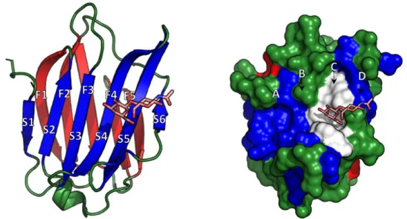
Gal-3 shares whit Gal-1 and Gal-2 ~25% sequence identity and their CRD have a similar three-dimensional structure and identical topology (Seetharaman et al. 1998). This latter, is composed of two anti-parallel β-sheets with five and six β-strands (F1-5 and S1-6) (Seetharaman et al. 1998), forming a globular structure that accommodates the carbohydrate-binding site, composed by the side chains of the aa forming the 6-stranded β-sheet (Hughes 2001). This structure is responsible for the lectin activity of Gal-3. In particular, the two β-sheets composing the CRD are arranged in a convex side (F1-5) and a concave side (S1-6) with this latter forming a groove able to accommodate a linear tetrasaccharide (Fig. 2) (Leffler et al. 2002). The CRD can be schematically divided into four sub-binding sites: A, B, C, D, where the C subsite is made of approximately 6 conserved aa and is responsible for the β-galactoside binding. The other sites participate in the binding activity, either by increasing or decreasing the protein affinity, depending on the saccharide moiety linked to the galactose bound in the C subsite (Salomonsson et al. 2010). Beside carbohydrate recognition/binding, the CRD is also involved in the anti-apoptotic activity associated with Gal-3 (for further details see paragraph 1.8.1). This domain, in fact, presents the NWGR aa sequence, which is also highly conserved within the BH1 domain of the anti-apoptotic B-cell lymphoma (Bcl)-2 family, and the interaction with this latter is responsible for the resistance to apoptotic stimuli (Yang et al. 1996). The importance of this sequence for Gal-3 is demonstrated by the fact that it is highly conserved among different species, and that a single amino acid substitution of Gly to Ala in the NWGR sequence abrogates the anti-apoptotic properties (Akahani et al. 1997). The NWGR motif is also essential for the carbohydrate recognition activity since the tryptophan (W) amino acid is directly involved in the sugar binding (Yang et al. 1998).
Finally, despite Gal-3 oligomerization has been proposed to occur through the N-terminal domain (type-N model), recently a new model (named type-C self-association) has been proposed. Accordingly, oligomerization would be mediated by the carbohydrate recognition site: the carbohydrate-binding site of one CRD would bind at the other side of the next CRD, and the N-terminal domain would participate by enhancing this association. Both models remain equally probable, and whether and when each model occurs in a cellular system remain to be demonstrated (Lepur et al. 2012).
Figure 2. High Resolution X-Ray Structure of Human Gal-3 in complex with LacNAc visualized as stick model (PDB: 1KJL). (A) secondary structure of the Gal-3 CRD; (B)
The solvent accessible surface of Gal-3 CRD with the position of A-D binding site shown above the binding groove, the C binding site was shown in white (adapted from Salomonsson et al. 2010; Hsieh et al. 2015).
1.4.2 The N-terminal domain
The N-terminal domain is a relatively flexible structure, consisting of two distinct portions: a collagenase sensitive, proline, glycine, alanine, and tyrosine rich motif, followed by 12 aa sequence also called the small N-terminal domain, containing a casein kinase I serine phosphorylation site. The former, consisting of 100–150 aa
residues, has homologous repeats (8 to 13 in vertebrates) (Ippel et al. 2016) of a consensus sequence Pro-Gly-Ala-Tyr-Pro-Gly, followed by three additional aa with a number of repetitions that vary among different species and lack of charged or large side-chain hydrophobic residues. This part of the N-terminus domain has 35.5% identity with collagen α1 (II) chain of bovine cartilage, and this explains the designed name of this domain as collagen-like N-terminal domain (Raz et al. 1989; Gupta 2012). In addition to collagen, this portion of the N-terminal domain has a 25% homology with some heterogeneous nuclear ribonucleoproteins (hnRPN) (Dumic et al. 2006).
At least four functional properties can be ascribed to the N-terminal domain:
i) Upon binding of specific glycoconjugates, Gal-3 changes its conformation, resulting in N-terminal domain self-assembly and consequent protein oligomerization (Sato et al. 2009). This process leads to the formation of a multimer (such as dimer or pentamer) (Gong et al. 1999;Ahmad et al. 2004), which possesses multivalent CRD (Sato and Nieminen 2002) even under non-reducing conditions or in the presence of sodium dodecyl sulfate (Henderson and Sethi 2009), leading to cross-linking of cell receptors;
ii) The N-terminal portion of the protein (small N-terminal domain) is involved in its secretion. In fact, the deletion of the initial 12 aa, completely abrogates secretion of this protein through a new mechanism independent from the classical secretory pathways (ER and Golgi system) (for further details see paragraph 1.6) (Dumic et al. 2006);
iii) Despite the carbohydrate binding is largely N-terminal independent (Dumic et al. 2006), molecular modeling and mutagenesis studies demonstrated that the aa present in the N-terminal domain can participate in sugar binding (Barboni et al. 2000). Particularly, the N-terminal domain would contribute to carbohydrate binding through the stabilization of the substrate-bound lectin complex (Barboni et al. 2000). These studies were performed with lipopolysaccharide (LPS) as binding partner,
demonstrating that Gal-3 is able to bind the side chain of the O-antigen of LPS, which contains polymers of LacNAc, a known ligand for the CRD of this lectin. Alternatively, for LPS devoid of beta-galactosides, Gal-3 can interact through the N-terminal domain, with the lipid A core region of LPS (Mey et al. 1996). The use of a monoclonal antibody (mAb) that recognizes an epitope within the N-terminal domain of Gal-3 completely abrogates the binding of this lectin to LPS (Fowler et al. 2006);
iv) Post-translational phosphorylation of a serine residue (Ser6) in the small N-terminal domain of the protein, has been associated with several Gal-3 functions: a) this modification acts as an "on/off" switch for its sugar-binding capability controlling downstream biological effects (Mazurek et al. 2000) b) similarly to Bcl-2, whose phosphorylation at Ser70 appears to be critical for its anti-apoptotic function, non-phosphorylated Gal-3 mutants result in loss of Gal-3 anti-apoptotic activity and fail to induce cell cycle arrest (Yoshii et al. 2002).
1.4.3 Galectin-3 ligand specificity
The minimal sugar unit recognized by galectins is represented by a galactose residue linked to an adjacent monosaccharide in the β configuration (β-galactoside), such as that found in the LacNAc residues (Galactose [Gal]β1-4N-acetylglucosamine [GlcNAc]). Each member of the galectin family presents slightly, yet significant, differences in the CRD that can tune the affinity of each galectin for its ligands (Sato et al. 2009).
Gal-3 can bind both N- and O-glycans (Sundblad et al. 2011) and it was originally identified to bind preferentially to Galβ13(4)GlcNAc (Cardoso et al. 2016); specific modifications of the β-galactoside core, however, can enhance or reduce the affinity towards Gal-3. α-N-acetylgalactosamine modifications of the Gal residue in the β-galactoside core, indeed, significantly increase the affinity for Gal-3, while modifications with α2-6- sialic acid reduce the affinity for the same protein (Hirabayashi et al. 2002). Moreover, the extension of the non-reducing end of the
disaccharide with 2- o 3- substituents on the outer galactose, such as N‐ acetylneuraminic acid (NeuNAc)α2,3 or GalNAcα1,3 and fucose (Fuc)α1,2 substituents greatly enhances the affinity for Gal-3 (Krześlak and Lipińska 2004). In addition, also galactomannans and polymannans can be bound by Gal-3 ( Díaz-Alvarez and Ortega 2017). Usually the affinity of Gal-3 toward its glycan ligands is lower (approximately 10-6 M) than those typically observed for protein-protein interactions (approximately 10-8 M); however, the affinity increases for branched
glycans and/or polylactosamine structures over simple saccharides. In fact, as previously mentioned, upon ligand binding Gal-3 can oligomerize through the N- terminal domain resulting in multiple CRD presentation. The multivalent binding of carbohydrate-containing glycoproteins or glycolipids by Gal-3 increases the avidity of the protein for its ligand, promoting the formation of organized glycan-galectin clusters, termed “lattices”, and mediating cross-linking of glycosylated molecules (Fortuna-Costa et al. 2014) (Fig. 1B).
Further, since Gal-3 possesses an extended binding site, when compared to Gal-1 (Sato and Nieminen 2002), this protein displays a high affinity for glycan presenting repeated LacNAc residues, as found in polylactosaminoglycans (polylactosamine effect). Amniotic fibronectin, which contains a tetra antennary complex composed of four parallel lactosamine residues presented by a mannose core structure, is an example (Sato and Hughes 1992). Interestingly, no effect is displayed against plasma fibronectin, that presents only two bi-antennary chains.
Mutational and structural studies of the human Gal-3 carbohydrate binding site (formed by β-strands S4–S6) in complex with LacNAc molecule revealed the aa directly implied in sugar binding. In particular, the galactose C-4 hydroxyl group interacts with His-158, Arg-162, Asn-160 and a water molecule (W1). The C-6 hydroxyl group interacts directly with Glu-184, Asn-174 and W3, while Arg144 can interact with the monomer linked to O-3 of the terminal galactose. Other favorable
stacking interactions are provided by Trp-181 with the hydrophobic galactose surface.
The N-acetylglucosamine moiety is more exposed to the solvent; therefore, only the C-3 hydroxyl group can interact with Glu-184 and Arg-162. The other interactions involving the GlcNAc moiety are mediated through its N-acetyl group bonded to Arg 186 and through a water molecule (W2) to Glu-165 and this probably accounts for the approximately 5-fold higher binding affinity of human Gal-3 for LacNAc over lactose (Seetharaman et al. 1998; Krześlak and Lipińska 2004).
Since position 4 and 6 of the galactose in the β-galactoside present key interactions with the protein, modifications of these positions are not tolerated. On the contrary, appropriate modifications of position 2 and 3 are well tolerated and can enhance the affinity of this protein for its ligand (Leffler et al. 2002; Salomonsson et al. 2010).
This chimera-type galectin not only interacts with glycan, but also with peptide motifs. Despite Gal-3 carbohydrate-binding activity appears to be critical for Gal-3 function, this lectin presents also many different molecular interactions that are largely carbohydrate independent, some of them involving the carbohydrate binding groove (Wang et al. 1995; Paron et al. 2003; Elad-Sfadia et al. 2004; Shimura et al. 2004; Shimura et al. 2005; Gupta 2012). For example, Gal-3 is able to interact with Bcl-2, an anti-apoptotic protein, which is not a glycoprotein (Yang et al. 1996).
1.5 Galectin-3 expression and distribution in human tissues
Data on Gal-3 expression in tissues of human adults and during embryonic stages are more limited if compared to mice. However, different studies (see below) have provided evidences about the expression pattern and distribution of this lectin in non-hematopoietic and non-hematopoietic human tissues.
1.5.1 Galectin-3 expression in human non-hematopoietic tissues
Gal-3 is broadly expressed in several human tissues and cells in a spatio-temporal fashion (Sundblad et al. 2011). During human embryogenesis, Gal-3 expression is mainly confined in epithelial cells of the skin, respiratory and digestive tract, excretory tubes of the kidney and to the uroepithelium. In the adult, Gal-3 expression resembles that found in embryogenesis, with protein expression restricted to the epithelial cells of different organs and to myeloid cells infiltrating the tissue. In particular, robust expression of this lectin was found during lung analysis in epithelial cells of the bronchus, chondrocytes of the bronchial cartilage, and in alveolar macrophages (Mathieu et al. 2005). Gal-3 expression is also described in the gastric epithelial cells, the colon, the colonic epithelial cells, and in the intestinal macrophages (Lippert et al. 2008). In the gut, Gal-3 expression is restricted to the crypt cells and macrophages of lamina propria (Mercer et al. 2009). Interestingly, the expression of this protein in endometrial and decidual tissues is tightly regulated through the menstrual cycle. Since Gal-3 has many immunomodulatory properties (for further details see paragraph 1.9), this observation led to hypothesize that Gal-3 might contribute not only to the regulation of the endometrial function, but also to the modulation of the endometrial immune system during the implantation process (von Wolff et al. 2005; Sundblad et al. 2011). Finally, Gal-3 expression has been detected also in prostate glands, in the cytoplasm of luminal cells, and intratubular fibroblasts of the breast, in the cytosolic and membrane-enriched fraction of chondrocytes, as well as in the coronary artery smooth muscle cells (Sundblad et al. 2011).
Under physiologic conditions, due to its ability to shape the immune response, Gal-3 expression in epithelia of the digestive tract and lungs might be involved in mucosal defense, modulating both host-pathogen interactions and immune attacks (Sundblad et al. 2011).
1.5.2 Galectin-3 expression and cellular localization in human
hematopoietic cells
Gal-3 has been reported to be ubiquitously expressed in many immune cells, where it contributes to regulate both the innate and adaptive immune responses (Chen et al. 2005; Liu 2005; Rabinovich et al. 2007). First identified in macrophages (Ho and Springer 1982), Gal-3 was found in monocytes (Liu et al. 1995), dendritic cells (DCs) (van Stijn et al. 2009), mast cells and basophils (Craig et al. 1995), eosinophils (Rao et al. 2007), neutrophils (Wu et al. 2017), T and B cells (Craig et al. 1995; Chen et al. 2005), with a level of relative expression which is tightly regulated by the cellular activation state.
In human monocytes, this lectin is constitutively expressed at low levels, both intracellularly and at the cell surface; however, its expression increases dramatically at both mRNA and protein levels, as monocytes differentiate into macrophages (Nangia-Makker et al. 1993; van Stijn et al. 2009). In this latter, Gal-3 was found in the cytoplasm/nucleus and at the plasma membrane surface. Interestingly, alternative macrophage polarization (M2) is associated whit higher levels of Gal-3 when compared to classically (M1) polarized macrophages, with a pattern of expression that follows the subsequent scheme: monocyte << M1 < M2 (Novak et al. 2012). Despite the difference in the levels of expression, cells of the monocyte/macrophage lineages are able to secrete this lectin in the extracellular media when properly stimulated (Liu et al. 1995)
In a similar manner, differentiation of human monocytes into DCs leads to an increment in the Gal-3 protein levels with only a moderate increase in Gal-3 mRNA. Moreover, during cellular differentiation, the monocyte surface expression of this lectin slowly disappears, with Gal-3 protein that mainly localizes intracellularly in immature DCs (van Stijn et al. 2009). Conversely, in activated DCs, this lectin is principally restricted to lipid raft domains of membrane ruffles and lamellipodia, where it contributes to regulate cell motility (Hsu et al. 2009). Similarly to monocytes
and macrophages, Gal-3 secretion was reported also for DCs. Proteomic analysis of DC-derived exosomes demonstrates, in fact, that these cells can release in the extracellular microenvironment, membrane vesicles containing Gal-3 (Théry et al. 2001).
Through light microscopy immunohistochemistry and ultrastructural immunogold labeling, Gal-3 expression and localization were studied in human mast cells and basophils. In particular, in mast cells, this lectin was detected in the nucleus (over heterochromatin whereas euchromatin was unlabeled) and/or the cytoplasm. Of note, cytoplasmic labeling was principally concentrated over secretory granules. Gal-3 localization in basophils was similar to that found in mast cells, but with a general low intensity of staining (Craig et al. 1995).
Finally, Gal-3 expression was also reported for two other populations of innate immune cells: neutrophils and eosinophils. In the former, Gal-3 localize mainly intracellularly, despite at a lower level compared to other innate cells (Wu et al. 2017). In addition, it and can be secreted by these cells upon treatment with mannan structure derived from microbial pathogens (Linden et al. 2013). Conversely, in human eosinophils, this chimeric protein is confined to the cells surface, with a level of expression that increases in allergic subjects (Rao et al. 2007).
In sharp contrast with the constitutive expression of Gal-3 in innate immune cells, this lectin is absent, or only scarcely expressed, in lymphocytes and lymphoid cell lines. Resting T and B cells, for example, do not express Gal-3, but this protein can be induced following interleukin (IL)-4 stimulation or CD40 cross-linking in B cells (Acosta-Rodríguez et al. 2004), and by TCR engagement, or mitogen exposure, in T cells (Joo et al. 2001; Dumic et al. 2006). Likewise, also virus and/or parasite infections are able to induce Gal-3 expression in these cells (Dumic et al. 2006).
1.5.3 Regulation of galectin-3 gene expression
Despite the amount of data available on Gal-3 expression in hematopoietic/non-hematopoietic tissues, the regulation of gene transcription remains poorly understood.
The analysis of the promoter sequence of LGALS3 revealed that this region contains, among others, two nuclear factor kappa B (NF-kB) and three putative activator protein (AP)-1 sites (Kadrofske et al. 1998). Particularly, in glioblastoma cells, the expression of this protein is regulated by Jun (a component of AP-1 transcription factor) and NF-kB, where Jun seems to be important for the basal expression of Gal-3, while NF-kB is implicated in the gene induction following cell damage (Dumic et al. 2000).
The presence in the promoter of cAMP-dependent response element (CRE) implies that cAMP-response element-binding protein (CREB) might participate in the Gal-3 gene regulation.
Particularly, the implication of CREB and NF-kB in Gal-3 gene regulation was studied in human T lymphotropic virus-I infected T cells (Hsu et al. 1996). In the latter, Gal-3 is under the regulation of both the CREB/activating transcription factor (ATF) and, to a lesser extent, the NF-kB transcription factor. Curiously, NF-kB regulation of Gal-3 expression is controlled by nuclin, an apoptosis-associated protein, which can interfere with NF-kB in order to inhibit the expression of Gal-3 (Liu et al. 2004). On the contrary, in macrophage cells, Gal-3 expression seems to be principally regulated by the rat sarcoma (Ras)/mitogen activated protein kinase (MAPK) signal transduction pathway (Kim et al. 2003).
Further analysis revealed that the promoter contains also numerous GC box motifs, which can be bound by the ubiquitously expressed specificity protein (Sp)1 transcription factor, a feature which is commonly observed in the promoters of the housekeeping genes (Fogel et al. 1999). Nevertheless, Gal-3 expression at both
mRNA and protein levels, following serum addition to serum-starved fibroblasts, represents a feature of early genes, and sis-inducible element (SIE) was suggested to be a potential candidate for the growth-induced activation of LGALS3 expression after serum addition (Dumic et al. 2006)
Finally, epigenetic mechanisms could also be involved in Gal-3 expression. The gene promoter and the first exon, in fact, present a high content of GcP island and the methylation status seems to contribute to Gal-3 expression in pituitary tumors (Ruebel et al. 2005).
As evidenced by the different regulatory elements found in the Gal-3 promoter and the different transcription factors involved in LGALS-3 transcription, Gal-3 expression results to be a complicated process probably depending on cell type, environmental stimuli and malignant transformation.
1.6 Cellular localization
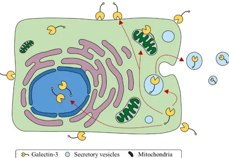
Gal-3 is a structurally unique protein expressed in many normal tissues and immune cells with a cellular localization that strongly depends on the cell type and its proliferation status. In addition, the cellular distribution of this protein can change during differentiation or neoplastic transformation (Dumic et al. 2006) (Fig. 3).
Figure 3. Intracellular trafficking of Gal-3 based on experimental evidence. Gal-3 can
be either found the extracellular space, attached to cell surface, in the cytoplasm, in secretory vesicles or in the nucleus. Moreover, it can be secreted from cells in a manner that is independent from classical secretory pathway (adapted from Hughes 2001).
Initially synthesized on free ribosomes in the cytoplasm (Mehul and Hughes 1997), Gal-3 is able to shuttle between the nucleus and the cytoplasm in a manner that is still unclear. Using a truncated form of Gal-3 was discovered that mutants containing truncations of the amino-terminal half of this protein, were dispensable for nuclear import (Davidson et al. 2006). On the contrary, mutants of the same construct, containing truncations from the carboxyl terminus, showed loss of nuclear localization. Then, site-directed mutagenesis of this latter portion of the polypeptide suggested that nuclear import was dependent on the ITLT sequence (residues 253-256). However, this sequence, by itself, is not sufficient to guarantee a correct nuclear localization: its activity, in fact, is strongly regulated by a neighboring leucine-rich nuclear export signal.
Conversely, another study suggested that Gal-3 could be either imported into the nucleus via active transport and/or passive diffusion (Nakahara et al. 2006). Concerning the active transport, the Authors of this study identified a nuclear localization signal (NLS)-like motif in its protein sequence, HRVKKL(residue
223-228), similar to those of p53 and c-Myc, respectively. Moreover, they suggest that nuclear localization of Gal-3 is, at least in part, due to the importin-α/β, with the Arg224 amino acid residue of human Gal-3 essential for its active nuclear translocation and molecular stability.
In contrast with nuclear import, nuclear export of this protein seems to be mediated by chromosome maintenance region 1 (CRM1), a nuclear export receptor. Candidate nuclear export signals, recognized by CRM1, can be found between residues 240 and 255 of murine Gal-3. Direct mutagenesis studies identified residues 240-255 of the Gal-3 protein, as containing fundamental aa for nuclear exportation, and partially overlaps with the region (residues 252-258), previously identified as important for nuclear localization (Li et al. 2006).
As previously reported, Gal-3 can undergo to post-translation modifications (see paragraph 1.4.2). In particular, this protein can be phosphorylated at both Ser6 and Ser12, of the NH2-terminal, even though the majority of phosphate is present at Ser6, while less than 10% at position Ser12 (Dumic et al. 2006). The protein responsible for Gal-3 phosphorylation seems to be casein kinase I and/or II, since the major acidic residues on both sides of Ser6 make it a good substrate for this kinase. The
presence of both unphosphorylated and phosphorylated Gal-3 has been reported in mouse 3T3 fibroblasts; where phosphorylated Gal-3 was found in both nucleus and cytoplasm, whereas unphosphorylated form was detected only in the cytoplasm. This observation suggests that the phosphorylation is a mechanism required for Gal-3 nuclear import, however, mutagenesis studies of Ser6 show that it is not essential for protein localization in this cellular compartment (Dumic et al. 2006).
Similarly to nuclear import/export, Gal-3 secretion mechanism is still under debating. Gal-3 lacks any recognizable canonical secretion signals able to direct this protein in the classical secretory pathways through the endoplasmic reticulum and Golgi system (Hughes 1999). Nevertheless, it has been extensively demonstrated that
this protein can be secreted by immune (Théry et al. 2001; van Stijn et al. 2009; Linden et al. 2013) and many other cells (Hughes 1999; Menon and Hughes 1999). It has been proposed that Ga-3 can be released by cells through an incompletely understood mechanism, called ectocytosis (Menon and Hughes 1999), in which the protein forms aggregates underlying the plasma membrane and the release takes place through membrane blebbing.
In particular, the first step in the Gal-3 secretion consists in protein accumulation and capture at the cytoplasmic side of the plasma membrane. This is a rate-limiting step and seems to be mediated by molecular chaperones and/or heat shock proteins. Essential requirements for this retention appear to be the acylation by the lymphocyte-specific protein tyrosine kinase (LcK). In particular, Cos cells transfected with a fusion protein containing within the N-terminal domain a Lck acylation sequence were efficiently acylated, retained at the cytoplasmic level and released in higher amount when compared to wild type lectin.
The following step consists in the evagination of the plasma membrane and secretion of the Gal-3-containing extracellular vesicles, in which the protein is protected against proteolysis. Under culture conditions, the Gal-3 release from these extracellular vesicles is quite fast, with a half-life of about 1 h. Conversely, isolated vesicles were more stable and this suggests that vesicles breakdown and cargo release requires specific factor(s) secreted by cells (Krześlak and Lipińska 2004).
The N-terminal domain of the protein seems to be fundamental for secretion and localization into secretory vesicles (Menon and Hughes 1999). The addition of the N-terminal domain to a cytosolic protein, the chloramphenicol acetyltransferase, leads to efficient export in transfected Cos cells. The 89–96 YP(90)SAP(93)GAY short sequence was identified as fundamental for Gal-3 secretion; however, this short segment alone is insufficient to induce the secretion of fusion proteins and results active only when larger portions of the Gal-3 N-terminal sequence are present.
1.7 Function of extracellular and intracellular galectin-3 in regulating
cellular homeostasis
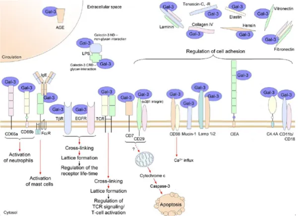
The many intra- and extra-cellular localizations of this protein, together with the ability to interact with numerous glycosylated or unglycosylated binding partners (Fig. 4 and 5), allow Gal-3 to modulate several physiological processes, such as cellular homeostasis, apoptosis, cell activation, cell adhesion, organogenesis, angiogenesis, immune response, and to act as chemoattractant for many immune cells (Sundblad et al. 2011).
1.7.1 Function of extracellular galectin-3
On cell surfaces, carbohydrates due to their structural and conformational diversity represent an incredible reservoir of biological information, even greater than DNA or peptides (Gabius 2000;Cardoso et al. 2016). Gal-3, as sugar binding proteins, can decipher this “sugar code” orchestrating a variety of biological processes (Sundblad et al. 2011).
In particular, once in the extracellular spaces, this protein can flow into the circulation, interact with the extracellular matrix or be associated with the cell surfaces, exhibiting numerous autocrine and paracrine effects (Sundblad et al. 2011). In particular, Gal-3 can modulate biological functions by forming ordered galectin-glycan arrays (lattices structure), cross-linking different cell surface glycoconjugates, or, alternatively, Gal-3 can engage specific cell surface ligands through the classical ligand-receptors interaction (Fig. 4) (Rabinovich and Toscano 2009; Sundblad et al. 2011).
1.7.2 Gal-3 function in endocytosis
In breast carcinoma cells, Gal-3 can participate in cellular endocytosis, through a caveolae-like pathway by means of which this protein promotes the endocytosis of CD29 (beta-1 integrin) in a lactose/temperature-dependent manner (Furtak et al.
2001). Interestingly, by the same pathway tumor cells can internalize from the extracellular spaces Gal-3 itself and this process is completely blocked by lactose (Furtak et al. 2001). In a similar manner, Gal-3 is involved in advanced glycation end products (AGE) and modified low density lipoproteins (LDL) endocytosis (Zhu et al. 2001).
In sharp contrast, Gal-3 can also interfere with endocytosis, extending the expression on cell surface of specific receptors and therefore altering cell responses during tumor progression. Gal-3 affinity for β-1,6-N-acetylglucosamine, in fact, mediates its binding to many glycoproteins expressed on cell surface, including lysosomal-membrane-associated glycoproteins (LAMPs)-1 and -2, carcinoembryonic antigen (CEA), mucin-1, macrophage antigen (Mac) -1/-3, transferrin receptor protein 1 (CD-71), protein tyrosine phosphatase receptor type C (CD45), and the glycosylated receptors for the vascular endothelial growth factor (VEGF), the epidermal growth factor (EGF), and the transforming growth factor (TGF)-β (Fortuna-Costa et al. 2014). Carcinoma transformation is often associated with the upregulation of the beta-1,6 N-acetylglucosaminyltransferase a Golgi enzyme which promotes the substitution of N-glycan with poly LacNac, the main ligand for Gal-3. This leads to Gal-3 cross-linking of N-glycans and lattice formation on EGF and TGF-β receptors at the cell surface, delaying their removal by constitutive endocytosis and prolonging cell activation (Partridge et al. 2004).
Of note, while extracellular Gal-3 plays a pivotal role in the balance of surface retention of glycosylated receptors against endocytosis, intracellular Gal-3 is equally important for proteins trafficking from the cytoplasm to the plasma membrane. In a physiologic contest, cytosolic Gal-3 controls the expression of EGF receptors in mouse keratinocytes. In particular, in the absence of Gal-3, EGF receptor expression results dramatically reduced with the protein that accumulates intracellularly. Of note, in these cells, Gal-3 can regulate this process through the interaction and
modulation of ALG-2 linked protein x (Alix), a member of the endosomal sorting complex required for transport (ESCRT) apparatus (Liu et al. 2012).
1.7.3 Cell adhesion
Recognition of extracellular glycoproteins and glycosylated components of the extracellular matrix by 3 plays a central role in cell adhesion/de-adhesion. Gal-3 was demonstrated to bind laminin, fibronectin, hensin, elastin, collagen IV, and tenascin-C/R (Dumic et al. 2006). Overexpression of this chimera-type protein in a human breast carcinoma cell line significantly enhanced adhesion to vitronectin, fibronectin and laminin (Materasse et al. 2000). Consistently, Gal-3 binding of Lamp-1/2, colon cancer mucin, the carcinoembryonic antigen, and the glycosylphosphatidylinositol (GPI)-anchored glycoprotein C4.4A were suggested to participate in cancer cell adhesion to extracellular matrix (Dumic et al. 2006). In addition, Gal-3 was found to interact with integrins, molecules involved in cell adhesion. Particularly, Gal-3 has been documented to associates with α1β1 integrin in a lactose-dependent manner (Ochieng et al. 1998). Moreover, through a Gal-3 affinity column purification of binding partners it was found that Gal-3 is able to interact with the alpha-subunit (CD11b) of the CD11b/CD18 integrin (CR3, Mac-1 antigen), and with the heavy chain of CD98, an integrin-associated protein (Dong and Hughes 1997). This latter is a cell surface heterodimeric glycoprotein that participates in cell-cell and cell-substratum interactions, expressed on monocytes/macrophages and in activated lymphocyte (T and B cells) (Cantor and Ginsberg 2012). Gal-3 appears to be able to induce cross-linking of CD98, leading to activation of integrin-mediated adhesion (Hughes 2001).
Besides the previously reported data, also a negative effect mediated by Gal-3 on cell adhesion was reported. In vitro, Gal-3-enriched media were demonstrated to inhibit thymocyte interactions with thymic microenvironmental cells (Villa-Verde et al. 2002).
Figure 4. A cartoon schematically representing some extracellular binding partners identified for Gal-3. Red arrows indicate positive effects (adapted from Dumic et al. 2006).
1.7.4 Function of nuclear and cytoplasmic galectin-3
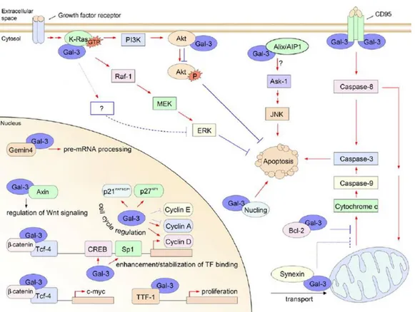
Depending on the cell type, differentiation and development status, Gal-3 can be predominantly nuclear, exclusively cytoplasmic, or spreads over these two compartments (Haudek et al. 2010). In the cytoplasmic compartment, Gal-3 interacts with numerous partners, implying a role for Gal-3 in the regulation of numerous intra-cytoplasmic events (Fig. 5).
1.7.5 Regulation of cell cycle
Cell cycle regulation is a complex process that requires intricate interactions among different proteins. Gal-3 expression, which is regulated by cell cycle (Moutsatsos et al. 1987), is also able to affect the cell cycle itself. Following expression of this lectin in BT549, a cell line that normally undergoes apoptosis induced by the loss of cell
anchorage (anoikis), Gal-3 mediates G1 phase arrest without detectable cell death. The acquisition of this anti-anoikis activity is accompanied by cyclin A and E (cyclins important for progression and maintenance of phase S) level downregulations, and upregulation of their inhibitory protein levels p21 and p27, as well as cyclin D.
These two phenomena may help the cells to bypass the critical apoptosis-sensitive point in early G1, arresting cells at an anoikis-resistant point (late G1). Furthermore, also the retinoblastoma protein, which is hyper-phosphorylated during the S/G2 phase and most of the M phase, in Gal-3 overexpressing cells, is kept in a hypophosphorylated form, showing that these cells fail to enter S phase (Kim et al. 1999). Similarly, in the same cell line overexpressing Gal-3, genistein, a soy-derived isoflavonoid able to induce apoptosis without any cell cycle arrest, induces arrest in G2/M phase without any apoptosis induction. The different phase arrests of the cell cycle might be dependent on genistein-induced upregulation of P21, but not P27 upregulation, thus leading to G2/M arrest (Lin et al. 2000).
These studies suggest that Gal-3 can regulate critical decision points through cell cycle progression, phase arrest (depending on the apoptotic stimuli) and induction of apoptosis.
1.7.6 Galectin-3 in gene regulation
Nuclear Gal-3 is also involved in gene transcription/regulation (Lin et al. 2002; Paron et al. 2003; Dumic et al. 2006). In particular, in human breast epithelial cells, Gal-3 is able to induce cyclin D promoter activity through stabilization and enhancement of the transcription factor binding to Sp1 and CRE sites. Moreover, in the same cells, Gal-3 overexpression is associated with changes in the expression levels of other proteins, such as cyclin A/E and p21/27. Since these cycle regulators contain in their promoter CRE and SP1 sites, they also represent possible candidates whose expression is regulated by Gal-3 (Lin et al. 2002).
The involvement of gene transcription was further demonstrated in papillary thyroid cancer cells, where Gal-3 directly interacts with the thyroid-specific transcription factor (TTF)-1 causing stimulation of TTF-1 binding activity (Paron et al. 2003; Dumic et al. 2006).
Further evidence came from a study demonstrating that, in the nucleus, Gal-3 co-localizes and binds to β-catenin/T cell factor (Tcf) complex in a lactose-depended manner (even if β-catenin is not a glycoprotein). This interaction induces the transcriptional activity of Tcf-4 regulating c-Myc expression and promoting cell growth and proliferation (Shimura et al. 2004). Moreover, analysis of the Gal-3 and β-catenin sequences revealed that these two protein share structural similarity. In particular, both proteins contain the S92XXXS96 consensus sequence for glycogen synthase kinase GSK-3β phosphorylation. Axin, a regulatory protein of the Wnt signaling pathway, can complex with both proteins promoting their GSK-3β phosphorylation. The Authors of this study, therefore, suggest that Gal-3 is a key regulator of the Wnt/ β-catenin signaling pathway and of its target gene expression (Shimura et al. 2005).
1.7.7 Galectin-3 as a factor in pre-mRNA splicing
Nuclear Gal-3 can directly participate to the splicing machinery. It was found that Gal-3 can associate, in the nucleus, with ribonucleoprotein complexes and be involved in spliceosome assembly, acting as a pre-mRNA splicing factor (Wang et al. 1995). Despite in the previous study was showed that Gal-3 can interact directly with single-stranded DNA (ssDNA) and with RNA, more recently it has been demonstrated that Gal-3 might interact, in a weak protein-protein manner, with another splicing component, identified in Gemin-4, rather than through the direct binding to the splicing substrate (Wang et al. 2006).
1.8 Galectin-3 at the edge between life and death
An increasing number of literature data have provided evidence about a role of Gal-3 in the regulation of apoptosis (Hsu and Liu 2002). Despite it remains largely unclear how this protein is able to modulate this process, the mechanism seems to involve both the extracellular and the intracellular side of the membrane. In particular, Gal-3 can act in a dual manner either protecting or inducing cell apoptosis, depending on whether this protein is localized intracellularly (generally anti-apoptotic) or extracellularly (mainly pro-anti-apoptotic) (Dumic et al. 2006).
1.8.1 The pro/anti-apoptotic effect of galectin-3
Yang and colleagues (1996) demonstrated that transfection of the T-cell line, Jurkat, with the Gal-3 gene promotes the growth of the cell line in suboptimal culture conditions (culture medium containing only 1% bovine serum), when compared to untransfected cells. Further, intracellular expression of the protein was able to confer protection to apoptosis induced by CD95 (apoptosis antigen [Apo]-1/Fas) receptor ligation or by staurosporine. The intracellular anti-apoptotic role of Gal-3 was also demonstrated in large B-cell lymphomas, where transfection with a Gal-3 expressing plasmids resulted in a markedly increased resistance to anti-Fas-induced cell death (Hoyer et al. 2004). Similarly, targeted disruption of Gal-3 gene in peritoneal macrophages resulted in cells more prone to undergo apoptosis than those from
Gal-3(+/+) mice when treated with apoptotic stimuli, suggesting that expression of Gal-3
in these cells may lead to longer cell survival (Hsu et al. 2000).
Gal-3 expression in the breast carcinoma cell line BT-549 and Evsa-T, which do not or slightly express Gal-3 respectively, protects cells from apoptotic insults (UV/anticancer drugs/tumor necrosis factor [TNF]-α). Interestingly, in BT-549 cells this latter phenomenon is also associated with phosphorylated Gal-3 transport from the nucleus to the cytoplasm (Matarrese et al. 2000; Takenaka et al. 2004).
The underlying mechanisms regulating the intracellular anti-apoptotic effect of Gal-3 are still under debate; many Authors identified several cytoplasmic binding partners and suggested that regulatory pathways are involved (Dumic et al. 2006). A possible mechanism seems to be the translocation of Gal-3, upon different apoptotic stimuli, from the cytoplasm/nucleus to the mitochondria, where it can interact with other apoptosis regulators and block the changes in mitochondrial membrane potential, the release of cytochrome C and therefore preventing apoptosis ( Nangia-Makker et al. 2007).
In fact, Gal-3 can interact with Bcl-2 (Yang et al. 1996), a death suppressor, probably through the NWGR sequence, designated as the anti-death domain, present in both proteins, which it is fundamental for Bcl-2/Bax heterodimerization, Bcl-2/Bcl-2 homodimerization (Hanada et al. 1995) and Gal-3 anti-apoptotic effect (Akahani et al. 1997).
Besides Bcl-2, Gal-3 prevents the mitochondrial damage following cytochrome C release through the interaction with synexin, a Ca2+/phospilipid binding protein. Synexin can bind and mediate Gal-3 translocation to the perinuclear membrane of mitochondria. Downregulation of this protein impairs the ability of Gal-3 to exert its anti-apoptotic effect (Yu et al. 2002).
In response to cellular stress, P53, a master regulator of apoptosis, can induce cell death through Gal-3 repression. In particular, Gal-3 is downregulated by the specific cooperation of the homeodomain-interacting protein kinase (HIPK)2 and P53, and the expression of a non-repressible Gal-3 prevents HIPK2 and p53-induced apoptosis (Cecchinelli et al. 2006). Similarly, it was demonstrated that Nuclin, an apoptosis-associated protein, can interact with Gal-3 (Dumic et al. 2006) in the cytoplasm and interfere with NF-kB in order to inhibit expression of Gal-3 at both mRNA and protein levels (Liu et al. 2004).
Another important partner for Gal-3 is the activated form of K-Ras, a small GTPase protein able to respond to different extracellular stimuli, modulating different biological consequences, such as senescence/proliferation, death/survival. Gal-3 acts
as a specific binding partner of activated K-Ras and depending on cellular contest can stimulate anchorage independent cell growth, cellular proliferation, and inhibition of apoptosis via K-Ras-mediated Raf/mitogen-activated protein (MEK)/ extracellular signal-regulated kinase (ERK) activation (Shalom-Feuerstein et al. 2005; Levy et al. 2011),as well as phosphoinositide 3-kinase (PI3K) (Elad-Sfadia et al. 2004).
Finally, Alix and ALG-2 interacting protein (AIP)-1 might be implied in the anti-apoptotic effect of Gal-3. These proteins are able to interact with ALG-2, a protein inhibiting paraptosis, a form of programmed cell death. Similarly, Alix and AIP-1, are also able to interact with Gal-3, as demonstrated in Jurkat cells, probably through the N-terminal of these proteins, containing a proline glycine alanine and tyrosine-rich sequence, homologous to the N-terminal part of Gal-3 (Nangia-Makker et al. 2007).
In sharp contrast, exogenously added Gal-3 has been shown to induce apoptosis in human T cells, neutrophils, peripheral blood mononuclear cells (PBMC) (Fukumori et al. 2003; Fernández et al. 2005; Stillman et al. 2006) and mouse activated T cells (Fukumori et al. 2003), while at the moment there is no evidence for an anti-apoptotic activity of exogenously added Gal-3 (Dumic et al. 2006). Concerning the mechanisms involved in Gal-3-induced apoptosis, at least in T cells, cell death seems to pass through the interaction with CD95, a member of the TNF superfamily. Activation of CD95 by apoptotic stimuli, can determine two different apoptotic signal pathways, one requires a large amount of activated caspase 8 at the cell death-inducing signaling complex (DISC) (mitochondria independent, type I cells), the other (mitochondria dependent, type II cells) depends on the apoptogenic activity at the mitochondria. It was demonstrated that the major difference among cells which respond to the apoptotic insult with the type I (such as SKW6.4 or H9) or type II (such as Jurkat or CEM) pathway is the intracellular level of Gal-3. Transfection of Gal-3 in Gal-3 null cells results in binding of this lectin to CD95, converting their
phenotype from type II in type I apoptotic cells (Fukumori et al. 2004). This suggests that Gal-3 can modulate the signaling through CD95 determining which apoptotic signaling pathway a cell will follow.
The differences in the intracellular levels between type I/II cells also reflect how these cells will react after extracellular 3 exposure. In particular, exogenous Gal-3 can induce apoptosis in type II cells with activation of intracellular signaling leading to cytochrome C release and caspase 3 activation, while type I are more resistant. This is probably due to the high level of endogenous Gal-3 in type I cells that can balance the pro-apoptotic effect of extracellular Gal-3 with an anti-apoptotic effect of endogenous Gal-3. Interestingly, the pro-apoptotic effect of Gal-3 passes through the carbohydrate-dependent interaction with surface glycosylated receptors, since lactose is able to reduce this effect in a dose-dependent manner. The receptors responsible for this effect were identified in C29 and CD7 (Fukumori et al. 2003). In particular, these two receptors may work as oligomers while delivering the Gal-3 death signal, through the clustering ability of Gal-3 (already observed for T cell; Hsu et al. 2009). This would be in line with the observation that oligomerization of Gal-3 is important for the pro-apoptotic effect, since the NH2 terminus removal abrogates this effect (Fukumori et al. 2003).
The behavior of Gal-3 in response to the tumor necrosis factor-related apoptosis-inducing ligand (TRAIL) is still elusive. This protein is a member of the TNF family of cytokines that promotes apoptosis, in a wide variety of tumor cells, but not in normal cells. Transfection of the TRAIL insensitive breast carcinoma cell line, BT-549, with Gal-3 gene, renders these cells sensitive to apoptosis suppressing the activity of Akt, a serine/threonine kinase, involved in resistance to TRAIL (Lee et al. 2003). Surprisingly, in J8 cells, a line of human bladder carcinoma, the levels of Gal-3 are elevated, and consistently the levels of constitutively activated Akt, rendering the cells resistant to apoptosis. This opposite result might be due to the use
of two different cell lines, containing different Gal-3 associated proteins (Oka et al. 2005).
Figure 5., A cartoon schematically representing some intracellular binding partners identified for Gal-3. Blue lines indicate negative effects, red arrows indicate positive effects
(adapted from Dumic et al. 2006).
1.8.2 Regulation of cell growth and differentiation by endogenous and
exogenous galectin-3
Besides the previously reported implications of Gal-3 in life/death regulation, this protein can be a crucial mediator for cell growth and proliferation. In particular, incubation of human lung fibroblasts with recombinant Gal-3 leads to cell proliferation and DNA synthesis, in a dose-dependent manner (Inohara et al. 1998). Similarly, the addition of exogenous Gal-3 to mesangial cells cultured without serum is able to prolong cell survival, protect the cells from the negative effect of TGF-β,
and to induce the synthesis and secretion of collagen type IV (Sasaki et al. 1999). In neural mouse cells, extracellular Gal-3 can serve as adhesion molecule, while it is also able to induce the outgrowth of neurites from dorsal ganglia explants (Pesheva et al. 1998). Still, Gal-3 stimulates not only the capillary tube formation of umbilical vein endothelial cells in vitro, but also angiogenesis in vivo (Nangia-Makker et al. 2000).
A role of endogenous Gal-3 in cell growth was also demonstrated for different cancer and normal cells with the use of antisense oligonucleotide or cDNA strategy (Hsu and Liu 2002). The blockage of Gal-3 expression in human breast cancer cells leads to a reversion of the transformed phenotype and a suppression of tumor growth (Vandenbrule et al. 1997; Honjo et al. 2001), while in T cells significantly decreases their proliferation (Joo et al. 2001). Similarly, the overexpression of Gal-3 in thyroid papillary carcinoma cells is necessary for the maintenance of anchorage independent growth (Yoshii et al. 2001).
Interestingly, opposite Gal-3 effects were observed for proliferation. In a cancer prostate cell line (LNCaP) transfection of Gal-3 gene reduces the proliferation rate in vitro compared to the vector control-transfected cell lines or to the parental LNCaP (Ellerhorst et al. 2002).
Finally, Gal-3 is able to inhibit the positive effect on proliferation of bone marrow cells given by recombinant granulocyte-macrophage colony-stimulating factor (GM-CSF) (Krugluger et al. 1997).
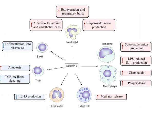
1.9. Function in immune system
During the last decades, compelling evidence has been accumulated regarding the ability of Gal-3 to affect both innate and adaptive immune system, modulating the course of immune responses, inflammation, cell growth, signaling and chemotaxis (Rabinovichet al.2002; Dumic et al. 2006; Sundblad et al. 2011). This effect can be