UNIVERSITY OF MOLISE
Department of Biosciences and Territory
DOCTORAL THESIS
S.D.S. BIO/19
Analysis and characterization of microbial biofilm associated
with the roots of Phragmites australis and Typha latifolia
UNIVERSITY OF MOLISE
Department of Biosciences and Territory - DiBT
PhD course in Biosciences and Territory (Bio-Environmental curriculum)
Cycle XXX – 2014/2015 – S.D.S. BIO/19
DOCTORAL THESIS
“Analysis and characterization of microbial biofilm associated with the
roots of Phragmites australis and Typha latifolia”
PhD student: LAURA PIETRANGELO
__________________________________
Supervisor: Professor Gino Naclerio Coordinator: Professor Gabriella Stefania Scippa
1
“Analysis and characterization of microbial biofilm associated with the
roots of Phragmites australis and Typha latifolia”
LAURA PIETRANGELO
University of Molise
Department of Biosciences and Territory
DOCTORAL THESIS Pesche (IS) 2018
2
“Analysis and characterization of microbial biofilm associated with the
roots of Phragmites australis and Typha latifolia”
LAURA PIETRANGELO
Supervisors:
Prof Gino Naclerio
University of Molise
Department of Biosciences and Territory (DiBT) Pesche (Italy)
Dr Davide Bulgarelli
University of Dundee
Plant Sciences, School of Life Sciences Dundee (United Kingdom)
Reviewers:
Prof Erika Tóth
Eötvös Loránd University Department of Microbiology Budapest (Hungary)
Prof Mogens Nicolaisen
Aarhus University
Department of Agroecology - Entomology and Plant Pathology Slagelse (Denmark)
3
“Analysis and characterization of microbial biofilm associated with the
roots of Phragmites australis and Typha latifolia”
LAURA PIETRANGELO
Abstract
Phragmites australis and Typha latifolia are two macrophytes commonly present in natural
and artificial wetlands. Roots of these plants engage in interactions with a broad range of microorganisms, collectively referred to as the microbiota. These interactions contribute to the natural process of phytodepuration, whereby pollutants are removed from contaminated water bodies through plants. The outermost layer of the root corpus, the rhizoplane, is a hot-spot for these interactions where microorganisms establish specialized aggregates designated biofilm. Earlier studies suggest that biofilm-forming members of the microbiota play a crucial role in the process of phytodepuration. However, the composition and recruitment cues of the Phragmites and Typha microbiota remain poorly understood. We therefore decided to investigate the composition and functional capacities of the bacterial microbiota thriving at the P. australis and T. latifolia root-soil interface. By using 16S rRNA gene Illumina MiSeq sequencing approach we demonstrated that, despite a different composition of the initial basin inoculum, the microbiota associated with the rhizosphere and rhizoplane of P. australis and T. latifolia tend to converge towards a common taxonomic composition dominated by members of the phyla Acidobacteria, Actinobacteria, Firmicutes, Proteobacteria and Planctomycetes. These differences were mirrored by a structural diversification of the microbiota at lower taxonomic ranks. This indicates the existence of a selecting process acting at the root-soil interface of these aquatic plants reminiscent of the one observed for land plants. The magnitude of this selection process is maximum at the level of the rhizoplane, where we identified different bacterial taxa enriched in and discriminating between rhizoplane and rhizosphere fractions in a species-dependent and –independent ways. This led us to hypothesize that the structural diversification of the rhizoplane community underpins specific metabolic capabilities of the microbiota. We tested this hypothesis by complementing the sequencing survey with a two-pronged approach. First, we inferred the functional potential of these communities through a predictive metagenomics approach using the software PICRUSt and we found that transporters and transcription
4 factors-encoding genes are a distinctive feature of the rhizoplane-enriched communities. In parallel, we used Scanning Electronic Microscopy, bacterial isolation and a biochemical assay to demonstrate that rhizoplane-enriched bacteria have a bias for biofilm-forming members. Together, our data will set the stage towards the rational exploitation of plant-microbiota interactions for phytodepuration.
5
CONTENTS
List of publications……….….7
1. INTRODUCTION……….8
1.1 Water, a resource to be safeguarded from pollution………...9
1.2 Wetland ecosystems………...9
1.3 The phytodepuration process……….……..10
1.3.1 The role of plants………...10
1.3.2 The microbial involvement………....12
1.4 The plant microbiota………....14
1.5 How to study the plant microbiota………17
1.5.1 Culture-dependent approaches………..……
17
1.5.2 Culture-independent
approaches:
DNA
sequencing-based
methodologies………...
17
1.5.2.1 Targeted amplicon sequencing for the characterization of
microbiota………19
1.5.2.2 Metagenomic approaches for microbiome investigations…...…….21
1.5.3 Establishing causality: the emergence of Synthetic Communities
(SynComs) of the plant microbiota………....24
2. OBJECTIVES……….
26
3. MATERIAL AND METHODS………..28
3.1 Samples collection and preparation………..…29
3.2 Biomolecular analyses……….29
3.2.1 16S rRNA gene sequence data analysis……….…32
3.2.2 PICRUSt predictive analysis of metagenomes.………..…34
6
4. RESULTS………..………...………38
4.1 Composition of the prokaryotic communities associated to Phragmites australis
and Typha latifolia………...39
4.1.1 Alpha-diversity calculations: the diversity within samples………39
4.1.2 Beta-diversity calculations: the diversity between samples…………...…41
4.2 The rhizoplane microbiota………...…43
4.2.1 Proteobacteria………....45
4.2.2 Planctomycetes………...48
4.2.3 Actinobacteria………...
48
4.2.4 Acidobacteria……….50
4.2.5 Firmicutes………..50
4.2.6 Chloroflexi……….51
4.2.7 Bacteroidetes……….
52
4.2.8 Verrucomicrobia………53
4.2.9 Cyanobacteria………....53
4.3 Conserved members of the rhizoplane communities………....54
4.4 Metagenomic analysis through PICRUSt………...……….56
4.5 The rhizoplane of P. australis and T. latifolia is a site for microbial
colonization….………...….62
4.6 Root-isolated bacteria have distinct biofilm-forming capabilities……..……..…63
5. DISCUSSION………...………68
6. CONCLUSIONS AND PERSPECTIVES……..………...74
Literature cited……….……….76
SUPPLEMENTARY MATERIAL………..……89
Supplementary figures……….…89
7
List of publications
This thesis is based on the work presented in the following paper:
8
Chapter 1
9
1.1 Water, a resource to be safeguarded from pollution
Water is an essential resource for our life, yet water pollution is one of the most serious ecological problems of the planet. Therefore, preserving water quality is major challenge that humanity is facing in the twenty-first century all over the world (Schwarzenbach et al., 2010). Water pollution is definable generally as the degrading of water quality due to toxic substances which accidentally or intentionally enter in water bodies such as rivers, lakes, seas and oceans, dissolve in them, remain floating on their surface or deposit themselves on the bed of water systems (Goel, 2006; Schwarzenbach et al., 2010).
The increasing of urbanization, industrialization and over population can be identified as main causes of water pollution, since industrial and residential wastes, agricultural and surface runoff exert the major impact on the quality of water bodies (Dhote and Dixit, 2009). To avoid the contamination of receiving water bodies various conventional methods are applied to remove pollutants from wastewaters, yet these conventional methods result costly and appear not sustainable in the long term (Dhote and Dixit, 2009). To overcome these problems, from many decades the most researched field in biological and environmental sciences all over the world has been the development of bioremediation techniques, i.e. biological processes performed naturally by eukaryotic and prokaryotic organisms or derived from their interaction, which are able to mitigate the environmental pollution using a lower amount of energy and thus resulting less expensive and more eco-friendly approaches (Srivastava et al., 2017).
Interestingly, many studies demonstrated that aquatic plants in the natural wetlands ecosystems play a relevant role in the removal of pollutants (Williams, 2002).
1.2 Wetland ecosystems
Wetlands are land areas wet during a part or all the year definable as the interface zones between freshwater and soil (Srivastava et al., 2017). The complex wetland ecosystem is based on the interaction between vegetation, microorganisms, animals, soil and water. In recent years the characterization of natural wetlands gained center stage in biological science owing to their contribution to the process of phytodepuration, whereby polluted sites are reclaimed to their natural
10 status through the use of plants (Stout and Nüsslein, 2010; Faußer et al., 2012; Gupta et al., 2012; Sharma et al., 2013).
Interestingly, artificial wetlands, mimicking the processes occurring in natural environments, have been designed and engineered to be used as a low-cost useful technology for wastewater treatment (Mthembu et al., 2013; Chong-Bang et al., 2010; Yongjun et al., 2010). More precisely, these artificial systems exploiting the natural processes of phytodepuration have been effectively used to remove pollutants from municipal, industrial, livestock farming wastewaters and from mine drainage (Basker et al., 2014; Stefanakis et al., 2011).
However, although the effectiveness of phytodepuration in such natural or recreated wetlands has been widely demonstrated, the process is not fully understood. Consequently, this knowledge gap is currently hampering rational biotechnological manipulations of phytodepuration processes to improve the water depuration efficiency.
1.3 The phytodepuration process
Phytodepuration is the process based on the combined action of aquatic plants and microorganisms which results in the removing of contaminants from water and sediments and finally in the improvement of water quality in natural and artificial wetlands (Domininguez-Patino et al., 2012).
1.3.1 The role of plants
A pivotal role for plants in phytodepuration processes has been reported in many informative overviews addressed both land and wetland plant species (Zhang et al., 2010; McCutcheon and Jørgensen, 2008; Williams, 2002; Dietz and Schnoor, 2001; Macek et al., 2000; Susarla et al., 2002). Plants have shown the capacity to withstand relatively high concentrations of contaminants without toxic effects (Zhang et al., 2010). They can uptake some chemicals as nutrients (i.e., N, P) and in some cases quickly convert toxic compounds to less toxic metabolites (i.e., phytotransformation) (Shelef et al., 2013; Zhang et al., 2010). Moreover, they release root exudates and enzymes which stimulate the degradation of the organic chemicals in the rhizosphere (i.e., rhizosphere
11 bioremediation) and represent a source of organic carbon for the microbial metabolism (Zhang et al., 2010). Also, an important role of plants was recognized in the uptake and recovery of metal contaminants into above-ground biomass (i.e., phytoextraction). Likewise, plants can act as a ‘biological filter’, sequestering at the root-soil interface water pollutants (i.e., rhizofiltration) (Zhang et al., 2010). Finally, it has been demonstrated that plants can ‘stabilize’ contaminated sites by reducing the risk of soil erosion and increasing the water evapotranspiration flux, both useful strategies to reduce the risk of contaminant dispersal to other sites (i.e., phytostabilization) (Zhang et al., 2010).
Regardless of aforementioned specific processes, phytodepuration in wetlands systems can be summarized as the net outcome of both direct and indirect interactions between plant roots and microorganisms. Indeed, the capability of the root system to oxygenate the sediment was demonstrated as a key in sustaining the metabolic activities of aerobic microorganisms such as the rhizobacteria (Faußer et al., 2012).
Interestingly, aquatic plants, mainly of the order Poales, Cyperaceae, Juncaceae, Typhaceae as well as other monocots, have evolved dedicated aeration systems which run through all plant organs (Brix et al., 1992). These include belowground rhizomes interconnecting individual plants (Klimešová and Čížková-Končalová, 1996). Anatomically, a specialized plant tissue, designated aerenchyma, empowers wetland plants to channel oxygen to submerged tissues and, at the same time, to partly oxygenate the rhizosphere surrounding belowground organs (Armstrong et al., 2000; Colmer, 2003). At the molecular level, this task is accomplished through the mechanism of radial oxygen loss (ROL) (Colmer et al., 2006; Matsui and Tsuchiya, 2006, 2008). The oxygen released via ROL in the rhizosphere underpins, at least in part, the biochemical reactions, catalyzed by both plants and microorganisms, degrading and recycling (into plant nutrients) phytotoxic compounds. Furthermore, the root system offers a wide surface to host microorganisms and, through the diffusion of exudates and other organic compounds, stimulates the degradation of pollutant by resident microorganisms (Trapp and Karlson 2001; Trapp et al., 2007).
The plant species more represented in the natural wetlands, and therefore more utilized in phytodepuration applications, are Phragmites spp. and Typha ssp. These plants can adapt to different abiotic conditions and, therefore, have a worldwide diffusion (Bellavance and Brisson, 2010; Li et al., 2013). In addition, these are perennial plants capable of performing the water cleaning process in the site of their rooting all year round (Tsyusko et al., 2005; Srivastava et al., 2014; Bonanno and Cirelli, 2017; Eller et al., 2017; Mthembu et al., 2013) and finally, thanks to a rhizomatous propagation, can promptly colonize wetlands areas (Dhir, 2013; Juneau and Tarasoff, 2013).
12
1.3.2 The microbial involvement
In the wetland ecosystems a wide range of microorganisms is commonly observed as detrital microbial mat, biofilm, and planktonic-microalgal-bacterial assemblages (Battin et al., 2003; Srivastava et al., 2017). These microorganisms contribute substantially to the nutrient cycling (e.g., nitrification, denitrification, sulfate reduction, methanogenesis, metal ion reduction or oxidation) and energy flow (Srivastava et al., 2017). In particular, the presence of biofilm microbial assemblages has commonly been detected on different plant surfaces, such as the leaves of submerged plants, in the rhizosphere on sediment and, more often, on the root surface, i.e. the area identifiable as rhizoplane (Srivastava et al., 2017; Giaramida et al., 2013; Calheiros et al., 2009). The tight and preferential association of microorganisms forming biofilm on the root surface suggests a functional interaction of microbial cells with the plant roots and with roots products diffused in the surrounding. For example, it has been proposed that oxygen and root exudates (carbon compounds) can be “traded” by the plants to fuel the microbial metabolism needed to degrade phytotoxic compounds (Srivastava et al., 2017). Consistently, this has recently been demonstrated by Srivastava et al. (2017) who reported the capability of the aquatic plant associated biofilm to degrade the algal-derived organics, i.e. chiefly amines, aliphatic aldehydes and phenolics (Simpson, 2008) and to use such algal derived carbon to grow and multiply efficiently (Gasol and Duarte, 2000). Moreover, the microorganisms constituting the plant associated biofilm have been demonstrated able to degrade the dissolved organic matter (DOM) (Tranvik, 1998) such as PCBs (poly-chlorinated biphenyls) (Ghosh et al., 1999) and atrazine (Guasch et al., 2007). The rhizoplane of aquatic plants resulted also being enriched for ubiquitous methanotrophs (α and γ proteobacteria) which use methane as carbon source for their metabolism removing it from the aquatic ecosystem (Semrau et al., 2010). Moreover, thanks to particulate methane monooxygenase (pMMO), some bacterial species (e.g., Methylosinus trichosporium,
Methylococcus capsulatus) resulted able to degrade a wide range of others toxic organic compounds
and among them especially chlorinated ethenes (Pandey et al., 2014), via a cascade of enzymatic reactions which end with CO2 as terminal product.
As reported from Hansel et al. (2001) and Carranza-Álvarez et al. (2008) microorganisms arranged as biofilms on the root surface perform also an important role in the removing of metal pollutants from water bodies. Sub-toxic levels of metals usually identified in wetlands can be the result of natural leaching of soil and sediments (Srivastava et al., 2017). However, these metals can reach toxic level when they are introduced into water bodies from industrial, agricultural and
13 municipal wastes (Zhou et al., 2008; Hansel et al., 2001; Carranza-Álvarez et al., 2008; Srivastava et al., 2017).
Interestingly, King and Garey (1999) and Hansel et al. (2001) reported that a consistent proportion of metal cations in water adheres to the negatively charged EPS of microbial biofilm matrix forming metal plaques around the roots of aquatic macrophytes and around all submerged plant parts. Through this mechanism, metals are sequestered from the water body and, not less important, the presence of plaques prevent other metals to enter and accumulate up to toxic level into plant tissues (Srivastava et al., 2017). Iron plaques are commonly detected around the roots of aquatic macrophytes and although their formation is principally due to iron oxidation process mediated by molecular O2, their presence is more consistent when iron oxidizing bacteria such as Ferroplasma sp.
and Leptospirillum ferroxidans are detected on the root surface (King and Garey 1999).
Sulfate reducing bacteria constituting biofilm on aquatic macrophytes roots also contribute to the metals removing process. Reducing sulfate in sulfides, they determine the lowering of water pH to values required from microbial cells to bioabsorb the metal ions from water (Han and Gu, 2010). Moreover, the hydrogen sulfide produced from these microorganisms reacts with metal ions and forms metal sulfide, which under acidogenic conditions precipitates sequenstring metal ions from water body (Webb et al., 1998; Machemer and Wildeman, 1992).
Together, these experiments clearly point to an active involvement of microorganisms in the phytodepuration process and an evident effective interaction of microbes with plants, particularly evident for microorganisms constituting biofilm on the rhizoplane. However, the knowledge about the entire microbial community underpinning the phytodepuration process and the factors which influence its composition are still incomplete.
These are becoming crucial questions in this research field, as advances in sequencing technologies and computational analysis have confirmed that plants are not autonomous entities but rather are sites of colonization for a myriad of microorganisms, collectively referred to as the plant microbiota, whose interactions at given plant sites define distinct biomes, the plant microbiome (Schlaeppi and Bulgarelli, 2015).
14
1.4 The plant microbiota
Microorganisms colonize almost all ecological niches. Plants represent, as the rest of most multicellular eukaryotic organisms, effectual provider of nutrients for microorganisms and thus result good hosts for them.
Along the plant structure diverse abiotic factors such as temperature, moisture, oxygen availability, wind exposure, etc. interact with biotic factors, such as the wide range of compounds produced by plant cells, creating outside and inside plant tissues different microhabitats for microbial colonization, epiphytes and endophytes respectively (Schlaeppi and Bulgarelli, 2015). One of the most characterized plant microhabitats is the rhizosphere, where microorganisms are associated to the thin soil layer around roots under the influence of numerous plant exudates (Walker et al., 2003). Instead, the rhizoplane microhabitat hosts microorganisms which live in a more tight interaction with roots, adhering on their surface. Then, endosphere is identifiable as the microhabitat which permits the microbial survival inside the plant tissue and finally the phyllosphere, the microhabitat colonized by microorganisms which proliferate on the stem and leaf surface (Hardoim et al., 2008; Lindow and Brandl, 2003).
Microorganisms interact with the host as their pathogens, mutualists or commensals. Although for economical reason the pathogens were the most studied ones, in the last decades great attention has been paid to other plant-associated microorganisms, especially after the demonstration of their important role in maintenance of plants health and for the improvement of their growth. Well know is the beneficial action of mutualists such as rhizobia for leguminous plants or of nitrogen fixing bacteria for their hosts (Masson-Boivin et al., 2009). Yet, in recent years also commensals, which by definition do not advantage the host plant, have been suggested as indirectly implicated in the plant protection and development under specific conditions.
Since it seems that plants can count for their survival not only on their own genes, but also on the accessorial pool of microbial genes as an “extended" trait to reach the adaptation to the environment, the plant and its microbiota are considered as an unique entity, an holobiont, whose genome, the holobiome, is composed both by plant genome and microbiome subjected to a mutual evolution process (Theis et al., 2016).
Effectively, the coevolution of plant host with its associated microbial community defines for each plant a consistent microbiota which differentiate it at specific and subspecific levels (Hartmann et al., 2009). The advantages obtained from the coevolution of plant genome and the associated microbiome appear clear since many plant hosts have shown their tight interaction with individual
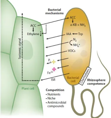
15 members of microbiota. This is the case of Plant Growth Promoting Rhizobacteria (PGPRs) which are able to confer protection to plants against pathogens and to enhance plant’s capabilities to uptake nutrients from soil (Lugtenberg and Kamilova, 2009). Specifically, it has been demonstrated that rhizosphere bacteria can contribute to the plant uptake of scarcely mobile minerals, such as iron and phosphorus, the biochemical fixation of atmospheric di-nitrogen into ammonia, the production of phytohormons, such as Indole Acetic Acid, and other signaling compounds such as volatile compounds (Berendsen et al., 2012, Turner et al., 2013, Lugtenberg and Kamilova, 2009; Yang et al., 2009; Berg et al., 2014). Likewise, non-pathogenic rhizobacteria can trigger plant immune responses in distal organs, a process known as Induced Systemic Resistance, and compete, either directly or indirectly, with pathogenic bacteria for the colonization of the root-soil interface (Reviewed by Bulgarelli et al., 2013, Figure 1).
FIGURE 1. Biochemical mechanisms by which rhizobacteria influence plant growth and health (from Bulgarelli et al., 2013).
Therefore, this positive influence of microbiota on plant could be considered an important manipulation target for crop improvement and management.
Yet, the knowledge about the composition of microbiota of major part of plants remains limited as well as the understanding of the molecular interaction between constituting
16 microorganisms with plant host. Moreover, about the picture of other abiotic and biotic determinants of the structure and function of the plant microbiota is far to be completed (Schlaeppi and Bulgarelli, 2015; Waldor et al., 2015).
In addition, the biological complexity of existing ecosystems makes it at the same time difficult and fascinating to infer general principles of plant-microbiota interactions (Alegria Terrazas et al., 2016). On these assumptions, the characterization of the microbiota of land plants has been gaining momentum both in basic and translational science (Hacquard et al., 2015). Yet, elucidating the functional significance of aquatic plants microbiome can be considered a research field in its infancy (Bowen et al., 2017; Cerri et al., 2017).
Consequently, one of the principal research targets in environmental science is the possible manipulation of the microbiota as eco-friendly strategy to increase the phytodepuration efficiency, an approach expressly similar to the one proposed to sustainably increase crop production and reduce the input of chemicals and the emission of greenhouses gases in natural ecosystems (Bakker et al., 2013; Adesemoye et al., 2009; Singh et al., 2010; Berg et al., 2014).
Thanks to its important contribution to the plant survival and development, the microbiome, as previous specified, may be considered a second plant genome whose composition is actually strongly influenced by the plant genome (Turner et al., 2013; Berg et al., 2014). Yet, only for a limited number of species the first principles underpinning the important interaction between plants and their microbiotas have been defined, whereas for the majority of other plants, the intertwined relationship host-microbiome is still poorly characterized (Turner et al., 2013). Moreover, also the abiotic conditions of different microenvironments such as temperature, soil properties, moisture, pH, and nutrients amount influence broadly, directly and indirectly, the composition of plant microbiota. According to that, also microbiomes associated with different compartments of the same plant such as above-ground, below-ground and internal tissues compartments are distinct from each other (Berg et al., 2014; Turner et al., 2013; Berg and Smalla, 2009). However, although microorganisms colonize most plant compartments, since the well demonstrated influence of soil habitat on plants productivity and the necessity to adapt the plant cultures to different soil conditions, the larger part of researches targets to the characterization of rhizosphere microbiome (Berg et al., 2014; Alegria Terrazas et al., 2016). Only few other plant compartments have been studied in this respect (Vorholt, 2012) and, with few notable exceptions (Edwards et al., 2015), the rhizoplane microbiota received limited attention.
17
1.5 How to study the plant microbiota
The study of plant microbiota ultimately aims at identifying the structural and functional composition of the entire microbial community associated to different plant compartments. Since each microbial community is constituted by a wide range of microorganisms which interact with each other and with the host, both culture- dependent and -independent approaches are required to capture as much as possible of the microbiota diversity (Turner et al., 2013).
1.5.1 Culture-dependent approaches
The culture-dependent approach for studying the microbial community associated to plant compartments involves the isolation of microorganisms from specific microenvironments on artificial media. Fundamental and critical in this type of studying approach is the choice of adequate media to use as substrates for the microorganisms growth. The useful and commonly applied strategy is the use of media which mimic as much as possible the environmental conditions of the ecological niche which microbes live in. However, no medium can perfectly reproduce all the abiotic and biotic factors and their interactions influencing the microbial life and survival. Historically, this technique has been considered limited by the fact that many soil bacteria, from which the plant microbiota is largely assembled, were considered recalcitrant to in-vitro cultivation: it was estimated that only a limited portion of microorganisms (i.e., less than 1%) can be obtained in pure culture through classic microbiology culture methods (Vieira and Nahas, 2005). However, recent breakthrough discoveries are challenging this vision and culture dependent approaches are regaining center-stage in the characterization of the plant microbiome (see paragraph 1.5.3).
1.5.2 Culture-independent
approaches:
DNA
sequencing-based
methodologies
Since the late 1970s the development and continuous implementation of culture-independent techniques targeted to the study of microbial phylogenetic markers, combined with the more recent advancement in computational analysis has permitted a pronounced outburst in characterization of
18 microbial communities associated to a wide range of different environments (Schlaeppi and Bulgarelli, 2015). In particular the introduction of high throughput sequencing (HTS) technologies which perform the sequencing of multiple DNA molecules in parallel, it is now possible the obtainment of thousands to millions of sequences in more than one sample at a time, revealing the abundances of even rare microbial species (Bentley et al., 2008; Margulies et al., 2005; Turner et al., 2013). Combined to this, the availability of many open source tools for data analysis has favored the rational organization and classification of complex sequencing data in consistent sequencing datasets (Caporaso et al., 2010; Meyer et al., 2008). Not less important, the development and availability of public databases where sequencing data are constantly annotated and implemented has facilitated much more the expansion of knowledge about a wide range of microbiotas (Cole et al., 2014; DeSantis et al., 2006; Fish et al., 2013), also permitting the comparison between microbial communities associated to different environments and the reduction of analysis costs.
These sequencing-based methodologies can be actually classified in the methods indicated as targeted amplicon sequencing and the metagenome approach. The target amplicon sequencing is applied to a specific set of genes of the studied microbial community, whereas the metagenome approach is aimed at providing a general overview about the functional role of microorganisms in a microbiota through the study of genes considered as genetic markers for specific functions and metabolic capabilities (Alegria Terrazas et al., 2016; Turner 2013). Since both approaches provide fundamental information to deeply understand the composition and organization of microorganisms in the whole community, the preferential strategy to study the microbiota should be the combination of both approaches. However, the application of such methods is often made difficult by the diversity and complexity of studied environments. In addition to the fact that the existing environments are all constituted by numerous different niches for microbial colonization and thus are associated to a multitude of microbiotas, each environment is also characterized by an intrinsic complexity due to the fact it is populated by organisms belonging to all domains of life (Turner et al., 2013, Alegria Terrazas et al., 2016). This means that sequencing-based methodologies must be set up and adapted for the study of microbes as diverse as multicellular eukaryotes (e.g., fungi), prokaryotes and viruses. Therefore, usually the characterization of the entire microbiota derives from the characterization of all portions of microorganisms represented in that. However, the prokaryotic and eukaryotic portions of microbiota represented by categories of bacteria, archaea and fungi are the most investigated ones for their supposed and in some cases demonstrated contribution to the maintenance of plant health and development (Turner et al., 2013, Alegria Terrazas et al., 2016).
19
1.5.2.1 Targeted amplicon sequencing for the characterization of the
microbiota
The targeted amplicon sequencing approach is the most applied strategy to study the microbiota. This technique allows scientists to identify the members of microbial communities or to compare the microbiota composition in different samples through the investigation of known phylogenetic markers. These phylogenetic markers permit the taxonomical classification of microorganisms grouping them on the basis of their phylogenetic similarity (Knief, 2014; Alegria Terrazas et al., 2016; Turner et al., 2013). The prokaryotic and eukaryotic genes encoding for the small subunit of ribosomes are the most commonly used phylogenetic markers in targeted amplicon sequencing surveys. These ribosomal genes are characterized by large sequence regions with a slow attitude to incur in sequence modifications thus only slowly subjected to evolutionary changing. Yet, these genes contain also regions with high attitude of being subjected to sequence modifications and to DNA evolving, indicated for this reason as hyper-variable regions. These aspects made the genes encoding for small ribosomal subunits the optimal candidates for the microbial phylogenetic study. Firstly, the presence of conserved sequences within microorganisms permits the usage of a wide range of PCR primers which can perform efficiently the amplification of the phylogenetic markers of all the different members of microbiota. Next, the sequence modifications which occur in the hyper-variable regions differentiate the microbes from each other generating ‘molecular fingerprints’ useful to their discrimination at a far more detailed taxonomic level. For these reasons, the 16S rRNA gene sequencing approach has been the method preferred to study the composition of bacterial and archeal communities since many decades, resulting in the availability of a large in silico dataset about 16S rRNA sequences (Schlaeppi and Bulgarelli, 2015). On the same principle, the 18S rRNA gene, which encodes for the small ribosomal subunit in eukaryotes, is used as phylogenetic marker to discriminate specific members of the microbiota, such as oomycetes, protists and nematodes. However, when the fungal community is the investigation target, the profiling of Internal Transcibed Spacer (ITS) is often applied. This exception for fungi is due to the fact, that the gene encoding for the fungal small ribosomal subunit is characterized by a short sequence subjected to the rapid mutation of DNA and this makes difficult its usage as phylogenetic marker. However, the region comprised between genes encoding for ribosomal subunits, this named ITS, presents the same characteristics of the other phylogenetic markers, i.e. the same proportion between conserved regions and hypervariable sequence (Alegria Terrazas et al., 2016).
20 Regardless to the target of PCR amplification, the principal limitation of this approach is the choice of primers which mainly influences the outcome of analysis. For this reason, each targeted amplicon sequencing procedure should pass through a preliminary set up of amplification protocol testing the amplification efficiency of diverse primer pairs before being applied to a full-scale analysis (Walters et al., 2015). Moreover, in the specific case of the study of microbiota associated with plants the majority of primer pairs generates also the undesired amplification of not targeted plant sequences together with the desiderated microbial ones. More precisely, since of their sequence similarity with sequence regions of 16S rRNA gene, host-derived plastidial and mitochondrial sequences are normally obtained as “contaminants” among the microbial amplicons pool, thus they need to be filtered from the final dataset to provide a realistic characterization of plant microbiota. The proportion of host derived “contaminant” sequences could be also reduced through suitable modification of PCR protocol regarding for example PCR steps temperature setting, number of amplification cycles and obviously the choice of primers pair which produce the lowest interfering amplification of host sequences (Lundberg et al., 2013). Intuitively, this fine tuning of PCR protocol is especially required for the study of endophytic microbiota since in this case the starting samples for the microbiota analysis are inevitably composed of a large portion of plant material and tissues.
The filtering of contaminant sequences and the analysis of entire sequencing dataset is normally conducted through in silico analysis. This process is based on the prior analysis of sequence quality and length, and the removal of all possible PCR artifacts. Subsequently, the highquality sequences are assigned to their source samples and clustered into Operational Taxonomical Units (OTUs) which identify closely related microorganisms, whose 16S rRNA gene sequences present a 97% identity threshold. Basically, OTUs can be considered the individual community members in amplicon sequencing surveys. Moreover, since each OTU is associated to a specific taxonomic classification, the pool of obtained OTUs permits to reconstruct the taxonomical classification of the entire microbial community associated to the investigated plant microbiota. Also, the association of sequences to specific OTUs simplifies the filtering process of those sequences belonging to host plastidial and mitochondrial DNA. The handling of obtained huge amount of sequencing data and the matching of enormous number of sequences with the available database of OTUs are made possible by the use of continually implemented software for the investigation of microbial ecology, such as QIIME (Quantitative Insights Into Microbial Ecology),the most employed one (Caporaso et al., 2010, 2012). Subsequently, after definition of OTUs constituting the studied microbiota, bioinformatics tools are often used to perform the statistical analysis of the OTUs properties such as their presence/absence, their relative abundance in the microbiota and whether these statistical parameters
21 are significantly associated to any given biotic or abiotic factor putatively influencing microbiome composition.
1.5.2.2 Metagenomic approaches for microbiome investigations
The metagenome approaches for studying the microbiome aim at identifying the functional genes detectable in the microbial community. Since the genes investigated are related to specific metabolic functions, the metagenome is the way to determine the putative role of microbes within the microbial community. This type of investigation can be conducted through the approach named gene-targeted metagenomic, a reduced complexity approach focused on the amplicon sequencing of genes encoding only for specific functions used as functional markers. This method is performed similarly to the one dedicated to the phylogenetic markers, but it results in the obtainment of specific database regarding the functional markers of interest in the considered microbiota (Fish et al., 2013, Alegria Terrazas et al., 2016).
Conversely, a methodology designated shotgun metagenomic approach aims a targeting of the totality of DNA collected from the studied environment. This approach is combined with dedicated
in silico analyses aimed at annotating (both taxonomically and metabolically) and characterizing the
putative function encoded by the sequenced material. Similar to the amplicon sequencing approach, the existence of open-access, dedicated analytical servers, such as the widely used MG-RAST (Meyer et al., 2008) are streamlining the associated analytical strategies.
However, this shotgun metagenomic suffers from some intrinsic limitations. In particular, the difficulty in defining the suitable sequencing depth (i.e., number of reads) per individual samples. Likewise, the high number of replicates required to obtain representative results contribute to inflate the costs of the analysis including those for the larger downstream computational effort (Knight et al., 2012).
Another approach to study the microbiome is the predictive metagenomic. This method aims at inferring the composition of a microbial community through available phylogenetic marker information (e.g. 16S rRNA gene profiles). This is achieved by deriving microbial metabolic capacities from database containing the entire genome of microorganisms associated to the same phylogenetic markers. The predictive analysis of microbiomes from samples associated to different niches is often performed through dedicated bioinformatics tools, such as the widely used PICRUSt
22 (Langille et al. 2013). This software predicts the functional composition of a microbial community from its 16S rRNA profile. The PICRUSt analysis process is organized onto two steps, the step of “gene content inference” and the step of “microbial inference” (see material and methods for details). The clear advantage of this method is that it is conducted fully in silico starting from the phylogenetic marker profiles which, nowadays, are routinely obtained in many laboratories. Conversely, the intrinsic limitation is that is based on an algorithm and probabilistic calculations, therefore it could fail to precisely characterize the functional genes in the microbiota (Bulgarelli et al., 2013). Nevertheless, it remains a useful preliminary analysis to evaluate the rate of diverse functions in a microbiota, to compare the functions detected in different microbiotas, understanding how much they differ from each other and also to establish the next steps of analysis (Alegria Terrazas et al., 2016). Figure 2 summarizes the main steps in cultivation-independent analysis of the plant microbiota and microbiome.
23
24
1.5.3 Establishing causality: the emergence of Synthetic Communities
(SynComs) of the plant microbiota
One of the perceived barriers severely impairing the advancement of this research field was the fact that the plant microbiota is represented by soil-dwelling bacteria (Bulgarelli et al., 2013) that, historically, have been considered recalcitrant to in vitro cultivation: it has been estimated that a very minor proportion, often less than 1%, of soil bacteria can be readily isolated on microbiological media (Vieira and Nahas, 2005). However, the realization that a) the plant microbiota is akin to a gated community, whereby a limited number of microorganisms can successfully thrive and b) these microorganisms are largely represented by members of taxa routinely isolated in laboratory (e.g., Actinobacteria, Bacteroidetes, Firmicutes and Proteobacteria (Hacquard et al., 2015)) challenged the paradigm of (un-)culturability of plant-associated microbial communities. For instance, a breakthrough study recently revealed that the majority (~58%) of the Arabidopsis thaliana root microbiota, identified in sequencing surveys, can be recapitulated in a bacterial collection (Bai et al., 2015). The establishment of such indexed bacterial collections is a fundamental pre-requisite allowing scientists to combine isolated members of the microbiota into ‘microbial consortia’ of known composition, designated Synthetic Communities (SynComs). These can be transplanted into germ-free plants and their impact on given plant phenotypes properly discerned. Combined with whole genome information, this is a powerful tool to formulate testable hypotheses and gain novel insights into plant-microbiota interactions. Perhaps not surprisingly, SynComs are taking center stage in microbiota science. In A. thaliana, SynComs have successfully been adopted to identify the host genetic determinants of the leaf-associated communities (Bodenhausen et al., 2014), demonstrate that components of the plant immune system shape the root microbiota (Lebeis et al., 2015) and infer the contribution of the microbiota to phosphorus starvation (Castrillo et al., 2017).
Operationally, the application of the SynComs approach is strictly dependent from a) the definition of the microbiota composition through cultivation-independent approaches, such as the 16S rRNA gene sequencing for bacteria and archaea, and b) the direct isolation of strains from the plant specimens. Although it results often difficult to translate OTUs to strains due to the variability within OTU (97% sequence identity for definition), selecting multiple strains per OTU can overcome this limitation (Callahan et al., 2016; Tikhonov et al., 2015). Therefore, both cultivation-independent and dependent approaches are necessary to ultimately determine whether structural and functional configurations of the microbiota are causally related to the given plant phenotypes.
25 Despite experiments conducted with the model A. thaliana have greatly enhanced our understanding of the interactions between plants and their associated microbial communities, the existence of species-dependent recruitment cues for these microbes (Bulgarelli et al. 2013, Hacquard et al., 2015) as well as the distinctive environmental variables impacting on these interactions are now calling for dedicated investigations of the plant microbiota in diverse natural and managed ecosystems, such as wetlands.
26
Chapter 2
27 Many studies demonstrated the potential and importance of microbiota for land plant nutrition, maintenance of their health state and development. Moreover, whereas the microbiota sustains indirectly plant growth, is also directly active in metabolizing and mobilization of chemicals and pollutants from the surrounding, usually performing a synergic action in association with plants which are the final up-taker of metabolized products in/on their tissues. Also in wetlands the important involvement of microorganisms associated with plants in removing pollutants from water and sediments has been demonstrated, in the process usually indicated as phytodepuration. In particular, microorganisms which colonize the rhizoplane of aquatic plants and form biofilm assemblages on their root surface have revealed an interesting ability in removal of pollutants from wetlands. Yet, the wetland plants microbiota is a research field in its infancy and in particular the microbiota of rhizoplane compartment received less attention compared to the communities inhabiting the rhizosphere. Therefore, the composition of the microbial assemblages on the root surface and their real potential contribution to phytodepuration remain largely unknown. Earlier studies suggest that Proteobacteria dominate the root-soil interface of Phragmites australis and Typha latifolia. However, these studies were conducted with low-resolution techniques and this makes it difficult to infer general principles.
To gain novel insights into the functional significance of the wetland plants microbiota, this doctoral thesis presents:
The characterization through Illumina MiSeq technology of the bacterial communities associated with the roots of Phragmites australis and Typha latifolia and comparison with the ones inhabiting the surrounding compartments, to understand the effect of microhabitat conditions in shaping the microbiome;
A comparison between the two plants microbiotas to point out the role of plant species factor in recruitment of microorganisms at root surface;
A predictive metagenomic investigation aimed at elucidating the functional potential of these communities;
A Scanning Electron Microscopy (SEM) analysis to go insight the spatial organization of the rhizoplane microbiota on the root surface;
The isolation of culturable portion of microorganisms from the rhizoplane of both plant species as a first step towards the establishment of SynComs for P. australis and T.28
Chapter 3
29
3.1 Samples collection and preparation
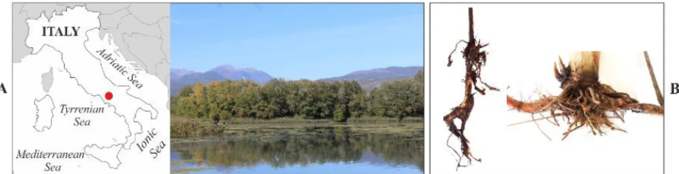
From the wetland located in the naturalistic area of “Le Mortine Oasis” (Campania, southern Italy) five root systems of five Phragmites australis and Typha latifolia plants respectively were sampled in sterile bags (Figure 3). Also four surface water samples (1L) were collected in sterile bottles from sampling points distributed along the Phragmites and Typha rooting sites. The samples were immediately transported to the laboratory in a portable cooler at 4 °C. 1 g of the soil surrounding the Phragmites australis and Typha latifolia roots was collected in sterile Petri dishes and used to investigate the rhizosphere microbial communities. Four root segments (2 cm in length and 0.3-0.5 cm in thickness) were obtained from each plant system. Subsequently, the root segments were washed by shacking three times in 10 ml of sterile tap water, twice in 10 ml of sterile distilled water and once in 20 ml of sterile 0.85% NaCl (Kirzhner et al., 2009; Li et al., 2013).
FIGURE 3. Sampling. The sampling site in the natural area of “Le Mortine Oasis” (41°28'11.4"N 14°05'26.6"E) (A) and examples of P. australis (B, left) and T. latifolia (B, right) root systems sampled from the wetland.
3.2 Biomolecular analyses
Water samples (1L) were filtered through sterile mixed esters of cellulose membranes (S-PakTM Membrane Filters, 47 mm diameter, 0.22 μm pore size, Millipore Corporation, Billerica, United States) and the DNA was extracted from the filters using the PowerWater® DNA Isolation Kit (MO BIO Laboratories, Inc., Carlsbad, United States) following manufacturer's recommendations. The DNA samples generated from the wetland water (W1, W2, W3, W4) were stored at -20°C until further use.
30 0.5 g of each soil sample was subjected to the DNA extraction using the PowerSoil® DNA Isolation Kit (MO BIO Laboratories, Inc., Carlsbad, United States) as the manufacture’s producer recommend. DNA extracted from Phragmites australis (Pr1, Pr2, Pr3, Pr4) and Typha latifolia (Tr1,
Tr2, Tr3, Tr4) rhizosphere was stored at -20°C until further use. Furthermore, three of the washed
root segments were subjected to a double step ultrasound treatment using the Vibra-Cell™ ultrasonic processor VCX 130 (Sonics and Materials, Inc. Newtown, United States) set at the constant frequency of 20 kHz and at the amplitude of 30%, with a 6 mm probe. Firstly, the roots were sonicated in 10 ml of sterile sonication buffer (0.85% NaCl and 0.1% Tween 80) for 2 min and 30 sec in 15 ml Falcon tube, then the roots were transferred into 10 ml new sterile sonication buffer and subjected to a second ultrasound treatment in the same conditions, for 5 min. This procedure ensured through the first sonication step the removal of cells not firmly attached onto the root surface and the detachment and subsequent collection of the rhizoplane cells during the second step. After the ultrasound treatments, the roots were recovered and fixed using 3% glutaraldehyde in a 0.1 M phosphate buffer at pH 7.2 for 24 hours. Therefore, samples were washed three times using the same buffer and dehydrated through an ethyl alcohol series (30, 50, 70, 95, and 100%, for 5 min at each step). After dehydration, they were dried using an Emitech K850 Critical Point Dryer (Quorum Technologies Ltd, England, United Kingdom), mounted on aluminum stubs and coated with gold using an Emitech K550 sputter coater (Quorum Technologies Ltd, England, United Kingdom). Finally, the prepared samples were observed using a ZEISS DSM-940A Scanning Electron Microscope (Carl Zeiss, Jena, Germany) at 10 kV and 30x and 2000x magnification images were acquired. Instead, the suspension of rhizoplane cells was divided in two aliquots (5 ml) representing replicates of each sample (indicated as “a” and “b”). The 5 ml aliquots were brought to a final volume of 100 ml with sterile MilliQ water and filtered through sterile mixed esters of cellulose membranes (S-PakTM Membrane Filters, 47 mm diameter, 0.22 μm pore size, Millipore Corporation, Billerica, United States). DNA was finally extracted from the filters using the PowerWater® DNA Isolation Kit (MO BIO Laboratories, Inc., Carlsbad, CA, United States) following the recommended protocol. More precisely, for each biofilm suspension two DNA samples were originated as replicates and they were indicated as “a” and “b” respectively. A total of 10 DNA samples were obtained from the rhizoplane of Phragmites australis (P1a and P1b;
P2a and P2b; P3a and P3b; P4a and P4b; P5a and P5b) and Typha latifolia (T1a and T1b; T2a and T2b; T3a and T3b; T4a and T4b; T5a and T5b). The DNA samples were stored at -20°C until further
use. DNA samples were quantitated using the NanoDrop 1000 spectrophotometer (Thermo Scientific, Wilmington, United States) and they were subjected to the amplification of the hypervariable V4 region of the 16S rRNA gene through a nested-PCR approach to generate amplicon libraries. The
31 PCR reactions were performed using Kapa HiFi HotStart PCR kit (Kapa Biosystems, Wilmington, United States) in a G-Storm GS1 Thermal Cycler (Gene Technologies, Somerton, United Kingdom). For the first amplification step the PCR mix contained 50 ng of DNA, 4 µl of 5X Kapa HiFi Buffer, 10 ng Bovine Serum Albumin (Roche, Mannheim, Germany), 0.6 µl of a 10 mM Kapa dNTPs solution, 0.6 µl of 10 µM solutions of the 27F (5’-AGAGTTTGATCMTGGCTCAG-3’) and 1392R (5’- ACGGGCGGTGTGTRC-3’) PCR primers, 0.25 µl of Kapa HiFi polymerase and sterile MilliQ water up to the final volume of 20 μl. The reaction was performed with an initial denaturation at 94°C for 3 min, then 20 cycles of denaturation at 98°C for 30 sec, annealing at 55°C for 30 sec, elongation at 72°C for 1 min and 30 sec and a final elongation step at 72°C for 10 min. The second amplification step was conducted using 2 µl of the first amplification product as template, 4 µl of 5X Kapa HiFi Buffer, 10 ng Bovine Serum Albumin (Roche, Mannheim, Germany), 0.6 µl of a 10 mM Kapa dNTPs solution, 0.6 µl of 10 µM solutions of the 515F (5’-GTGCCAGCMGCCGCGGTAA-3’) and 806R (5’-GGACTACHVG GGTWTCTAAT-3’) PCR primers, 0.25 µl of Kapa HiFi polymerase and sterile MilliQ water up to the final volume of 20 μl. To generate the amplicon libraries both primers used in this PCR step presented flow cell adapter sequences at their 5’ termini and the primers 806R also 12-mer unique ‘barcode’ sequences to provide the simultaneously sequencing of several samples (Caporaso et al., 2012). This PCR reaction was performed using the following conditions: initial denaturation at 94°C for 3 min, 25 cycles of denaturation at 98°C for 30 sec, annealing at 50°C for 30 sec, elongation at 72°C for 1 min and a final elongation step at 72°C for 10 min. Reaction negative controls (rNTCs) were generated in all the individual PCR reactions and for all the barcodes used in the second amplification. Furthermore, four no-template samples were amplified through both nested-PCR steps and thus they were tagged by their own barcodes in the second amplification step to be used as sequencing negative controls (sNTCs). 5 µl of amplified samples and controls were checked on 1.5% agarose gel. The samples which showed the expected size amplicon and whose rNTCs presented no detectable amplicon were used for the amplicons library construction. The four sNTCs were also used to generate the amplicons library. The amplicons and the sequencing negative controls (sNTCs) were purified using Agencourt AMPure XP kit (Beckman Coulter, Brea, United States) with a ratio of 0.7 µl AMPure XP beads per 1 µl of sample and then 3 µl of each sample were quantified using Picogreen (Thermo Fisher, United Kingdom) according to the manufacturer’s recommendations. After that, individual barcode samples were pooled at equimolar ratios to generate the amplicon libraries. All library QC and processing was carried out by the Genome Technology group at James Hutton Institute (Invergowrie, United Kingdom) and high-quality libraries were run
32 at 10 pM final concentration on an Illumina MiSeq system with paired-end 2 × 150 bp reads for FASTQ file generation (Caporaso et al., 2012).
3.2.1 16S rRNA gene sequence data analysis
The FASTQ files obtained from the MiSeq machine were processed using the QIIME software version 1.9.0 (Caporaso et al., 2010). Firstly, forward and reverse files from libraries were decompressed and merged through the command join_paired_ends.py setting the minimum overlap of 5 bp between reads. Then the overlapping paired end (PE) reads were subjected to demultiplexing and quality filtering running the command split_libraries_fastq.py with a minimum PHRED score of 20. Subsequently, the high quality PE reads were matched with Operational Taxonomic Units (OTUs) at 97% sequence identity collected in the chimera-checked Greengenes database (DeSantis et al., 2006), version 13_5, using the “closed reference” approach. For the OUT-picking the SortMeRNA algorithm (Kopylova et al., 2012) was used. The singleton OTUs, OTUs associated only to a single read, were filtered in silico and using the command merge_otu_tables.py the OTU tables obtained from the two independent sequencing runs were merged to obtain an unique OTUs table. Then, from this OTUs pool were in silico filtered the OTUs assigned to host- derived sequences, i.e. plastidial or mitochondrial DNA. Through the command summarize_taxa.py, the taxonomy matrix correspondent to the OTUs table was generated. The taxonomy matrix, reporting the number of reads for each identified taxonomy, was finally merged to the OTUs table generating a unique file reporting identified OTUs with correspondent taxonomies and number of reads for each taxonomy in the individual libraries. This file has been used for the statistical analysis performed in R using the R Phyloseq package (McMurdie and Holmes, 2013) as follow described.
The alpha and beta-diversity calculations were performed for two samples sets in parallel, each one composed of all the rhizosphere and water samples plus respectively the first set of rhizoplane replicates (set1) or the second set of replicates (set2). Therefore, two independent OTUs tables were obtained. Firstly, low abundance OTUs were filtered from the datasets, referring to those OTUs observed for less than 25 reads in at least the 20% of samples. This represents a modification of an abundance threshold previously adopted for a comparable sequencing protocol applied to rice (Edwards et al., 2015). The adjustment has been conceived considering the characteristic of the obtained dataset, targeting to discard the poorly reproducible OTUs and retain the ones which mainly describe the microbiota composition. Then, the residual reads were rarefied at the sequencing depth
33 of 66,000 sequencing reads per sample. After filtering, we obtained 1,906 unique OTUs for the samples set1 and 1901 for the samples set2. For the alpha-diversity calculation, the richness within samples was evaluated through number of Observed OTUs and Chao1 index whereas the evenness was estimated through Shannon index using the function estimate_richness. Data were visualized using the function ggplot from the package ggplot2. For each dataset, the normality of rhizosphere and rhizoplane data distribution was evaluated applying the Shapiro–Wilk test through the function shapiro.test to evaluate the microbial diversity between the two more closely related microhabitats. We imposed the alpha level to infer whether the data tested were normally distributed establishing a p-value <0.01 for the richness parameters, i.e. Observed OTUs and Chao1 index, and a p-value <0.05 for the evenness calculation through Shannon index. For datasets whose Shapiro–Wilk test generated a p-value lower than the established alpha levels, and consequently resulted not normally distributed, a not-parametric analysis of variance was performed through Wilcox test, run by the function wilcox. test, to evaluate the microhabitat effect on the microbial diversity. For the beta-diversity calculation firstly the OTUs counts were transformed to relative abundance using the function transform_sample_counts and then running the function ordinate the distance between samples was calculated using both the Bray-Curtis index, which is sensitive to the OTU relative abundance only, and the weighted UniFrac index, sensitive to OTU relative abundance and also to phylogenetic assignment (Lozupone and Knight, 2005). Distance matrices were represented through principal component analysis (PCoA). In order to evaluate the effect of microhabitat and plant species on the samples distancing, the analysis of variance using the distance matrices was performed through the adonis function of the package Vegan and the p-values were calculated for 5000 permutations. Furthermore, a differential analysis of the count data was executed to identify individual bacteria differentially recruited between the rhizoplane and the rhizosphere of the two studied plants using negative binomial generalized linear models and the package DESeq2 (Love et al., 2014).
For each sample set the OTU count and sample information were collapsed to generate two DESeq objects using the function DESeqDataSetFromMatrix. Then, running the function DESeq the DESeq objects were subjected to the differential analysis to enumerate and identify the OTUs enriched in the rhizoplane of Phragmites australis and Typha latifolia respect to their rhizosphere.
Through the DESeq function we extracted as rhizoplane enriched OTUs only OTUs whose adjusted p-value in the considered comparison was <0.05 and fold change >0.
After that, we obtained for each plant two sets of enriched OTUs in the rhizoplane from the two samples sets. We considered as rhizoplane enriched OTUs of Phragmites australis and Typha
34 we compared the rhizoplane enriched OTUs of the two plant species between each other and we enumerated and identified the conserved ones as the OTUs enriched in the rhizoplane of both plants. Therefore, to compare the proportion of enriched OTUs in the rhizoplane of each and both plants we generated a Venn diagram using the R package VennDiagram. The complete script used to perform the data analysis of the present study and to generate the related figures is available at https://github.com/BulgarelliD-Lab.
3.2.2 PICRUSt predictive analysis of metagenomes
The OTUs table generated using QIIME (software version 1.9.0) as describe above, was also used to perform the PICRUSt analysis (Langille et al., 2013). The OTUs table in biom format was uploaded through the directory “Get Data” into the online platform Galaxy Version 1.1.1 (http://galaxy.morganlangille.com/). Then, the uploaded OTUs table was set as input file into the panel of PICRUSt analysis dedicated to normalization step, thus the command 'Normalize by Copy Number' was run to correct the OTUs table for multiple 16S copy number setting the GreenGenes version “GG 13.5” as reference database. Subsequently, using the normalized OTUs table generated as output from previous step, the 'Predict Metagenome' command was executed to obtain the metagenome prediction, setting the GreenGenes version “GG 13.5” as reference database and the KEGG Ortholog as the type of functional prediction. This module produced a 'virtual' metagenome of KEGG Ortholog abundances for each sample in the given OTUs table. A text file containing accuracy metrics for the predicted metagenome has been also generated (NSTI values). As defined by Langille et al. (2013) NSTI represents the sum of phylogenetic distances of each organism of the OTUs table by the nearest relative from the reference genome. The phylogenetic distance for each organism is measured in term of number of substitutions per site in the 16S rRNA gene, weighted by the frequency of the organism in the OTUs table. Experimentally estimated values for this index defined the NSTI greatest values for phylogenetically diverse hypersaline mat microbiome (mean NSTI = 0.23 ± 0.07 s.d.), lowest values for the well-covered Human Microbiome Project metagenomes (HMP) (mean NSTI = 0.03 ± 0.02 s.d.), mid-range values for the soils samples (mean NSTI = 0.17 ± 0.02 s.d.) and varied for the mammals ones (mean NSTI = 0.14 ± 0.06 s.d.).
35
FIGURE 4. PICRUSt workflow. Predictive functional profiling of microbial communities
through PICRUSt analysis (from Langille et al., 2013).
Finally, the generated metagenome prediction file was used for analyzing the metagenomic profile through the software package STAMP (Statistical Analysis of Metagenomic Profiles). STAMP permits to statistically analyze data regarding detected functions since it supports statistical hypothesis tests for pairs of samples or groups of samples along with a wide range of exploratory plots (Parks et al., 2014). Therefore, to infer the biological relevance of features in the metagenomic profile the exploration of statistical results and generation of plots were performed.
3.3 Culture-dependent approach
For studied plants a composite sample of five roots was generated pooling one washed root from each root system. The five roots sample was subjected to the ultrasound treatment following the same procedure described above. After the ultrasound treatment, the roots were removed and the rhizoplane suspension was divided in two series of 4 ml, 400 µl, 40 µl and 4µl aliquots. Sterile MilliQ
36 water was added to the aliquots to reach the final volume of 100 ml. The samples were filtered through sterile mixed esters of cellulose membranes (S-PakTM Membrane Filters, 47 mm diameter, 0.22 μm pore size, Millipore Corporation, Billerica, United States). The filters obtained were placed on 2% Nutrient (Difco-BD, Sparks, United States) and R2A (Lab M, Lanchashire, United Kingdom) agarised media and plates were incubated for 48 hours at 25°C and 37°C, respectively (Calheiros et al., 2009; Kirzhner et al., 2009). An aliquot of the samples (50 µl) was also directly spread without filtering on the surface of each medium and incubated in the same conditions. Because of the presumed complexity of the investigated microbial community and diverse nutritional requirements of constituting microorganisms, for their isolation two different media commonly used for isolation of microorganisms from water environments were simultaneously used. The Nutrient agar was used as nutrient medium to isolate the majority of nonfastidious microorganisms whereas R2A was used as low nutrient medium to reduce the growth rate of nonfastidious microorganisms permitting also the isolation of oligotrophic microorganisms otherwise overcome in nutrient medium (Mina et al., 2011; Kirzhner et al., 2009; Calheiros et al., 2009).
After incubation the colonies grown on the membrane surface were discriminated on the basis of their morphological characteristics and color. The selected colonies were picked and re-streaked onto 2% agarised TY medium (1,6% Tryptone, 1% yeast extract, 0,5% NaCl) to obtain pure cultures and to confirm the maintenance of distinct colonies aspect regardless to the medium they were isolated on. Then, colonies were inoculated in TY broth at 37°C in shaking condition (rpm 200) and culture aliquots were stored in 20% glycerol stocks at -80 °C originating a collection of rhizoplane isolates for each plant species. The isolates were tested for their ability to form biofilm in vitro through a modified Stepanović biofilm formation assay (Stepanović et al., 2004). They were statically grown over-weekend in TY broth at room temperature and subsequently their O.D. was measured using the spectrophotometer UV-1601 (SHIMADZU, Kyoto, Japan) at the wavelength of 600 nm. The cultures were diluted in triplicates to the O.D. of 0.2 in a final volume of 0.2 ml of TY broth in sterile 0.5 ml Eppendorf tubes. Controls were generated in triplicates using 0.2 ml of TY broth only. The replicates of each isolate and also the controls were incubated statically at 37°C for 24, 48 and 72 hours respectively. At the end of each incubation time the culture was removed and the tubes were washed three times with 300 µl of distilled water. The biofilm attached to the tube walls was fixed with 250 µl of methanol. After 15 min the tubes were emptied and dried under the laminar flow hood. The dried tubes were stained with 250 µl of 2% Crystal violet solution from the Gram staining kit (Biolife Italiana srl., Milano, Italy) for 5 min. The excess of stain was removed firstly using a pipette and then rinsing out under flowing tap water. The tubes were dried in upside down position under the laminar