Università degli Studi del Piemonte Orientale “Amedeo Avogadro”
Dipartimento di Medicina Traslazionale
Corso di Dottorato di Ricerca in Scienze e Biotecnologie Mediche
XXX ciclo – MED/11
Impact of 719 Trp>Arg polymorphism of KIF 6 gene on coronary artery
disease, contrast induced nephropathy and modulation of statin therapy
effectiveness
Coordinatore: Prof. Marisa Gariglio
PhD candidate: Dr. Lucia Barbieri
UOC Cardiology, Ospedale San Paolo, Polo Universitario - ASST Santi Paolo e Carlo, Milano Università degli Studi del Piemonte Orientale (UPO), Novara
Contents
Part 1 Introduction
Part 2 Cardiovascular risk assessment
Chapter 2 Impact of sex on uric acid levels and its relationship with the extent of coronary artery disease: A single-centre study
Atherosclerosis. 2015 Jul;241(1):241-8
Chapter 3 Neutrophil to Lymphocyte Ratio and the Extent of Coronary Artery Disease: Results From a Large Cohort Study.
Angiology. 2016 Jan;67(1):75-82.
Part 3 Contrast induced nephropathy
Chapter 4 Pre-diabetes and the risk of contrast induced nephropathy in patients undergoing coronary angiography or percutaneous intervention
Diabetes Res Clin Pract. 2014 Dec;106(3):458-64
Chapter 5 Elevated homocysteine and the risk of contrast induced nephropathy: a cohort study
Angiology. 2015 Apr;66(4):333-8
Chapter 6 Uric acid levels and the risk of contrast induced nephropathy in patients undergoing coronary angiography or PCI
Nutr Metab Cardiovasc Dis. 2015 Feb;25(2):181-6
Chapter 7 The role of statins in the prevention of contrast induced nephropathy: a meta-analysis of 8 randomized trials
J Thromb Thrombolysis. 2014 Nov;38(4):493-502
Part 4 Impact of Trp719Arg polymorphism on patients undergoing coronary
angiography or PCI
Chapter 8 KIF 6 Polymorphism and coronary artery disease Chapter 9 KIF 6 Polymorphism and contrast induced nephropathy
Chapter 10 KIF 6 and the modulation of the protective effects of statin in naïve patients
Part 1 Introduction
Coronary artery disease (CAD) is a multifactorial and complex condition resulting from the interaction between genes and environmental factors. In the last decades several studies have tried to identify major cardiovascular risk factors, therapies and preventive measures in order to fight against this common and severe disease. Statin treatment plays a central role in the prevention of cardiovascular disease, in fact, in addition to their impact on cholesterol levels, statins have shown multiple non lipid-lowering pleiotropic effects such as anti-oxidant, anti-inflammatory and anti-thrombotic properties with enhancement of endothelial nitric oxide production and reducing of endothelin secretion (1-3). Therefore, they are highly recommended both as chronic therapy in patients with CAD, but also with a loading dose in the early phase of acute coronary syndrome to stabilize the plaque and reduce acute complication, especially if a percutaneous coronary intervention (PCI) is performed. Genetics plays an important role in determining the inherent CAD vulnerability and in
affecting the response and potentially the beneficial effects of statin therapy. Contrast-induced nephropathy (CIN) is a common complication of procedures that foresee
the use of contrast media and is known as the third leading cause of hospital-acquired acute kidney injury, accounting for 11% of all cases (4). In last decades several therapies for the prevention of CIN, such as different hydration and alkalinization measures (5,6), N-acetylcysteine (NAC) (7), Fenoldopam, hemodyalisis and hemofiltration (8,9) have been explored in randomized clinical trial with conflicting results. Due to the important pleiotropics effect of statins, a large number of studies have assessed their role for the prevention of CIN. Recent studies (10,11) showed a protective effect of statin therapy in
patients treated before coronary angiography/PCI, while other studies suggested that statin
therapy is not useful for the prevention of CIN (12,13). Kinesis-Like Protein 6 (KIF 6) is an omodimeric protein expressed in coronary arteries and
other vascular tissues, that is involved in cellular microtubular transport (14). The impact of KIF 6 gene on cardiovascular risk modulation has been investigated since 2007 due to the presence of a single nucleotide polymorphism (non synonym replacement A>G) related with the replacement of Trp 719 with arginine (Arg). Several prospective trials and meta-analysis (15-17) assessed the association between this genetic variant (expressed in 60% of European population) and a significant increase of cardiovascular risk, anyway these results were not confirmed in a large study (Heart Protection study that involved more than 18000 patients (18). Several mechanism such as a modification in the particles binding capacity (19), a modulation in the endothelial cells progenitors growth (20) and an increased expression of KIF 6 in the population with the risk allele (21) has been proposed to explain this association. Particular attention in last years has been focused on the role of the Trp719ARg polymorphism in the modulation of response to statin treatment. Several clinical trials showed a significant association between anti-inflammatory, metabolics and vasoprotective effects of statin therapy and a reduction in cardiovascular events in the population with the risk allele (22,23). Moreover these protective effects with a significant reduction (about 13%) of LDL-cholesterol level and risk of cardiovascular events of statin therapy has been recently confirmed by two meta-analysis (24,25).
Therefore, the aim of the current thesis was to identify new prognostic factor for cardiovascular risk assessment and the risk of contrast induced nephropathy, with a special
focus on the association between of KIF-6 polymorphism (Trp719Arg polymorphism) and the prevalence and extent of CAD, the occurrence of CIN and modulation of the protective effects of statin therapy.
References
1. Nicholls SJ, Ballantyne CM, Barter PJ et al.”Effect of two intensive statin regimens on progression of coronary disease.” N Engl J Med 2011;365:2078–87.
2. Ridker PM, Rifai N, Clearfield M et al. “Measurement of C-reactive protein for the targeting of statin therapy in the primary prevention of acute coronary events”. N Engl J Med 344:1959-65.
3. Zhang BC, Li WM, Xu YW et al. “High-dose statin pre-treatment for the prevention of contrast induced nephropathy: a meta analysis.” Can J Cardiol 2011;27:851-58
4. Nash K, Hafeez A, Hou S e al. Hospital acquired renal insufficiency. Am J Kidney Dis 2002;39:930-6.
5. Merten GJ et al. Prevention of Contrast-Induced Nephropathy with Sodium Bicarbonate. JAMA 2004;291:2328-2334
6. From AM, Bartholmai BJ, Williams AW et al. Sodium Bicarbonate is associated with an increased incidence of Conrast Nephropathy: A retrospective study. Clin J Am Soc Nephrol 2008;3:10-18
7. Marenzi G, Assanelli E, Marana I et al. N-Acetylcysteine and Contrast-Induced Nephropathy in Primary Angioplasty. NEJM 2006;354:2773-82
8. Stone GW, McCullough PA, Tumlin JA, et al. CONTRAST Investigators. Fenoldopam mesylate for the prevention of contrast-induced nephropathy: a randomized controlled trial. JAMA 2003;290: 2284-2291 9. Marenzi G, Marana I, Lauri G, et al. The prevention of radiocontrast-agent-induced nephropathy by
hemofiltration. N Engl J Med 2003; 349:1333-1340
10. Barbieri L, Verdoia M, Schaffer A et al. The role of statins in the prevention of contrast induced nephropathy: a meta-analysis of 8 randomized trials. J Thromb Thrombolysis. 2014 Nov;38(4):493-502.
11. Leoncini M, Toso A, Maioli M et al. Early high-dose Rosuvastatin for contrast induced nephropathy prevention in acute coronary syndrome.J Am Coll Cardiol 2014;63:71-9.
12. Yoshida S, Kamihata H, Nakamura S et al.”Prevention of contrast induced nephropathy by chronic pravastatin treatment in patients with cardiovascular disease and renal insufficiency” J Cardiol 2009;54:192-198.
13. Kandula P, Shah R, Singh N et al.”Statins for prevention of contrast induced nephropathy in patients undergoing non-emergent percutaneous coronary intervention” Nephrology 2010;15:165-170.
14. Miki H, Okada Y, Hirokawa, N, et al. “Analysis of the kinesin superfamily: Insights into structure and function.” Trends Cell Biol. 2005, 15, 467–476.
15. A.C. Morrison, L.A. Bare, L.E. Chambless et al. “Prediction of coronary heart disease risk using a genetic risk score: the Atherosclerosis Risk in Communities study” Am J Epidemiol, (2007), 166:28–35
16. Shiffman D, O’Meara ES, Bare LA et al.”Association of gene variants with incident myocardial infarction in the Cardiovascular Health Study.” Arteriosclerosis, thrombosis, and vascular biology 2008, 28:173-179. 17. Shiffman D, Chasman DI, Zee RYL et al.“A kinesin family member 6 variant is associated with coronary heart
disease in the Women’s Health Study.” Journal of the American College of Cardiology 2008, 51:444-448. 18. Hopewell JC, Parish S, Clarke R et al.”MRC/BHF Heart Protection Study Collaborative Group. No impact of
KIF6 genotype on vascular risk and statin response among 18,348 randomized patients in the heart protection study.” J Am Coll Cardiol. 2011 May 17;57(20):2000-7.
19. Seiler S, Kirchner J. Horn C et al.”Cargo binding and regulatory sites in the tail of fungal conventional kinesin.” Nat. Cell Biol. 2000, 2, 333–338.
20. Davani S, Gozalo C, Gambert S et al.”The polymorphism Trp719Arg in the kinesin-like protein 6 is associated with the presence of late outgrowth endothelial progenitor cells in acute myocardial infarction.”
Atherosclerosis 2010, 210, 48–50.
21. Shiffman D, O’Meara E.S, Rowland C.M et al.”The contribution of a 9p21.3 variant, a KIF6 variant, and C-reactive protein to predicting risk of myocardial infarction in a prospective study.” BMC Cardiovasc. Disord. 2011, 11, 10.
22. Iakoubova OA, Tong CH, Rowland CM, et al.”Association of the Trp719Arg polymorphism in kinesin-like protein 6 with myocardial infarction and coronary heart disease in 2 prospective trials: the CARE and WOSCOPS trials.” Journal of the American College of Cardiology 2008, 51:435-443
23. Shiffman D, Sabatine MS, Louie JZ, et al.”Effect of pravastatin therapy on coronary events in carriers of the KIF6 719Arg allele from the cholesterol and recurrent events trial.” Am J Cardiol 2010;105:1300 –5.
24. Ference BA, Yoo W, Flack JM et al.”A common KIF6 polymorphism increases vulnerability to low-density lipoprotein cholesterol: two meta-analyses and a meta-regression analysis.” PLoS One. 2011;6(12):e28834. 25. Peng P, Lian J, Huang RS, Xu L et al. “Meta-analyses of KIF6 Trp719Arg in coronary heart disease and statin
Part 2
Chapter 2
Impact of sex on uric acid levels and its relationship with the extent of
coronary artery disease: A single-centre study
Lucia Barbieri a, Monica Verdoia a, Alon Schaffer a, Paolo Marino a, Harry Suryapranata b,Giuseppe De Luca a, *, on behalf of the Novara Atherosclerosis Study Group (NAS)
a Division of Cardiology, Eastern Piedmont University, Novara, Italy
b Department of Cardiology, UMC St Radboud, Nijmegen, HS, The Netherlands
Atherosclerosis. 2015 Jul;241(1):241-8
Abstract
Background. Serum uric acid (SUA) elevation has been largely addressed in the past as a
possible risk factor for cardiovascular disease. However, uric acid has not clearly emerged as independent risk factor for coronary artery disease. Several studies in literature have assessed sex-related differences in the association between elevated SUA levels and cardiovascular events with conflicting results. Therefore, aim of the current study was to evaluate the relationship between uric acid levels and the extent of coronary artery disease in male and female patients undergoing coronary angiography.
Methods. Our population is represented by 3520 consecutive patients undergoing coronary
angiography from March 2007 to October 2012. Patients were divided according to Tertiles of SUA (Males, Group 1, < 5.5mg/dL-0.33mmol/mol, n=762, Group 2, 5.5-6.8mg/dL–0.33-0.40mmol/mol, n=829 and Group 3 > 6.8mg/dL-5.5-6.8mg/dL–0.33-0.40mmol/mol, n=851), (Females, Group 1, < 4.8mg/dL-0.28mmol/mol, n=349, Group 2, 4.8-6.3mg/dL–0.28-0.37mmol/mol, n=359 and Group 3 > 6.3mg/dL-0.37mmol/mol, n=370). Fasting samples were collected for uric acid levels assessment. Coronary disease was defined for at least 1 vessel stenosis > 50% as
evaluated by QCA. Severe coronary disease was defined as three-vessel disease and/or left main disease.
Results. Among 3520 patients, we identified 2442 men (69.4%) and 1078 women (30.6%).
Males had higher levels of uric acid than women (6.33 + 1.7 vs 5.8 + 1.9 - p<0.001). The association between elevated uric acid (≥ 7 mg/dl or 0.42 mmol/l) and male gender was confirmed after correction for baseline confounding factors (Adjusted OR=1.28 [1.01-1.62], p=0.004). Males displayed a significantly higher prevalence and extent of CAD (p<0.001) and more complex coronary lesions (p<0.001). However, no significant relationship was observed between uric acid and CAD (Adjusted OR [95%CI] = 0.90 [0.76-1.06], p = 0.22) or severe CAD (Adjusted OR [95%CI] = 0.89 [0.79-1.01], p = 0.08). Among females, higher SUA levels were significantly associated with higher prevalence of severe CAD (p<0.001) (Adjusted OR [95%CI] = 1.29 [1.03-1.62], p = 0.03).
Conclusion. Our study showed that uric acid levels are significantly higher in men.
However, high uric acid levels are associated with severe CAD only in women. Future large studies are certainly needed to confirm our findings and to evaluate the effects of SUA lowering therapies on cardiovascular prevention and outcome, especially in women.
Introduction
Coronary artery disease (CAD) still represents the leading cause of death in developed countries (1,2). The improvement in pharmacological therapies and percutaneous revascularization procedures have greatly contributed to the relevant reduction in mortality observed in the last decades for coronary artery disease, particularly in the setting of acute myocardial infarction (3-5), however, the results are still unsatisfactory in high-risk subgroups such as patients with diabetes or with impaired renal function (6,7). Therefore, large interests have been focused on the identification of new risk factors for CAD and its prevention.
Serum uric acid (SUA), a degradation metabolite of purines, has been addressed in the past as a possible risk factor for cardiovascular disease (8,9) on the basis that hyperuricemia could induce atherosclerosis progression increasing oxidative stress and endothelial dysfunction (10,11). However, in the last decades several studies have assessed the role of SUA as a risk factor for CAD with conflicting results. We previously found no association between uric acid and the extent of CAD (12). However, even though it is well know that SUA levels are lower in women than in men (13), some, but not all studies, have reported an association between SUA, cardiovascular events and CAD related mortality only in women. Plausible explanation for such a mechanism is still lacking (14,15).
Therefore, aim of the current study was to evaluate the relationship between uric acid levels and the extent of coronary artery disease in male and female patients undergoing coronary angiography.
Methods
Our population is represented by 3520 consecutive patients undergoing coronary angiography at Catheterization Laboratory of AOU “Maggiore della Carità”, Novara, from March 2007 to October 2012.
All demographic and clinical data were collected after obtaining written informed consent from the patient and included in a dedicated database, in adherence to rules for protection of human subjects. No exclusion criteria were applied. Hypertension was defined as systolic pressure > 140 mmHg and/or diastolic pressure > 90 mmHg or if the individual was taking an antihypertensive medication. The diagnosis of diabetes was based on previous history of diabetes treated with or without drug therapies, fasting glycaemia > 126 mg/dL, random glycaemia > 200 mg/dL or HbA1c > 6.5%. ACS was defined as an elevation of cardiac biomarkers beyond the upper limit of normal (ULN) (respectively 0,04 µg/l for Troponin I and 5,00 µg/l for CK-MB) due to angiographically documented critical coronary stenosis (>70%). Hyperuricemia was defined as SUA levels >7mg/dL or 0.42mmol/mol.
Biochemical measurements
Blood samples were drawn at admission in patients undergoing elective (following a fasting period of 12 h) or urgent coronary angiography. Glucose, creatinine, uric acid, blood cells count and lipid profile were determined by standard methods. Cardiac biomerakers (Troponin I and CK-MB) were assessed by sandwich immunoassay with direct chemiluminescence.
Coronary angiography
Coronary angiography was routinely performed, preferring a radial approach, using 6-French right and left heart catheters. Quantitative coronary angiography was performed by
experienced interventional cardiologists by automatic edge-detection systems (Siemens Acom Quantcor QCA, Erlangen, Germany) as previously described (16). After the visual inspectionof the coronary artery, the frame of optimal clarity was selected,showing lesion at maximal narrowing and arterial silhouette in sharpest focus. After the calibration of guiding catheter,analysed arterial segment with coronary lesion was defined bymoving the cursor from the proximal to the distal part of coronary artery to ensure adequate determination of reference diameter. We have measured minimal luminal diameter, reference diameter,percent diameter stenosis, and length of the lesion.
Significant coronary artery disease was defined as at least 1 coronary stenosis more than 50%. Severe coronary disease was defined as three-vessel disease and/or left main disease. For patients who had previously undergone a percutaneous coronary intervention, even though no restenosis was observed, the treated vessel was considered as significantly diseased. In previously bypassed patients, native arteries and grafts were taken into account in the evaluation of extension of artery disease (number of diseased vessels).
Statistical analysis
Continuous data were expressed as mean + SD and categorical data as percentage. Analysis of variance and chi-square test were used for continuous and categorical variables, respectively. A trend analysis was performed across tertiles of SUA according to sex (17). The relationship between uric acidand coronary artery disease was evaluated at multivariate analysis separately in males and females after correction for baseline confounding factors that were entered in the model in block for each analysis. Results were considered
statistically significant at two-sided p< 0.05. Statistical analysis was performed using the SPSS 17.0 statistical package.
Table 1. Clinical and demographical characteristics according to sex. Baseline Clinical Characteristics Female
(n=1078) Male (n=2442) P value Arterial hypertension (%) 75.3 69.7 0.001 Age (Mean +-SD) 70.8+/-10.6 66.2+/-11.3 <0.001 Smokers (%) <0.001 Active smokers 15.8 31.7 Previous smokers 8.0 24.1 Dyslipidemia ( %) 55.3 55.9 0.73 Diabetes (%) 37.3 36.8 0.77
Family history of CAD (%) 27.8 28.9 0.51
History of MI (%) 15.7 28.5 <0.001
Previous PCI (%) 16.1 27.3 <0.001
Previous CABG (%) 6.4 14.3 <0.001
Previous Stroke (%) 4 7.9 <0.001
Renal failure (%) 39.2 23.4 <0.001
Indication for angiography p<0.001
Stable angina or silent ischemia (%) 19.2 25.7
Acute Coronary Syndrome (%) 60.4 57.4
DCM or valvular disease (%) 20.4 17
Biochemistry
White blood cells (10^3/µl) 7.64+/-2.5 8.01+/-3.1 0.001
Platelets Count (10^5/ml) 237.3+/-72.4 206.9+/-60.3 <0.001 Haemoglobin (g/dl) 12.4+/-1.5 13.8+/-1.6 <0.001 Creatinine (mg/dl) 0.98+/-0.4 1.01+/-0.36 <0.001 Glycaemia (mg/dl) 126.3+/-54.7 124.6+/-48.8 0.35 Glycosylated Haemoglobin (%) 6.3+/-1.3 6.2+/-1.3 0.08 Total cholesterol (mg/dL) 170.1+/-43.5 160.7+/-40.5 <0.001 Tryglicerides (mg/dL) 125.9+/-67.7 137.7+/-85 <0.001 HDL cholesterol 44.7+/-13.6 39.4+/-11.6 <0.001 LDL cholesterol 104.5+/-41.6 100.5+/-46 0.01 Reactive protein C (mg/dL) 1.53+/-3.1 1.25+/-2.6 0.009 Uric Acid (mg/dl) 5.8+/-1.9 6.3+/-1.7 <0.001 Fibrinogen (mg/dL) 464.4+/-143.1 429+/-152.6 <0.001 Therapy at admission ACE inhibitors (%) 35.5 39.1 0.04 ARB (%) 24.5 18.5 <0.001
Nitrate (%) 35.6 35.7 0.94 Beta blockers (%) 50.8 51 0.95 Calcium antagonists (%) 20.1 20.2 0.93 Diuretics (%) 36.3 27.7 <0.001 Statins (%) 41.8 50.9 <0.001 ASA (%) 52.9 59.8 <0.001 Clopidogrel (%) 21.6 23.7 0.17
Table 2. Angiographic characteristics (per lesion) according to sex.
Variable Female
(n=3528)
Male (n=8054)
P value
Coronary artery disease (%)§ 65.1 81.5 <0.001
Left main/trivessel disease (%)§ 21.5 32 <0.001
Left main coronary (%)§ 7.6 10.1 0.02
Left anterior descending coronary (%)§
45.4 56.3 <0.001
Circumflex coronary (%)§ 30.7 45 <0.001
Anterolateral branch (%) § 5.5 8.8 0.002
Right coronary(%)§ 35.7 50 <0.001
Complex type C lesions (%) 27.5 37.1 <0.001
Proximal vessel tortuosity (%) 3.6 3.6 0.952
Spontaneous dissection (%) 0.5 0.4 0.492 Lesion lenght (mm) 18.43 11.57 20.39 13.12 <0.001 Vessel diameter (mm) 2.88 0.76 2.97 1.24 0.014 Stenosis %(mean SD) 85.0 15.2 86.9 14.7 <0.001 Calcified lesions(%) 24.2 24.2 0.976 Bifurcation (%) 22 21.5 0.654 Intracoronary Thrombus (%) 4.4 5.8 0.05 In-stent Restenosis (%) 3.4 4.5 0.067 TIMI flow <0.001 3 (%) 76.2 68.3 2 (%) 4.8 5 1 (%) 2.6 3.2 0 (%) 16.4 23.5 Results
Among 3520 patients undergoing coronary angiography, we identified 2442 men (69.4%) and 1078 women (30.6%). Main demographic and clinical characteristics in females and males are listed in Table 1.
Males were younger (p<0.001), smokers (p<0.001) with previous history of myocardial infarction (p<0.001), previous PCI (p<0.001), CABG (p<0.001) and cerebrovascular
accident (p<0.001), but with lower prevalence of hypertension (p=0.001) and renal failure (p<0.001). Male sex was related to higher haemoglobin (p<0.001), white blood cells (p=0.001), creatinine (p<0.001) and tryglicerides (p<0.001), but lower platelets count (p<0.001), total cholesterol (p<0.001), HDL-cholesterol (p<0.001), LDL-cholesterol (p=0.01), reactive protein C (p=0.009) and fibrinogen (p<0.001). Males were more often on therapy with angiotensin coverting enzyme (ACE) inhibitors (p=0.04), statins (p<0.001) and ASA (p<0.001), but less often with angiotensin II receptor blockers (ARBs) (p<0.001) and diuretics (p<0.001) at admission. Stable angina or silent ischemia was more often related to male sex as indication for angiography (p<0.001). Males had higher levels of uric acid than women (6.33 + 1.7 vs 5.8 + 1.9 - p<0.001) (Figure 1).
The association between elevated uric acid (≥ 7 mg/dl or 0.42 mmol/l) and male gender was confirmed after correction for baseline confounding factors (hypertension, age, smoke, renal failure, previous AMI, previous PCI, previous CABG, previous stroke, indication for angiography, platelets count, white blood cells, haemoglobin, basal creatinine, total cholesterol, LDL cholesterol, HDL cholesterol, tryglycerides, fibrinogen, reactive C protein, therapy with ARB, ACE inhibitors, diuretics, ASA and statins); Adjusted OR=1.28[1.01-1.62], p=0.004). Males displayed a significantly higher prevalence and extent of CAD (p<0.001) and more complex coronary lesions (p<0.001) such as left main/three vessel disease (p<0.001), complex type C lesions (p<0.001) and the presence of intracoronary thrombus (0.05) (Table 2). Moreover males had more often a low TIMI flow grade at angiography (p<0.001). All angiographic characteristics according to gender are listed in Table 2.
Uric acid and CAD in women
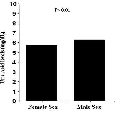
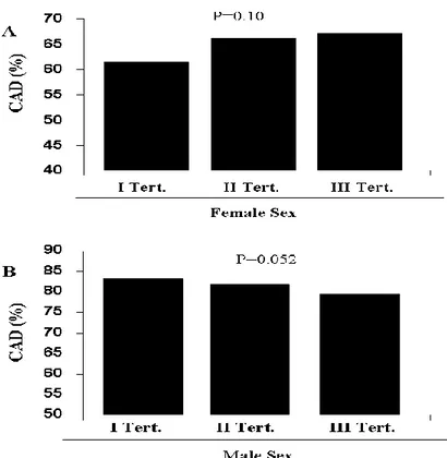
Baseline clinical characteristics and angiographic characteristics according to Tertiles of serum uric acid in females (Group 1, < 4.8mg/dL or 0.28mmol/mol, n = 349, Group 2, 4.8-6.3mg/dL – 0.28-0.37mmol/mol, n = 359 and Group 3 > 4.8-6.3mg/dL or 0.37mmol/mol, n = 370) are listed in Table 3. Patients with elevated SUA were older (p<0.001), with higher prevalence of hypertension (p=0.02), diabetes (p<0.001) and renal failure (p<0.001), but with lower family history of CAD (p=0.001). They had higher levels of white blood cells (p=0.01), tryglicerides (p=0.002) and fibrinogen (p<0.001) and higher creatinine (p<0.001), glycaemia (p<0.001) and glycosilated haemoglobin (p<0.001) at admission, but lower haemoglobin (p=0.05) and HDL-cholesterol (p<0.001). Patients of the Third Tertile were more often in therapy with ARBs (p=0.006) and diuretics (p<0.001) at admission, but less often with Clopidogrel (p=0.04) and had more frequently dilated cardiomyopathy and valvular disease as indication for angiography (p<0.001). Women with higher SUA had a higher prevalence of severe CAD (OR [95% CI] = 1.38 [1.16-1.64], p<0.001; age-adjusted OR [95% CI] = 1.32 [1.11-1.57], p = 0.002) at coronary angiography with a more frequent involvement of left main (p=0.01), left anterior descending (p=0.03) and right coronary artery (p=0.006). No other differences were found in other biochemical parameters or angiographic characteristics. The significant relationship between uric acid levels and prevalence of severe CAD (p<0.001) (Figure 3A) was also confirmed after correction for baseline confounding factors (hypertension, age, diabetes, family history of CAD, renal failure, indication for angiography, white blood cells, haemoglobin, creatinine, glycaemia, glycosylated haemoglobin, tryglicerides, HDL cholesterol, fibrinogen, ARB, diuretics, clopidogrel) (adjusted OR [95%CI] = 1.29 [1.03-1.62], p = 0.03), despite no significant
relationship was observed with the prevalence of CAD (OR [95% CI] = 1.16 [0.99-1.34], p=0.10; age-adjusted OR [95% CI] =1.11 [0.95-1.28[, p = 0.22) (Figure 2A), also after correction for all baseline confounding factors (adjusted OR [95%CI] = 0.97 [0.79-1.18], p = 0.76).
Table 3. Baseline clinical and angiographic characteristics (per lesion) according to Tertile of SUA in Females.
Baseline Clinical Characteristics Females 1° Tertile SUA<4.8mg/dL (0.28mmol/mol) (n = 349) 2° Tertile SUA 4.8-6.3mg/dL (0.28-0.37mmol/mol) (n = 359) 3° Tertile SUA > 6.3mg/dL (0.37mmol-mol) (n = 370) P value Arterial hypertension (%) 72.5 73.2 80.2 0.02 Age (Mean +-SD) 68.4+10.7 70.3+10.7 73.6+9.7 <0.001 Smokers (%) 0.57 Active smokers 19.5 14.2 13.7 Previous smokers 6.3 9.8 7.9 Dyslipidemia ( %) 55.5 55.6 54.9 0.88 Diabetes (%) 29.5 33.1 48.8 <0.001
Family history of CAD (%) 35 25.4 23.4 0.001
History of MI (%) 14 17.9 15.3 0.67
Previous PCI (%) 14.7 16.5 17.2 0.37
Previous CABG (%) 4.9 6.4 7.9 0.10
Previous Stroke (%) 3.4 5.6 53 0.74
Renal failure (%) 18.5 31.7 65.5 <0.001
Indication for angiography <0.001
Stable angina or silent ischemia (%)
22.2 18.5 17.2
Acute Coronary Syndrome (%) 64.9 66.8 49.9
DCM or valvular disease (%) 12.9 14.7 33
Biochemistry
White Blood Cells (10^5/ml) 7.36+2.42 7.64+2.42 7.92+2.5 0.01 Platelets Count (10^5/ml) 238.5+62.6 239.7+75.8 233.8+77.5 0.5 Haemoglobin (g/dl) 12.6+1.46 12.4+1.5 12.3+1.66 0.05 Creatinine (mg/dl) 0.83+0.32 0.92+0.34 1.2+0.45 <0.001 Glycaemia (mg/dl) 122+50.9 120.8+45 135.8+64.6 <0.001 Glycosylated Haemoglobin (%) 6.13+1.25 6.16+1.12 6.6+1.5 <0.001 Total cholesterol (mg/dL) 173.3+45 168.7+43 168.4+42.2 0.243 Tryglicerides (mg/dL) 117.2+58.8 125+62 135+79.1 0.002 HDL cholesterol (mg/dL) 47.1+14.2 45.3+13.9 41.6+12 <0.001
LDL cholesterol (mg/dL) 105.8+41.5 100.8+39 107+43.9 0.11 Reactive protein C (mg/dL) 1.34+3.05 1.56+3.5 1.68+2.7 0.35 Uric Acid (mg/dl) 3.8+3.9 5.4+5.5 7.7+8 <0.001 Fibrinogen (mg/dL) 446.8+149.3 457+132.2 488.2+144.6 <0.001 Therapy at admission ACE inhibitors (%) 36.3 33.9 36.3 0.98 ARB (%) 19.3 25.8 28.2 0.006 Nitrate (%) 35.3 36.7 34.7 0.85 Beta blockers (%) 51.4 49.6 51.5 0.98 Calcium antagonists (%) 19.7 20.7 19.8 0.97 Diuretics (%) 21.7 32.2 53.9 <0.001 Statins (%) 43.4 42 40.2 0.39 ASA (%) 52.2 54.9 51.8 0.9 Clopidogrel (%) 24.3 22.7 18.2 0.04 Angiographic characteristics
Coronary artery disease (%)§ 61.5 66.3 67.3 0.10
Left main/trivessel disease (%)§ 17.1 18.1 29 <0.001
Left main coronary (%)§ 5.5 6.8 10.3 0.01
Left anterior descending coronary (%)§
40.2 47.3 48.4 0.03
Circumflex coronary (%)§ 29.2 30.5 32.2 0.39
Anterolateral branch (%) § 5 6.1 5.3 0.85
Right coronary(%)§ 3 0 0 0.006
Complex type C lesions (%) 30.7 24.5 25.3 0.08
Proximal vessel tortuosity (%) 3.9 4.3 2.5 0.22
Spontaneous dissection (%) 0 1.4 0.6 0.34 Lesion lenght (mm) 19+11.8 17.9+11.3 18.4+12.1 0.44 Vessel diameter (mm) 2.86+0.62 2.88+0.56 2.86+0.6 0.86 Stenosis %(mean SD) 85.5+14.8 84.2+16 84.7+15.1 0.44 Calcified lesions(%) 21.4 23.8 23.6 0.45 Bifurcation (%) 19.9 22.9 22.6 0.34
Chronic total occlusion (%) 11.7 9.1 13.2 0.39
Intracoronary Thrombus (%) 4.7 4.7 3.1 0.2 In-stent Restenosis (%) 3.7 2.5 3.7 0.95 TIMI flow 0.39 3 (%) 76 76.2 77.9 2 (%) 3.9 5.6 4.2 1 (%) 2.2 3.3 2.3 0 (%) 17.9 14.9 15.5
Uric acid and CAD in men
Baseline clinical characteristics and angiographic characteristics according to Tertiles of serum uric acid in males (Group 1, < 5.5mg/dL or 0.33mmol/mol, n = 762, Group 2, 5.5-6.8mg/dL – 0.33-0.40mmol/mol, n = 829 and Group 3 > 5.5-6.8mg/dL or 0.40mmol/mol, n = 851) are listed in Table 4. Patients with elevated SUA were older (p<0.001), with higher prevalence of hypertension (p<0.001), renal failure (p<0.001) and previous smoke (p=0.009). They had a lower incidence of previous PCI (p=0.008), but they were more often in therapy with ACE inhibitors (p=0.001), calcium antagonist (p=0.01) and diuretics (p<0.001) at admission. About baseline chemistry patients of the Third Tertile had higher level of baseline creatinine (p<0.001), glycaemia (p=0.006), glycosilated haemoglobin (p=0.02), total cholesterol (p=0.004), tryglicerides (p<0.001), reactive C protein (p=0.006) and fibrinogen (p<0.001) but lower HDL-cholesterol (p<0.001) and haemoglobin (p=0.02). Moreover they had more frequently dilated cardiomyopathy and valvular disease as indication for angiography (p<0.001). Interestingly elevated Uric acid was associated with a trend towards lower prevalence of CAD (OR [95% CI] = 0.91 [0.78-1.01], p = 0.052; age-adjusted OR [95% CI] = 0.92 [0.79-1.01 = 0.055]) (Figure 2B) that was not confirmed after correction for baseline confounding factors (hypertension, age, smoke, previous PCI, renal failure, indication for angiography, haemoglobin, basal creatinine, glycaemia, glycosylated haemoglobin, total cholesterol, tryglicerides, HDL-cholesterol, reactive protein C, fibrinogen, ACE inhibitors, calcium antagonist, diuretics, lesion length, calcified lesions, intracoronary thrombus) (Adjusted OR [95%CI] = 0.90 [0.76-1.06], p = 0.22; p gender interaction = 0.46). Similarly, no association was found between SUA level and the presence of severe CAD (Figure 3B) (OR [95% CI] = 1.01 [0.92-1.12], p = 0.76;
age-adjusted OR [95% CI] = 1.0 [0.91-1.11] = 0.96), even after correction for baseline confounding factors (adjusted OR [95%CI] = 0.89 [0.79-1.01], p = 0.08; p gender interaction = 0.002).
Table 4. Baseline clinical and angiographic characteristics (per lesion) according to Tertile of SUA in Males.
Baseline Clinical Characteristics Males 1° Tertile SUA<5.5mg/dL (0.33mmol/mol) (n =762) 2° Tertile SUA 5.5-6.8mg/dL (0.33-0.40mmol/mol) (n =829) 3° Tertile SUA > 6.8mg/dL (0.40mmol-mol) (n =851) P value Arterial hypertension (%) 64.2 65.9 78.4 <0.001 Age (Mean +-SD) 65.4+11.7 65.4+11.4 67.7+11.7 <0.001 Smokers (%) 0.009 Active smokers 32.6 34.3 28.3 Previous smokers 20.1 24.1 27.6 Dyslipidemia ( %) 53.2 58.3 56.1 0.28 Diabetes (%) 37.9 34 38.6 0.71
Family history of CAD (%) 28.3 31.6 26.8 0.48
History of MI (%) 28.4 27.5 29.5 0.60
Previous PCI (%) 30.6 27 24.7 0.008
Previous CABG (%) 13.4 12.8 16.5 0.07
Previous Stroke (%) 7.6 6.3 9.8 0.10
Renal failure (%) 12.4 18.3 38.1 <0.001
Indication for angiography <0.001
Stable angina or silent ischemia (%)
27.6 26 23.6
Acute Coronary Syndrome (%) 60.6 60 52
DCM or valvular disease (%) 11.8 14 24.4
Biochemistry
White Blood cells (10^5/ml) 7.9+3.2 7.9+3.2 8.2+3 0.22
Platelets Count (10^5/ml) 205.3+59.6 203.8+56.6 211.6+64.2 0.19 Haemoglobin (g/dl) 13.8+1.6 13.9+1.6 13.7+1.7 0.02 Creatinine (mg/dl) 0.99+0.32 1.06+0.31 1.22+0.4 <0.001 Glycaemia (mg/dl) 128.7+53.2 120.8+42.5 129.6+50.3 0.006 Glycosylated Haemoglobin (%) 6.19+1.3 6.10+1.3 6.3+1.2 0.02 Total cholesterol (mg/dL) 156.6+38.9 162.2+40.7 162.7+41.3 0.004 Tryglicerides (mg/dL) 122.6+73.5 137.1+77.7 151.7+98.3 <0.001 HDL cholesterol 40.9+12.1 39.7+11.5 38+11 <0.001 LDL cholesterol 98+45.2 101.1+45.7 102+46.8 0.21 Reactive protein C (mg/dL) 1.2+2.5 1.07+2.5 1.48+2.89 0.006 Uric Acid (mg/dl) 4.6+0.65 6+0.37 8.1+1.3 <0.001
Fibrinogen (mg/dL) 414.4+153 417+146 453.2+155.8 <0.001 Therapy at admission ACE inhibitors (%) 34.5 40 42.5 0.001 ARB (%) 17.3 17.4 20.7 0.07 Nitrate (%) 35.2 34.7 37.1 0.4 Beta blockers (%) 50.7 50.1 52.1 0.56 Calcium antagonists (%) 18.4 18.6 23.4 0.01 Diuretics (%) 18.9 22 39.7 <0.001 Statins (%) 52.2 52.1 48.5 0.13 ASA (%) 60.6 59.5 59.5 0.66 Clopidogrel (%) 23.3 24.5 23.4 0.99 Angiographic characteristics
Coronary artery disease (%)§ 83.4 81.9 79.6 0.052
Left main/trivessel disease (%)§ 32.8 29.8 33.3 0.76
Left main coronary (%)§ 9.6 9.1 11.5 0.19
Left anterior descending coronary (%)§
60.1 53.6 55.5 0.08
Circumflex coronary (%)§ 46.3 45.1 43.8 0.30
Anterolateral branch (%) § 10.6 7.6 8.3 0.13
Right coronary(%)§ 49.5 49 51.4 0.43
Complex type C lesions (%) 36.8 34.6 39.4 0.155
Proximal vessel tortuosity (%) 3.3 4 3.6 0.74
Spontaneous dissection (%) 0.5 0.4 0.1 0.08 Lesion length (mm) 21.6+14 19.6+12.3 20+12.8 0.001 Vessel diameter (mm) 2.94+0.6 2.92+0.62 2.95+0.62 0.36 Stenosis %(mean SD) 86.4+15.6 87+14 87+14.7 0.52 Calcified lesions(%) 19.8 22.5 26.9 <0.001 Bifurcation (%) 22.2 21.9 20.7 0.34
Chronic total occlusion (%) 19.1 18.2 22 0.053
Intracoronary Thrombus (%) 6.8 5.3 4.4 0.005 In-stent Restenosis (%) 5.3 4.7 4.3 0.21 TIMI flow 0.138 3 (%) 69 70.4 66.6 2 (%) 4.9 4.7 4.8 1 (%) 3.1 2.9 3.5 0 (%) 23 21.9 25.1
Figure 2. Bar graph showing the prevalence of coronary disease according to uric acid tertiles in females (Figure 2A) and males (Figure 2B).
Figure 3. Bar graph showing the prevalence of severe (left main and/or trivessel) coronary disease according to uric acid tertiles in females (Figure 3A) and males (Figure 3B).
Discussion
The main findings of our study are that: 1) male sex, is independently associated with higher SUA; 2) high uric acid levels are independently associated with more severe CAD in women. Despite the great improvement in pharmacological and revascularization therapies to treat and reduce cardiovascular mortality in the last decades (3-5), especially in the acute setting, the results are still suboptimal, in particular among high-risk subgroups of patients (6, 7, 18). Therefore, large interest has been focused on the identification of new risk factors in order to prevent CAD. Uric acid represents a metabolite of the degradation pathway of purines, with renal elimination (19). Its catabolism via the xanthine oxidase has been related to increased production of reactive oxygen species and reduced availability of nitric oxide, with consequent oxidative damage (20). Therefore, elevated serum uric acid (SUA) has been addressed in literature as a potential contributor of cardiovascular risk, but the results were conflicting. In the past several studies showed an association between hyperuricemia and an increased rate of cardiovascular events and higher mortality (21,22), recently confirmed in a large study by Ndrepepa G et al. (23). On the contrary, other large studies, such as the Framingham Heart Study, (24) and the study from Panero et al, (25) concluded that uric acid was not a causal risk factor for cardiovascular disease. We previously found no association between uric acid and the extent of CAD (12). Moreover, it is know that serum uric acid levels are lower in women than in men and several studies in the past assessed the association between elevated uric acid levels and the extension of CAD, cardiovascular events and CAD related mortality in association to sex with important contradictions. Fang J et al. showed that increased serum uric acid levels are independently and significantly associated with risk of cardiovascular mortality both in women and men
(26). A recent study by Onat et al. (27) showed among more than 1500 patients that CAD risk was independently predicted by elevated SUA in nondiabetic men and was modulated by metabolic syndrome and gender. Subsequently, Tuttle et al. (15) suggested a significant association between high uric acid levels and CAD in woman but not in men and these results were also confirmed in a more recent meta-analysis by Kim et al. (28) in which among 402,997 patients, subgroup analyses showed no significant association between hyperuricemia and CAD incidence/mortality in men, but an increased risk for CAD mortality in women (RR 1.67, 95% CI 1.30-2.04). Moreover, a recent study by Ndrepepa et al. (29) showed between 13,273 patients a stronger association between hyperuricemia and an increased risk of mortality in both sex, with a stronger association in women. In line with most of the studies, we found that despite the higher uric acid levels observed in men a significant association between uric acid and the severity of CAD was observed only in women. In our population women were older, with higher prevalence of the most important cardiovascular risk factors and this finding can probably explain the strong association between severe CAD and SUA in the female population. Anyway, this association remained significant even after correction for all baseline confounders, underlying the presence of an independent correlation between elevated SUA and CAD. Therefore, the complex gender-interaction in the relationship between uric acid and CAD can not only be explained by the major cardiovascular risk factors in women, such as diabetes, hypertension and chronic renal failure (30). A recent study (31) has shown a significant gender-difference in the association between uric acid and MPV, with larger platelets observed in the presence of increased uric acid only among women. In fact, MPV has recently been shown to affect platelet reactivity (32,33) and may therefore contribute to explain our findings. Future large
studies are certainly needed to confirm our findings, to provide physiopathologic insights on the observed gender interaction and to evaluate gender-specific benefits from SUA lowering therapies on cardiovascular prevention and cardiovascular outcome. In fact, so far weak benefits have been demonstrated and therefore their use is, till now, very modest in the subset of asymptomatic patients (34-36).
Limitations
Our study is not able to provide data about long term effect of SUA on cardiovascular outcome. Our study has a cross sectional design, therefore we have no data are regarding the follow up of this high risk subgroup of patients. Finally the use of intravascular imaging, as with IVUS technique, would have improved the definition of CAD, especially in patients with complex eccentric plaques and with negative remodelling, which has been more often demonstrated in older patients, as our patients with higher SUA (37).
Conclusion
Our study showed that even though uric acid levels are significantly higher in men, high uric acid levels are associated with severe CAD only in women. Future large studies are certainly needed to confirm our findings and to evaluate the effects of SUA lowering therapies on cardiovascular prevention and outcome, especially among women.
References
1. Rosamond W, Flegal K, Friday G et al. Heart disease and stroke statistics-2007 update: a report from the American Heart Association statistics committee and stroke statistics subcommittee. Circulation 2007; 115: e69-171.
2. Allender S, Scarborough P, Peto V et al. European cardiovascular disease statistics:2008 edition (online). Available from URL:http://www.enheart.org/files/statistics%202008%20web-161229A.pdf
3. De Luca G, Bellandi F, Huber K et al. Early glycoprotein IIb-IIIa inhibitors in primary angioplasty-abciximab long-term results (EGYPT-ALT) cooperation: individual patient's data meta-analysis. J Thromb Haemost. 2011; 9: 2361-70.
4. Navarese EP, De Luca G, Castriota F et al. Low-molecular-weight heparins vs. unfractionated heparin in the setting of percutaneous coronary intervention for ST-elevation myocardial infarction: a meta-analysis. J Thromb Haemost 2011; 9: 1902-15.
5. Navarese EP, Kubica J, Castriota F et al. Safety and efficacy of biodegradable vs. durable polymer drug-eluting stents: evidence from a meta-analysis of randomised trials. EuroIntervention 2011; 7: 985-94.
6. Natali A., Boldrini B, Baldi S et al. Impact of mild to moderate reductions of glomerular filtration rate on coronary artery disease severity. Nutr Metab Cardiovasc Dis. 2013 Dec 24. [Epub ahead of print]
7. De Luca G, Gibson CM, Bellandi F et al. Diabetes mellitus is associated with distal embolization, impaired myocardial perfusion, and higher mortality in patients with ST-segment elevation myocardial infarction treated with primary angioplasty and glycoprotein IIb-IIIa inhibitors. Atherosclerosis. 2009; 207: 181-5.
8. Tomita M, Mizuno S, Yamanaka H, et al. Does hyperuricemia affect mortality? A prospective cohort study of Japanese male workers. J Epidemiol 2000;10:403–9.
9. Reunanen A, Takkunen H, Knekt P et al. Hyperuricemia as a risk factor for cardiovascular mortality. Acta Med Scand Suppl 1982;668:49–59.
10. Levine W, Dyer AR, Shekelle RB et al. Serum uric acid and 11.5-year mortality of middle-aged women: findings of the Chicago Heart Association Detection Project in Industry. J Clin Epidemiol. 1989; 42(3): 257-67.
11. Lazzeri C, Valente S, Chiostri M et al. Uric acid in the acute phase of ST elevation myocardial infarction submitted to primary PCI: its prognostic role and relation with inflammatory markers: a single center experience. Int J Cardiol 2010;138:206–9.
12. De Luca G, Secco GG, Santagostino M et al. Uric acid does not affect the prevalence and extent of coronary artery disease. Results from a prospective study. Nutr Metab Cardiovasc Dis. 2012; 22: 426-33.
13. Holme I, Aastveit AH, Hammar N et al. Uric acid and risk of myocardial infarction, stroke and congestive heart failure in 417734 men and women in the Apolipoprotein Mortality RISk study (AMORIS) J Intern Med 2009; 266: 558-70.
14. Strasak AM, Kelleher CC, Brant LJ et al. Serum uric acid is an independent predictor for all major forms of cardiovascular death in 28613 elderly women: a prospective 21-year follow up study. Int J Cardiol 2008; 125: 232-9.
15. Tuttle KR, Short RA, Johnson RJ. Sex differences in uric acid and risk factors for coronary artery disease. Am J Cardiol 2001; 87: 1411-4.
16. De Luca G, Verdoia M, Cassetti E et al. High fibrinogen level is an independent predictor of presence and extent of coronary artery disease among Italian population. J Thromb Thrombolysis 2011; 31: 458-63.
17. De Luca G, van 't Hof AW, de Boer MJ et al. Time-to-treatment significantly affects the extent of ST-segment resolution and myocardial blush in patients with acute myocardial infarction treated by primary angioplasty. Eur Heart J. 2004; 25: 1009-13.
18. De Luca G, Dirksen MT, Spaulding C, Kelbæk H, Schalij M, Thuesen L, van der Hoeven B, Vink MA, Kaiser C, Musto C, Chechi T, Spaziani G, Diaz de la Llera LS, Pasceri V, Di Lorenzo E, Violini R, Suryapranata H, Stone GW; DESERT cooperation. Impact of diabetes on long-term outcome after primary angioplasty: insights from the DESERT cooperation. Diabetes Care 2013; 36: 1020-5.
19. Boushey CJ, Beresfors SA et al. A quantitative assessment of plasma homocysteine as a risk factor for vascular disease : probable benefits of increasing folic acid intakes. JAMA 1995; 274:1049-1057.
20. Zweier L, Kuppusamy P, Lutty GA. Measurement of endothelial cell free radical generation: evidence for a central mechanism of free radical injury in postischemic tissues. Proc Natl Acad Sci U S A 1988;85:4046e50
21. Nieto FJ, Iribarren C, Gross MD et al. Uric acid and serum antioxidant capacity: a reaction to atherosclerosis? Atherosclerosis 2000;148:131e9.
22. Mazza A, Pessina AC, Pavei A et al. Predictors of stroke mortality in elderly people from the general population. The CArdiovascular STudy in the ELderly. Eur J Epidemiol. 2001;17(12):1097-104
23. Ndrepepa G, Braun S, King L et al. Association of uric acid with mortality in patients with stable coronary artery disease. Metabolism. 2012;61(12):1780-6.
24. Culleton BF, Larson MG, Kannel WB, Levy D. Serum uric acid and risk for cardiovascular disease and death: the Framingham Heart Study. Ann Intern Med 1999;131:7e13.
25. Panero F, Gruden G, Perotto M et al. Uric acid is not an independent predictor of cardiovascular mortality in type 2 diabetes: a population-based study. Atherosclerosis 2012;221:183e188.
26. Fang J, Alderman MH. Serum uric acid and cardiovascular mortality the NHANES I epidemiologic follow up study, 1971-1992. National Health and Nutrition Examination Survey. JAMA 2000;283:2404-10.
27. Onat A, Can G, Örnek E et al. Elevated serum uric acid in non diabetic people mark pro-inflammatory state and HDL dysfunction and independently predicts coronary disease. Clin Rheumatol. 2013 Dec;32(12):1767-75.
28. Kim SY, Guevara JP, Kim KM et al. Hyperuricemia and coronary heart disease: a systematic review and meta-analysis. Arthritis Care Res. 2010 Feb;62(2):170-80.
29. Ndrepepa G, Cassese S, Braun S et al. A gender-specific analysis of association between hyperuricaemia and cardiovascular events in patients with coronary artery disease. Nutr Metab Cardiovasc Dis. 2013 Dec;23(12):1195-201.
30. Neogi T. Asymptomatic hyperuricemia: perhaps not so benign? J Rheumatol. 2008; 35(5):734–7.
31. Fang JI, Wu JS, Yang YC et al. High uric acid level associated with increased arterial stiffness in apparently healthy women. Atherosclerosis. 2014 Oct;236(2):389-93.
32. Tavil Y, Sen N, Yazici HU, Hizal F, Abaci A, Cengel A. Tavil Y, Sen N, Yazici HU, Hizal F, Abaci A, Cengel A. Mean platelet volume in patients with metabolic syndrome and its relationship with coronary artery disease. Thromb Res. 2007; 120: 245-50.
33. Tsiara S, Elisaf M, Jagroop IA, Mikhailidis DP. Platelets as predictors of vascular risk: is there a practical index of platelet activity? Clin Appl Thromb Hemost 2003;9:177–90.
34. Milionis HJ, Kakafika AI, Tsouli SG et al. Effects of statin treatment on uric acid homeostasis in patients with primary hyperlipidemia. Am Heart J. 2004; 148: 635-40.
35. Strazzullo P, Puig JG. Uric acid and oxidative stress: relative impact on cardiovascular risk? Nutr Metab Cardiovasc Dis. 2007; 17: 409-14.
36. Savarese G, Ferri C, Trimarco B et al. Changes in serum uric acid levels and cardiovascular events: A meta-analysis. Nutr Metab Cardiovasc Dis. 2013;23(8):707-14.
37. Hassani SE, Mintz GS, Fong HS et al. Negative remodeling and calcified plaque in octogenarians with acute myocardial infarction: an intravascular ultrasound analysis. J Am Coll Cardiol 2006; 47: 2413-9.
Chapter 3
Neutrophil to Lymphocyte Ratio and the extent of coronary artery disease:
results from a large cohort study
Monica Verdoiaa, MD, Lucia Barbieria, MD, Gabriella Di Giovine, MD, Paolo Marinoa,
MD, Harry Suryapranatab, MD, PhD, Giuseppe De Lucaa, MD, PhD on behalf of the Novara Atherosclerosis Study Group (NAS)
aDepartment of Cardiology, Ospedale “Maggiore della Carità”, Eastern Piedmont University, Novara, Italy; bDepartment of Cardiology, UMC St Radboud, Nijmegen, The Netherlands
Angiology. 2016 Jan;67(1):75-82.
Abstract
The neutrophil to lymphocyte ratio (NLR), an inflammatory biomarker, may be of predictive and prognostic value for cardiovascular (CV) events. We evaluated the relationship of NLR with the prevalence and extent of coronary artery disease (CAD) in consecutive patients undergoing elective or urgent coronary angiography. Our population (n=3738 patients) was divided into NLR quartiles. Higher NLR was associated with ageing and established CV risk factors, previous percutaneous coronary revascularization, acute presentation and more complex pharmacological therapy. NLR was related with platelet count, WBC count, creatinine, glycemia, uric acid and C reactive protein (all p=0.001) levels, but inversely related with hemoglobin (p<0.001), total cholesterol (p=0.005) and triglycerides (p<0.001) levels. NLR was associated with multivessel disease (p<0.001), anterior descending, right coronary arteries (p<0.001) or circumflex branch lesions (p=0.01), percentage stenosis (p<0.001), coronary calcification (p<0.001) and intracoronary thrombus (p<0.001) but inversely with instent restenosis (p<0.001) and Thrombolysis-In Myocardial-Infarction flow (p=0.04). NLR was directly related with the prevalence of CAD (p=0.001) and severe CAD (p<0.001). In patients undergoing coronary angiography, the NLR is independently associated with the prevalence and severity of CAD.
Keywords: white blood cells, neutrophils, lymphocytes, coronary artery disease; coronary
angiography
Introduction
Atherosclerosis is an inflammatory process leading to vascular wall degeneration as a response to various risk factors.1 Considerable interest has been given to the field of cardiovascular (CV) disease prevention and major advances have been achieved in mechanical reperfusion and antithrombotic therapies 2-6, especially in acute myocardial
infarction. However, the outcome is still unsatisfactory in a relatively large proportion of patients 7-9, thus shifting the focus on the identification of new indicators, better allowing to
stratify the risk of CV events. 10-12 Several inflammatory biomarkers, such as high
sensitivity C-reactive protein (hsCRP), interleukin-6 and lipoprotein associated phospholipase A2, have been associated with coronary artery disease (CAD) 13-15. The WBC count has emerged as one of the easiest to obtain, cheapest and moreover, best predictive indicators of CV risk 16. In fact, leukocytes play a crucial role in the progression of
atherosclerosis and in destabilization and rupture of a plaque, leading to thrombotic events.
17 More recently, attention has shifted to leukocyte subtypes and to the Neutrophil to
Lymphocyte Ratio (NLR), potentially being more accurate and stable than absolute blood cell counts, especially in patients with acute presentation, where it has been associated with clinical outcome and procedural results after percutaneous revascularization. 18, 19 However,
few reports have evaluated the relationship between NLR and the prevalence and extent of CAD; this was the aim of the present study.
Methods
Our population consisted of consecutive patients undergoing elective or urgent coronary angiography between April 2007 and December 2013 at the Ospedale “Maggiore della Carità”, Novara, Italy. Informed consent was obtained from all patients before angiography. The study was approved by our local Ethical Committee. All demographic and clinical data were prospectively collected in a dedicated database. Hypertension was defined as systolic pressure >140 mmHg and/or diastolic pressure >90 mmHg or if the individual was taking antihypertensive medication. Diabetes mellitus was defined as previous diagnosis, specific treatment (oral drug or insulin), fasting glycemia >126 mg/dL or glycosylated hemoglobin (HbA1c) >6.5%. 20 Chronic renal failure was considered as a history of renal failure or an
admission glomerular filtrate (GFR) <60 ml/min/1.73m2 as defined by Modifying Diet in Renal Disease (MDRD) formula. Non-ST-elevation myocardial infarction was defined as chest pain lasting more than 5 min, associated with elevation of cardiac biomarkers beyond the upper limit of normal (ULN) (0.04 µg/l for Troponin I and 5.00 ng/ml for creatine-kinase-MB, respectively), with or without ECG changes.
Biochemical measurements
Blood samples were drawn at admission in patients undergoing (following a fasting period of 12 h) or urgent coronary angiography. Glucose, creatinine, HbA1c and lipid profile were determined by standard methods.
The WBC counts were measured in a blood sample collected in Ethylenediaminetetraacetic acid (EDTA) (7.2 mg) tubes. These blood samples were analyzed within 2 h of venipuncture using an automatic blood counter (Sysmex XE-2100, Sysmex Corporation, Kobe, Japan). 21
Coronary angiography
Coronary angiography (Siemens AXIOM ARTIS dTC, Erlangen, Germany) was routinely performed by the Judkins technique using 6-French right and left heart catheters. Quantitative coronary angiography was performed, by an automatic edge-detection systems (Siemens Acom Quantcor QCA, Erlangen, Germany) as previously described. 22 The measured parameters were minimal luminal diameter, reference diameter, percent diameter stenosis and length of the lesion. Significant CAD was defined as at least 1 coronary stenosis >50%. Severe CAD was defined as 3-vessel disease and/or left main disease. In cases who had previously undergone percutaneous coronary intervention (PCI), even if no restenosis was observed, the treated vessel was counted as significantly diseased. In previously bypassed patients, native arteries and grafts were taken into account in the evaluation of the extent of artery disease (number of diseased vessels).
Statistical analysis
Statistical analysis was performed using SPSS 15.0 statistical package. Continuous data were expressed as mean ± SD and categorical data as percentage. Analysis of variance and the chi-square test were used for continuous and categorical variables, respectively. Patients were grouped according to quartiles of NLR. Multiple logistic regression analysis was performed to evaluate the relationship between NLR and CAD, even in higher risk subsets of patients, after correction for baseline confounding factors that were entered in the model in block. A two-sidedp value < 0.05 was considered significant.
Table I. Clinical and demographic characteristics according to NLR quartiles. Baseline Characteristics I quart
(n= 933) II quart (n=919) III quart (n=954) IV quart (n=932) P value Age (mean +/- SD) 64.3 11.2 66.9 10.9 68.7 10.9 70.4 11.4 <0.001 Male Sex (%) 67.5 69.9 70.8 69 0.49 Dyslipidemia (%) 62.1 59.5 54.6 45.4 <0.001 Diabetes mellitus (%) 37.1 33.5 36.9 39.5 0.13 Renal failure (%) 9.6 14.6 17.8 24.1 <0.001 Smokers (%) <0.001 Active smokers 30.9 26.9 25.5 23.2 Previous smoker 19.2 20.8 17.1 16 Hypertension (%) 69.4 71.4 72.6 71.5 0.26 History of MI (%) 25.1 24.9 23.5 24 0.44 Previous PCI (%) 27.8 26.3 22 18.9 <0.001 Previous CABG (%) 10.4 13 11 12.4 0.38 Biochemistry Platelet Count (10^3/µl) 212 58 214 57 217 67 224 79 0.001 Hemoglobin (g/dl) 13.7 1.6 13.6 1.6 13.4 1.8 12.9 1.9 <0.001 White blood cell count
(10^3/µl) 7.1 2.7 7.2 2 7.8 2.2 9.5 3.1 <0.001 Creatinine (mg/dl) 0.99 0.3 1.04 0.3 1.08 0.4 1.13 0.5 <0.001 Glycaemia (mg/dl) 121 46 121 45 125 50 137 61 <0.001 Glycosylated hemoglobin (%) 6.3 1.4 6.2 1.3 6.2 1.2 6.2 1.4 0.73 Total cholesterol (mg/dL) 167 41 165 42 163 42 160 42 0.005 Triglycerides (mg/dL) 140 86 140 85 134 82 115 61 <0.001 HDL cholesterol (mg/dl) 41 12 41 12 41 12 41 13 0.82 Uric acid (mg/dl) 6.0 1.6 6.1 1.7 6.2 1.8 6.3 2 0.001 C-reactive protein (mg/dl) 1.1 0.04 2.0 0.07 2.5 0.8 4.1 0.14 <0.001 Indication to angiography <0.001 Stable angina (%) 31.8 27.9 22.4 11.7
Acute coronary syndrome (%) 53 53.2 58.8 69
Arrhythmias/Valvulopathy/LV dysfunction (%) 15.2 18.9 18.8 19.3 Therapy at admission ACE inhibitors (%) 37.7 36.3 38.9 37.7 0.74 ARB (%) 19 22.4 21.7 17.6 0.43 Beta-blockers (%) 55.6 53.9 50.3 42.1 <0.001 Calcium antagonists (%) 19.2 21.1 21.7 17.2 0.53 Nitrates (%) 34.6 36.2 36.7 33.4 0.67 Diuretics (%) 25.5 28.2 33.4 32.4 <0.001 Statins (%) 53.2 50.5 48.7 38.7 <0.001 ASA (%) 62.7 62.1 56.9 47.8 <0.001
Clopidogrel (%) 23.4 23.4 24.8 19.2 0.08
Results
Our population consisted of 3738 patients undergoing coronary angiography. They were divided according to quartiles values of NLR (< 1.8; 1.8-2.49; 2.5-3.69; >3.7). The main clinical and demographical features of included patients are displayed in Table 1. Higher NLR values were associated to ageing (p<0.001), dyslipidemia (p<0.001), renal failure (p<0.001), smoking (p<0.001), previous PCI (p<0.001), acute presentation (p<0.001), therapy at admission with beta-blockers, diuretics, statins and acetylsalicylic acid (p<0.001, respectively). NLR was directly related with the platelet count (p=0.001), WBC count, creatinine, glycemia (p<0.001), uric acid (p=0.001) and C-reactive protein (p<0.001) levels, while inversely with hemoglobin levels (p<0.001), total cholesterol (p=0.005) and triglycerides (p<0.001) levels. Table 2 shows main angiographic features according to NLR quartiles.
Table 2. Angiographic characteristics (per lesion) according to Neutrophil to Lymphocyte Ratio.
Variable I quart (n= 1368) II quart (n=1468) III quart (n=1444) IV quart (n=1626) P value Multivessel (%)§ 40.9 45.6 45.9 51.4 <0.001
Left main coronary artery (%)§ 9 8.3 9.8 9.9 0.33
Left anterior descending coronary artery (%)§
48 53.4 52.8 57.9 <0.001
Circumflex coronary artery (%)§ 36.7 40.1 39.4 44.9 0.01
Right coronary artery (%)§ 40 44.8 43.9 50.7 <0.001
Type C lesions (%) 34.4 34.1 33.7 36.6 0.25
Lesion length, mm (mean SD) 19.8 13 19.9 12.8 19.9 12.7 19.9 12.8 0.99 Vessel diameter, mm (mean
SD)
2.9 1 3 1.3 2.9 0.7 3 1 0.94
Stenosis % (mean SD) 85.2 15.7 85.7 15.7 85.7 14.3 87.9 13.9 <0.001
Calcified lesions (%) 18.1 22.2 24.9 28.1 <0.001
Intracoronary Thrombus (%) 3.7 2.9 5.6 9.1 <0.001 Chronic occlusion (%) 18.3 18.8 17.1 16.7 0.07 In-stent Restenosis (%) 6.3 5.4 3.7 2.5 <0.001 TIMI flow 0.04 3 71.9 70.6 72.4 67.6 2 4.7 4.6 5.1 5.1 1 2.7 3 3.1 3.5 0 20.7 21.8 19.3 23.8
Higher NLR was associated with the extent of multivessel coronary disease (p<0.001) and with lesion location (p<0.001). NLR was related with the percentage of stenosis (p<0.001), the presence of coronary calcification (p<0.001), intracoronary thrombus (p<0.001) and inversely with instent restenosis (p<0.001) and TIMI flow (p=0.04).
Figure 1. Bar graphs showing the relationship between Neutrophil to lymphocyte ratio (NLR) quartiles values and the prevalence of coronary artery disease (CAD).
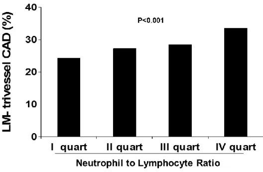
As shown in Figure 1, NLR was directly related with the prevalence of CAD (73.1 vs 76.4 vs 75.6 vs 80.4, p=0.001; OR [95%CI] = 1.12 [1.05-1.2], p=0.001) and with the prevalence of severe CAD (24.3 vs 27.3 vs 28.5 vs 33.6%, p<0.001; OR [95%CI] = 1.16 [1.08-1.23], p<0.001) (Figure 2).
Figure 2. Bar graph showing the relationship between Neutrophil to lymphocyte ratio (NLR) quartiles values and the prevalence of severe coronary artery disease (CAD)
Results were confirmed after correction for baseline differences for both prevalence of CAD (adjusted OR [95%CI] = 1.11 [1.01-1.22], p=0.04) and severe CAD (adjusted OR [95%CI] = 1.12 [1.03-1.23], p=0.007). Similar results were confirmed in the majority of high risk subsets of patients when considering high (4th quartile) vs all lower NLR quartiles. Data for
p=0.006) or young patients (<75 years old, OR=1.23 [0.97-1.54], p=0.08, p interaction=0.23), males (OR=1.26 [1-1.60], p=0.05) and females (OR=1.59 [1.19-2.14], p=0.002, p interaction=0.21), according to diabetes (diabetics: OR=1.59 [1.13-2.25], p=0.01 and non-diabetics: OR=1.25 [1.001-1.56], p=0.05, p interaction=0.24), renal function (renal failure: OR=1.37 [1-1.88], p=0.05; normal function: OR=1.38 [1.09-1.74], p=0.007, p interaction=0.97), hypertension (hypertension: OR=1.34 [1.07-1.67], p=0.01, no hypertension: OR= 1.39 [1.002-1.92], p=0.048, p interaction =0.85).
Figure 3 Forest plot showing the risk of coronary artery disease (CAD) for Neutrophil to lymphocyte ratio (NLR) in IVth quartile vs lower values in higher risk subsets of patients (CKD = chronic kidney disease).
Discussion
This is the largest single-center cohort study conducted so far that evaluated the relationship between the NLR and angiographically defined CAD. Our main finding is that higher NLR values are associated with the extent and severity of coronary lesions, independently from the main established CV risk factors and in the majority of higher risk subsets of patients. Pharmacological and technical innovations have changed the approach to patients with CAD, extending the indication for PCI to more complex subsets of patients, especially those with acute coronary syndromes (ACS), reducing the rate of repeated target vessel revascularization and increasing event-free survival. 23-29 Indeed, potent antiplatelet agents,
new anti-ischemic drugs and high-dose statins have contributed to reduce the progression of atherosclerosis and CV disease, although a considerable residual risk of acute events remains. 30-32 Therefore, there is a need to identify new markers to allow better risk stratification and also as potential pharmacological targets. Special attention has been dedicated to inflammatory biomarkers, with contrasting results. 33 Indeed, inflammation
plays a central role in the pathogenesis of atherosclerosis and an elevation of inflammatory markers, indicates the activation of vascular damage. 34 However, a modest potential of
predicting CV events has been reported in patients without established CAD, even with complex combinations of indicators of acute response and endothelial dysfunction. 17, 35 Moreover, initial attempts to target selective inhibition of cytokines in CV prevention has resulted in inconsistent results. 36,37 The WBC count represents a cheap, widely available
and early indicator of the inflammatory response and initial studies showed that an elevation of total WBC was associated with increased mortality and worse outcomes after acute
myocardial infarction (MI). 38,39 Thereafter, with the recognition of a more relevant role for
immune rather than inflammatory response, in the pathogenesis of CAD, attention has been addressed to leukocyte subtypes and especially the NLR, combining the effects of the non-specific inflammatory response, mediated by neutrophils, and the subsequent regulatory immune response, involving lymphocytes. 40 Neutrophils have been claimed for every step leading to acute coronary events and can release pro-oxidant and pro-thrombotic substances, leading to endothelial damage and platelet aggregation. A low lymphocyte count has also been associated with worse prognosis in patients with CAD and unstable angina 41, as
certain subset of lymphocytes, have been shown to play an inhibitory role in atherosclerosis, possibly by controlling and regulating the inflammatory response. 40 An increased NLR has
been related with arterial stiffness and with indirect indicators of atherosclerosis, such as coronary calcium score and carotid intima-media thickness. Moreover, this hematological index has been associated with thrombus formation in ACS 42, where it has been suggested
to influence short and long- outcome, especially in patients with ST elevation MI (STEMI) undergoing primary PCI. 43, 44 In particular, in a large cohort of STEMI patients, a NLR
>6.97 was associated with increased in-hospital and long-term CV mortality 45, and similar
results were identified in a Korean population. 46 Recent studies, in addition, have reported a
prognostic role of NLR in patients undergoing elective PCI or surgical coronary revascularization 22,47 and NLR was associated with worse outcome at 3 years follow-up
independently from the therapeutic strategy selected for CAD. 48 In addition, the authors 48
suggested a more advanced obstructive CAD (OR = 2.45, CI 95% 1.76, 3.42, p < 0.001) in patients with higher NLR. However, only few small studies have evaluated the relationship between NLR and angiographic findings, thus not providing conclusive results. We present