dema is caused in most cases by the obstructions or the stenosis of lymph vessels in relation with the oncological therapies1. Other causes are
ac-cidental or iatrogenic lesions of the lymphatic vessels.
Lymphoedema usually occurs in 6-63% of patients who received a radiotherapy and/or sur-gery as cancer therapy. The rise of the pathology may occur immediately after this therapy or may be delayed2. Even if the lymphoedema does not
represent a death risk for patients, the impact of the external changes in a lower quality of life is significative3. Lymphangiosarcoma has been
de-scribed as a rare complication, too4. Additionally,
a very common and severe complication is cellu-litis of the affected limb5.
The use of penicillin is one of the most com-mon treatments of symptomatic cellulitis; howev-er, this is not a decisive treatment and cellulitis may relapse after or even during the therapy6.
Another common treatment is drainage. Complex physical therapy (CPT) is a recent treatment in-troduced for lymphoedema that reduces the limb circumference and the occurrence of cellulitis7,8.
The lymphaticovenular anastomosis (LVA) is a microsurgical technique very effective in improv-ing limb circumference and alleviatimprov-ing dermal sclerosis9-14.
The LVA technique rapidly evolved in recent years. In 2014, Yamamoto et al15 described a
ladder-shaped LVA, 3 lymphatic vessels and 1 vein were anastomosed in a side-to-side fashion; further, in 2015 Yamamoto et al16 reported the
parachute technique, a simplified technique to perform the side to end supramicrosurgical lym-phaticovenular anastomosis (s-LVA) while main-taining efficacy comparable to the conventional technique.
Abstract. – OBJECTIVE: Aim of this paper is to present our reduction of the frequency of celluli-tis before and after supramicrosurgical lympha-ticovenular anastomosis (s-LVA) in lymphoede-ma patients, and discuss the possibility to per-form this technique outside Japan.
PATIENTS AND METHODS: 37 patients affect-ed by lymphoaffect-edema were enrollaffect-ed. All patients received preoperative indocyanine green lym-phography. Under local anaesthesia s-LVA was performed on all patients. All patients were fol-lowed for 1 year. Lymphoedema was staged us-ing the lymphoedema stagus-ing classification rec-ommended by the International Society of Lym-phology. Cellulitis rate was recorded for all pa-tients the year before and after the s-LVA. A
t-test was used to evaluate differences in the
fre-quency of cellulitis the year before surgery and the year following surgery.
RESULTS: Cellulitis incidence decreased in all patients, with a mean 1.7 cases the year before s-LVA and 0.1 the year after s-LVA. A significant difference between preoperative and postopera-tive cellulitis rate was found (p = 0.0012).
CONCLUSIONS: This study reports our s-LVA case series of lymphoedema patients. With the proper learning curve, s-LVA may be repro-duced and lymphoedema patients may gain a better quality of life and a reduced cellulitis rate.
Key Words:
Supramicrosurgical, Lymphaticovenular, Anastomo-sis, LVA, Cellulitis, Lymphoedema.
Introduction
Lymphoedema is a pathology characterized by a limb accumulation of subcutaneous protein-rich fluid due to a lymphatic system disorder. While the primary lymphoedema is commonly caused by congenital abnormalities, secondary
lymphoe-P. GENNARO
1, G. GABRIELE
1, C. SALINI
1, G. CHISCI
1, F. CASCINO,
J.-F. XU
3, C. UNGARI
21Department of Maxillofacial Surgery, University of Siena, Siena, Italy
2Department of Maxillofacial Surgery, “Sapienza” University of Rome, Rome, Italy 3Department of Dentistry, Tongde Hospital, Hangzhou, Zhejiang Province, China
Our supramicrosurgical experience of
lymphaticovenular anastomosis in
The limitation of this technique is its reproduci-bility; commonly, mainly Japanese authors reported the efficacy of LVA and very few papers reported the effect of s-LVA on cellulitis incidence. Campisi et al17 in 2007 reported his experience with LVA,
with 87% reduction of cellulitis incidence; Mihara et al12 in 2014 reported a significant rate of reduction
after LVA in lymphedema patients.
The purpose of this paper is to compare the frequency of cellulitis before and after minimally invasive s-LVA in patients with arm/leg lym-phoedema and discuss the reproducibility of this technique.
Patients and Methods
This is a retrospective observational study that reports the clinical data of preoperative and postoperative cellulitis rate occurred in 37 lym-phoedema patients (M:2, F:35) operated with s-LVA from February 2012 to April 2013 at the Department of Maxillofacial Surgery, Uni-versity of Siena, Italy. The participants signed an informed consent agreement. Among the 37
patients, 10 patients presented upper limb lym-phoedema and 27 lower limb lymlym-phoedema. 4 patients presented primary lymphoedema, while 26 patients reported lymhpoedema due to breast cancer, and 7 patients reported lymhpoedema due to pelvic cancer.
The diagnostic criteria of lymphoedema were based on indocyanine green (ICG) lymphography findings, clinical criteria, lymphoscintigraphy as well as limb circumference18-21. Lymphoedema
was staged using the lymphoedema staging clas-sification recommended by the International So-ciety of Lymphology.
The diagnostic criteria for cellulitis were a fever of 38.5°C or higher and warmth/redness in the affected limb.
Lymphography was performed with two injec-tions with green indocyanine (Pulsion®) between
first and second, fourth and fifth toes of the limb; a superficial vein mapping with Accuvein device was performed before surgery too.
Surgical Technique
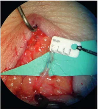
Under local anaesthesia with 1% adrenaline xylocaine, a skin incision was performed for each site. The subcutaneous tissue was dissected under a high magnification operating micro-scope. Lymphatic collectors and adjacent sub-cutaneous veins were identified. Vessels were dissected and 4 or 5 11/0 endoluminal sutures with a 50-micron needle were performed (Fig-ure 1). After each anastomosis, the milk-test was performed to check the patency22 (Figure 2).
Afterwards, the skin was sutured with
continu-Figure 1. Image of an anastomosis between a lymphatic of approximately 0.5 mm diameter (at the top) and a venule of the same size. Inside the anastomosis a stent made with 7/0 ethilon suture was present: this stent was used to ensure the patency of the anastomosis and removed before the last suture. This lambda-shaped anostomosis allows efficient lymphaticovenu-lar anastomoses to be performed simultaneously by a team of
per-ous 6/0 absorbable microsutures. The purpose of this technique was to drain the accumulated lymphatic fluid into the venous system. The anastomotic configuration was chosen on the basis of the vessels calibre; the most performed was the end-to-end configuration, while the side-to-end configuration was performed if the venular calibre was twice the lymphatic vessel calibre23. The average number of anastomosis
performed was 3 per patient, with an average operation time of 3 hours.
The incidence of cellulitis before surgery was recorded in the patient history collection, while postoperative cellulitis was collected with tele-phone interviews.
Standardized CPT with compression garments was performed 1 year before surgery with 20 cycles of therapy with lymphatic drainages, fol-lowed by bandage with progressive compression; two weeks after s-LVA CPT was continued post-operatively for 1 year. Oral amoxicillin (1 g) was prescribed for 2 days after s-LVA.
The evaluated parameters consisted in: age at the time of surgery, sex, radiotherapy, affected limb, lymphoedema stage, time of lymphoede-ma before surgery, follow-up period, number of cellulitis occurred in the year before surgery, number of cellulitis after surgery.
The frequency of cellulitis in the year be-fore and after s-LVA was compared.
Statistical Analysis
The t-test was used to evaluate if the de-crease of cellulitis was significant. If not
otherwise specified two-tailed p-values < 0.05 were considered significant.
Results
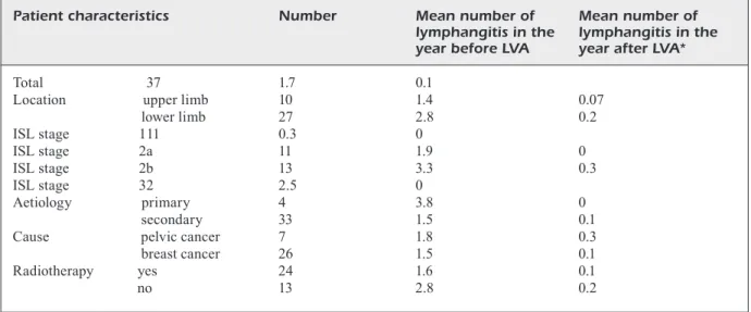
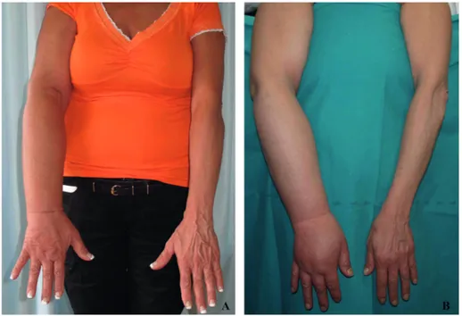
S-LVA resulted successful in all patients as the intraoperative wash-out of the LVA resulted positive in all patients. Table I reports popula-tion data of the patients. The incidence of cellu-litis significantly decreased in all patients; pass-ing from an average of 1.7 cases the year before s-LVA to 0.1 the year after surgery (p = .0012). Cellulitis incidence in upper limb lymphedema dropped from an average of 1.4 cases the year before s-LVA to 0.07 cases the year after surgery while in lower limb lymphedema cellulitis inci-dence decreased from an average of 2.8 cases the year before s-LVA to 0.2 cases the year after surgery. All patients with ISL stage 1, 2a and 3 showed a reduction of cellulitis incidence to zero cases the year after LVA while 2b ISL stage patients showed a cellulitis incidence decreased from an average of 3.3 the year before s-LVA to an average of 0.3 the year after surgery. s-LVA appeared to reduce the instances of cellulitis on all patients. Two typical cases are reported in Figure 3 and Figure 4.
Discussion
Cellulitis is a very serious episode that de-grades the quality of life of many patients. Every
Table I.This table shows the p-values of patients before and after LVA, and patients characteristics. Two-tailed p-values of <0.05 were considered significant if not otherwise specified.
Patient characteristics Number Mean number of Mean number of
lymphangitis in the lymphangitis in the
year before LVA year after LVA*
Total 37 1.7 0.1
Location upper limb 10 1.4 0.07
lower limb 27 2.8 0.2 ISL stage 1 11 0.3 0 ISL stage 2a 11 1.9 0 ISL stage 2b 13 3.3 0.3 ISL stage 3 2 2.5 0 Aetiology primary 4 3.8 0 secondary 33 1.5 0.1
Cause pelvic cancer 7 1.8 0.3
breast cancer 26 1.5 0.1
Radiotherapy yes 24 1.6 0.1
fibrosis, leading to a vicious cycle where the lym-phoedema worsens and cellulitis becomes easier and easier to develop24.
Minimizing the lymphoedema progression is required in order to prevent and reduce the frequency of cellulitis. LVA has proven its re-liability in reducing the incidence of cellulitis, as recently reported by Mihara et al12. Campisi
et al25 reported good results with LVA, too.
Onoda et al26 reported satisfactory results in
the reduction of cellulitis in lymphoedema patients with LVA, and Boccardo et al27
re-ported a positive outcome with the use of the LYMPHA method.
All papers that report the role of s-LVA in lymphoedema patients are usually Japanese pa-pers; the main argument against this technique is its difficult reproducibility out of Japan.
In the present study, we performed s-LVA on 37 Italian patients and reduced cellulitis inci-dence from an average of 1.7 episodes per year before s-LVA to an average of 0.1 cases per year after s-LVA. Patient with 2a and 2b ISL stage reported the most marked decrease.
The traditional LVA technique uses lymphatic vessels at the root of the limb, while s-LVA con-sists in a supramicrosurgical endoluminal anas-tomosis performed on small veins with 0.2 mm diameter all over the limb; with s-LVA smaller
Figure 3. Clinical appearance of right lower limb before (A) and after (B) s-LVA. This patient reported lymphedema of the lower right limb after ovarian cancer surgery; after the s-LVA the limb circumference reduced and no cellulitis were reported in the year after s-LVA.
Figure 4. Clinical appearance of a upper limb in a lymphoedema case after the s-LVA (A). Clinical appearance before s-LVA (B).This patient reported lymphedema after breast cancer surgery, with numerous cellulitis: after s-LVA the limb circumference reduced and no cellulitis were reported in the year after s-LVA.
case damages the lymphatic vessels and may worsen the lymphoedema. Lymph vessels and subcutaneous tissue undergo to cellulitis-induced
lymphatic vessels may be detected and used for the supramicrosurgical anastomosis with smaller veins.
S-LVA reduces cellulitis rate in lymphoedema patients due to the formation of anastomosis and lets the lymph flow into the peripheral venular circulation, with a reduction of stagnant lymph. The concomitant use of CPT and s-LVA with their different mechanisms may also have a syn-ergistic effect on improving lymphatic stasis.
A standard prophylaxis protocol for cellulitis in lymphoedema patients is yet to be present in literature; s-LVA may play an important role in the prevention of future cellulitis cases after can-cer surgery and/or radiotherapy.
In this study, we report a consistent number of lymphoedema patients operated in Italy with s-LVA. This paper dispels the myth of an irre-producible technique and opens the gates for a worldwide dissemination of the technique as the reported results similar to Mihara et al12 suggest
the possibility of diffusion of this technique. S-LVA is a standardized, reliable and repro-ducible technique that finds its main indication in lymphoedema treatment. The reduction of celluli-tis in these patients after s-LVA adds credit to this technique while validating it.
The recent introduction of ICG lymphography and the possibility to perform the LVA operation via the supramicrosurgical technique and under local anesthesia makes this a minimally invasive approach10. ICG lymphography is a useful
imag-ing technique for both beginner and advanced mi-crosurgeon to investigate lymphatic disorders and to decide the best sites for skin incisions. Even though the number of patients in this study was relatively small, a positive trend was evidenced in all subclasses reported in Table I. Further, in pa-tients with prolonged follow-up (2 years), the cel-lulitis trend kept going towards even lower rates. The limitation of the present study is repre-sented by the fact that it is a retrospective study and no consideration was given to the volume reduction. Our study group is now working to another article with a greater population focused on limb volume reduction after s-LVA28.
Conclusions
A significant reduction of cellulitis after s-LVA was reported in all patients enrolled in this study. S-LVA is a standardized, minimally invasive and easily accepted by patients operation that
influ-ences the incidence of cellulitis in lymphedema patients. The main argument against s-LVA in lymphoedema patients is that the positive results present in literature come only from Japanese microsurgeons. This paper reports similar results out of Japan, suggesting the possibility of diffu-sion of this technique.
Conflict of Interest
The authors declare no conflicts of interest.
References
1) Olszewski w. On the pathomechanism of develop-ment of postsurgical lymphoedema. Lymphology 1973; 6: 35-51.
2) COrmier JN, Askew rl, muNgOvAN ks, XiNg Y, rOss
mi, Armer Jm. Lymphoedema beyond breast
can-cer: a systematic review and meta-analysis of cancer-related secondary lymphoedema. Cancer 2010; 116: 5138-5149.
3) mCwAYNe J, HeiNeY sP. Psychologic and social sequelae of secondary lymphoedema: a review. Cancer 2005; 104: 457-66.
4) COzeN w, BerNsteiN l, wANg F, Press mF, mACk
tm. The risk of angiosarcoma following primary
breast cancer. Br J Cancer 1999; 81: 532-536. 5) simON ms, COdY rl. Cellulitis after axillary lymph
node dissection for carcinoma of the breast. Am J Med 1992; 93: 543-548.
6) Olszewski wl, JAmAl s, mANOkArAN g, triPAtHi Fm, zAleskA m, stelmACH e. The effectiveness of long-acting penicillin (penidur) in preventing re-currences of dermatolymphangioadenitis (DLA) and controlling skin, deep tissues, and lymph bacterial flora in patients with “filarial” lymphede-ma. Lymphology 2005; 38: 66-80.
7) vigNes s, POrCHer r, ArrAult m, duPuY A. Long-term management of breast cancer-related lymphedema after intensive decongestive phys-iotherapy. Breast Cancer Res Treat 2007; 101: 285-290.
8) FOldi e. Prevention of dermatolymphangioadenitis by combined physiotherapy of the swollen arm af-ter treatment for breast cancer. Lymphology 1996; 29: 48-49.
9) miHArA m, HArA H, NArusHimA m, HAYAsHi Y, YAmAmOtO t, OsHimA A, kikuCHi k, murAi N, kOsHimA i. Lower limb lymphoedema treated with lymphatico-venous anastomosis based on pre- and intraoperative ICG lymphography and non-contact vein visualization: A case report. Microsurgery 2012; 32: 227-230. 10) miHArA m, HArA H, kikuCHi k, YAmAmOtO t, iidA t,
NArusHimA m, ArAki J, murAi N, mitsui k, geNNArO P, gABriele g, kOsHimA i. Scarless lymphatic venous anastomosis for latent and early-stage lymphoe-dema using indocyanine green lymphography
and non-invasive instruments for visualising sub-cutaneous vein. J Plast Reconstr Aesthet Surg 2012; 65: 1551-1558.
11) miHArA m, HArA H, HAYAsHi Y, iidA t, ArAki J, YAmAmO -tO t, tOdOkOrO t, NArusHimA m, murAi N, kOsHimA
i. Upper-limb lymphoedema treated
aesthetical-ly with aesthetical-lymphaticovenous anastomosis using in-docyanine green lymphography and noncontact vein visualization. J Reconstr Microsurg 2012; 28: 327-332.
12) miHArA m, HArA H, FurNiss d, NArusHimA m, iidA t, kikuCHi k, OHtsu H, geNNArO P, gABriele g, murAi
N. Lymphaticovenular anastomosis to prevent
cellulitis associated with lymphoedema. Br J Surg 2014; 101: 1391-1396.
13) kOsHimA i, NANBA Y, tsutsui t, tAkAHAsHi Y, itOH s, FuJitsu m. Minimal invasive lymphaticovenular anastomosis under local anesthesia for leg lym-phoedema: is it effective for stage III and IV? Ann Plast Surg 2004; 53: 261-266.
14) kOsHimA i, NANBA Y, tsutsui t, tAkAHAsHi Y, itOH
s. Long-term follow-up after lymphaticovenular
anastomosis for lymphoedema in the leg. J Re-constr Microsurg 2003; 19: 209-215.
15) YAmAmOtO t, kikuCHi k, YOsHimAtsu H, kOsHimA i. Ladder-shaped lymphaticovenular anastomosis using multiple side-to-side lymphatic anastomo-ses for a leg lymphedema patient. Microsurgery 2014; 34: 404-408.
16) YAmAmOtO t, CHeN wF, YAmAmOtO N, YOsHimAtsu H, tAsHirO k, kOsHimA i. Technical simplification of the supramicrosurgical side-to-end lymphaticovenu-lar anastomosis using the parachute technique. Microsurgery 2015; 35: 129-134.
17) CAmPisi C, erettA C, Pertile d, dA riN e, CAmPisi C, mACCiò A, CAmPisi m, ACCOgli s, BelliNi C, BO -NiOli e, BOCCArdO F. Microsurgery for treatment of peripheral lymphedema: long-term outcome and future perspectives. Microsurgery 2007; 27: 333-338.
18) YAmAmOtO t, YOsHimAtsu H, NArusHimA m, YAmAmO -tO N, HAYAsHi A, kOsHimA i. Indocyanine Green Lymphography Findings in Primary Leg Lymphedema. J Reconstr Microsurg 2016; 32: 72-79.
19) YAmAmOtO t, NArusHimA m, dOi k, OsHimA A, OgAtA F, miHArA m, kOsHimA i, muNdiNger gs. Character-istic indocyanine green lymphography findings in lower extremity lymphedema: the generation of a novel lymphedema severity staging system using dermal backflow patterns. Plast Reconstr Surg 2011; 127: 1979-1986.
20) YAmAmOtO t, YAmAmOtO N, dOi k, OsHimA A, YOsHi -mAtsu H, tOdOkOrO t, OgAtA F, miHArA m, NArusHimA m, iidA t, kOsHimA i. Indocyanine green-enhanced lymphography for upper extremity lymphedema: a novel severity staging system using dermal backflow patterns. Plast Reconstr Surg 2011; 128: 941-947
21) YAmAmOtO t, mAtsudA N, dOi k, OsHimA A, YOsHimAtsu H, tOdOkOrO t, OgAtA F, miHArA m, NArusHimA m, ii -dA t, kOsHimA i. The earliest finding of indocyanine green lymphography in asymptomatic limbs of lower extremity lymphedema patients secondary to cancer treatment: the modified dermal back-flow stage and concept of subclinical lymphede-ma. Plast Reconstr Surg 2011; 128: 314e-321e. 22) YAmAmOtO t, NArusHimA m, kikuCHi k, YOsHimAtsu H,
tOdOkOrO t, miHArA m, kOsHimA i. Lambda-shaped anastomosis with intravascular stenting method for safe and effective lymphaticovenular anasto-mosis. Plast Reconstr Surg 2011; 127(5): 1987-92. 23) YAmAmOtO t, YOsHimAtsu H, NArusHimA m, seki Y,
YAmAmOtO N, sHim tw, kOsHimA i. A modified side-to-end lymphaticovenular anastomosis. Microsur-gery 2013; 33: 130-133.
24) miHArA m, HArA H, HAYAsHi Y, NArusHimA m, YAmAmO -tO t, tOdOkOrO t, iidA t, sAwAmOtO N, ArAki J, kikuCHi k, murAi N, Okitsu t, kisu i, kOsHimA i. Pathological steps of cancer-related lymphoedema: histolog-ical changes in the collecting lymphatic ves-sels after lymphadenectomy. PLoS One 2012; 7: e41126.
25) CAmPisi C, BelliNi C, CAmPisi C, ACCOgli s, BONiOli e, BOCCArdO F. Microsurgery for lymphoedema: clini-cal research and long-term results. Microsurgery 2010; 30: 256-260.
26) ONOdA s, tOdOkOrO t, HArA H, AzumA s, gOtO A. Minimally invasive multiple lymphaticovenular anastomosis at the ankle for the prevention of lower leg lymphedema. Microsurgery 2014; 34: 372-376.
27) BOCCArdO F, CAsABONA F, deCiAN F, FriedmAN d, murelli F, Puglisi m, CAmPisi CC, mOliNAri l, sPiNACi s, dessAlvi s, CAmPisi C. Lymphatic microsurgical preventing healing approach (LYMPHA) for pri-mary surgical prevention of breast cancer-related lymphedema: over 4 years follow-up. Microsur-gery 2014; 34: 421-424.
28) geNNArO P, gABriele g, miHArA m, kikuCHi k, sAliNi C, ABOH i, CAsCiNO F, CHisCi g, uNgAri C. Supramicro-surgical lymphatico-venular anastomosis (LVA) in treating lymphoedema: 36-months preliminary report. Eur Rev Med Pharmacol Sci 2016; 20: 4642-4653.


