Proposal for ultrasound staging of placenta accreta spectrum disorders
Giuseppe Cali1,2,Francesco Forlani1,Cristoph Lees3,4,5, Ilan Timor-Trisch6,Josè Palacios-Jaraquemada7, Andrea Dall’Asta3,8, Amar Bhide9, Maria Elena Flacco10, Lamberto Manzoli11, Francesco Labate2, Antonio
Perino2, Giovanni Scambia12, Francesco D’Antonio13,14
1: Department of Obstetrics and Gynaecology, Arnas Civico Hospital, Palermo, Italy
2: Department of Obstetrics and Gynaecology, Azienda Ospedaliera Villa Sofia Cervello, Palermo, Italy 3:Centre for Fetal Care, Queen Charlotte's and Chelsea Hospital, Imperial College Healthcare NHS Trust,
London, United Kingdom.
4:Department of Surgery and Cancer, Imperial College London, United Kingdom 5: Department of Development and Regeneration, KU Leuven, Belgium
6: Department of Obstetrics and Gynaecology, Division of Maternal-Fetal Medicine, New York University SOM, New York, NY, USA
7: Centre for Medical Education and Clinical Research (CEMIC), University Hospital, Buenos Aires, Argentina
8: Department of Medicine and Surgery, Obstetrics and Gynecology Unit, University of Parma, Italy 9: Fetal Medicine Unit, Division of developmental Sciences, St. George’s University of London, London,
United Kingdom
10: Local Health Unit of Pescara, Pescara, Italy 11: Department of Medical Sciences, University of Ferrara
12: Department of Obstetrics and Gynecology, Catholic University of The Sacred Heart
13: Department of Clinical Medicine, Faculty of Health Sciences, UiT-The Arctic University of Norway, Tromsø, Norway
14: Department of Obstetrics and Gynaecology, University Hospital of Northern Norway, Tromsø, Norway
Corresponding Author: Francesco D’Antonio, MD, PhD Department of Clinical Medicine Faculty of Health Sciences
UiT - The Arctic University of Norway Hansine Hansens veg 18
9019 Tromsø, Norway [email protected]
Running head: US staging of PAS
Keywords: Placenta accreta spectrum disorders, Outcome, Prenatal diagnosis
This article has been accepted for publication and undergone full peer review but has not been through the copyediting, typesetting, pagination and proofreading process, which may lead to differences between this version and the Version of Record. Please cite this article as doi: 10.1002/uog.20246
This article is protected by copyright. All rights reserved.
ABSTRACT
Objective: To develop an ultrasound (US) staging system for placenta accreta spectrum disorders
(PAS) and to ascertain whether it may stratify the risk of surgical outcome before birth.
Methods: Secondary retrospective analysis of a prospective collected data on women with placenta
previa. We propose the following classification of PAS based upon the distribution of the different ultrasound signs in women with placenta previa:
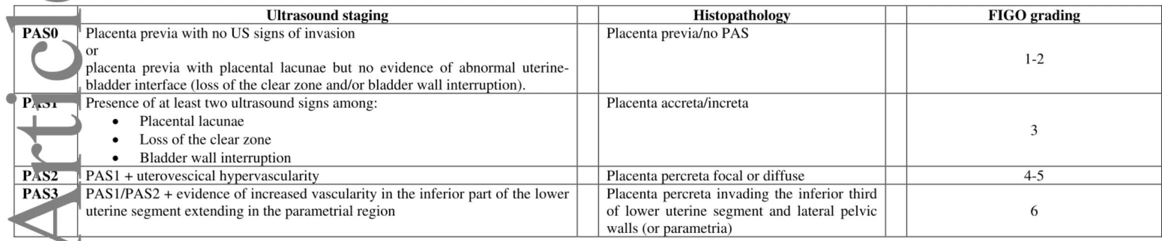
PAS0: Placenta previa with no US signs of invasion or placenta previa with placental lacunae but no evidence of abnormal uterine-bladder interface
PAS1: Presence of at least two ultrasound signs among: placental lacunae, loss of the clear zone or bladder wall interruption.
PAS2: PAS1 + uterovescical hypervascularity
PAS3: PAS1/PAS2 + evidence of increased vascularity in the inferior part of the lower uterine segment potentially extending in the parametrial region.
We explored whether this ultrasound staging system was correlated with surgical outcomes [estimated blood loss (EBL, ml), units of packed red blood cells (PRBC), fresh frozen plasma (FFP) and platelets (PLT) transfused, operative times (minutes), surgical complications, defined as the occurrence to any damage in either bladder, ureters or bowel, length of in hospital stay (days) and admission to intensive care unit (ICU)], and depth of placental invasion. Finally, we assessed the correlation between the present ultrasound staging and the clinical grading system proposed by the International Federation of Gynaecology and Obstetrics (FIGO). Prenatal and surgical management were not based on such prenatal ultrasound staging of PAS disorders. Linear and multiple regression models were used to analyse the data.
Results: Two-hundred and fifty-nine women were included in the analysis. Mean EBL was
516151 ml in women with PAS0, 609146 in PAS1, 950190 in PAS2 and 132353 in PAS3 and significantly increased with increasing severity of ultrasound PAS staging. Mean units of PRBC transfused were 0.050.21 in PAS0, 0.100.45 in PAS1, 1.191.11 in PAS2 and 4.482.06; likewise, there was a progressive increase in the mean units of FFP transfused with increasing severity of ultrasound staging of PAS (0.050.21 in PAS=, 0.00.0 in PAS1, 0.251.0 in PAS2 and 3.632.67 in PAS3. Women presenting with PAS3 on ultrasound had a significantly higher PLT transfused (2.372.40) compared to either those with PAS0, PAS1 and PAS2. Mean surgical time was longer in women PAS3 (18432) compared to PAS1 (15338) and PAS2 (16128). Likewise, women with PAS3 (7.42.1 days) had a longer LOS in the hospital compared to PAS0 (3.406 days), PAS1 (6.41.3 days) and PAS2 (5.90.8 days). On linear regression analysis, after adjusting for all potential confounders, increase severity in ultrasound staging for PAS was independently
This article is protected by copyright. All rights reserved.
associated with a significant increase in EBL (314 mL per 1-category increase, 95% CI 234-291; p<0.001), units of PRBC (1.74 U per 1 category increase, 95% CI 1.33-2.15; p<0.001), FFP (1.19 U per 1 category increase, 95% CI 0.61-1,77; p<0.001), PLT (1.03 U per 1 category increase, 95% CI 0.59-1.47; p<0.001) transfused, surgical time (38.8 minutes per 1 category increase, 95% CI 31.6-46.1; p<0.01) and length of in hospital stay (0.83 days per1-catergory increase, 95% CI 46-1.27, p<0.001). Likewise, on logistic regression analysis, increase in PAS severity was independently associated with surgical complications with an OR of 3.14 (95% CI 1.36-7.25; p:0.007), while only PAS3 was associated with admission to NICU (p<0.001).
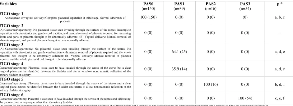
When assessing the correlation between the present ultrasound staging and the clinical grading proposed by FIGO, all women labelled as PAS0 on ultrasound were stage 1 according to FIGO grading system. Conversely, of the women presenting with PAS1 on ultrasound, 64.1% (95% CI 48.4-77.3) were classified as stage 3, while 35.9 (95% CI 22.7-51.6) as stage 4 according to FIGO grading system. Finally, all women with PAS2 were categorized as stage 5 and all those with PAS3 as stage 6 according to the clinical classification proposed by FIGO.
Conclusion: Ultrasound staging of PAS disorders is feasible and correlates with surgical outcome,
depth of invasion and FIGO clinical grading system.
This article is protected by copyright. All rights reserved.
INTRODUCTION
Placenta accreta spectrum (PAS) disorders encompasses a heterogenous group of anomalies characterized by an abnormal adhesion or invasion of the trophoblastic tissue into the myometrium
1-3
.
Advances in prenatal imaging techniques and improved knowledge of the natural history of these anomalies have led to an increase in the detection rate of PAS disorders prenatally4. Prenatal diagnosis of PAS disorders is fundamental as it has been shown to reduce the burden of maternal morbidities by allowing a pre-planned treatment in centers with high expertise in surgical management5,6.
The large majority of studies on prenatal diagnosis of PAS disorders has focused on the diagnostic performance of ultrasound in detecting these anomalies, with no attempt in reporting a whether it can stratify the risk of severe maternal morbidity.
PAS disorders are commonly classified according to depth of trophoblastic invasion into the myometrium, with its most severe type, placenta percreta, being associated with a higher risk of massive hemorrhage, need for blood transfusion and admission to intensive care unit7. However, although depth of invasion represents one of the major determinants of surgical outcome in PAS, large variability in the clinical course of women presenting with the same degree of placental invasion may be observed8,9.
In oncology, stratification of the severity of cancer, based upon several characteristics of the tumor, such as size, spread and cellular abnormalities, helps in classifying the disease, understanding its seriousness, making an informed prediction on prognosis, planning the most appropriate treatment and identifying clinical trials that may be applicable in treating an individual.
The International Federation of Gynecology and Obstetrics (FIGO) has recently introduced a clinical grading system of PAS3. However, despite its importance, this grading system is performed at delivery and it cannot be used to counsel women prenatally. Conversely, only few studies have looked at the feasibility of a prenatal staging system for the detection of the presence and severity of PAS disorders10-12.
The aims of this study were to develop an ultrasound staging system for PAS disorders and to ascertain whether it may stratify the risk of surgical outcome before birth.
This article is protected by copyright. All rights reserved.
METHODS
This is a secondary retrospective analysis of a prospective longitudinal study on the diagnostic performance of ultrasound in detecting PAS disorders including women with placenta previa between 2009 and 201813. All women had a longitudinal bimonthly assessment in the second and third trimester of pregnancy as per local guidelines, in order to detect PAS. STROBE guidelines were followed14.
Ultrasound assessment was performed via transvaginal and transabdominal ultrasound in all cases. All examinations were originally performed using a 4.0-6.0 MHz curved transabdominal or 5.0-7.0 MHz transvaginal transducers (GE Voluson® 730, General Electrics and Samsung WS80A with Elite, Samsung); when using Colour Doppler ultrasound, the pulsed rate frequency (PRF) was initially set at 1.3 KHz but it was lowered in order to identify the presence of placental lacunar flow.
Prenatal diagnosis of PAS disorders was performed according to the recently proposed guidelines for ultrasound detection of PAS, using the following ultrasound signs (Supplementary Figures 1-3)15:
1. Loss of clear zone, defined as a loss, or irregularity, of hypoechoic plane in myometrium underneath placental bed (‘clear zone’).
2. Placental lacunae, defined as the presence of numerous lacunae, often containing turbulent flow visible on grayscale or Color Doppler ultrasound.
3. Bladder wall interruption, defined as loss or interruption of bright bladder wall (hyperechoic band or ‘line’ between uterine serosa and bladder lumen)
4. Uterovescical hypervascularity, defined as striking amount of Color Doppler signal seen between myometrium and posterior wall of bladder, including vessels appearing to extend from placenta, across myometrium and beyond serosa into bladder or other organs; often running perpendicular to myometrium.
5. Increased vascularity in the parametrial region, defined as the presence of hypervascularity extending beyond the lateral uterine walls and involving the region of the parametria16.
We propose the following classification of PAS based upon the distribution of the different ultrasound signs in women presenting with placenta previa:
PAS0: Placenta previa with no US signs of invasion or placenta previa with placental lacunae but
no evidence of abnormal uterine-bladder interface (loss of the clear zone and/or bladder wall interruption).
This article is protected by copyright. All rights reserved.
PAS1: Presence of at least two ultrasound signs among:
Placental lacunae Loss of the clear zone Bladder wall interruption
PAS2: PAS1 + uterovescical hypervascularity
PAS3: PAS1/PAS2 + evidence of increased vascularity in the inferior part of the lower uterine
segment extending in the parametrial region.
The last ultrasound examination prior to surgery to assess the presence and the distribution of the different ultrasound signs and to build the staging system.
We aimed to explore whether such ultrasound staging system was correlated with: Surgical outcome
Depth of placental invasion
FIGO staging system of PAS disorders3
The surgical outcomes explored were: Estimated blood loss (ml)
Units of packed red blood cells (PRBC) transfused Units of fresh frozen plasma (FFP) transfused Units of platelets (PLT) transfused
Operative times (minutes)
Surgical complications, defined as the occurrence to any damage in either bladder, ureters or bowel
Length of in hospital stay (days) Admission to intensive care unit (ICU)
Furthermore, we explored the correlation between the present ultrasound staging system with the clinical grading of PAS disorders recently proposed by FIGO and assessed at delivery3. According to this clinical staging system, PAS disorders can be categorized in the following sub-groups3: 1: At caesarean or vaginal delivery: Complete placental separation at third stage. Normal adherence of placenta.
2: (A) Cesarean/laparotomy: No placental tissue seen invading through the surface of the uterus. Incomplete separation with uterotonics and gentle cord traction, and manual removal of placenta
This article is protected by copyright. All rights reserved.
required for remaining tissue and parts of placenta thought to be abnormally adherent; (B) Vaginal delivery: Manual removal of placenta required, and parts of placenta thought to be abnormally adherent.
3: (A) Cesarean/laparotomy: No placental tissue seen invading through the surface of the uterus. No separation with uterotonics and gentle cord traction with manual removal of placenta required and the whole placental bed thought to be abnormally adherent: (B) Vaginal delivery: Manual removal of placenta required and the whole placental bed thought to be abnormally adherent.
4: Cesarean/laparotomy: Placental tissue seen to have invaded through the serosa of the uterus but a clear surgical plane can be identified between the bladder and uterus to allow nontraumatic reflection of the urinary bladder at surgery.
5: Cesarean/laparotomy: Placental tissue seen to have invaded through the serosa of the uterus and a clear surgical plane cannot be identified between the bladder and uterus to allow nontraumatic reflection of the urinary bladder at surgery.
6: Caesarean/laparotomy: Placental tissue seen to have invaded through the serosa of the uterus and infiltrating the parametrium or any organ other than the urinary bladder.
Depth of placental invasion was defined as the degree of trophoblastic invasion through the myometrium and assessed on histopathological analysis of the removed uterus for those cases undergoing hysterectomy. Placenta accreta was diagnosed when anchoring placental villi were attached to myometrium rather than decidua, but without completely invading it; placenta increta when chorionic villi penetrated the myometrium, while the diagnosis of placenta percreta was considered when chorionic villi penetrated through the myometrium to the uterine serosa and/or adjacent organs. All uterine specimens were assessed by the same research pathologist blinded to the ultrasound and surgical findings; furthermore, because different degrees of placental invasion may co-exist in the same uterus, every case was labelled according to the maximum depth of placental invasion observed. Conversely, in women not undergoing hysterectomy, the presence of PAS was defined according the FIGO clinical grading system and cases with PAS0 as those presenting with complete placenta separation at the third stage3.
For the purpose of the analysis, we used the data from the ultrasound assessments performed at 30-32 weeks of gestation. We have previously demonstrated that the distribution of the different ultrasound signs in women affected by PAS is similar between the second and third trimester of pregnancy17. In the collective authors’ experience, assessment of parametrial invasion is more
This article is protected by copyright. All rights reserved.
accurately performed at 30-32 weeks of gestation. Early assessment may not allow a proper assessment of abnormal vascularization in the parametrial region, while late assessment may be technically affected by the increase in uterine size and difficulties in visualizing parametrial regions. The correlation between the present and FIGO clinical grading system of PAS was performed by retrospectively analysing the surgical notes of each woman because at the time the study was conducted, the FIGO staging system of PAS disorders was not available yet.
Clinical management of PAS disorders
All women presenting with at least two ultrasound signs suggestive of PAS were treated with caesarean hysterectomy and pre-operative temporary occlusion of internal iliac arteries with balloon catheters. For those cases showing ultrasound evidence of PAS and no clear placental invasion at direct visualization, after the delivery of the fetus and clamping of the cord, the balloon catheters were inflated and an attempt to expel the placenta performed by administering carbetocin and a controlled cord traction. In the event of failed placental detachment, hysterectomy was performed, preserving the adnexa and leaving the placenta in situ. Conversely, for cases showing placental tissue protruding through the uterine serosa, a longitudinal incision on the uterine fundus was performed to deliver the fetus, followed by cesarean hysterectomy without any attempt to remove the placenta.
Finally, women presenting with placenta previa with no ultrasound evidence of PAS had caesarean delivery with an incision performed on the lower uterine segment without the use of interventional radiology techniques. The multidisciplinary team remained the same through the study period. No attempt was made to leave the placenta in situ in order to delay surgery and no women undergoing conservative surgical technique (including TRIPLE-P or one step conservative surgery) was included in the study in order not to bias the analysis.
There was no change in the management of women according to the ultrasound grading of PAS as regard as prenatal follow-up, time at delivery and surgical technique.
Data analysis
We investigated the relationship between selected maternal and gestational characteristics and six continuous and two categorical outcomes, in women with different grades of placenta accreta spectrum disorders (PAS), ranging from PAS0 (the least severe form) to PAS3 (the most severe form). The continuous outcomes were: (1) estimated blood loss during delivery; (2) units of red blood cells, (3) units of platelets; (4) units of fresh frozen plasma transfused during surgery; (5) length of surgical time; (6) length of in-hospital stay. The categorical outcomes were: (1) onset of
This article is protected by copyright. All rights reserved.
maternal surgical complications and (2) admission to Intensive Care Unit (ICU). Furthermore, we assessed the depth of placental invasion in women presenting with PAS1, PAS2 and PS3. Finally, we assessed the distribution of the different sub-groups of PAS disorders as proposed by FIGO into the different classes of the present ultrasound staging.
We evaluated the potential association between all recorded maternal and gestational characteristics and the continuous outcomes, first using standard univariate analyses (Spearman test for continuous parameters and Kruskal-Wallis test for categorical ones), and then fitting six multiple regression models. We included a priori all recorded covariates, which were previously tested for multicollinearity, and investigated potential transformation, interaction and/or quadratic/cubic terms18. Parity status, gravidity status and number of previous Cesarean sections (CS) were collinear, as well as PAS and histopathological diagnosis. We thus chose to include the most relevant covariates from a clinical point of view, namely parity status and PAS. Both parity status and PAS were treated both as continuous and ordinal, categorical variables, identifying three categories for parity (nulliparous, primiparous and multiparous), and four categories for PAS (PAS0 to PAS3). All remaining outcomes were included as covariates in all models, with the exception of the model predicting the amount of blood loss, in which the units of red blood cells, plasma and platelets transfused were excluded because they are a consequence of blood loss. The validity of each final regression model was assessed as follows: the assumption of constant error variance was checked graphically, plotting Pearson residuals vs. fitted values, and formally, using the Cook-Weisberg test for heteroskedasticity. High leverage observations were identified by computing Pearson, standardized and studentized residuals, and Cook's D influence. In all models, we found less than 10 high-leverage observations, excluding which we noted no substantial changes.
The potential association between the recorded maternal and gestational parameters and the two categorical outcomes were first evaluated with standard univariate analyses (chi-squared test for categorical variables; Kruskal-Wallis test for continuous variables), and then fitting two logistic regression models. Both models were built using a stepwise forward process, including only significant covariates and maternal age, which was forced to entry. To reduce the potential overfitting due to the small number of successes (n=19 women with surgical complications and n=17 women admitted to ICU), the number of covariates was limited to four in every phase of both models fitting19. The goodness-of-fit was checked using Hosmer-Lemeshow test, and the predictive power assessed through C-statistics (area under the Receiving Operator Curve). Standard post-estimation tests were used to check the validity of both final models, performing multicollinearity and influential observation analyses (using standardized residuals, change in Pearson and deviance chi-square).
This article is protected by copyright. All rights reserved.
We explored further the relationship between pregnancy outcomes and PAS severity comparing the distribution of all recorded outcomes and gestational parameters across various PAS severity categories: (1) women with PAS0 vs women with PAS1; (2) PAS0 vs PAS2: (3) PAS0 vs PAS3: (4) PAS1 vs PAS2; (5) PAS1 vs PAS3; (6) PAS2 vs PAS3.
Statistical significance was defined as a two-sided p-value<0.05 for all analyses, which were carried out using Stata, version 13.1 (Stata Corp., College Station, Texas, USA, 2013).
This article is protected by copyright. All rights reserved.
RESULTS
Two-hundred and fifty-nine women were included in the analysis. General characteristics of the study population in the study are reported in table 1. Mean maternal age was 31.6±5.6 years, while gestational age at birth 35.6±1.7 weeks. Hysterectomy was performed in all women affected by PAS1-3 compared to none of those presenting with PAS0; likewise, none of the included cases was treated leaving the placenta in situ or partially resective surgical techniques. Mean estimated blood loss was 725±427 mL, while mean units of PRBC, FFP and PLT transfused was 1.1±2.1, 0.8±1.2 and 0.5±1.5 respectively. Surgical complications occurred in 7.3% (95% CI 4.8-11.2), while 6.6% (95% CI 4.1-10.3; 17/259) of women were admitted to intensive care unit (Table 1). At histopathological or clinical assessment, placenta accreta was diagnosed in 8.9% (95% CI 6.0-13.0; 23/259) of women, placenta increta in 6.2% (95% CI 3.8-9.8; 16/259) and placenta percreta in 27.0% (95% CI 22.9-32.8; 70/259) of included cases, while 57.9% (95% CI 51.8-64.0) did not show any sign of PAS.
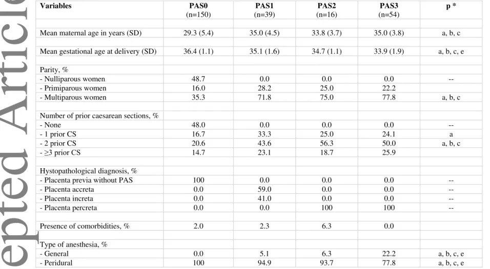
According to the distribution of the different ultrasound signs, 57.9% (95% CI 51.6-64.0; 150/259) of women were labelled as PAS0, 15.1% (95% CI 11.2-19.9; 39/259) as PAS1, 6.2% (95% CI 3.8-9.8; 16/259) as PAS2 and 20.9% (95% CI 18.2-26.4; 54/259) as PAS3 (Table 1). General characteristics of the study population according to the ultrasound staging of PAS are reported in Table 2. Women with PAS0 were significantly younger (mean maternal age. 29.35.49 than those with PAS1 (35.04.5), PAS2 (33.83.7) and PAS3 (35.03.8) while there was no difference between those with PAS1, PAS2 and PAS3. Likewise, women with PAS0 were delivered at later gestational ages (36.41.1), compared to those with PAS1 (35.11.6), PAS2 (34.71.19 and PAS3 (33.91.9).
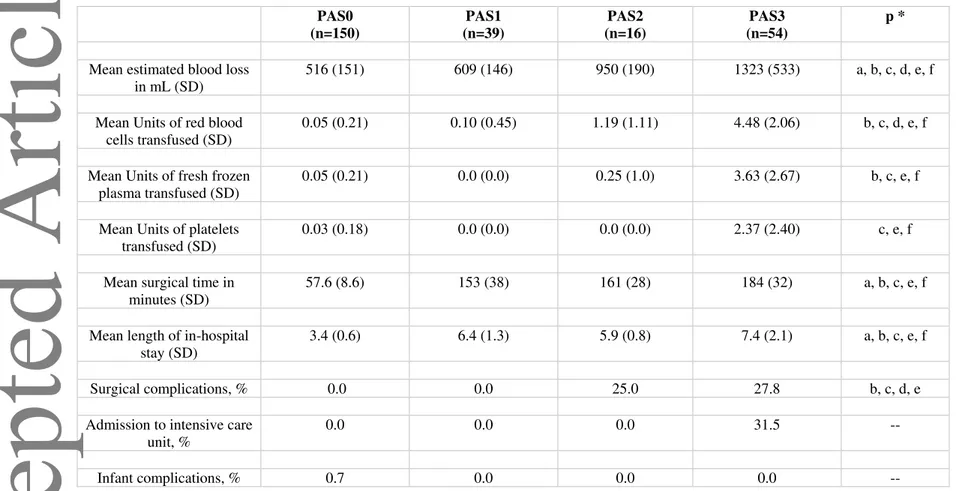
Mean EBL was 516151 ml in women with PAS0, 609146 in PAS1, 950190 in PAS2 and 132353 in PAS3 and significantly increased with increasing severity of ultrasound PAS staging (Table 3). Mean units of PRBC transfused were 0.050.21 in PAS0, 0.100.45 in PAS1, 1.191.11 in PAS2 and 4.482.06; likewise, there was a progressive increase in the mean units of FFP transfused with increasing severity of ultrasound staging of PAS (0.050.21 in PAS=, 0.00.0 in PAS1, 0.251.0 in PAS2 and 3.632.67 in PAS3 (Table 3). There was no difference in the mean units of PLT transfused between women with PAS0 (0.030.18), PAS1 (0.00.0) and PAS2 (0.00.0), while women presenting with PAS3 on ultrasound had a significantly higher PLT transfused (2.372.40) compared to either those with PAS0, PAS1 and PAS2.
This article is protected by copyright. All rights reserved.
Mean surgical time was longer in women with PAS1 (15338) and PAS2 (16128) compared to PAS0 (57.68.6), while that of PAS3 (18432) was longer than PAS1 and PAS2 respectively (Table 3). Likewise, women with PAS1 (6.41.3 days) and PAS2 (5.90.8 days) had a longer LOS in the hospital compared to PAS0 (3.406 days), while LOS in women presenting with PAS3 (7.42.1 days) was longer than PAS1 and PAS2 respectively (Table 3).
Surgical complications, consisting in bladder and ureteral damage, occurred in none of the cases with PAS0 and PAS1 and in 25.0% and 27.8% with PAS2 and PAS3, while 31.5% of women with PAS3 were admitted to ICU compared to none in PAS0, PAS1 and PAS2 (Table 3). Finally, there was no case of neonatal complication in the study population.
On linear regression analysis, after adjusting for all potential confounders, increase severity in ultrasound staging for PAS was independently associated with a significant increase in EBL (314 mL per 1-category increase, 95% CI 234-291; p<0.001), units of PRBC (1.74 U per 1 category increase, 95% CI 1.33-2.15; p<0.001), FFP (1.19 U per 1 category increase, 95% CI 0.61-1,77; p<0.001), PLT (1.03 U per 1 category increase, 95% CI 0.59-1.47; p<0.001) transfused, surgical time (38.8 minutes per 1 category increase, 95% CI 31.6-46.1; p<0.01) and length of in hospital stay (0.83 days per1-catergory increase, 95% CI 46-1.27, p<0.001)(Supplementary Tables 1-6). Likewise, on logistic regression analysis, increase in PAS severity was independently associated with surgical complications with an OR of 3.14 (95% CI 1.36-7.25; p= 0.007), while only PAS3 was associated with admission to NICU (p<0.001) (Supplementary Tables 7-8).
When assessing the distribution of the depth of placental invasion among the different groups, 59% (95% CI 42.1-74.4) of cases with PAS1 has placenta accreta compared to none of the cases with PAS2 and PAS3, while all cases of PAS2 and PAS3 had placenta percreta detected on histopathological analysis.
Correlation between the present ultrasound staging and the clinical grading system recently proposed by FIFO was affected by the retrospective nature of the analysis because at the time the study was conducted, the latter had not been published yet. All women labelled as PAS0 on ultrasound were confirmed to be stage 1 according to FIGO grading system. Conversely, of the women presenting with PAS1 on ultrasound, 64.1% (95% CI 48.4-77.3) were classified as stage 3, while 35.9% (95% CI 22.7-51.6) as stage 4 according to the clinical grading system. Finally, all
This article is protected by copyright. All rights reserved.
women with PAS2 were categorized as stage 5 and all those with PAS3 as stage 6 according to the clinical classification proposed by FIGO (Table 4).
This article is protected by copyright. All rights reserved.
DISCUSSION
Main findings
The findings from this study show that a prenatal ultrasound staging of PAS disorders is feasible. Increase severity in ultrasound staging for PAS was independently associated with a significant increase in EBL, units of PRBC, FFP, PLT transfused, surgical time, length of in hospital stay and surgical complications. When assessing the depth of invasion, all women in PAS1 had placenta accreta or increta while those with PAS2 and PAS3 had exclusively placenta percreta. Despite presenting with the same depth of placental invasion, women with PAS3 were at significantly higher risk of hemorrhage and need for transfusion compared with PAS2. Finally, the present ultrasound staging of PAS disorders showed a good correlation with the clinical grading provided by FIGO.
Comparison with previous studies, strengths and limitations
Only few studies have tried to explore the feasibility and the diagnostic performance of an ultrasound-based scoring system in detecting the presence and the severity of PAS disorders10-12. Tobvin et al. reported that a scoring system including the number of placental lacunae and the presence of bladder wall interruption had a high diagnostic performance for PAS, with an area under the receiver operating characteristic curve of 0.94 (95% CI 0.86-1.0), while in the study by Rac et al., the combination of smallest sagittal myometrial thickness, lacunae, and bridging vessels, in addition to number of caesarean deliveries and placental location, yielded an area under the curve of 0.87 (95% CI 0.80-0.95)10,11.
Prospective designs, large sample size and longitudinal assessment of the included women since the first trimester of pregnancy in the large majority of included cases represent the major strengths of the study. Furthermore, we correlated the present staging system for PAS disorders not only with surgical outcome but also with the depth of placental invasion and FIGO grading system. Finally, all cases affected by PAS were managed by the same multidisciplinary team and treated with hysterectomy, thus reducing the bias related to operator’s experience and type of surgical approach adopted.
The main limitation of the study is represented by the fact that we did not explore whether the application of such staging system in clinical practice may affect prenatal management and prognosis of women with PAS disorders, being the study a secondary retrospective analysis. Furthermore, the correlation between the present ultrasound staging and the clinical grading system proposed by FIGO was affected by the retrospective nature of the analysis, because at the time the
This article is protected by copyright. All rights reserved.
study was conducted, the FIGO grading system was not available yet. Finally, we did not explore whether first trimester assessment gestational sac position can improve the risk stratification provided by the present staging system21-25.
Implications for clinical practice and research
Risk stratification in women affected by PAS disorders is challenging. Although advances in prenatal imaging have led to an increase in the detection rate of these anomalies, there is still a limited evidence on how to identify cases at higher risk of increased surgical morbidity.
Depth of placental invasion is one of the major determinants of surgical outcome in PAS disorders, with women affected by placenta percreta being at higher risk of intra-surgical complications such as massive hemorrhage, need for blood transfusion and damage to adjacent organs7. Despite this, there might be high variability in the surgical outcome even in women presenting with the same depth of placenta invasion.
In the present study, women with PAS and PAS3 were affected exclusively by placenta percreta; despite this, PAS3 carried a significantly higher risk of severe hemorrhage and need for transfusion. These findings confirm that, although fundamental, depth of placental invasion cannot completely stratify the surgical risk of women affected by PAS.
Topography of placental invasion has been recently proposed to be reliable predictor of surgical morbidity in women affected by PAS disorders. Invasions in the inferior third of lower uterine segment, posterior bladder and parametria carry a high risk of surgical morbidity while upper invasions are commonly associated with a more favorable clinical outcome and a relatively easier vascular control surgical9. Assessment of the topography of invasion has been reported only on MR and it is still unclear whether such staging can be reproduced on ultrasound9.
In the present study, all cases with PAS3 showed S2 invasion at surgery; more importantly, there was no case of parametrial invasion in cases labelled as PAS2. Therefore, ultrasound evidence of increased vascularization in the lateral walls of inferior part of the lower uterine segment may identify a sub-group of PAS disorders at higher risk of surgical morbidity due to invasion of the posterior bladder wall and parametria.
Recently, FIGO has proposed a clinical grading system performed at delivery, which helps in determining the presence and severity of PAS disorders. Despite not having been validated yet, the FIGO grading system represents one of the most robust attempts to objectively categorize PAS
This article is protected by copyright. All rights reserved.
disorders. When exploring the correlation between the ultrasound and clinical staging systems, all women with PAS0 on ultrasound were labelled as stage 1 according to FIGO grading system; conversely, of the women presenting with PAS1 on ultrasound, 64.1% (95% CI 48.4-77.3) were classified as stage 3, while 35.9% (95% CI 22.7-51.6) as stage 4 according to the clinical grading system. Finally, all women with PAS2 were categorized as stage 5 and all those with PAS3 as stage 6 according to the clinical classification proposed by FIGO. Despite being affected by the retrospective nature of the analysis, these findings show that prenatal ultrasound staging system of PAS may correlate with the post-natal clinical grading system proposed by FIGO (Figure 1, Table 5).
Conclusions
This study demonstrates that an ultrasound staging of PAS disorders based upon the distribution of different imaging signs is feasible and is correlated with surgical outcome irrespective of the depth of placental invasion.
Further large prospective studies are needed in order to validate the present ultrasound staging system and to explore whether its inclusion in clinical practice may help in deciding the optimal surgical approach and improving the outcome of women affected by PAS disorders.
This article is protected by copyright. All rights reserved.
References
1. Timor-Tritsch IE, Monteagudo A. Unforeseen consequences of the increasing rate of cesarean deliveries: early placenta accreta and cesarean scar pregnancy. A review. Am J Obstet Gynecol 2012; 207:14-29.
2. Belfort MA. Placenta accreta. Am J Obstet Gynecol 2010; 203:430-439.
3. Jauniaux E, Chantraine F, Silver RM, Langhoff-Roos J; FIGO Placenta Accreta Diagnosis
and Management Expert Consensus Panel. FIGO consensus guidelines on placenta accreta spectrum disorders: Epidemiology. Int J Gynaecol Obstet 2018; 140: 265-273.
4. D'Antonio F, Palacios-Jaraquemada J, Lim PS, Forlani F, Lanzone A, Timor-Tritsch I, Cali G. Counseling in fetal medicine: evidence-based answers to clinical questions on morbidly adherent placenta. Ultrasound Obstet Gynecol 2016; 47:290-301.
5. Buca D, Liberati M, Calì G, Forlani F, Caisutti C, Flacco ME, Manzoli L, Familiari A, Scambia G, D'Antonio F. Influence of prenatal diagnosis of abnormally invasive placenta on maternal outcome: systematic review and meta-analysis. Ultrasound Obstet Gynecol 2018;
52: 304-309.
6. Silver RM, Fox KA, Barton JR, Abuhamad AZ, Simhan H, Huls CK, Belfort MA, Wright
JD. Center of excellence for placenta accreta. Am J Obstet Gynecol 2015; 212:561-568. 7. Marcellin L, Delorme P, Bonnet MP, Grange G, Kayem G, Tsatsaris V, Goffinet F. Placenta
percreta is associated with more frequent severe maternal morbidity than placenta accreta. Am J Obstet Gynecol 2018; 219: 193.e1-193.e9.
8. Cali G, Forlani F, Timor-Trisch I, Palacios-Jaraquemada J, Foti F, Minneci G, Flacco ME, Manzoli L, Familiari A, Pagani G, Scambia G, D'Antonio F. Diagnostic accuracy on ultrasound in detecting the depth of invasion in women at risk of abnormally invasive placenta: A prospective longitudinal study. Acta Obstet Gynecol Scand 2018; 97: 1219-1227. 9. D'Antonio F, Palacios-Jaraquemada J, Lim PS, Forlani F, Lanzone A, Timor-Tritsch I, Cali
G. Counseling in fetal medicine: evidence-based answers to clinical questions on morbidly adherent placenta. Ultrasound Obstet Gynecol 2016; 47: 290-301.
10. Rac MW, Dashe JS, Wells CE, Moschos E, McIntire DD, Twickler DM. Ultrasound predictors of placental invasion: the Placenta Accreta Index. Am J Obstet Gynecol 2015; 212: 343.e1-7.
11. Tovbin J, Melcer Y, Shor S, Pekar-Zlotin M, Mendlovic S, Svirsky R, Maymon R. Prediction of morbidly adherent placenta using a scoring system. Ultrasound Obstet Gynecol 2016; 48: 504-510.
This article is protected by copyright. All rights reserved.
12. Gilboa Y, Spira M, Mazaki-Tovi S, Schiff E, Sivan E, Achiron R. A novel sonographic scoring system for antenatal risk assessment of obstetric complications in suspected morbidly adherent placenta. J Ultrasound Med 2015; 34: 561-567.
13. Cali G, Forlani F, Timor-Trisch I, Palacios-Jaraquemada J, Foti F, Minneci G, Flacco ME, Manzoli L, Familiari A, Pagani G, Scambia G, D'Antonio F. Diagnostic accuracy of ultrasound in detecting the depth of invasion in women at risk of abnormally invasive placenta: A prospective longitudinal study. Acta Obstet Gynecol Scand 2018; 97:1219-1227.
14. von Elm E, Altman DG, Egger M, Pocock SJ, Gøtzsche PC, Vandenbroucke JP; STROBE
Initiative. The Strengthening the Reporting of Observational Studies in Epidemiology (STROBE) statement: guidelines for reporting observational studies. J Clin Epidemiol 2008; 61: 344-349.
15. Collins SL, Ashcroft A, Braun T, Calda P, Langhoff-Roos J, Morel O, Stefanovic V, Tutschek B, Chantraine F; European Working Group on Abnormally Invasive Placenta (EW-AIP). Proposal for standardized ultrasound descriptors of abnormally invasive placenta (AIP). Ultrasound Obstet Gynecol 2016; 47: 271-275.
16. Calì G, D'Antonio F, Forlani F, Timor-Tritsch IE, Palacios-Jaraquemada JM. Ultrasound Detection of Bladder-Uterovaginal Anastomoses in Morbidly Adherent Placenta. Fetal Diagn Ther 2017; 41: 239-240.
17. Calì G, Timor-Trisch IE, Palacios-Jaraquemada J, Monteaugudo A, Forlani F, Minneci G, Foti F, Buca D, Familiari A, Scambia G, Liberati M, D'Antonio F. Changes in ultrasonography indicators of abnormally invasive placenta during pregnancy. Int J Gynaecol Obstet 2018; 140: 319-325.
18. Pizzi C, Costa GM, Santarella L, Flacco ME, Capasso L, Bert F, Manzoli L. Depression symptoms and the progression of carotid intima-media thickness: a 5-year follow-up study. Atherosclerosis 2014; 233: 530-536.
19. Faustino M, Pizzi C, Capuzzi D, Agricola T, Costa GM, Flacco ME, Marzuillo C, Nocciolini M, Capasso L, Manzoli L. Impact of atrial fibrillation termination mode during catheter ablation procedure on maintenance of sinus rhythm. HeartRhythm 2014; 11: 1528-1535.
20. Timor-Tritsch IE, Monteagudo A, Cali G, Palacios-Jaraquemada JM, Maymon R, Arslan AA, Patil N, Popiolek D, Mittal KR. Cesarean scar pregnancy and early placenta accreta share common histology. Ultrasound Obstet Gynecol 2014; 43: 383-395.
This article is protected by copyright. All rights reserved.
21. Timor-Tritsch IE, Monteagudo A, Cali G, Vintzileos A, Viscarello R, Al-Khan A, Zamudio S, Mayberry P, Cordoba MM, Dar P. Cesarean scar pregnancy is a precursor of morbidly adherent placenta. Ultrasound Obstet Gynecol 2014; 44: 346-53.
22. Cali G, Forlani F, Timor-Tritsch IE, Palacios-Jaraquemada J, Minneci G, D'Antonio F. Natural history of Cesarean scar pregnancy on prenatal ultrasound: the crossover sign. Ultrasound Obstet Gynecol 2017; 50: 100-104.
23. Calì G, Forlani F, Minneci G, Foti F, Di Liberto S, Familiari A, Scambia G, D'Antonio F. First-trimester prediction of surgical outcome in abnormally invasive placenta using the cross-over sign. Ultrasound Obstet Gynecol 2018; 51: 184-188.
24. Kaelin Agten A, Cali G, Monteagudo A, Oviedo J, Ramos J, Timor-Tritsch I. The clinical outcome of cesarean scar pregnancies implanted "on the scar" versus "in the niche". Am J Obstet Gynecol 2017; 216: 510.e1-510.e6.
This article is protected by copyright. All rights reserved.
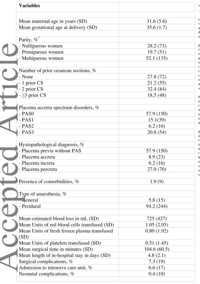
Table 1. General characteristics of the sample (n=259). * : c r u d e p r o p o r t i o n s b e t w e e n p a r e n t h e s e s Variables
Mean maternal age in years (SD) 31.6 (5.6)
Mean gestational age at delivery (SD) 35.6 (1.7) Parity, %*
- Nulliparous women 28.2 (73)
- Primiparous women 19.7 (51)
- Multiparous women 52.1 (135)
Number of prior cesarean sections, %
- None 27.8 (72)
- 1 prior CS 21.2 (55)
- 2 prior CS 32.4 (84)
- ≥3 prior CS 18.5 (48)
Placenta accreta spectrum disorders, %
- PAS0 57.9 (150)
- PAS1 15.1(39)
- PAS2 6.2 (16)
- PAS3 20.8 (54)
Hystopathological diagnosis, %
- Placenta previa without PAS 57.9 (150)
- Placenta accreta 8.9 (23) - Placenta increta 6.2 (16) - Placenta percreta 27.0 (70) Presence of comorbidities, % 1.9 (9) Type of anaesthesia, % - General 5.8 (15) - Peridural 94.2 (244)
Mean estimated blood loss in mL (SD) 725 (427) Mean Units of red blood cells transfused (SD) 1.05 (2.05) Mean Units of fresh frozen plasma transfused
(SD)
0.80 (1.92) Mean Units of platelets transfused (SD) 0.51 (1.45) Mean surgical time in minutes (SD) 104.6 (60.5) Mean length of in-hospital stay in days (SD) 4.8 (2.1)
Surgical complications, % 7.3 (19)
Admission to intensive care unit, % 6.6 (17)
Neonatal complications, % 0.4 (10)
This article is protected by copyright. All rights reserved.
Table 2. Selected maternal and gestational characteristics by level of placenta accreta spectrum (PAS) disorders. Variables PAS0 (n=150) PAS1 (n=39) PAS2 (n=16) PAS3 (n=54) p *
Mean maternal age in years (SD) 29.3 (5.4) 35.0 (4.5) 33.8 (3.7) 35.0 (3.8) a, b, c Mean gestational age at delivery (SD) 36.4 (1.1) 35.1 (1.6) 34.7 (1.1) 33.9 (1.9) a, b, c, e Parity, %
- Nulliparous women 48.7 0.0 0.0 0.0 --
- Primiparous women 16.0 28.2 25.0 22.2
- Multiparous women 35.3 71.8 75.0 77.8 a, b, c
Number of prior caesarean sections, %
- None 48.0 0.0 0.0 0.0 --
- 1 prior CS 16.7 33.3 25.0 24.1 a
- 2 prior CS 20.6 43.6 56.3 50.0 a, b, c
- ≥3 prior CS 14.7 23.1 18.7 25.9
Hystopathological diagnosis, %
- Placenta previa without PAS 100 0.0 0.0 0.0 --
- Placenta accreta 0.0 59.0 0.0 0.0 -- - Placenta increta 0.0 41.0 0.0 0.0 -- - Placenta percreta 0.0 0.0 100 100 -- Presence of comorbidities, % 2.0 2.3 6.3 0.0 Type of anesthesia, % - General 0.0 5.1 6.3 22.2 a, b, c, e - Peridural 100 94.9 93.7 77.8 a, b, c, e
Chi-squared test for categorical variables; t-test and Kruskal-Wallis test for normally-distributed and non-normally distributed continuous variables, respectively. a = p<0.05 for the comparison between women with a diagnosis of PAS0 and women with a diagnosis of PAS1; b = p<0.05 for the comparison between women with a diagnosis of PAS0 and women with a diagnosis of PAS2; c = p<0.05 for the comparison between women with a diagnosis of PAS0 and women with a diagnosis of PAS3; d = p<0.05 for the comparison between women with a diagnosis of PAS1 and women with a diagnosis of PAS2; e = p<0.05 for the comparison between women with a diagnosis of PAS1 and women with a diagnosis of PAS3; f = p<0.05 for the comparison between women with a diagnosis of PAS2 and women with a diagnosis of PAS3. All p-values that are not indicated were >0.05.
This article is protected by copyright. All rights reserved.
Table 3.Comparison of maternal and gestational outcomes across different levels of placenta accreta spectrum (PAS) disorders. PAS0 (n=150) PAS1 (n=39) PAS2 (n=16) PAS3 (n=54) p *
Mean estimated blood loss in mL (SD)
516 (151) 609 (146) 950 (190) 1323 (533) a, b, c, d, e, f
Mean Units of red blood cells transfused (SD)
0.05 (0.21) 0.10 (0.45) 1.19 (1.11) 4.48 (2.06) b, c, d, e, f
Mean Units of fresh frozen plasma transfused (SD)
0.05 (0.21) 0.0 (0.0) 0.25 (1.0) 3.63 (2.67) b, c, e, f
Mean Units of platelets transfused (SD)
0.03 (0.18) 0.0 (0.0) 0.0 (0.0) 2.37 (2.40) c, e, f
Mean surgical time in minutes (SD)
57.6 (8.6) 153 (38) 161 (28) 184 (32) a, b, c, e, f
Mean length of in-hospital stay (SD)
3.4 (0.6) 6.4 (1.3) 5.9 (0.8) 7.4 (2.1) a, b, c, e, f
Surgical complications, % 0.0 0.0 25.0 27.8 b, c, d, e
Admission to intensive care unit, %
0.0 0.0 0.0 31.5 --
Infant complications, % 0.7 0.0 0.0 0.0 --
Chi-squared test for categorical variables; t-test and Kruskal-Wallis test for normally-distributed and non-normally distributed continuous variables, respectively. a = p<0.05 for the comparison between women with a diagnosis of PAS0 and women with a diagnosis of PAS1; b = p<0.05 for the comparison between women with a diagnosis of PAS0 and women with a diagnosis of PAS2; c = p<0.05 for the comparison between women with a diagnosis of PAS0 and women with a diagnosis of PAS3; d = p<0.05 for the comparison between women with a diagnosis of PAS1 and women with a diagnosis of PAS2; e = p<0.05 for the comparison between women with a diagnosis of PAS1 and women with a diagnosis of PAS3; f = p<0.05 for the comparison between women with a diagnosis of PAS2 and women with a diagnosis of PAS3. All p-values that are not indicated were >0.05.
This article is protected by copyright. All rights reserved.
Table 4. Correlation between the ultrasound and clinical staging system of PAS disorders. Variables PAS0 (n=150) PAS1 (n=39) PAS2 (n=16) PAS3 (n=54) p * FIGO stage 1
At caesarean or vaginal delivery: Complete placental separation at third stage. Normal adherence of placenta
100 (150) 0 (0) 0 (0) (0) a, b, c
FIGO stage 2
(A) Caesarean/laparotomy: No placental tissue seen invading through the surface of the uterus. Incomplete separation with uterotonics and gentle cord traction, and manual removal of placenta required for remaining tissue and parts of placenta thought to be abnormally adherent; (B) Vaginal delivery: Manual removal of placenta required, and parts of placenta thought to be abnormally adherent.
0 (0) 0 (0) 0 (0) 0 (0) --
FIGO stage 3
(A) Caesarean/laparotomy: No placental tissue seen invading through the surface of the uterus. No separation with uterotonics and gentle cord traction with manual removal of placenta required and the whole placental bed thought to be abnormally adherent: (B) Vaginal delivery: Manual removal of placenta required and the whole placental bed thought to be abnormally adherent.
0 (0) 64.1 (25) 0 (0) 0 (0) a, d, e
FIGO stage 4
Caesarean/laparotomy: Placental tissue seen to have invaded through the serosa of the uterus but a clear surgical plane can be identified between the bladder and uterus to allow nontraumatic reflection of the urinary bladder at surgery.
0 (0) 35.9 (14) 0 (0) 0 (0) a, d, e
FIGO stage 5
Caesarean/laparotomy: Placental tissue seen to have invaded through the serosa of the uterus and a clear surgical plane cannot be identified between the bladder and uterus to allow nontraumatic reflection of the urinary bladder at surgery.
0 (0) 0 (0) 100 (16) 0 (0) b, d, f
FIGO stage 6
Caesarean/laparotomy: Placental tissue seen to have invaded through the serosa of the uterus and infiltrating the parametrium or any organ other than the urinary bladder.
0 (0) 0 (0) 0 (0) 100 (54) c, e, f
Chi-squared test for categorical variables; a = p<0.05 for the comparison between women with a diagnosis of PAS0 and women with a diagnosis of PAS1; b = p<0.05 for the comparison between women with a diagnosis of PAS0 and women with a diagnosis of PAS2; c = p<0.05 for the comparison between women with a diagnosis of PAS0 and women with a diagnosis of PAS3; d = p<0.05 for the comparison between women with a diagnosis of PAS1 and women with a diagnosis of PAS2; e = p<0.05 for the comparison between women with a diagnosis of PAS1 and women with a diagnosis of PAS3; f = p<0.05 for the comparison between women with a diagnosis of PAS2 and women with a diagnosis of PAS3. All p-values that are not indicated were >0.05.
This article is protected by copyright. All rights reserved.
Table 5. Ultrasound staging of PAS and correlation with histopathology and FIGO staging system.
Ultrasound staging Histopathology FIGO grading
PAS0 Placenta previa with no US signs of invasion or
placenta previa with placental lacunae but no evidence of abnormal uterine-bladder interface (loss of the clear zone and/or uterine-bladder wall interruption).
Placenta previa/no PAS
1-2
PAS1 Presence of at least two ultrasound signs among: Placental lacunae
Loss of the clear zone Bladder wall interruption
Placenta accreta/increta
3
PAS2 PAS1 + uterovescical hypervascularity Placenta percreta focal or diffuse 4-5
PAS3 PAS1/PAS2 + evidence of increased vascularity in the inferior part of the lower uterine segment extending in the parametrial region
Placenta percreta invading the inferior third of lower uterine segment and lateral pelvic walls (or parametria)
6
This article is protected by copyright. All rights reserved.
Figure legend.
Figure 1 (a, b c, d). Ultrasound and surgical views of different types of
PAS. Figure a): placenta previa showing lacunae but no other signs of invasion. At surgery, the lower uterine segment seems unaffected and there is no macroscopic evidence of PAS disorders.
Figure b) woman affected by PAS1 according to the ultrasound staging
system. Placental shows the presence of lacunae and abnormal bladder interface but with no utero-vesical hypervascularity. At surgery, there is placental adherence but no sign macroscopic invasion through the uterine serosa can be identified. Figure 1c): Woman with PAS2 showing signs of utero-vescical hypervascularity. At surgery, there is placental percreta invading the S1 and S2 segment but with no evidence of parametrial invasion. Figure 1e): PAS3 showing increased vascularity in the inferior part of the lower uterine segment extending in the parametrial region. At surgery there is evidence of parametrial invasion.
Supplementary Figure 1: Sonographic markers of PAS with histologically
proven placentae percreta after hysterectomy .Disappearance of the “clear space” is also evident on all the pictures.
A. Lacunae demonstrated by 2D gray scale US at 28 weeks 1 day. B and C. 3D gray scale tomographic images using several sections in two perpendicular scanning planes to each other at 28 weeks 1 day.
D -E. Power and color Doppler images of increased blood flow at the placenta /bladder interface. Some distortion of the bladder-line is also seen in this placenta percreta at 21 weeks and 1 day. After repeat cesarean delivery hysterectomy was performed and massive transfusion was necessary.
G – I. Massive involvement of the cervix is demonstrated. G. grayscale with large lacunae.
H. power Doppler and
I. color Doppler. Both Color Doppler images show a plume of flow in the large lacunae.
This article is protected by copyright. All rights reserved.
Supplementary Figure 2: Serial transvaginal US images from 18 weeks
through 23 weeks ending with 33 weeks of a patient with placenta percreta (proven by histology after cesarean hysteretomy) demonstrating progressive cervical involvement, invasion of the bladder (proven at surgery) also showing lacunae, interruption of the bladder line and no “clear space”.
Supplementary Figure 3: Demonstration of the effect of placental invasion
distorting the bladder line.
A, 2D color Doppler of the bulge into the bladder.
B. C. and D. Three consecutive rotational stages around the longitudinal axis of the uterus using 3D surface rendering of the placental bulge into the bladder.
E. F. and G. Three consecutive rotational stages around the longitudinal axis of the same uterus using 3D color Doppler rendering of the placental blood vessels along the incisional line of the previous cesarean section.
This article is protected by copyright. All rights reserved.
For Peer Review
a) b) c) d) PAS0 PAS1 PAS2 PAS3This article is protected by copyright. All rights reserved.