11 June 2021
Pastore, G., Carraro, M., Pettini, E., Nolfi, E., Medaglini, D., & Ciabattini, A. (2019). Optimized protocol for the detection of multifunctional epitope-specific CD4+ T cells combining MHC-II tetramer and intracellular cytokine staining
technologies. FRONTIERS IN IMMUNOLOGY, 10, 1-10. This is the peer reviewd version of the followng article:
Optimized protocol for the detection of multifunctional epitope-specific CD4+ T cells combining MHC-II tetramer and intracellular cytokine staining technologies
Published:
DOI:10.3389/fimmu.2019.02304 Terms of use:
Open Access
(Article begins on next page)
The terms and conditions for the reuse of this version of the manuscript are specified in the publishing policy. Works made available under a Creative Commons license can be used according to the terms and conditions of said license. For all terms of use and more information see the publisher's website.
Availability:
This version is available http://hdl.handle.net/11365/1079892 since 2019-09-23T12:12:42Z Original:
1
Optimized protocol for the detection of multifunctional epitope-specific CD4+ T cells combining 1
MHC-II tetramer and intracellular cytokine staining technologies 2
3
Gabiria Pastore, Monica Carraro, Elena Pettini, Emanuele Nolfi, Donata Medaglini and Annalisa 4
Ciabattini* 5
6
Laboratory of Molecular Microbiology and Biotechnology (LA.M.M.B.), Department of Medical
7
Biotechnologies, University of Siena, Siena, Italy.
8 9
*Correspondence: 10
Università di Siena, Dipartimento di Biotecnologie Mediche 11
Policlinico Le Scotte, V lotto piano 1, 12 Viale Bracci, 4 13 Siena 53100, Italy 14 e-mail: [email protected] 15 16
Running title: Multifunctional tetramer-specific CD4+ T cells 17
18
Keywords: MHC-II tetramers, ICS, cytokines, multifunctional T cells, flow cytometry, immune 19
response, vaccination 20
2 Abstract
21 22
Analysis of multifunctional CD4+ T cells is fundamental for characterizing the immune responses to 23
vaccination or infections. Peptide-MHC tetrameric complexes represent a powerful technology to 24
detect antigen-specific T cells by the specific binding to their T cell receptor, and their combination 25
with functional assays is fundamental for characterizing the antigen-specific immune response. Here 26
we optimized a protocol for the detection of multiple intracellular cytokines within epitope-specific 27
CD4+ T cells identified by the MCH class II tetramer technology. The optimal procedure for assessing 28
the functional activity of tetramer-binding CD4+ Tis based on the simultaneous intracellular staining 29
with both MHC tetramers and cytokine-specific antibodies upon in vitro restimulation of cells with 30
the vaccine antigen. The protocol was selected among procedures that differently combined the steps 31
of cellular restimulation and tetramer staining with intracellular cytokine labelling. 32
This method can be applied to better understand the complex functional profile of CD4+ T cell 33
responses upon vaccination or infection. 34
3 1. Introduction
35 36
The study of the CD4+ T cell activation and effector function is fundamental in the 37
characterization of immune responses to vaccination (1). 38
CD4+ T cells play a central role in mediating vaccine immune responses by shaping both the 39
humoral and cellular immunity (2). Activated CD4 T cells are critically involved in providing 40
cognate help to B cells for production of protective antibodies, and modulate the functions of 41
macrophages and CD8+ cytotoxic T cells through cytokines secretion. The characterization of the 42
cytokine production of antigen-specific T cells is therefore of critical importance to profile 43
vaccine immune response. The direct and specific method for identifying antigen-specific CD4+ 44
T cells is based on the major histocompatibility complex (MHC) tetramer staining technique (3). 45
This procedure allows the identification of specific T cells due to the selective and multivalent 46
binding of tetramer MHC–peptide complexes to the T cell receptors (TCR) (3,4) and has been 47
used for characterizing the primary and recall antigen-specific CD4+ T cell responses in many 48
pre-clinical and human studies (1,5–9). 49
The effector function of antigen-reactivated T cells is commonly measured by flow cytometry-50
based intracellular cytokine staining (ICS) that allows the simultaneous phenotypic 51
characterization and cytokine detection within single cells (10,11). The characterization of 52
intracellular cytokines allows to identify activated CD4+ T cells capable of producing more than 53
one cytokine, and the analysis of these multifunctional/polyfunctional cells is important for 54
characterizing the immune response elicited by the vaccination or natural infections (12). 55
Polyfunctional CD4+ T cells secreting IFN‐γ, TNF‐α and IL‐2 have been proposed as a major 56
component of immune response that correlates with mouse protection against challenge with 57
Leishmania major (13). In tuberculosis (TB), it has not been clarified if the frequency and quality 58
of polyfunctional CD4+ T cell responses elicited in mice by different types of vaccines correlate 59
with protective immunity (14–17), while human studies have shown that a consistent response of 60
CD4+ T cells co-expressing IFN-γ, TNF-α and IL-2 was associated with acute TB infection (18). 61
Tetramer labeling and intracellular cytokine staining are generally not recommended to be 62
performed concurrently since the in vitro antigen restimulation can induce TCR internalization, 63
thus losing the possibility of detecting epitope-specific CD4+ T cells using tetramers (19). 64
In order to identify a protocol for the detection of intracellular cytokine production within the 65
activated epitope-specific CD4+ T cells, we assessed different strategies that combined cellular 66
restimulation (with the vaccine antigen or tetramers) and tetramer staining (extracellular or 67
intracellular) with intracellular cytokine labelling. The different procedures were tested in 68
4
splenocytes from mice immunized with the chimeric tuberculosis vaccine antigen H56 (20) mixed 69
with the adjuvant CAF01 (21), a model vaccine formulation deeply characterized in preclinical 70
studies for its capacity of inducing both humoral and cellular responses (9,22–24). H56 is a fusion 71
protein of M. tuberculosis antigens Ag85B, ESAT-6, and Rv2660, and the H56-specific CD4+T 72
cell response can be monitored by employing Ag85B280–294-complexed MHC class II tetramers 73
(8).The different procedures were tested also in another experimental setting, in which mice were 74
immunized with the model chicken ovalbumin antigen, and the CD4+ T cell response was assessed 75
employing tetramers specific for the epitope325-335 (25). 76
The comparative analysis of the different protocols has permitted to optimize the procedure for 77
identifying the multifunctional profile of tetramer-specific CD4+ T cells performing intracellular 78
staining with both tetramers and cytokine-specific antibodies, upon antigen restimulation. This 79
method represents a helpful tool for identifying epitope-specific CD4+ T cells and analyzing their 80
specific effector function. 81
5 2. Materials and methods
83 84
2.1 Mice 85
Female C57BL/6 mice, purchased from Charles River (Lecco, Italy) were housed under specific 86
pathogen-free conditions in the animal facility of the Laboratory of Molecular Microbiology and 87
Biotechnology (LA.M.M.B.), Department of Medical Biotechnologies at University of Siena, and 88
treated according to national guidelines (Decreto Legislativo 26/2014). The protocol was approved 89
by the Italian Ministry of Health (authorization n° 1004/2015-PR, 22 September 2015). 90
91
2.2 Immunizations 92
Groups of 10-12 mice were immunized by the subcutaneous route at the base of the tail with the 93
chimeric tuberculosis vaccine antigen H56 (2 µg/mouse) combined with the adjuvant CAF01 (250µg 94
dimethyldioctadecylammonium and 50µg trehalose dibehenate/mouse, both kindly provided by 95
Statens Serum Institut, Denmark) and boosted with a lower dose of H56 alone (0.5 µg/mouse) 4 96
weeks later. Another experiment was performed immunizing mice with albumin from hen egg white 97
(OVA, 25 µg/mouse, Sigma-Aldrich) combined with the adjuvant CAF01, and boosted with OVA 98
alone. The formulations containing antigens and CAF01 were injected in a volume of 150µl/mouse 99
of Tris 10 mM, while the formulations containing H56 and OVA alone in a volume of 100µl/mouse 100
of 1X Dulbecco’s Phosphate Buffered Saline (1X PBS). Mice were sacrificed 5 days after boosting. 101
102
2.3 Sample collection and cell preparation 103
Spleens collected from mice were mashed onto 70µm nylon screens (Sefar Italia, Italy) and washed 104
in complete RPMI (cRPMI) medium [RPMI (Lonza, Belgium), 100 U/ml penicillin/streptomycin, 105
and 10% fetal bovine serum (Gibco, USA)] for 10 min at 300 g at 4°C. Splenocytes were treated with 106
red blood cell lysis buffer (1X, eBioscience, USA) for 4 min. Following centrifugation at 300 x g at 107
4˚C for 10 min, cells were washed with 1X PBS and counted with cell counter (Bio-Rad, USA). 108
109
2.4 Protocols and reagents 110
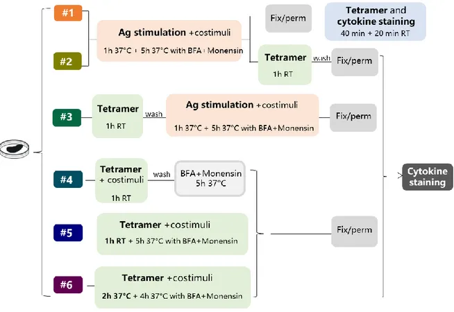
Six different protocols for detecting intracellular cytokines within activated epitope-specific CD4+ T 111
splenocytes that differently combined cellular restimulation, tetramer staining and cytokine labelling, 112
were assessed (Figure 1). Protocols were assessed in two different experimental settings, in which 113
mice were immunized with H56 or OVA antigens, and the CD4 T cell response was analysed 114
employing two different tetramers, specific for the H56 or OVA epitopes, respectively. 115
6
Protocol 1: Splenocytes (2x10⁶/well) were cultured in a round-bottom 96-well plate with H56 protein 116
(2 µg/ml) or OVA (50 g/ml), anti-CD28 and anti-CD49d costimuli (both 2 µg/ml, eBioscience) at 117
37°C, 5%CO2 for 6h, with Brefeldin A (BFA, 5 µg/ml, Sigma-Aldrich) and monensin solution (1×, 118
eBioscience) added during the last 5 h of incubation. Cells were washed with cRPMI for 7 min at 300 119
g at 4°C, labelled with Fixable Viability Stain 780 staining (FVS780, BD Biosciences, 1:1000, 100 120
µl/well) for 20 min at RT in the dark, and washed twice in PBS. Cells were fixed and permeabilized 121
for 20 min at 4°C with BD Cytofix/Cytoperm (Becton Dickinson). Samples were blocked for 30 min 122
at 4°C in Fc-blocking solution (5 µg/ml of CD16/CD32 mAb, eBioscience, USA) and stained for 1 h 123
at room temperature (RT) with PE-conjugated I-A (b) M. tuberculosis Ag85B precursor 280-294 124
(FQDAYNAAGGHNAVF) tetramer (diluted 1:80, hereafter Tet-Ag85B) or with PE-conjugated I-A 125
(b) chicken ova 325-335 (QAVHAAHAEIN) tetramer (diluted 1:50, hereafter Tet-OVA; both 126
tetramers were kindly provided by NIH MHC Tetramer Core Facility, Emory University, Atlanta, 127
GA, USA) diluted in Perm/wash buffer. In the last 20 minutes of tetramer incubation, the following 128
mix of fluorescent antibodies was added: APC-conjugated anti-CD3 (clone 145-2C11), BB700-129
conjugated anti-CD4 (clone RM-5), APC-R700-conjugated, anti-CD44 (clone IM-7), BV786-130
conjugated anti-IFN-γ (clone XMG1.2), BV650-conjugated anti-TNF-α (clone MP6-XT22), BV421-131
conjugated anti-IL-17A (clone TC11-18H10), PE-CF594-conjugated anti-IL-2 (clone JES6-5H4), 132
(all antibodies were purchased from BD Biosciences). All antibodies and tetramer were titrated for 133
optimal dilution. 134
Protocol 2: Splenocytes were cultured with the respective antigens and costimuli as in protocol 1,
135
then were washed and stained with the respective tetramers for 1h at RT. Cells were labelled with 136
FVS780, fixed and permeabilized with BD Cytofix/Cytoperm and stained with the mix of fluorescent 137
antibodies for 20 minutes at RT. 138
Protocol 3: Splenocytes were stained with the respective tetramers for 1h at RT, washed and
139
stimulated with the respective antigens and anti-CD28 and anti-CD49d costimuli at 37°C for 6h, with 140
Brefeldin A and monensin solution added during the last 5 h of incubation. Cells were labelled with 141
FVS780, fixed and permeabilized with BD Cytofix/Cytoperm and stained with the mix of fluorescent 142
antibodies for 20 minutes at RT. 143
Protocol 4: Splenocytes were cultured with the respective tetramers and costimuli for 1h at RT,
144
washed and added with BFA and monensin solution at 37°C for 5 h. Cells were labelled with FVS780, 145
fixed and permeabilized with BD Cytofix/Cytoperm and stained with the mix of fluorescent 146
antibodies for 20 minutes at RT. 147
Protocol 5: Splenocytes were cultured with the respective tetramers and costimuli for 1h at RT and
148
for 5h at 37°C in the presence of BFA and monensin. Cells were labelled with FVS780, fixed and 149
7
permeabilized with BD Cytofix/Cytoperm and stained with the mix of fluorescent antibodies for 20 150
minutes at RT. 151
Protocol 6: Splenocytes were cultured with the respective tetramers and costimuli for 6h at 37°C, 152
with BFA and monensin during the last 4h of incubation.Cells were labelled with FVS780, fixed and 153
permeabilized with BD Cytofix/Cytoperm and stained with the mix of fluorescent antibodies for 20 154 minutes at RT. 155 156 157 158 2.5 Flow cytometry 159
About 7x10⁵ stained cells from each protocol were acquired on BD™ LSRFortessa X20 flow 160
cytometer (BD Biosciences) and stored. Data analysis was performed using FlowJo v10 (TreeStar, 161
USA), and the evaluation of different cytokines co-expression was performed using the FlowJo 162
Boolean gate platform. Fluorescence minus one (FMO) controls were performed for all fluorescence 163
and used for gating setting. 164
165 166
2.6 Statistical analysis 167
Kruskal-Wallis test, followed by Dunn’s post test for multiple comparisons, was used to assess the 168
statistical difference between protocols. A P value ≤ 0.05 was considered significant. Analysis were 169
performed using GraphPad Prism v7 (GraphPad Software, USA). 170
8 3. Results
171 172
In order to optimize the protocol for the detection of intracellular cytokines within activated epitope-173
specific CD4+ T cells, we tested different procedures in splenocytes from mice parenterally 174
immunized with two different antigens, the chimeric TB vaccine antigen H56 or OVA, combined 175
with the liposome adjuvant CAF01, 5 days after the booster immunization. The induction of Ag-176
specific CD4+ T cells producing cytokines was assessed combining antigen restimulation and 177
tetramer staining, followed by intracellular cytokine detection (Figure 1). In protocols 1-3 splenocytes 178
were restimulated with the respective antigens, added before (protocols 1 and 2) or after (protocol 3) 179
tetramer staining. In protocols 4-6 the restimulation step was performed directly with epitope-180
complexed MHC II tetramers, that were therefore used not only as staining tool, but also as functional 181
stimulus. The comparison of results obtained following the different strategies, and tested with two 182
different antigens, has permitted to optimize the procedure for identifying the cytokine profile of 183
tetramer-specific CD4+ T cells. 184
185
3.1 Identification of tetramer-specific CD4+ T cells producing cytokines 186
H56-specific CD4+ T cells were identified using the Ag85B280–294-complexed MHC class II tetramers 187
specific for the immunodominant epitope of Ag85B (26), which is part of the chimeric H56 protein, 188
while OVA-specific CD4+ T cells using the chicken OVA325-335 -complexed MHC class II 189
tetramersTetramer positive cells (Tet-Ag85B+ or Tet-OVA+) were identified as live single CD3+ 190
CD4+ CD44+ cells, and TNF-α, IFN-γ, IL-17 and IL-2 cytokines were detected within gated Tet-191
Ag85B+ cells. All gates were defined on the bases of the respective FMO controls. Staining specificity 192
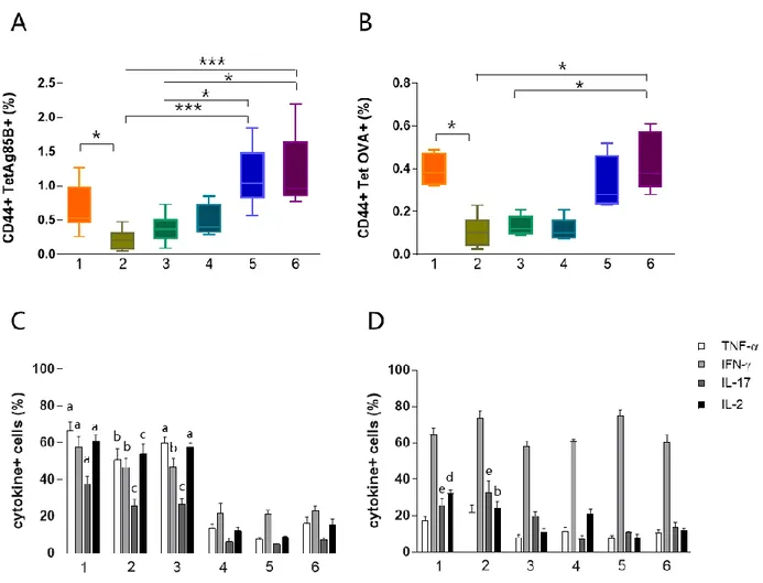
was determined using a control tetramer complexed with an unrelated antigen that showed a level of 193
staining below 0.02% (data not shown). The identification of Tet-Ag85B+ cells in the different six 194
protocols (Figure 2 A), and their intracellular cytokine production (Figure 2 B) are shown. 195
ICS protocol typically includes an antigen stimulation step, that is crucial for activation of effector 196
function of CD4+ T cells. Nevertheless, this step induces the internalization of TCR molecules, thus 197
negatively impacting on the tetramer staining procedure. To overcome this limitation, we assessed a 198
strategy based on the antigen stimulation phase followed by permeabilization and fixation of cells 199
and subsequent tetramer staining (Figure 1, protocol #1). Using this procedure, that allows to identify 200
both extra and intracellular TCR molecules, we detected 0.53% of Tet-Ag85B+ cells (Figure 3A, 201
orange box). This frequency was significantly higher compared to protocol 2, in which splenocytes 202
were firstly stimulated with H56 antigen and then labelled with the specific tetramer (Figure 1, 203
9
protocol #2) allowing an identification of only a 0.2 % of tetramer-positive T cells (Figure 3A, light 204
green). 205
The impact of the tetramer staining performed before antigen restimulation was also evaluated (Figure 206
1, #3). This procedure allowed to detect a frequency of 0.37% of Tet-Ag85B+ T cells that was higher 207
compared to protocol 2 while was lower respect to protocol 1(Figure 3A dark green box). The higher 208
number of Tet-Ag85B+ T cells detected in protocol 1 could be due to the effect of prior antigen 209
restimulation that is known to induce the formation of large clusters of TCR molecules thus increasing 210
tetramer binding avidity (27). 211
In protocols 4, 5 and 6, there was no antigen stimulation and the Ag85B280–294-complexed MHC class 212
II tetramers were used not only for identifying but also for stimulating antigen-specific CD4+ T cells 213
(Figure 1, protocols #4, 5 and 6). As expected, the frequencies of Tet-Ag85B+ T cells detected in 214
protocol 4 were comparable to those of protocol 3 (Figure 3A, light blue and dark green box). In 215
protocols 5 and 6, in which a tetramer incubation phase of 6 hours was performed, frequencies of 216
1.07 and 1% of Tet-Ag85B+ T cells were observed, significantly higher respect to H56-stimulated 217
samples (Figure 3A, P ≤ 0.05 and P ≤ 0.001 compared to protocols 3 and 2, respectively). A similar 218
behavior was observed in mice immunized with the OVA antigen, in which the OVA-specific CD4+ 219
T cells were identified using the OVA325-335 peptide-complexed tetramer (Figure 3B). This shows that 220
tetramers with different peptide specificity, respond in a very similar way to the in vitro staining 221
procedures assessed in the different protocols. 222
The effector function of both Tet-Ag85B+ and Tet-OVA+ T cells identified with the different 223
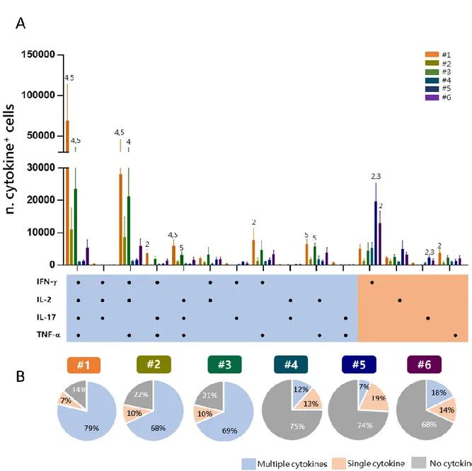
strategies, was analyzed by measuring the intracellular production of 4 different cytokines using 224
multiparametric flow cytometry. As shown in figure 3 C and D, the highest percentage of cells 225
producing intracellular cytokines was detected for both tetramers in protocols 1, 2 and for Tet-226
Ag85B+ also in protocol 3, compared to protocols 4, 5 and 6. These data show that in absence of 227
antigen stimulation, despite the high frequencies of tetramer-binding CD4+ T cells (protocols 5 and 228
6, Figure 3 A and B), a significant lower cytokines production is induced (Figure 3 C and D) 229
highlighting the importance of the in vitro re-stimulation step with the vaccine antigen to effectively 230
stimulate the effector function of antigen-specific CD4+ T cells. 231
232
3.2 Evaluation of tetramer-specific CD4+ T cells mutlifunctional profile. 233
In order to have a picture of the multifunctional profiles of T cells elicited by immunization and 234
detected by the different experimental procedures, a Boolean analysis of data was performed within 235
Tet-Ag85B+ cells (Figure 4A). A significant amount of cells positive for all the four cytokines or for 236
TNF-α, IFN-γ, and IL-2 were observed in protocols 1 and 3 compared to protocols 4 and 5 (P ≤ 0.05). 237
10
Cells producing only IFN-γ were instead significantly higher in protocols 5 and 6 compared to 238
protocol 2 (P ≤ 0.01). The analysis of the frequency of cells producing two or more cytokines 239
(multifunctional), a single one (single) or no cytokines in each protocol shows that the frequency of 240
multifunctional Tet-Ag85B+ T cells was higher in protocols 1, 2 and 3 (79, 68, 69 % respectively) 241
that included the antigen restimulation. Lower frequencies of multifunctional cells were observed in 242
protocols 4, 5 and 6 (12, 7, 18 % respectively) in which most of cells did not produce any cytokine 243
(75, 74, 68 %) (Figure 4B). The intracellular staining with the tetramer performed in protocol 1, 244
allowed to detect the highest percentage of multifunctionality, while no differences were observed 245
among protocols 2 and 3. Therefore the optimal strategy of staining that allows to identify 246
multifunctional T-helper cells among tetramer-specific CD4+ T cells was the protocol #1. 247
248
In conclusion, our comparative analysis, confirmed for two different antigens and their respective 249
tetramers, has shown that the optimal strategy for identifying the multifunctional cytokine profile of 250
tetramer-specific CD4+ T cells is the procedure #1, in which the antigen restimulation phase is 251
followed by the intracellular tetramer staining. Indeed, this protocol allows to detect a significant 252
amount of tetramer-specific T cells, their multifunctional activity, and it allows to reduce the staining 253
time, by adding cytokine-specific antibodies in the last 20 minutes of tetramer incubation. A detailed 254
description of protocol #1 is reported in Figure 5. 255
11 Discussion
256 257
In this study we optimized a flow cytometric protocol for identifying at the single-cell level 258
multifunctional epitope-specific CD4+ T cells, elicited by immunization. Demonstrating pros and 259
cons of different protocols, we showed that the optimal procedure for the simultaneous detection of 260
epitope-specific CD4+ T cells and their effector function is based on the antigenic stimulation of cells 261
combined with a single step of cytokine and tetramer staining in permeabilized cells (Figure 5). Our 262
analysis was based on the comparison of different experimental procedures, tested with two different 263
epitope-specific tetramers, in which the steps of antigen restimulation, tetramer and cytokine staining 264
were differently combined. The systematic analysis of different procedures performed in the same 265
samples has offered the possibility of selecting the optimal protocol among different strategies. The 266
results have been confirmed with tetramers specific for two different antigens, thus strengthening the 267
possible application of the selected procedure to the characterization of the complex functional profile 268
of CD4+ T cell responses upon vaccination or infection. 269
Most of the studies of intracellular cytokine production within tetramer positive cells, have been 270
conducted in CD8+ T cells (28–30), while few works have been performed in CD4+ T cells with MHC 271
class II tetramers (31–33) and none has compared different protocols in a systematic way. Even though 272
a direct comparison with the present study is difficult due to different experimental settings, i.e the 273
use of T cell clones or human CD4+ T cells, prolonged incubation with antigen for cell activation, 274
magnetic bead enrichment of tetramer positive cells before ICS staining, we can generally observe 275
that the tetramer staining was performed extracellularly, often before the antigen stimulation step. 276
Our analysis clearly demonstrates that antigenic stimulation is necessary for an efficient reactivation 277
of the cellular effector function, and the same stimulation effect can not be obtained with the direct 278
incubation of cells with epitope-complexed MHC tetramers, also when prolonged for 6 hours 279
(protocols 5-6). Nevertheless, many studies have demonstrated that ligation of TCR by processed 280
antigen induces TCR internalization and a subsequent down-modulation of its cell surface expression 281
(19,34). Indeed, in protocol 2, in which antigen stimulation was performed before the extracellular 282
tetramer staining the frequency of tetramer positive cells was significantly lower compared to 283
protocol 1. 284
Here, using tetramers specific for two different antigens, we have shown the efficiency of tetramer 285
staining performed in permeabilized cells (protocol 1) that allows to detect both surface expressed 286
and internalized TCR molecules resulting in the identification of the highest percentages of tetramer-287
binding cells. This procedure stained epitope-specific CD4+ T better than protocol 3 in which labeling 288
with tetramer was performed before antigen stimulation. This can be due to the lower avidity of 289
12
tetramer binding to TCR molecules in the absence of cell activation by antigen stimulation. Indeed, 290
cellular activation is known to induce the TCR reorganization with the generation of large clusters 291
of TCR molecules (27) that increase the strength of tetramer binding. 292
Analysis of multifunctional CD4+ T cells is of critical importance for in depth characterization of 293
immune responses to vaccination both in pre-clinical and clinical studies. It is therefore essential to 294
have a protocol that optimally combines the identification of antigen-specific T cells with the analysis 295
of their cytokine profile. Here, we show the possibility to combine, upon antigen stimulation, tetramer 296
and intracellular cytokine staining in permeabilized cells allowing the identification of an higher 297
number of polyfunctional tetramer positive CD4 T cells. The amount of cells producing all the four 298
cytokines, or coexpressing two or three cytokines (especially TNF-α, IFN-γ, and IL-2) was indeed 299
higher compared to the other protocols tested. Significant lower levels of multifunctional cells were 300
observed when tetramers were used both as stimulus and as staining (protocols 4, 5 and 6). Indeed, 301
even though a higher percentage of tetramer-binding T cells was identified by protocols 5 and 6, about 302
70% were negative for cytokine production, respect to 14% observed in protocol 1, demonstrating 303
that the binding of epitope-complexed MHC class II molecules to TCR in the presence of CD49d and 304
CD28 costimuli is not sufficient for effectively reactivating multifunctional antigen-specific CD4+ T 305
cells. 306
The functional characterization of CD4+ T cells described by the protocols analyzed here, is 307
particularly suitable for pre-clinical studies, in which sufficient quantity of CD4+ T cells can be easily 308
identified in draining lymphoid organs such as lymph nodes or spleens, while in humans it is generally 309
more complicated because the frequency of antigen-specific CD4+ T cells in blood are low and often 310
undetectable (6). Moreover, the use of MHC tetramers requires prior knowledge of the peptide epitope 311
and host MHC haplotype, a limitation that can be easily circumvented in inbred animals. 312
In conclusion, in the present work we have selected an optimized protocol for identifying epitope-313
specific CD4+ T cells and their effector function, combining antigenic stimulation of cells with the 314
intracellular staining of TCR molecules and cytokines. Antigenic restimulation, performed at the 315
beginning of the procedure, allows the activation of cells and elicits multiple cytokine production, 316
but at the same time it promotes the down regulation of surface TCR expression that is resolved by 317
the intracellular tetramer staining. This procedure allows also to reduce the total protocol time, since 318
tetramer, surface marker and cytokine staining are combined in a single staining step. 319
This protocol allows to better understand the complex functional profile of T cell responses upon 320
vaccination or natural infection, and it can be instrumental for the dissecting the immune response to 321
vaccination. 322
13 Acknowledgments
323
The authors acknowledge the NIH Tetramer Core Facility (contract HHSN272201300006C) for 324
provision of MHC class II tetramers, and Staten Serum Institute for provision of H56 and CAF01 325
reagents. 326
This study has been carried out with financial support from the Commission of the European 327
Communities, Seventh Framework Programme, contract HEALTH-2011-280873 “Advanced 328
Immunization Technologies” (ADITEC), and Horizon 2020 Framework Programme, grant number 329 730964 (TRANSVAC). 330 331 332 333 Author Contributions 334
GP, DM, AC conceived and designed the experiments; GP, MC, EP: performed the experiments; GP, 335
MC, AC, EP, EN: analyzed the data; GP, AC: wrote the paper; DM, AC: critically revised the 336
manuscript. All authors read and approved the final manuscript. 337
14 References
338
1. Ciabattini A, Pettini E, Medaglini D. CD4(+) T Cell Priming as Biomarker to Study Immune Response 339
to Preventive Vaccines. Front Immunol (2013) 4:421. doi:10.3389/fimmu.2013.00421 340
2. Jelley-Gibbs DM, Strutt TM, McKinstry KK, Swain SL. Influencing the fates of CD4 T cells on the path 341
to memory: lessons from influenza. Immunol Cell Biol (2008) 86:343–352. doi:10.1038/icb.2008.13 342
3. Altman JD, Moss PA, Goulder PJ, Barouch DH, McHeyzer-Williams MG, Bell JI, McMichael AJ, Davis 343
MM. Phenotypic analysis of antigen-specific T lymphocytes. Science (1996) 274:94–96. 344
4. Moon JJ, Chu HH, Pepper M, McSorley SJ, Jameson SC, Kedl RM, Jenkins MK. Naive CD4(+) T cell 345
frequency varies for different epitopes and predicts repertoire diversity and response magnitude. 346
Immunity (2007) 27:203–213. doi:10.1016/j.immuni.2007.07.007 347
5. Cameron TO, Norris PJ, Patel A, Moulon C, Rosenberg ES, Mellins ED, Wedderburn LR, Stern LJ. 348
Labeling antigen-specific CD4(+) T cells with class II MHC oligomers. J Immunol Methods (2002) 349
268:51–69. 350
6. Vollers SS, Stern LJ. Class II major histocompatibility complex tetramer staining: progress, problems, 351
and prospects. Immunology (2008) 123:305–313. doi:10.1111/j.1365-2567.2007.02801.x 352
7. Nepom GT. MHC class II tetramers. J Immunol (2012) 188:2477–2482. doi:10.4049/jimmunol.1102398 353
8. Prota G, Christensen D, Andersen P, Medaglini D, Ciabattini A. Peptide-specific T helper cells identified 354
by MHC class II tetramers differentiate into several subtypes upon immunization with CAF01 adjuvanted 355
H56 tuberculosis vaccine formulation. Vaccine (2015) 33:6823–6830. 356
doi:10.1016/j.vaccine.2015.09.024 357
9. Ciabattini A, Pettini E, Fiorino F, Pastore G, Andersen P, Pozzi G, Medaglini D. Modulation of Primary 358
Immune Response by Different Vaccine Adjuvants. Front Immunol (2016) 7:427. 359
doi:10.3389/fimmu.2016.00427 360
10. Schmittel A, Keilholz U, Scheibenbogen C. Evaluation of the interferon-gamma ELISPOT-assay for 361
quantification of peptide specific T lymphocytes from peripheral blood. J Immunol Methods (1997) 362
210:167–174. 363
11. Freer G, Rindi L. Intracellular cytokine detection by fluorescence-activated flow cytometry: basic 364
principles and recent advances. Methods (2013) 61:30–38. doi:10.1016/j.ymeth.2013.03.035 365
12. De Rosa SC, Lu FX, Yu J, Perfetto SP, Falloon J, Moser S, Evans TG, Koup R, Miller CJ, Roederer M. 366
Vaccination in humans generates broad T cell cytokine responses. J Immunol (2004) 173:5372–5380. 367
13. Darrah PA, Patel DT, De Luca PM, Lindsay RWB, Davey DF, Flynn BJ, Hoff ST, Andersen P, Reed 368
SG, Morris SL, et al. Multifunctional TH1 cells define a correlate of vaccine-mediated protection against 369
Leishmania major. Nat Med (2007) 13:843–850. doi:10.1038/nm1592 370
14. Lewinsohn DA, Lewinsohn DM, Scriba TJ. Polyfunctional CD4+ T Cells As Targets for Tuberculosis 371
Vaccination. Front Immunol (2017) 8:1262. doi:10.3389/fimmu.2017.01262 372
15. Lindenstrøm T, Agger EM, Korsholm KS, Darrah PA, Aagaard C, Seder RA, Rosenkrands I, Andersen 373
P. Tuberculosis subunit vaccination provides long-term protective immunity characterized by 374
multifunctional CD4 memory T cells. J Immunol (2009) 182:8047–8055. 375
doi:10.4049/jimmunol.0801592 376
15
16. Derrick SC, Yabe IM, Yang A, Morris SL. Vaccine-induced anti-tuberculosis protective immunity in 377
mice correlates with the magnitude and quality of multifunctional CD4 T cells. Vaccine (2011) 29:2902– 378
2909. doi:10.1016/j.vaccine.2011.02.010 379
17. Tchilian EZ, Desel C, Forbes EK, Bandermann S, Sander CR, Hill AVS, McShane H, Kaufmann SHE. 380
Immunogenicity and protective efficacy of prime-boost regimens with recombinant (delta)ureC hly+ 381
Mycobacterium bovis BCG and modified vaccinia virus ankara expressing M. tuberculosis antigen 85A 382
against murine tuberculosis. Infect Immun (2009) 77:622–631. doi:10.1128/IAI.00685-08 383
18. Caccamo N, Guggino G, Joosten SA, Gelsomino G, Di Carlo P, Titone L, Galati D, Bocchino M, 384
Matarese A, Salerno A, et al. Multifunctional CD4(+) T cells correlate with active Mycobacterium 385
tuberculosis infection. Eur J Immunol (2010) 40:2211–2220. doi:10.1002/eji.201040455 386
19. Liu H, Rhodes M, Wiest DL, Vignali DA. On the dynamics of TCR:CD3 complex cell surface expression 387
and downmodulation. Immunity (2000) 13:665–675. 388
20. Aagaard C, Hoang T, Dietrich J, Cardona P-J, Izzo A, Dolganov G, Schoolnik GK, Cassidy JP, Billeskov 389
R, Andersen P. A multistage tuberculosis vaccine that confers efficient protection before and after 390
exposure. Nat Med (2011) 17:189–194. doi:10.1038/nm.2285 391
21. Agger EM, Rosenkrands I, Hansen J, Brahimi K, Vandahl BS, Aagaard C, Werninghaus K, Kirschning 392
C, Lang R, Christensen D, et al. Cationic liposomes formulated with synthetic mycobacterial cordfactor 393
(CAF01): a versatile adjuvant for vaccines with different immunological requirements. PLoS ONE 394
(2008) 3:e3116. doi:10.1371/journal.pone.0003116 395
22. Ciabattini A, Prota G, Christensen D, Andersen P, Pozzi G, Medaglini D. Characterization of the 396
Antigen-Specific CD4(+) T Cell Response Induced by Prime-Boost Strategies with CAF01 and CpG 397
Adjuvants Administered by the Intranasal and Subcutaneous Routes. Front Immunol (2015) 6:430. 398
doi:10.3389/fimmu.2015.00430 399
23. Ciabattini A, Pettini E, Fiorino F, Lucchesi S, Pastore G, Brunetti J, Santoro F, Andersen P, Bracci L, 400
Pozzi G, et al. Heterologous Prime-Boost Combinations Highlight the Crucial Role of Adjuvant in 401
Priming the Immune System. Front Immunol (2018) 9:380. doi:10.3389/fimmu.2018.00380 402
24. Santoro F, Pettini E, Kazmin D, Ciabattini A, Fiorino F, Gilfillan GD, Evenroed IM, Andersen P, Pozzi 403
G, Medaglini D. Transcriptomics of the Vaccine Immune Response: Priming With Adjuvant Modulates 404
Recall Innate Responses After Boosting. Front Immunol (2018) 9:1248. doi:10.3389/fimmu.2018.01248 405
25. Moon JJ, Dash P, Oguin TH, McClaren JL, Chu HH, Thomas PG, Jenkins MK. Quantitative impact of 406
thymic selection on Foxp3+ and Foxp3− subsets of self-peptide/MHC class II-specific CD4+ T cells. Proc 407
Natl Acad Sci U S A (2011) 108:14602–14607. doi:10.1073/pnas.1109806108 408
26. Bennekov T, Dietrich J, Rosenkrands I, Stryhn A, Doherty TM, Andersen P. Alteration of epitope 409
recognition pattern in Ag85B and ESAT-6 has a profound influence on vaccine-induced protection 410
against Mycobacterium tuberculosis. Eur J Immunol (2006) 36:3346–3355. doi:10.1002/eji.200636128 411
27. Cecconi V, Moro M, Del Mare S, Dellabona P, Casorati G. Use of MHC class II tetramers to investigate 412
CD4+ T cell responses: problems and solutions. Cytometry A (2008) 73:1010–1018. 413
doi:10.1002/cyto.a.20603 414
28. Appay V, Nixon DF, Donahoe SM, Gillespie GMA, Dong T, King A, Ogg GS, Spiegel HML, Conlon 415
C, Spina CA, et al. HIV-Specific Cd8+ T Cells Produce Antiviral Cytokines but Are Impaired in Cytolytic 416
Function. J Exp Med (2000) 192:63–76. 417
16
29. Wilmschen S, Banki Z, Laer D von, Kimpel J. Simultaneous Quantification of vector and Anti-418
transgene-Specific CD8+ T Cells Via MHC I Tetramer Staining After Vaccination with a Viral Vector. 419
JoVE (Journal of Visualized Experiments) (2018)e58680. doi:10.3791/58680 420
30. Han YW, Aleyas AG, George JA, Yoon HA, Lee JH, Kim BS, Eo SK. Intracellular CD154 expression 421
reflects antigen-specific CD8+ T cells but shows less sensitivity than intracellular cytokine and MHC 422
tetramer staining. J Microbiol Biotechnol (2007) 17:1955–1964. 423
31. Meyer AL, Trollmo C, Crawford F, Marrack P, Steere AC, Huber BT, Kappler J, Hafler DA. Direct 424
enumeration of Borrelia-reactive CD4 T cells ex vivo by using MHC class II tetramers. Proc Natl Acad 425
Sci USA (2000) 97:11433–11438. doi:10.1073/pnas.190335897 426
32. Uchtenhagen H, Rims C, Blahnik G, Chow I-T, Kwok WW, Buckner JH, James EA. Efficient ex vivo 427
analysis of CD4+ T-cell responses using combinatorial HLA class II tetramer staining. Nat Commun 428
(2016) 7:12614. doi:10.1038/ncomms12614 429
33. Tesfa L, Volk HD, Kern F. A protocol for combining proliferation, tetramer staining and intracellular 430
cytokine detection for the flow-cytometric analysis of antigen specific T-cells. J Biol Regul Homeost 431
Agents (2003) 17:366–370. 432
34. Sims S, Willberg C, Klenerman P. MHC-peptide tetramers for the analysis of antigen-specific T cells. 433
Expert Rev Vaccines (2010) 9:765–774. doi:10.1586/erv.10.66 434
17 436
Figure 1. Study design. Six different protocols, combining antigen stimulation and tetramer staining with 437
intracellular cytokine labelling, were used for detecting antigen-specific CD4+ T cell producing cytokines in 438
splenocytes of mice immunized with two different antigens, H56 or OVA, and CAF01 adjuvant, 5 days after 439
booster immunization. In protocols 1-3 splenocytes were restimulated with the respective antigen (Ag, pink 440
box), added before (protocols 1 and 2) or after (protocol 3) tetramer staining (green box), while in protocols 441
4-6 the restimulation step was performed directly with Ag85B or OVA epitope-complexed MHC II tetramers. 442
Anti-CD28 and anti-CD49d (co-stimuli) were added with Ag (protocol 1-3) or with tetramers (protocols 4-6). 443
After Ag or tetramer incubation, cells were treated with Brefeldin A and Monensin for 4-5 h at 37°C, fixated 444
and permeabilized (gray box) and finally stained with anti-cytokines antibodies (dark gray box), except for 445
protocol 1, in which tetramer staining was performed in fixed and permeabilized cells together with cytokine 446
staining. In protocol 6, tetramer staining was performed for 2 h at 37°C. 447
18 448
Figure 2. Flow cytometric analysis of Tet-Ag85B+ T cells producing cytokines. A. Ag85B-tetramer 449
binding T cells were identified among live single CD3+ CD4+, as CD44high Tet-Ag85B+ cells in the six 450
different protocols, and the frequencies of positive cells are reported within the dot plots. B. Intracellular 451
production of TNFα, IFNγ, IL-17 and IL-2 cytokines assessed within the Tet-Ag85B+ T cells in the six 452
different protocols. Frequencies of positive cells are reported within the dot plots. Gates were defined on the 453
respective FMO controls. 454
19 455
Figure 3. Identification of tetramer-specific CD4+ T cells and their cytokine production. Tetramer-456
specific CD4+ T cells and their cytokine production were assessed in splenocytes treated with the different 457
protocols reported in Figure 1. A-B. Box plots of the frequencies of Tet-Ag85B+ (A) and Tet-OVA+ (B) T 458
cells respect to CD4+ T cells, detected employing protocols 1-6, as reported in x axis. Values are reported as 459
mean ± SEM of 10-12 mice, obtained in three independent experiments. Kruskal-Wallis test, followed by 460
Dunn’s post test for multiple comparisons, was used to assess the statistical difference between protocols (*P 461
≤ 0.05, **P ≤ 0.01, ***P ≤ 0.001). C-D. Frequencies of TNF-α, IFNγ, IL-17 and IL-2 positive cells among 462
Tet-Ag85B+ (C) and Tet-OVA+ (D) cells, employing protocols 1-6, as reported in x axis. Values are reported 463
as mean ± SEM of 10-12 mice, obtained in three independent experiments. The significant difference between 464
each cytokine among the different protocols, according to the Kruskal-Wallis test followed by Dunn’s post test 465
for multiple comparisons (P ≤ 0.05), is reported with letters above the error bars; “a”, significant difference 466
versus protocols 4, 5 and 6; “b” versus protocol 5; “c” versus protocols 4 and 5; “d” versus protocols 3, 5 and 467
6; “e” versus protocol 4. 468
20 469
Figure 4. Multifunctional response of Tet-Ag85B+ T cells. Multifunctional profiles of Tet-Ag85B+ T cells 470
detected by the different experimental procedures. A. Histograms represent the number of Tet-Ag85B+ T cells 471
producing different combinations of cytokines shown on the x axis, detected employing the different protocols. 472
Responses are grouped and color coded according to the functionality (orange for single cytokine, light blu for 473
two or more cytokines). Values are reported as mean ± SEM of 10-12 mice, obtained in three independent 474
experiments, and the numbers above the error bars indicate which protocols are significantly different 475
according to the Kruskal-Wallis test, followed by Dunn’s post test for multiple comparisons (P ≤ 0.05). B. Pie 476
charts of the 6 protocols, in which each slice of the pie represents the fraction of Tet-Ag85B+ T cells producing 477
two or more cytokines (multiple cytokines, light blue), a single one (orange) or none (grey). Frequencies are 478
reported within each slice. 479
21 480
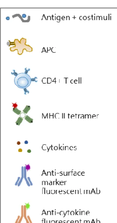
Figure 5. Optimal procedure for identifying multifunctional tetramer-specific CD4+ T cells. Schematic 481
overview of the protocol optimized for the detection of multifunctional epitope-specific CD4+ T cells. 482
Splenocytes are cultured in 96-well plates with antigen and costimuli for 1h at 37°C in order to allow antigen 483
presentation by APC to cognate epitope-specific CD4+ T cells. Antigen stimulation elicits reactivation of 484
effector function of CD4+ cells, and TCR internalization. Brefeldin A (BFA) and monensin solution are added 485
for the last 5 h of incubation to block cytokines secretion. Cells are fixed and permabilized for 20 min at 4°C, 486
and then simultaneously stained with MHC II tetramers (1 h, at RT) and surface markers/cytokine-specific 487
antibodies (last 20 minutes). This allows to detect both surface expressed and internalized TCR molecules, 488
with intracellular cytokines. The single-step staining procedure allow to reduce the complex time of the 489
protocol. Stained samples are then analysed by flow cytometry. 490