DEPARTMENT OF MOLECULAR MEDICINE
PhD course on Immunological, Hematological and Rheumatological Sciences XXXII cycle
Curriculum: Clinical Immunology
Coordinator: Prof. Angela Santoni
PhD THESIS
B cell receptor endocytosis induced by divalent
or multivalent ligands differs in human Naïve
and Memory B cells
Tutor: Prof. Massimo Fiorilli PhD Student: Giovanna Radicchio
ABBREVIATION LIST
BCR= B Cell receptor
BL= Bivalent ligation
CLM= Cross-linking Model
CME= Clatrin-mediated endocytosis
DAM= Dissociation Activation Model
DT= Double Triggering
EE= Early endosomes
FC= Flow Cytometry
FCS= fetal calf serum
FL-H= Fluorescence Height
FRET= fluorescence resonance energy transfer
FSC-H= Forward Scatter Height
GAM= Goat Anti-Mouse
Ig= Immunoglobulin
LE= Late endosomes
ITAM= immunoreceptor tyrosine-based activation motif mAbs= Monoclonal Antibodies
MFI= mean fluorescence intensity
mIg= membrane immunoglobulin
NMS= normal mouse serum
PBS= phosphate-buffered saline 1X
PL= Polyvalent ligation
PBMC= peripheral blood mononuclear cells Syk= spleen tyrosine kinase
SSC-H= Side Scatter Height
Summary
1. INTRODUCTION 3
1.1 An overview of endocytosis phenomenon 3
1.2 The cross-linking model (CLM) of B cell activation 5
1.3 The dissociation activation model (DAM) of B cell activation 9
2. OBJECTIVE OF THE STUDY 11
3. MATERIALS AND METHODS 12
3.1 Reagents 12
3.2 Purification of B cell subsets 12
3.3 BCR endocytosis assay 12
3.3.1 Polyvalent stimulation protocol 13
3.3.2 Bivalent stimulation protocol 13
3.3.3 Sequential bivalent and polyvalent “double triggering” protocol 13
3.4 Flow cytometry and Image Stream 14
3.5 Statistical analysis 14
4. RESULTS 15
4.1 BCR endocytosis by polyvalent ligation in different B cell subsets 15 4.2 BCR endocytosis by bivalent ligation in different B cell subsets 18 4.3 BCR endocytosis by sequential bivalent and polyvalent ligation “double triggering” in different B cells
subsets 20
4.4 Comparison of BCR endocytosis induced by polyvalent, bivalent and double triggering models 21 4.5 Imaging flow cytometry to visualize BCR clustering and endocytosis 22
5. DISCUSSION 24
6. REFERENCES 28
2
ABSTRACT
Antigen binding to the B cell antigen receptor (BCR) initiates endocytosis, which represents the first step of antigen processing and presentation to helper T cells. The receptor crosslinking model (CLM), in which BCRs freely diffuse on the cell surface and are clustered after multivalent antigen binding, was considered for a long time the only mechanism responsible for lymphocyte activation and BCR-antigen complex endocytosis. Recently, a different model of B cell activation, called dissociation-activation model (DAM), was described. In the DAM model, BCRs are organized in auto-inhibiting oligomers that, subsequently to antigen binding, dissociate and become activated. Here we investigated BCR endocytosis in human B cells by a novel flow cytometric assay making use of monoclonal
antibodies (mAbs) for ligating surface Igs. We found that the binding of mAbs, a situation that might perhaps reproduce the DAM model of BCR activation, induced significant BCR endocytosis in naïve, IgM+ memory and switched B cells. Crosslinking of mAb-coated BCRs with F(ab’)2 goat-anti-mouse
IgG (GAMIgG), which putatively reproduces the CLM model of activation by multivalent antigens, increased BCR endocytosis in all B cell subsets. Interestingly, when endocytosis was sequentially induced by DAM (addition of Ig-specific mAb and first step at 37°C) and then by CLM (addition of GAMIgG and second step at 37°C), a model defined as “double triggering”, nearly 100% endocytosis was obtained in switched memory but not in the other B cell subsets, suggesting distinctive features of BCR activation and endocytosis in these subsets. Thus, exploiting mAbs rather than the commonly used polyclonal antibodies for ligating surface Igs provides a simple and accurate way for the analysis of BCR endocytosis potentially useful for untangling the interplay of CLM and DAM.
3 1. INTRODUCTION 1.1 An overview of endocytosis
By the term "endocytosis" we comprise all the active transport processes of the molecules from the outside towards the inside of the eukaryotic cell that cannot passively pass through the membranes [Doherty, 2009]. Endocytic mechanisms control the lipid and protein composition of the plasma membrane, thereby regulating the interaction of the cells with their environments and assuring both their own cellular homeostasis maintenance and their being a part of a multicellular community. These activities include the transmission of diverse signals, the uptake of nutrients and the ability to trigger an effective defense against invading microorganisms [Mellman, 1996]. As regarding exocytosis, it provides the opposite function by pushing molecules out of the cell, so enabling an equal flow of molecules in and out of the cell. The two processes combined ensure a balance of nutrients and waste for regular cell life and function.
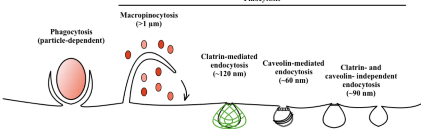
The essential small molecules can traverse the plasma membrane through the action of integral membrane protein pumps or channels, while macromolecules must be carried into the cell in membrane-bound vesicles derived by the invagination of the plasma membrane through endocytosis. On this basis, it is possible to distinguish this uptake in two broad categories: the ‘phagocytosis’ or cell eating, to characterize the large particles uptake and the ‘pinocytosis’ or cell drinking, for the fluid and solutes uptake. Pinocytosis can be further sub-classified according to the mechanism adopted by the cell as: i) macropinocytosis, ii) clathrin-mediated endocytosis (CME), iii) caveolae-mediated endocytosis, and iv) clathrin- and caveolae-independent endocytosis as shown in Fig. 1. These endocytic pathways are highly regulated to control all the physiological processes to assure both homeostasis and adequate guarantees responses to the environment. [Conner, 2003].
4
Cells accomplish CME to internalize the foreign antigen through the B cell receptor (BCR) at the initiation of the immunological response. The BCR is a protein complex composed of membrane immunoglobulin (mIg) molecules associated with heterodimeric Igα/Igβ glycoproteins through non-covalent binding [Hombach, 1990]; this complex forms a type 1 transmembrane receptor [Stoddart, 2002]. The mIg monomers are "Y"-shaped molecules in turn composed by four (two heavy and two light) chains connected by disulfide bonds, and for each chain there are structural domains called “immunoglobulin domains” containing about 70–110 amino acids[Woof MJ, 2004]. This portion of the BCR allows the specific antigen detection and triggers B cell activation. As regards Igα (or CD79a) and Igβ (or CD79b) heterodimers, each of them containing an immunoreceptor tyrosine-based activation motif (ITAM) that, once phosphorylated by other proteins, is capable of signal transduction. [Reth, 1992; Reth 1994; Sanchez, 1993]. A typical human B cell will have 50,000 to 100,000 antibodies bound to its surface. Thus, BCR exploits two crucial functions upon the interaction with the antigen: signal transduction (mediated by Igα and Igβ heterodimers) leading to cell activation, and endocytosis for subsequent processing of the antigen and presentation of peptides to helper T cells.
Figure 1: Modality of entrance into the cell. The endocytic pathways differ with regard to the size of the endocytic
5
1.2 The cross-linking model (CLM) of B cell activation
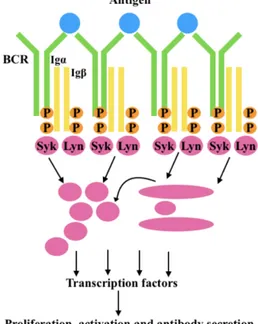
As previously mentioned, B cell responses are initiated by recognition of antigens by mIgs of the BCRs which activates Igα/Igβ heterodimers inducing a cascade of intracellular signaling events that results in B cell activation in a very dynamic process [Hoogeboom, 2016]. BCRs are freely diffused and highly mobile as monomers on the membrane surface of resting B cells, and the binding of multivalent antigen leads to both conformational changes in the BCRs and simultaneous phosphorylation of the ITAMs of Igα/Igβ heterodimers (Fig. 2)[Tolar, 2005]. The phosphorylation of conserved tyrosines present within ITAM allow the Syk-receptor interaction [Reth, 1989;Cambier, 1995], but to obtain both an efficient activation and internalization of the BCRs it is required Lyn, a member of Src kinase family, after BCR aggregation. [Ma, 2001]. Once Syk couples the BCRs, multiple downstream signaling pathways are activated including the mobilization of intracellular stores of calcium, the activation of the mitogen-activated protein (MAP) kinase cascade, and generation of phosphatidylinositide 3-phosphates [Cambier, 1995;Kurosaki, 1999; Chan and Shaw, 1995; De Franco, 1997].
Figure 2: B-cell receptor response to antigen stimulation. This process involves the phosphorylation of two tyrosine
residues in the ITAMs of Igα and Igβ by Lyn (SRC- family kinases). This results in the recruitment and activation of Syk (spleen tyrosine kinase), which in turn phosphorylates other key downstream substrates resulting in productive BCR signaling, activation and productive interactions with helper T cells.
6
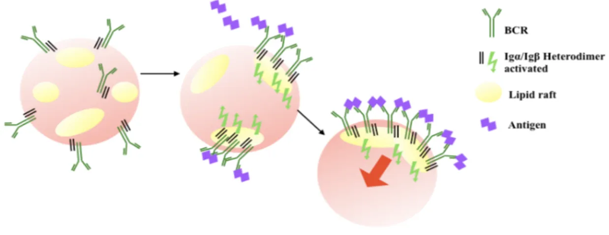
In 1999, the role of lipid rafts in both the initial steps of BCR signaling and antigen targeting was defined [Cheng, 1999]. Lipid rafts are detergent-insoluble microdomains of the plasma membrane enriched in cholesterol and sphingolipids, which play a crucial role in the initiation and organization of signaling cascades since they spatially concentrate components of the signaling machinery (Fig. 3), such as the Src-family kinases, with ligated receptors [Stoddart, 2002] and excluding inhibitor membrane proteins, such as CD45 and CD22 [Cheng, 1999; Pierce, 2002].
Figure 3: Schematic representation of the role of lipid raft in BCR-clusterization. After antigen binding, the
oligomerized BCR associates with rafts and signaling is initiated in rafts. Rafts will exclude inhibitor membrane proteins.
At the same time, antigen binding also triggers the internalization of the BCR. The BCR–antigen complexes are centralized and myosin IIa pulls them inward by the CME mechanism. Then, Rab5 protein converts the internalized vesicle into an early endosome (EE) while Rab7 replaces Rab5, marking the conversion from an early into a late endosome (LE) where BCR signaling is terminated. The late endosome fuses with a lysosome, resulting in degradation of the antigen. Antigen is processed into peptides and loaded onto MHCII molecules for presentation to T cells as shown in Fig. 4 [Hoogeboom, 2016].
7
Taking into account all of these aspects, the model proposed for both the activation and the internalization of BCR-antigen complexes was called the Crosslinking Model (CLM), through which the BCR fulfils two interrelated functions in B-cell activation: the first is the initiation of signal cascades that result in the transcription of a variety of genes associated with B cell activation, and the second is the uptake and targeting of multivalent antigen to MHC class II for presentation to T cells [Pierce, 2002].
Endocytosis of the BCR has been studied by several techniques. For instance, in 2001 Batista et al showed by confocal microscopy that the B cell, while interacting with the immobilized antigens on a target cell, forms a synapse which enhances antigen processing and subsequent presentation to T cells with high efficacy [Batista, 2001]. With regard to the BCR spatial distribution and the role of CD45 membrane receptor of B lymphocytes, Tolar and colleagues [Tolar, 2005] used quantitative fluorescence resonance energy transfer imaging (FRET) to provided evidence that BCRs are organized as monomers and that, once the antigens bind them, they cluster so to exclude the inhibitory receptor CD45. On the other hand, in 2008, Depoil and colleagues [Depoil, 2008] made use of total internal reflection fluorescence microscopy (TIRFM) to provide evidence that CD19 plays an important role in the clusterization of BCR; in fact, both BCR signaling and the microcluster formation were significantly reduced in CD19 deficient B cells.
Figure 4: BCR-antigen steps for presenting the antigen to helper T
cells.
8
Flow cytometry was employed for the first time by Metezeau and colleagues [Metzeau, 1982] for studying endocytosis of ligand-bound BCRs within a heterogeneous population of mouse splenic B cells.
A subsequent important observation, based on mathematical models, was that tyrosine-based motifs mediate signaling when they are phosphorylated, while BCR internalization occurs when they are not phosphorylated; thus, each BCR can undergo only one of two mutually exclusive fates, signaling for B cell activation or internalization for antigen presentation [Hou, 2006].
Since the CLM model was widely accepted as the mechanism of both B cell activation and endocytosis, most of the flow cytometric assays reported in literature exploited F(ab’)2 fragments of
polyclonal anti-BCR antibodies to induce extensive BCR crosslinking [Stoddart, 2002; Caballero, 2006; Malhotra, 2009]. In fact, it is well known that F(ab’)2 fractions of polyclonal anti-BCR
antibodies promote clustering by the binding of multiple antibodies to multiple copies of the BCR (i.e. two antibodies can bind a single BCR, and one antibody is capable of binding two BCRs) [Puffer, 2006]. The commonly used assays measure the rate of endocytosis of BCRs cross-linked with polyclonal antibodies as the ratio between the residual antibody/BCR complexes (detected using fluorescent anti-IgG antibodies specific for the species in which the polyclonal anti-BCR antibody was raised) remaining at the cell surface in samples kept at 37°C (a temperature at which endocytosis takes place) compared to samples kept at 4°C (a temperature at which endocytosis is supposed not to occur).
9
1.3 The dissociation activation model (DAM) of B cell activation
CLM has stood for a long time as the sole mechanistic model for BCR endocytosis [Lemmon, 2010]. This model is based on the assumption that BCRs are dispersed on the cell surface as inactive monomers and that signaling occurs when two or more of these monomers are cross-linked together, for example by bivalent or multivalent ligands.
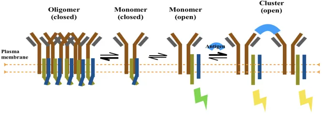
At variance with this view, Reth’s group proposed that BCRs are not freely diffusing as inactive monomers in resting B cells, but are rather disposed as auto-inhibited oligomers and the opening of these oligomers drives BCR activation [Schamel, 2000; Yang, 2010 a]. Thus, based on a number of experimental observations, Yang and Reth proposed [Yang and Reth, 2010 b] a new model of BCR activation, called Dissociation Activation Model (DAM), according to which an antigen, either bivalent or multivalent, dissociates spatially the auto-inhibited oligomers leading to their activation and signaling (Fig. 5).
Figure 5: (from Yang, 2010b). Schematic drawing of the dissociation activation model (DAM). According to this
model the BCR has an auto-inhibitory oligomeric structure on resting B cells. In this structure, the ITAM tyrosines are not accessible to kinase (closed). In the presence of antigen, the equilibrium between closed BCR oligomers and monomers is shifted towards the open clustered monomers. The dissociation of BCR oligomers leads to B cell activation.
10
To analyze the BCR conformation in the nanoscale range, Kläsener et al in 2014, employed different versions of the PLA method using DNA-oligo-coupled antibodies, a rolling circle amplification and detection by fluorescence-coupled oligonucleotideshe IgM–IgM PLA studies which suggested that, upon B cell activation, mIgM molecules move further than 20 nm apart from each other so that their proximity can be still detected by 1-PLA but no longer by Fab-PLA as shown in Fig. 6 [Kläsener, 2014].
Figure 6: (from Kläsener 2014) Schematic drawing showing the spatial organization of the BCR as monitored by
11
2. OBJECTIVE OF THE STUDY
BCR endocytosis induced by oligovalent or multivalent ligands has been investigated using either nominal antigens in BCR transgenic mice [Puffer 2007, Kim 2006], antigen mimics such as F(ab’)2
fragmens of isotype-specific polyclonal anti-Ig antibodies [Metzeau, 1982, Stoddart, 2002; Caballero, 2006; Malhotra, 2009; Thyagarajan, 2003], or anti-idiotype monoclonal antibodies (mAbs) [Elliot 1987]. The use of polyclonal anti-Ig antibodies for ligating the BCR represents, at present, the only way to investigate BCR endocytosis in human heterogeneous B cell populations; however, antibodies may not accurately reproduce the events that take place when ligation of BCR is induced by the cognate antigen and this approach suffers from the limited specificity, the variability of the titres of antibodies to different Ig isotypes in different reagents, and the unpredictability of the binding of polyclonal anti-Ig antibodies to diverse Ig epitopes. Furthermore, oligovalent and multivalent binding to BCR cannot be dissected.
Trying to overcome these limitations, we developed a flow cytometric assay for BCR endocytosis based on the ligation of mIg with isotype-speficic mAbs followed or not by hyper-crosslinking with polyclonal goat-anti-mouse Ig antibodies. Using this assay, which might potentially dissect the relationship of the CLM and DAM models of activation with BCR endocytosis, we compared BCR endocytosis in distinct subsets of normal circulating human B cells.
12
3. MATERIALS AND METHODS 3.1 Reagents
All murine mAbs used were from Becton Dickinson-Pharmingen. Biotinylated anti-human IgG (clone G18-145), IgA (clone G20-359), IgM (clone G20-127) and IgD (clone IA6-2). Unlabeled purified anti-human IgM (clone G20-127), IgD (clone IA6-2), IgA (clone G18-1), IgG (clone G18-145), and control irrelevant IgG1 (clone X40). Fluochrome-labeled mAbs to human k light chain (clone TB28-2, fluorescein isothiocyanate, FITC), l light chain (clone 1-155-2, phycoerythrin, PE), CD19 (clone SJ25C1, peridinin chlorophyll protein, PerCP), CD27 (clone L128, allophycocianin, APC), and CD3 (clone SK7, FITC or PE). Secondary antibodies were PE-conjugated (code R0480, Dako) or unlabelled (code 31170, Thermo Fischer Scientific) F(ab’)2 fragments of goat anti-mouse IgG (GAMIgG)
polyclonal antibodies. Hoechst 33342 (code H3570, Thermo Fischer Scientific) was used for nucleic acid staining.
3.2 Purification of B cell subsets
Human peripheral blood mononuclear cells (PBMC) were obtained from buffy coats of healthy donors by density gradient centrifugation using Lymphoprep (cod. 1114545, Sentinel Diagnostics, Milan, Italy). Resting B cells were isolated from PBMC by Dynabeads untouched human B cells kit (cod. 11351D, Thermo Fisher Scientific, Rome, Italy), according to the manufacturer's instructions with the exception of the spheres/cells incubation step that was carried out at 4°C. To purify total IgM+IgD+ B cells, biotinylated mouse mAbs against human IgG and IgA were added to the antibody mix provided by the Dynabeads kit. Conversely, to purify IgG+IgA+ switched B cells biotinylated mouse mAbs against IgM and IgD were used. The purity of B cell subpopulations, tested before each experimental session, was >95%.
3.3 BCR endocytosis assay
The procedures for inducing BCR endocytosis through either the paucivalent or multivalent ligation of activation are made as follows. After the induction of endocytosis, irrespective of the protocol used, the cells were surface stained with anti-κ chain (FITC), anti-λ chain (PE) and anti-CD27 (APC) mAbs. The extent of BCR endocytosis was calculated as the decrease of the anti-κ or of anti-λ geometric mean fluorescence intensities (MFI) in experimental samples compared to control samples according to the formula: 100 - [(MFI experimental/MFI control) x 100].
13
3.3.1 Polyvalent stimulation protocol
Total, IgM+IgD+ or IgG+IgA+ B cells (2x10⁵) were incubated on ice for 20 min with mixes of anti-IgM/IgD or of anti-IgG/IgA unlabeled mAbs (Becton-Dickinson) at 10 µg/ml each, and then washed to remove unbound antibodies. Cells were then incubated with 5 µg/ml of goat anti-mouse GAMIgG on ice for 20 min, washed and resuspended in RPMI 1640 (cod. 3187-017, Thermo Fisher Scientific, Rome, Italy) with 20% heat inactivated fetal bovine serum (FBS) (cod. ECS0180L, Euro Clone, Milan, Italy), and further incubated at 37°C for 30 min to induce endocytosis. The internalization process was stopped by transferring the samples on ice for 5 min. After one wash, the cells were incubated on ice for 15 min with normal mouse serum (NMS) (Thermo Scientific, Milan, Italy) diluted 1:25 in phosphate buffer saline (PBS) to saturate free sites of GAMIgG and surface stained as described below. Finally, the cells were stained on ice for 20 min with fluorochrome-conjugated mAbs against k (FITC) and l (PE) light chains, CD19 (PerCP) and CD27 (APC). Control samples were initially incubated with irrelevant unlabeled murine IgG1 mAb instead of anti-IgM/IgD or anti-IgG/IgA, and thereafter subjected to the same procedure.
3.3.2 Bivalent stimulation protocol
Total, IgM+IgD+ or IgG+IgA+ B cells (2x10⁵) were incubated on ice for 20 min with mixes of
anti-IgM/IgD or of anti-IgG/IgA unlabeled mAbs (10 µg/ml), or with unlabeled irrelevant IgG1 mAb as control (10 µg/ml each), and washed. After resuspension in RPMI with 20% FBS cells were incubated at 37°C for 30 min to induce endocytosis. The internalization process was blocked on ice for 5 min, and after one wash the cells were stained as in the polyvalent ligation protocol.
3.3.3 Sequential bivalent and polyvalent “double triggering” protocol
Total, IgM+IgD+ or IgG+IgA+ B cells (2x10⁵) were incubated on ice for 20 min with both mixes of
anti-IgM/IgD and of anti-IgG/IgA unlabeled mAbs (10 µg/ml each), or purified mouse IgG1 mAb as control. After washing and resuspension in RPMI 20% FBS, cells were incubated at 37°C for 15 min (bivalent ligation step). Thereafter, GAMIgG was added for additional 15 min at 37°C to induce the crosslinking and internalization of residual BCRs at the cell surface (polyvalent ligation step). All subsequent treatments were done as for the polyvalent ligation protocol.
14
3.4 Flow cytometry and Image Stream
Cells were analyzed using a FACSCalibur cytometer (Becton Dickinson, Mountain View, CA) equipped with a 15 mW, 488nm, air-cooled argon ion laser for excitation of FITC (FL1), PE (FL2) and PerCP (FL3), and with a 10 mW, 635nm, red diode laser for excitation of APC (FL4). The cytometer stability and sensitivity were checked before each acquisition session, by measuring the intensity and the variation coefficient of scatters and fluorescence signals of microbeads Nile Red (Becton Dickinson, Milan, Italy). FL4 detection was optimized by time delay calibration using APC microbeads (Becton Dickinson, Milan, Italy). Samples were acquired and analyzed using CELLQuest 3.3 (Becton Dickinson, Mountain View, CA) and FlowJo (TreeStar, Ashland, Ore) software.
Surface Ig clustering and endocytosis were also analyzed by an ImageStream X Mk II Flow Cytometer (Amnis Corporation), equipped with a 405nm violet laser for excitation of Hoechst 33342, a 488nm blue laser for excitation of FITC and PE, and a 642nm red laser for excitation of APC. The INSPIRE software was used to define cell parameters and to collect data files for image analysis. The IDEAS software was used for spectral compensation and image analysis.
3.5 Statistical analysis
Comparisons were done with the Kruskal-Vallis test or the Mann-Whitney test run by the Prism-Graph-Pad version 7 software (La Jolla CA USA). A p-value of less than 0.05 was considered significant.
15
4. RESULTS
4.1 BCR endocytosis by polyvalent ligation in different B cell subsets
According to the CLM model, BCRs are freely distributed and highly mobile as monomers on the membrane of resting B cells. The binding of multivalent antigens to the BCR initiates two concurrent processes, signaling and receptor internalization [Tolar, 2005; Ma, 2001]. The assay that we developed for inducing BCR endocytosis by paucivalent or polyvalent ligands has been compared using mAbs against single Ig isotypes, rather than of the commonly used F(ab’)2 fragments of anti-human Ig
polyclonal antibodies, that are then cross-linked by F(ab’)2 polyclonal anti-mouse IgG antibody.
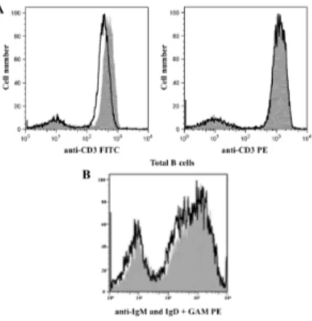
Preliminarily, we verified the reliability of our method particularly to ensure that no shedding of BCR occurred. Thus, we treated whole B cells with purified mAbs to IgM and IgD followed by conjugated GAMIgG-PE; cells were then kept either at 4°C or at 37°C for 30 min. We found that the PE MFIs remained nearly identical under the two incubation conditions (Fig 7A); thus, based on the notion that PE fluorescence is not quenched after internalization (Fig 7 B), and that shedding of BCR at 4°C is presumably negligible, we conclude that BCR is not significantly shed under our experimental conditions.
Figure 7: Fluorochrome stability and analysis of BCR shedding after cross-linking. A) overlaid histograms of human
lymphocytes stained with fluorochrome conjugated mAb anti CD3 FITC (on the left) and anti CD3 PE (on the right) after incubated at 37 °C (line) or stored on ice (solid grey) for 30 min. The fluorescence intensity of CD3 positive cells stained with FITC decreases after incubation at 37 °C while fluorescence of negative cells is unchanged. By contrast, the overlaid histograms of PE labeled sample are unvarying both for positive and negative cells. B) Total B cells were incubated with mAbs to human IgM and IgD, washed and incubated with PE-conjugated GAMIgG at 4°C, and then split and transferred to either 37°C (open histogram) or 4°C (gray histogram) for 30m min and analyzed by flow cytometry. Overlaid histograms show very similar PE fluorescence intensities in the samples, indicating negligible shedding at 37°C.
16
Furthermore, the physical parameters [forward scatter (FSC) and side scatter (SSC)] and the immunophenotypic features of B cells were well preserved (Fig. 8).
Figure 8: Gating strategy for the analysis of BCR endocytosis in B cell subpopulations. (A) Physical parameters (FSC
and SSC) and percentages of naïve and marginal zone B cell subpopulations in purified IgM+IgD+ B cells in the control
(upper panel) and in the sample undergoing endocytosis (lower panel). Dot plots for k+ and l+ BCRs in naïve and marginal
zone B cells are shown in the control sample (upper panel) and in the sample undergoing endocytosis (lower panel). (B) Physical parameters (FSC and SSC) and percentages of total CD19+ CD27+/- in IgA+ IgG+ purified B cells in the control
(upper panel) and in the sample undergoing endocytosis (lower panel). Switched CD27- negative B cells were included in
the analysis because, when analyzed separately they did not show differences in the percentages of endocytosis compared to switched CD27+ B cells. Dot plots for k+ and l+ BCRs in switched B cells are shown in the control sample (upper panel)
and in the sample undergoing endocytosis (lower panel).
Finally, we found that ligation of BCRs with mAbs against IgM and IgD plus GAMIgG was effective in inducing endocytosis in naïve and in IgM+ memory B cells but not in switched B cells (data not
17
shown), thus overcoming the risk of spurious results due to possible cross-reactivity of commercial isotype-specific polyclonal antibodies with other Ig isotypes.
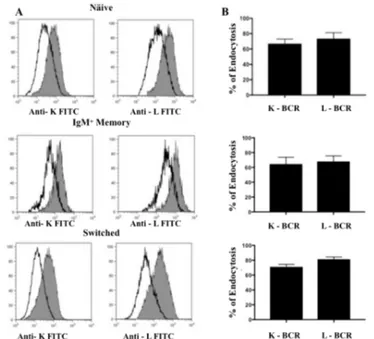
Next, we investigated BCR endocytosis by polyvalent ligation in purified subpopulations of IgM+IgD+CD27-naïve, IgM+IgD+CD27+ unswitched memory and IgG+IgA+CD27+ switched memory B cells. Naïve and IgM memory B cells were incubated with mAbs against IgM and IgD as primary ligands, and switched B cells with mAbs against IgG and IgA; each subpopulation was also incubated with irrelevant IgG1 mAb as control. Cells were subsequently treated with unconjugated GAMIgG and transferred to 37°C to induce endocytosis; then cells were stained with mAbs against k (FITC) and l (PE) light chains to reveal residual Igs at the cell surface. Figure 9 illustrates representative histograms and the cumulative results obtained in each B cell subpopulation with the polyvalent ligation protocol. We found that the extent of endocytosis was similar, irrespectively of the light chain carried by the BCR, in naïve (mean±SD, k 67±6%, l 73±8%), IgM memory (k 65±9%, l 68±8%) and switched memory B cells (k 71±3%, l 81±3%). Hyper-crosslinking (or polyvalent ligation) of BCR with GAMIgG resulted in similar level of internalizationin all B cell subsets.
Figure 9: (A) Representative overlaid histograms of the MFI of anti-k FITC and anti-l PE light chains between the control (grey histograms) and the sample undergoing endocytosis with the polyvalent stimulation protocol (solid line histograms) in naïve, IgM memory and switched B cells. (B) Cumulative results (means and SDs) from 7 separate experiments.
18
4.2 BCR endocytosis by bivalent ligation in different B cell subsets
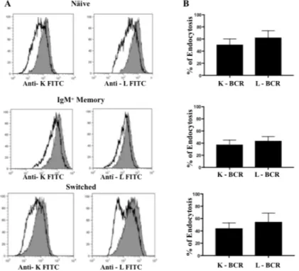
We next investigated whether BCR endocytosis also occurs, and to what extent, when BCR is not grossly aggregated by polyclonal antibodies but is bound by bivalent mAbs, a situation that has been investigated with therapeutic mAbs [Shan, 1998, Cragg, 1999] or with synthetic antigens with low valency [Puffer, 2007]. We loaded purified naïve and IgM+ memory B cells with mAbs to IgM and IgD, and switched memory B cells with mAbs to IgG and IgA; as control, cells were treated with an irrelevant IgG1 mAb. Cells were then directly transferred from 4°C to 37°C and BCR internalization was examined. Triggering with mAbs alone induced similar rates of BCR internalization in all B cell subsets, irrespectively of the light chain expressed (Fig. 10).
Figure 10: BCR endocytosis induced by the bivalent ligation protocol. (A) Representative overlaid histograms of the
MFI of anti-k FITC and anti-l PE light chains between the control (grey histograms) and the sample undergoing endocytosis by bivalent ligation protocol (solid line histograms) in naïve, IgM memory and switched memory B cells. (B) Cumulative results (means and SDs) from 7 separate experiments.
19
However, BCR endocytosis induced by the bivalent ligation was significantly less extensive than that induced by the polyvalent ligation in IgM memory (p=0.0179) and in switched memory (p=0.0167) B cells, but not in naïve B cells (Fig. 11).
Figure 11: Bars denotes the Means and SD of cumulative BCR endocytosis calculated as the mean between k positive and l positive BCR endocytosis in naïve, IgM memory and switched B cells obtained by polyvalent ligation (PL), bivalent ligation (BL) and “double triggering” (DT) protocols from 7 experiments.
BL vs PL, p<0.02 in IgM memory and in switched memory B cells.
DT vs PL in switched B cells, p=0.03; DT in switched B cells vs DT in naïve B cells, p=0.01; DT in switched B cells vs DT in IgM memory B cells, p=0.006.
20
4.3 BCR endocytosis by sequential bivalent and polyvalent ligation “double triggering” in different B cells subsets
To determine whether ligands of different valence may modulate the capacity to internalize BCRs under dynamic conditions, we performed experiments with sequential rounds of endocytosis by bivalent and subsequently the polyvalent ligation with GAMIgG. This assay was named “double triggering”. The rate of BCR endocytosis induced by “double triggering” (Fig. 12) was similar in naïve and in IgM memory B cells as it was observed with the polyvalent protocol (Fig. 9), which differs from “double triggering” solely for the fact that endocytosis by multivalent binding (addition of GAMIgG) takes place at a single step at 37°C rather than sequentially after induction by bivalent ligation (mAb alone) at 37°C followed by addition of GAMIgG and a second step at 37°C. At variance with the polyvalent protocol however, “double triggering” induced significantly more extensive BCR endocytosis in switched memory B cells rather than in naïve (p≤0.0111) or IgM memory B cells (p≤0.0063), a phenomenon not observed when using the previous protocols.
Figure 12: Endocytosis induced by the double triggering protocol. (A) Representative overlaid histograms of the MFI of
anti-k FITC and anti-l PE light chains between the control (solid grey) and the sample undergoing endocytosis with the
double triggering protocol (solid line) in naïve, IgM memory and switched B cells. (B) Cumulative results (means and SDs from 7 separate experiments) on BCR endocytosis by the double triggering.
21
4.4 Comparison of BCR endocytosis induced by bivalent, polyvalent and double triggering binding models
Figure 11 compares total BCR endocytosis (calculated as the mean value of the percent endocytosis of k+ and l+ BCRs) obtained with the polyvalent ligation (PL), bivalent ligation (BL) and “double
triggering” protocols in different B cell subpopulations. BCR endocytosis induced by mAb alone (bivalent ligation protocol) was lower than that induced by the polyvalent ligation or the “double triggering” protocols in all B cell subsets. Intriguingly, the rate of BCR endocytosis induced by the “double triggering” model (a first step with mAbs alone at 37°C followed by a second step at 37°C after crosslinking with GAMIg) or by the polyvalent ligation (a single step at 37°C after treatment with mAbs plus GAMIg at 4°C) was similar in naïve and in IgM memory B cells; by contrast, in switched memory B cells BCR endocytosis was significantly more extensive using the “double triggering” rather than the polyvalent ligation protocol. This suggests that in the latter cells BCR triggering by bivalent ligation and polyvalent ligation might interplay differently than in other B cell types. Intrinsic differences in the initiation of BCR responses between naïve and switched memory B cells are suggested by the observation that the latter cells form antigen-induced and spontaneous sub-microscopic BCR oligomers, and activate signalling-associated kinases, more efficiently than the former [Pierce, 2012]. Also, the observation in switched memory, and not in naïve, B cells of a bimodal pattern of phosphorylation of extracellular signal regulated kinase (ERK) following BCR crosslinking with F(ab′)2 anti-Ig [Visentini, 2012] suggests in these cells a heterogeneous population of BCRs with
22
4.5 Imaging flow cytometry to visualize BCR clustering and endocytosis
To visualize BCR clustering and endocytosis induced in B cell subpopulations by different stimuli we used imaging flow cytometry. Figure 13 shows representative images.
Figure 13: BCR clustering and endocytosis visualized by ImageStream. Naïve, IgM memory and switched B cells were
treated with irrelevant IgG1 plus GAMIgG as control (CTRL), mAbs alone as bivalent ligation protocol (BL), or mAbs followed by GAMIgG according to the “double triggering” protocol (DT). After treatments, cells were stained with Hoechst, anti-k chain-FITC mAb or anti-CD27-APC mAb; results with anti-l (not shown) were similar to those obtained with anti-k.
23
When treated with the IgG1 control mAb, naïve, marginal-zone and switched B cell subpopulations show a homogeneous surface arrangement of the BCR, indicating that no endocytosis occurred. Also, treatment with Ig-specific mAbs did not induce endocytosis of CD27 by any of the protocols. Bivalent ligation with anti-Ig mAbs alone determines virtually no clustering in naive B cells, small clusters in IgM memory B cells and extensive clustering and capping in switched B cells. Polyvalent crosslinking with GAMIgG according to the “double triggering” protocol, which consists of two sequential rounds of endocytosis at 37°C first by bivalent and then by polyvalent ligation with GAMIgG, led to the formation of larger patches and caps, although internalization of BCRs was only slightly increased compared to the polyvalent ligation (Fig. 9). In switched memory B cells, double triggering resulted in the disappearance of surface BCRs.
By comparing BCR clustering (Fig. 13) with internalization detected by flow cytometry (Fig. 11), it appears that there is no correlation between these outcomes of BCR ligation. In fact, bivalent ligation in naive B cells led to minimal BCR clustering while endocytosis was nearly as high as that obtained with polyvalent ligation or double triggering. By contrast, in IgM memory and, especially, in switched memory B cells bivalent ligation induced remarkable BCR clustering but the rate of internalization was significantly lower that that obtained with polyvalent ligation or double triggering. These findings suggest different dynamics of BCR activation, clustering and endocytosis in human naïve and memory B cells, consistent with the findings of Davey and Pierce on differences in the initiation of B Cell receptor signaling in these subsets [Davey, 2012].
24
5. DISCUSSION
Endocytosis of BCR has been thoroughly studied by confocal microscopy [Batista, 2001], fluorescence resonance energy transfer (FRET) [Tolar, 2005], total internal reflection fluorescence microscopy (TIRFM) [Depoil, 2008] and flow cytometry [Metzeau, 1982]. This last method was used for the first time by Metezeau to investigate BCR endocytosis in murine splenic B cells [Metzeau, 1982]. Nearly all of the flow cytometric assays for BCR endocytosis in heterogeneous B cell populations subsequently described in the literature exploited F(ab’)2 fragments of polyclonal anti-BCR antibodies that bind mIgs
heavy chains leading to multivalent BCR crosslinking [Stoddart, 2002; Caballero, 2006; Malhotra, 2009]. When cells are transferred to 37 °C the BCR internalizes and the receptors remaining on the cell surface are detected by fluorochrome conjugated antibodies specific for polyclonal anti-BCR antibodies. The percentage of internalized BCR is calculated using the ratio between the fluorescence of the sample incubated at 37 °C and the fluorescence of the control sample incubated at 4 °C for preventing internalization. This approach to the study of B cell endocytosis by flow cytometry suffers from some important inaccuracies and several reasons justify the need for new assays. First, F(ab’)2
fragments of anti-human Ig polyclonal antibodies, commonly used in the protocols reported in literature and in our own previous experience [Visentini, 2012; Visentini, 2014], are intrinsically characterized by limited specificity and by variable titres of antibodies to different Ig isotypes. Second, it would be desirable to keep control and experimental samples at the same temperature, since keeping the control sample at 4°C does not necessarily avoid some degree of endocytosis during the balancing time especially if the reaction volume is relatively high [Depoil, 2008; Metzeau, 1982; Stoddart, 2002]. Finally, the use of F(ab’)2 polyclonal antibodies prevents investigating B cell endocytosis induced by
ligands of low or high valency. Thus, our assay overcomes some of these pitfalls, such as those related to temperature control, to poor specificity of the antibodies used, and to the difficulty in properly investigating endocytosis of BCRs made of distinct Ig isotypes. Moreover, since mAbs may bind the BCR either in a bivalent “monogamous” or “bigamous” way [Elliott, 1987], our experimental model might under certain conditions reproduce the DAM model of B cell activation.
Few studies have investigated the capacity of anti-idiotype mAbs to induce endocytosis of the BCR; to our knowledge, anti-Ig isotype-specific mAbs have not been thoroughly investigated as to their capacity to induce BCR internalization. A study of a panel of nine anti-idiotype mAbs directed to guinea pig leukemic lymphocytes revealed striking differences in their ability to induce BCR internalization [Elliott, 1987]. These differences were independent on affinity or binding
25
characteristics, but rather appeared to depend on the time required for achieving bivalent binding and on the rate of dissociation from the cell surface. On the basis of their findings, the authors speculated that there are two distinct types of anti-idiotype antibody: those that form predominantly intra-Ig bridges, with each antibody Fab being linked to an Fab on one target molecule (“monogamous" binding) and not favoring internalization, and those that form predominantly inter-Ig bridges ("bigamous" binding) and favor internalization.
Anti-CD20 antibodies have been investigated because the rate of their internalization may correlate with clinical efficacy. Indeed, mAbs that are internalized more efficiently (type I anti-CD20, eg rituximab) are less effective in inducing B cell depletion than those that are internalized at a lower rate (type II anti-CD20, eg tositumumab). The rate of endocytosis induced by anti-CD20 mAbs was independent on intrinsic characteristics of the antibodies, eg their isotype, or on the density of BCR at the B cell surface, while correlated with the amount of Fc︎RIIb expressed by normal or malignant B cells [Lim, 2011; Reddy, 2015]. In our experiments, we could not exclude that differences in BCR endocytosis induced by the CLM, DAM or double triggering protocols in different B cell subsets could be due to differences in the expression of Fc︎RIIb, although this seems unlikely since Fc︎RIIb is expressed at similar levels in human transitional, naïve and switched memory B cells [Karnell, 2014].We show that bivalent binding of isotype-specific anti-Ig mAbs can induce significant BCR endocytosis in all B cell subsets investigated, and that multivalent crosslinking of BCR-bound mAbs with F(ab’)2 GAMIgG remarkably increases BCR internalization especially in memory B cells. This is in contrast with the finding that multivalent antigens are more efficient than low-valency antigens in inducing BCR clustering, localization to membrane microdomains and calcium flux, but are equally efficient in inducing BCR internalization [Puffer, 2007]. This discrepancy might depend on the fact that anti-BCR mAbs and nominal antigens have different properties, first of which the fact that they bind different regions of Ig, namely the Fc region and the antigen binding site. Indeed, monovalent antigen, while inducing little internalization, efficiently triggers the BCR whereas monovalent Fab fragments of anti-Ig do not [Kim, 2006]; also, internalization of policlonal anti-µ Fc F(ab’)2 antibody-BCR complexes is more susceptible to inhibition of signaling and highly sensitive to disruption of lipid rafts and the actin cytoskeleton compared to antigen-BCR complexes [Caballero, 2006]. The capacity of anti-idiotype mAbs to induce BCR internalization appears to be limited to mAbs that bind “bigamously” the BCR, that is what determines inter-Ig bridges and thus BCR crosslinking, while mAbs predominantly capable of “monogamous” binding, that is the non crosslinking formation of
26
intra-Ig bridges, are unable to induce internalization [Elliott, 1987]. Thus, the Fc specific anti-isotype mAbs used in our study are likely to predominantly perform a “bigamous” kind of binding, since they are able to induce, although at a much lower level than after hyper-crosslinking with GAMIgG, clustering and internalization of the BCR. The bivalent nature of mAbs could lead to reduced clustering and to a distinct endocytic pathway, as suggested by the experiments described by Davenport and Colleagues [Davenport, 2018]. In these experiments, the capacity of high concentrations of anti-human IgM F(ab’)2 antibody to induce clustering and endocytosis was compared with that of low
concentrations of antibody, a condition that for a stoichiometric mechanism should tend to the formation of small BCR aggregates similar to that induced by mAbs. The results showed that BCR cluster size increased with F(ab’)2 concentration and the mechanism of internalization switched in
response to BCR cluster size, as at low concentrations BCR clusters were internalized by classical clathrin-mediated endocytosis whereas at high Fab’2 concentrations B cells retrieve clathrin-bound
BCR clusters using large invaginations of the plasma membrane.
The distinctive effects of anti-Ig mAbs on BCR internalization led us to hypothesize that they might trigger BCRs by a modality more closely recalling DAM rather than CLM. Experiments with naïve B cells were particularly relevant in this regard; in fact, in these cells treatment with mAbs alone was able to induce rates of BCR internalization close to those obtained with hyper-crosslinking using mAbs+GAMIgG, despite the fact that clustering of BCRs at the cell surface was minimal using mAbs alone. Another finding suggesting that mAbs may exploit an endocytic pathway distinct from that produced by the CLM model is that, in switched B cells, the sequential induction of endocytosis by mAbs alone and then by hyper-crosslinking with GAMIgG results in significantly higher BCR internalization than when cells are triggered by the CLM mimic (mAbs+GAMIgG in a single step). A possible explanation for this phenomenon is that switched memory B cells are, upon triggering with anti-human Ig, more robust than naïve B cells at each step of the BCR signalling, from interrogation of the lipid bilayer to the formation of submicroscopic BCR oligomers and to the recruitment and activation of kinases in the BCR signaling cascade [Davey, 2012]. Thus, IgA+ and/or IgG+ BCRs of switched B cells might be intrinsically more susceptible than IgM+ BCRs to be activated and to form small clusters after ligation with mAbs alone, as supported by our observation of a more evident capping in switched rather than in IgM+ B cells in this experimental condition. The virtually complete endocytosis of BCRs in switched B cells after “double triggering” might be explained by “cluster spreading”. It has been shown [Puffer, 2007] that BCRs ligated by multivalent antigen form clusters in which unligated BCRs are also retained, and this has been proposed as a mechanism of signal
27
amplification. Thus, it is possible that BCRs that are not destined to endocytosis, for example those that are phosphorylated upon receptor ligation [Hou, 2006], are recruited into the clusters formed by mAb-responsive BCRs of switched B cells and thus be passively co-internalized.
In conclusion, while the hypothesis that our model of BCR endocytosis induced by mAbs predominantly may reproduce activation by DAM needs to be confirmed, the novel assay that we exploited for our experiments highlights important differences in BCR internalization between naive and memory B cells that may be of importance for further understanding the mechanisms of activation and antigen presentation by human B cells.
28
6. REFERENCES
Batista F.D., Iber D., Neuberger M.S., “B cells acquire antigen from target cells after synapse formation”. Nature 2001; 411(6836):489-94.
Caballero A., Katkere B., Wen X.Y., Drake L., Nashar T.O., Drake J.R., “Functional and structural requirements for the internalization of distinct BCR-ligand complexes”. Eur J Immunol 2006; 36(12):3131-45.
Cambier, J. C., “Antigen and Fc receptor signaling: the awesome power of the immunoreceptor tyrosine-based activation motif (ITAM)”. J. Immunol. 1995; 155: 3281.
Chan A.C., Shaw A.S., “Regulation of antigen receptor signal transduction by protein tyrosine kinases”. Curr. Opin. Immunol 1995; 8:394.
Cheng P.C., Dykstra M.L., Mitchell R.N., Pierce S.K., “A role for lipid rafts in B cell antigen receptor signaling and antigen targeting”. J Exp Med 1999; 190(11):1549-60.
Cheng P. C., Brown B. K., Song W. and Pierce S. K., “Translocation of the B cell antigen receptor into lipid rafts reveals a novel step in signalling”. J. Immunol. 2001. 166: 3693-3701.
Conner S.D., Schmid S.L., “Regulated portals of entry into the cell”. Nature 2003 422(6927):37-44. Cragg M.S., Zhang L., French R.R., Glennie M.J., “Analysis of the interaction of monoclonal antibodies with surface IgM on neoplastic B cells” British Journal of Cancer 1999; 79(5/6), 850–857. Davenport T.M., Dickey A.M., Ahn R., Roberts A.D., Sochacki K.A., Taraska J.W., “Structurally distinct endocytic pathways for B cell receptors in B lymphocytes”. Bio. Rxiv preprint first posted
29
Davey A.M., Pierce S.K., “Intrinsic Differences in the Initiation of B Cell Receptor Signaling Favor Responses of Human IgG+ Memory B Cells over IgM+ Naive B Cells”. J Immunol 2012; 188:3332-3341.
De Franco A. L., “The complexity of signaling pathways activated by the BCR”. Curr. Opin. Immunol.
1997; 9:296.
Depoil D., Fleire S., Treanor B.L., Weber M., Harwood N.E., Marchbank K.L., Tybulewicz V.L., Batista F.D., “CD19 is essential for B cell activation by promoting B cell receptor-antigen microcluster formation in response to membrane-bound ligand”. Nat Immunol 2008; 9(1):63-72.
Doherty G.J., McMahon H.T., “Mechanisms of Endocytosis” Annu. Rev. Biochem. 2009; 78:857–902. Elliot T.J., Glennie M.J., McBride H.M., Stevenson G.T., “Analysis of the interaction of antibodies with immunoglobulin idiotype of neoplastic B lymphocytes; implications for immunotherapy”. J
Immunol 1987;138: 981–98.
Hokazono, Y., Adachi, T., Wabl, M., Tada, N., Amagasa, T., and Tsubata, T. (2003) Inhibitory coreceptors activated by antigens but not by anti-Ig heavy chain antibodies install requirement of co-stimulation through CD40 for survival and proliferation of B cells, J. Immunol. 171, 1835–1843. Hombach J., Tsubata T., Leclercq L., Stappert H., Reth M., “Molecular components of the B-cell antigen receptor complex”. Nature 1990; 343, 760–762.
Hoogeboom R., Tolar P., “Molecular Mechanisms of B Cell Antigen Gathering and Endocytosis”. Curr
Top Microbiol Immunol 2016; 393, 45-63.
Hou P., Araujo E., Zhao T., Zhang M., Massenburg D., Veselits M., Doyle C., Dinner A.R., Clark M.R., “B cell antigen receptor signaling and Internalization Are Mutually Exclusive Events”. Plos
30
Karnell J.K., Dimasi N., Karnell F.G III., Fleming R., Kuta E., Wilson M., Wu H., Gao C., Herbst R., Ettinger R., “CD19 and CD32b Differentially Regulate Human B Cell Responsiveness”. J Immunol
2014; 192:1480-1490.
Kim Y.M., Pan J.Y., Korbel G.A., Peperzak V., Boes M., Ploegh H., “Monovalent ligation of the B cell receptor induces receptor activation but fails to promote antigen presentation”. Proc Natl Acad. Sci. U
S A. 2006;103(9):3327-32.
Kläsener K., Maity P.C., Hobeika E., Yang J., Reth M., “B cell activation involves nanoscale receptor reorganizations and inside-out signaling by Syk”. Elife. 2014; Jun 24;3.
Kurosaki T., “Genetic analyses of B cell antigen receptor signaling”. Annu. Rev. Immunol.
1999;17:593.
Lemmon M.A., Schlessinger J., “Cell signaling by receptor tyrosine kinases”. Cell 2010;141, 1117– 1134.
Lim S.H., Vaughan A.T., Ashton-Key M., Williams E.L., Dixon S.V., Chan H.T., Beers S.A., French R.R., Cox K.L., Davies A.J., Potter K.N., Mockridge C.I., Oscier D.G., Johnson P.W., Cragg M.S., Glennie M.J., “Fc gamma receptor IIb on target B cells promotes rituximab internalization and reduces clinical efficacy”. Blood. 2011;118(9):2530-40.
Ma H., Yankee T.M., Hu J., Asai D.J., Harrison M.L., Geahlen R.L., “Visualization of Syk-antigen receptor interactions using green fluorescent protein: differential roles for Syk and Lyn in the regulation of receptor capping and internalization”. J Immunol 2001;166(3):1507-16.
Malhotra S., Kovats S., Zhang W., Coggeshall K.M., “B cells antigen receptor endocytosis and antigen presentation to T cells require Vav and dynamin”. J Biol Chem 2009; 284(36):24088-97.
31
Metezeau P., Djavadi-Ohaniance L., Goldberg M.E., “The kinetics and homogeneity of endocytosis of a receptor-bound ligand in a heterogeneous cell population studied by flow cytometry”. J Histochem.
Cytochem. 1982;30(4):359-63.
Pierce S.K., “Lipid rafts and B cell activation”. Nat Rev Immunol 2002;2(2):96-105.
Puffer E.B., Pontrello J.K., HollenbeckJ.J., Kink J.A., Kiessling L.L., “Activating B cell signaling with defined multivalent ligands”. ACS Chem. Biol. 2007;2(4):252-262.
Reddy V., Cambridge G., Isenberg D.A., Glennie M.J., Cragg M.S., Leandro M., “Internalization of rituximab and the efficiency of B Cell depletion in rheumatoid arthritis and systemic lupus erythematosus”. Arthritis Rheumatol. 2015;67(8):2046-55.
Reth M., “Antigen receptors on B lymphocytes”. Ann Rev Immunol 1992; 10:97-121. Reth M., “Antigen Receptor Tail Clue” Nature 1989; 338: 383−384.
Reth M., “B cell antigen receptors”. Curr Opin Immunol 1994; 6(1):3-8
Salisbury J.L., Condeelis J. S., Satir P., “Role of coated vesicles, microfilaments, and calmodulin in receptor-mediated endocytosis by cultured B lymphoblastoid cells”. J. Cell Biol. 1980; 87: 132-141. Sanchez M., Misulovin Z., Burkhardt A.L., Mahajan S., Costa Thias., Franke Roland., Bolen J.B., Nussenzweig M., “Signal transduction by immunoglobulin is mediated through Igα and Igβ” J Exp.
Med 1993; 178:1043-1055.
Schamel W.W., Arechaga I., Risueno R., Santen H.M., Cabezas P., Risco C., Valpuesta J.M., Alarcon B., “Coexistence of multivalent and monovalent TCRs explains high sensitivity and wide range of response”. J. Exp. Med 2005; 202, 493–503.
Schamel, W.W. Reth M., “Monomeric and oligomeric complexes of the B cell antigen receptor”.
32
Shan X., Wange R.L., “Itk/Emt/Tsk activation in response to CD3 cross-linking in Jurkat T cells requires ZAP-70 and Lat and is independent of membrane recruitment”. J. Biol. Chem. 1999; 274:29323–30.
Stoddart A., Dykstra M.L., Brown B.K., Song W., Pierce S.K., Brodsky F.M., “Lipid rafts unite signaling cascades with clathrin to regulate BCR internalization”. Immunity 2002;17(4):451-62.
Thyagarajan R., Arunkumar N., Song W., “Polyvalent Antigens Stabilize B Cell Antigen Receptor Surface Signaling Microdomains” J Immunol. 2003; 170: 6099–6106.
Tolar P., Sohn H.W., Pierce S.K., The initiation of antigen-induced B cell antigen receptor signaling viewed in living cells by fluorescence resonance energy transfer”. Nat Immunol 2005; 6(11):1168-76. Visentini M., Cagliuso M., Conti V., Carbonari M., Cibati M., Siciliano G., Cristofoletti C., Russo G., Casato M., Fiorilli M., “Clonal B cells of HCV-associated mixed cryoglobulinemia patients contain exhausted marginal zone-like and CD21 low cells overexpressing Stra13”. Eur J Immunol.
2012;42(6):1468-76.
Visentini M., Marrapodi R., Conti V., Mitrevski M., Camponeschi A., Lazzeri C., Carbonari M., Catizone A., Quinti I., Fiorilli M. “Dysregulated extracellular signal-regulated kinase signaling associated with impaired B-cell receptor endocytosis in patients with common variable immunodeficiency”. J Allergy Clin Immunol 2014;134(2):401-10.
William E.P., “Fundamental Immunology” 7th Edition. 2008;(chapter 7):183-214.
Woof J.M., Burton D.R., “Human-antibody-Fc receptor interaction illuminated by crystal structures”
Nature Reviews Immunology 2004; Vol 4
Yang J., Reth M., (a) “Oligomeric organization of the B-cell antigen receptor on resting cells”. Nature
33
Yang J., Reth M., (b) “The dissociation activation model of B cell antigen receptor triggering”. FEBS
34
7. ACKNOWLEDGEMENTS
Giunta la conclusione di questo dottorato, troverei imbarazzante non ringraziare le persone che mi hanno accompagnato in questo lungo percorso formativo. Innanzitutto, i miei ringraziamenti vanno al Professor Fiorilli per avermi dato la possibilità di far parte del team di Immunologia Clinica; lo ringrazio per la sua umanità, bontà d’animo, per la sua incessante disponibilità oltre che alla sua grande passione e preparazione per la scienza. Voglio ringraziare il Dr. Maurizio Carbonari per avermi fornito in questi tre anni, gli strumenti per valutare analiticamente e scientificamente tutto quello che mi circonda: ho avuto modo di comprendere il valore del rigore scientifico e ad “entrare” nelle provette di qualsiasi esperimento. Voglio inoltre ringraziare, tutto il team di Immunologia Clinica: la Dottoressa Visentini per i suoi consigli sempre validissimi, la Dottoressa Marrapodi e la Dottoressa Del Padre per la sua amicizia oltremodo preziosa in questi tre lunghi anni.
Sento inoltre, il bisogno di ringraziare altre persone esterne al Dipartimento che mi hanno sempre sostenuta in questo percorso impegnativo. Ringrazio la mia famiglia, i miei fantastici amici storici, i miei nuovi favolosi amici romani (e non), il Signor Velluso (esperto nel setacciare farina) e ultimo ma non ultimo, il mio futuro marito.
Ringrazio in modo figurato Roma che, oltre a donarmi un piccolo pezzetto della sua immensa bellezza, mi ha regalato momenti che porterò sempre nel mio cuore e la possibilità di migliorarmi come persona e come professionista, sebbene abbia ancora molta strada da percorrere in tutti e due gli aspetti.