UNIVERSITY OF PISA
RESEARCH DOCTORATE IN
NEUROBIOLOGY AND CLINIC OF AFFECTIVE DISORDERS
THESIS
ASSOCIATIONS BETWEEN BRAIN-DERIVED
NEUROTROPHIC FACTOR PLASMA LEVELS AND
SEVERITY OF THE ILLNESS, RECURRENCE AND
SYMPTOMS IN DEPRESSED PATIENTS
CANDIDATE: SUPERVISING PROFESSOR:
DR. CAROLINA BIANCHI PROF. LILIANA DELL’OSSO
PRESIDENT:
PROF. ANTONIO LUCACCHINI
TABLE OF CONTENTS
FOREWARD pag.4
INTRODUCTION pag.7
Major depressive disorder: socio-epidemiologic and clinical background pag.7
Neurotrophins and Brain-Derived Neurotrophic Factor pag.17
Neurobiological mechanisms in major depressive disorder: pag.22
the role of BDNF
BDNF response in depressed patients before and after treatment: pag.29
clinical studies
Neurobiological and clinical implications of BDNF in mood disorders: pag.34
AIMS OF THE PRESENT STUDY pag.40
METHODS pag.42
Subjects pag.42
Clinical assessment pag.44
BDNF assay pag.45
Statistical analyses pag.46
RESULTS pag.48
DISCUSSION AND CONCLUSIONS pag.52
REFERENCES pag. 59
TABLES pag. 89
FOREWORD
The dogma of adult neural stability who, until recently, reported the uniqueness of the brain in its lack of ability to repair itself once it had maturated to adulthood (Ramon y Cajal, 1969), had influenced the view of neurobiological research as well as the clinical research and the accepted methods for treating brain illnesses. This dogma has been progressively challenge by the discovery of the ability of certain areas of the adult brain (e.g., the hippocampus) to occur structural changes, including the generation of new neurons and other brain cells and connections between and among neurons, a process known as adult neurogenesis. Animal studies demonstrated for the first time this ability of adult brain, reporting data on rats (Altman and colleagues, 1960s and 1970s) , on birds (Goldman and Nottebohm, 1980s) and later in nonhuman primates. Human evidences came from studies conducted in the 1990s (Eriksson et al., 1998). Besides, the neural plasticity, structural, synaptic and morphological, has been proposed like another critical neuronal
function playing an important role in the appropriate adaptative responses to environmental stimuli (Duman, 2004). Structural brain changes (atrophy and cell loss of limbic structures) demonstrating in postmortem and neuroimaging studies have been represented the neurobiological basis of stress-related illnesses and contributed to the neurotrophic hypothesis of mood disorders, that could include alterations in brain structure and neural plasticity (Duman et al., 1997). The maladaptive neurobiological changes occurring in depression (atrophy of limbic brain structures, decreased dendrite length and branching, and impaired neurogenesis) and impaired neuroplasticity are fundamental for the development of pathology. Investigations into the neurobiology of mood disorder have traditionally focused on the monoamine neurotransmitters to elucidate that depression is caused by decreased monoamine function in the brain (Hindmarch, 2002). However, even though monoaminergic antidepressants are generally used for first-line treatment, they do not exert their clinical benefit immediately and for
some people they do not provide any benefit at all. The time lag betweenthe start of and response to antidepressants in patients whose symptoms improve can be explained by the effects of these drugs on brain-derived neurotrophic factor (BDNF) and other growth-regulating systems. Decreased BDNF expression can have serious consequences for the function of limbic structures that control mood and cognition and carry out reduced neurogenesis and atrophy of neurons. The effects of these drugs on neuroplasticity are particularly relevant given that treatment, increasing BDNF levels, may reverse or even prevent structural brain abnormalities. Research about the role of neurotrophic factors in neuropsychiatric disorders, efforting to define new treatment strategies to treat severe mood disorders and prevent the underlying disease progression, has already led to a number of innovative targets for therapeutic intervention. For example, BDNF has been administered intrathecally in amyotrophic lateral sclerosis or decreasing neurogenesis may be beneficial in some disorders, such as epilepsy (Stein et al., 2008).
INTRODUCTION
Major depressive disorder: socio-epidemiologic and clinical background
Depressive disorder is highly prevalent and affects about 15% of the population at some point in their lives (in United States the 12-month and lifetime prevalences of major depressive disorder, MDD, are 5.3% and 13.3%, respectively) (Blazer DG, 2000; Hasin et al., 2005). Major depression is twice as common in women as in men (Murphy et al., 2000; Kessler et al., 1994) and its risk increases 1.5 to 3.0 times if the illness is present in a first-degree relative as compared with no such illness in a first-degree relative (Bland et al., 1997; Sadovnick et al., 1994). The association between age and onset is debated: it has been suggested that the highest rates of first onset of depression occur among young adults (aged 12 to 24) and lower rates among people 65 years of age or more (Patten, 2000). Depression is
the leading cause of disability worldwide (World Health Organization, 1996) and is associated with significant morbidity and mortality (Keller et al., 1998; Wells et al., 1989). Depression is correlated with an increased number of suicide attempts and increased lethality (Bostwick et al., 2000; Soloff et al., 2000). Suicide accounts for almost 2% of the world’s deaths (World Health Organization Health Systems, 2000). In most of the developed world, suicide is among the top 10 leading causes of death for individuals of all ages, and is the third leading cause of death among adolescents, after motor vehicle accidents and homicide (Mao et al., 1990; Mann J., 2002).
Depressive illnesses are associated with poor work productivity; according to the World Health Organization Global Burden of Disease Study depression is the fourth most costly of all medical illnesses worldwide and predicted to rank second by the year 2010 (Murray and Lopez,1997).
The classification of depressive disorders is controversial: in the current diagnostic systems, the Diagnostic and Statistical Manual of Mental Disorders (DSM-IV-TR, 2000) and the International Classification of Diseases (ICD-10, (WHO, 1992) the model is unitarian, with disorders differentiated largely on the basis of severity: mild, moderate and severe (Parker, 2000). Another binary model, i.e. endogenous/melancholic vs. neurotic/reactive, had been proposed earlier: according to the ICD-10 Glossary of Mental Disorders (WHO, 1994) endogenous depression (manic-depressive psychosis, depressive type) was defined as “an affective psychosis in which there is reduced activity and widespread depressed mood, self-reproachful and hypocondrical delusions, shame and disorders of sleep, appetite, sexual desire and capacity. Diurnal variations may be marched”. Actually, findings from recent studies suggest that there is no clear demarcation between mild, moderate and severe depression, pointing toward continuity rather than categories of illness. Moreover, it has been observed that depressive symptoms may
change over time, fulfilling criteria for major depression, minor depression, dysthymia, and subsyndromal states. Finally, recent genetic studies seem to confirm that the majority of depressions develop from the interaction between genes and life events, while “reactive” and “endogenous” depressions appear to occur at a lower frequency (Kessing, 2007). The approach of conceptualizing depression as an unitary disorder with a single pathophysiology has been criticized by several authors (Klein, 1974; Parker, 2000; VanPraag, 1993) who advocate the tradition of research addressed to validating distinct phenotypes of depression by treatment specificity and longitudinal outcome; four types of depression can be identified: a) melancholic (endogenous) depression, with a typical symptom profile, psychomotor changes, a comparatively lower rate of placebo response, and no clear evidence of response to the psychotherapies (Klerman, 1971; Paykel, 1971; Thase and Friedman, 1999); b) psychotic depression, with additional delusions or hallucinations; c) anxious (neurotic) depression, with associated symptoms of anxiety
and particular personality or temperamental variables (Winokur, 1985); and d) atypical depression, defined by reactivity of mood, reversed neurovegetative symptoms and interpersonal sensitivity to rejection (Liebowitz and Klein, 1979).
Several correlates for a worse outcome following an episode of major depression have been taken into account: socio-demographic characteristics (such as non-Caucasian ethnicity, female gender, older age, worse education, loneliness, retirement and financial problems) (Trivedi et al., 2006; Cohen et al., 2006), and clinical variables (including depression subtypes: atypical, psychotic and bipolar depression, comorbidity with anxiety disorders, in particular panic disorder, obsessive-compulsive and post-traumatic stress disorders, alcohol and substance use/dependence disorders, presence of personality disorders, and medical comorbidity, such as hypothyroidism, stroke, diabetes, coronary artery disease, Parkinson’s disease, immunodeficiency, cancer and chronic pain) (Keller et al., 1986; Paykel et al., 1973; Alexopoulus et al., 2001;
Liebowitz et al., 1988; Charney and Nelson, 1981; Kupfer et al., 2000; Frank et al., 2000, 2002; Trivedi et al., 2006; DeBattista and Mueller, 2001; Nunes et al., 1996; O’Reardon and Amsterdam, 2001).
Major depressive episodes are characteristic of both unipolar depressive and bipolar disorder. Diagnostic criteria rely on clinical features to distinguish between the two diagnoses. However initial misdiagnosis is common (Ghaemi et al., 1999; Ghaemi et al., 2000; Hirschfeld et al., 2003), and delayed or inappropriate treatment can be associated with consequences, including switching into mania, precipitation of a mixed state, more frequent mood episodes, or poorer outcome in general (Altshuler et al., 1995; Wehr et al., 1987; Goldberg et al., 2002). Therefore, distinguishing patients with major depressive disorder from patients with bipolar disorder in a depressive episode is of profound clinical importance and a number of studies have attempted that.
From a clinical point of view, it has been suggested that bipolar depression differs from unipolar depression because it is more characterized by behavioral symptoms such as hypersomnia, lethargy and apathy, higher rates of suicide attempts, more psychomotor retardation, hypersomnia and psychotic symptoms, in comparison to the pessimistic thoughts and feelings of worthlessness characterizing unipolar depression (Mitchell and Malhi, 2004; Potter, 1998). Compared with patients with major depressive disorder, those with bipolar depression are more likely to have an earlier age of onset (the lifetime risk of bipolar disorder is at least twice as high for those who develop recurrent depression before age 20) and a family history of bipolar disorder or mania (Goodwin and Jamison, 1990, 2007; Akiskal et al., 1983; Winokur et al., 1995).
In bipolar depression, a greater prevalence of atypical features or reverse neurovegetative symptoms, such as hypersomnia or hyperphagia, was reported by most studies (Akiskal et al., 1983; Perugi et al., 1998; Benazzi F, 1999; Benazzi F., 2003) . Likewise, a
greater prevalence of melancholic symptoms among bipolar depressed patients was identified in several reports (Mitchell et el., 2001; Parker et al., 2000). Finally, irritability (Benazzi et al., 2004; Deckersbach et al., 2004), anger (Perlis et al., 2004; Mammen et al., 2004), subthreshold mixed symptoms, such as overactivity (Benazzi F., 2003), and psychosis (Akiskal, 1995; Mitchell et al., 2001) have also been associated with bipolar depression. It has been suggested that unrecognized bipolar disorder may be common among patients with a history of poor response to antidepressant therapy (Ghaemi et al., 2000). In addition, antidepressant-induced mood switches are considered the strongest indicator of bipolarity among patients with “unipolar” depression (Chun and Dunner, 2004) and antidepressants may induce adverse outcomes in bipolar disorder such as treatment-emergent agitation, worsening insomnia, grandiose ideas, or racing thoughts despite the persistence of the depressive symptoms (Akiskal, 2005). Perlis and collegues (2006) concluded that bipolar depression and major depressive disorder exhibit subtle differences
in presentation, which may help guide the initial diagnosis. In particular, bipolar depression was associated with family history of bipolar disorder, an earlier age at onset, a greater previous number of depressive episodes, and , individual symptoms (particularly those related to anxiety, both somatic and cognitive). Fears were more common in patients with bipolar disorder, whereas sadness, insomnia, intellectual (cognitive), somatic (muscular), respiratory, genitourinary complaints, and depressed behavior were more common in patients with unipolar depression.
Major depressive episodes and depressive symptoms burden bipolar patients more than any other mood state: results show that time spent in episodes of major depression cover about 31.9% and 50.3% of weeks for bipolar I and bipolar II, respectively, while patients of both groups spend only about 8.9% and 1.3% in manic or hypomanic states, respectively (Judd et al., 2002; 2008). Symptoms of depression in bipolar disorder may cause more disruption than mania, involving a greater disability and economic burden (Bowden
and Krishnan, 2004; Mitchell and Malhi, 2004) and risk for suicide attempts and completed suicide (estimated rates of suicide in bipolar disorder are around 15%) (Simpson and Jamison, 1999).
Actually, a dimensional construct for mood disorders has been suggested as an alternative to the DSM categorical approach (Goodwin and Jamison, 1990; Akiskal, 1996; Dunner, 2003). This classification, referred to as bipolar spectrum disorder, is inspired by an analogy from general medicine, such as hypertension, that can also be viewed along a continuum. The hypothesis of the bipolar spectrum is that less conventional forms of bipolar illness may exist outside of classical manic-depressive illness or type I bipolar disorder (Akiskal, 1996; Cassano et al., 1999; Ghaemi et al., 2002; Katzow et al., 2003; Angst and Cassano, 2004). These authors advocate the Kraepelinian concept of manic-depressive illness whose broad scheme included not only the DSM-IV categories of bipolar disorder but also much of the large terrain of recurrent major depressions (Akiskal & Pinto, 1999).
Neurotrophins and Brain-Derived Neurotrophic Factor
Neurotrophins are a family of regulatory factors, structurally and functionally homologous, that mediate the differentiation and survival of peripheral and central neurons, as well as the modulation of synaptic plasticity (Patapoutian and Reichardt 2001). The neurotrophin family includes nerve growth factor (NGF), Brain Derived Neurotrophic Factor (BDNF), Neurotrophins (NT) 3, NT4/5 and NT6 (Patapoutian and Reichardt 2001). These various proteins are related in terms of sequence homology and receptor specificity. They bind and activate specific receptor tyrosine kinases belonging to the Trk family of receptors, including TrkA, TrkB, and TrkC, and a pan-neurotrophin receptor P75 (Patapoutian and Reichardt 2001). Neurotrophins can be secreted constitutively or transiently, and often in an activity-dependent manner. Recent observations support a model wherein neurotrophins are secreted locally from the dendrites and generally act retrogradely at presynaptic terminals to induce
long-lasting modifications (Poo, 2001). In addition to their classical roles in neuronal differentiation and survival NTs have also been implicated in axon pathfinding and synaptic plasticity (Thoenen, 1995). NTs were shown to increase the length and complexity of the dendritic trees in cortical neurons (McAllister et al., 1995), however, this effect could be abolished if spiking, synaptic transmission, or L type Ca++ channels were blocked (Mc Allister et al., 1996). The NT hypothesis proposes that repetitive neuronal activity enhances the expression, secretion and/or actions of neurotrophins at the synapse to modify synaptic transmission and connectivity, and thus provides a connection between neuronal activity and synaptic plasticity. NTs can play either an instructive or permissive role in activity-dependent synaptic modification of developing and adult brains. In the instructive role, modification is a consequence of NTs acting at the synapse to directly modify presynaptic transmitter release, postsynaptic sensitivity or synaptic morphology, thus leading to a persistent synaptic modification. In the permissive role, modification
is induced by other factors that are associated with neuronal activity, whereas NTs carry out housekeeping functions that are necessary for the modification of the synapse. Within the neurotrophin family, BDNF is a potent physiological survival factor that is recognized as playing an important role in the survival, differentiation, and outgrowth of select peripheral and central neurons during development and in adulthood (Schinder et al., 2000). It is the most widely distributed trophic factor in the brain and participates in plasticity mechanisms such as long-term potentiation, learning, and memory (Schinder et al., 2000). The cellular actions of BDNF are mediated through two types of receptors: - a high-affinity tyrosine receptor kinase (TrkB) and - a low-affinity pan neurotrophin receptor (P75). BDNF also has a number of much more acute effects on synaptic plasticity and neurotrasmitters release and it facilitates the release of glutamate, γ-aminobutyric acid (GABA), dopamine, and serotonin (Goggi et al., 2002; Schinder et al., 2000). It is noteworthy that although endogenous neurotrophic factors have traditionally
been viewed as increasing cell survival by providing necessary trophic support, it is now clear that their survival-promoting effects are mediated in large part by an inhibition of cell death cascades (Riccio et al., 1999). Increasing evidence suggest that neurotrophic factors inhibit cell death cascades by activating the mitogen activated protein (MAP) kinase signalling pathway and the phosphotidylinositol-3 kinase (PI-3K)/Akt pathway. BDNF is best known for its long-term neurotrophic and neuroprotective effects, which may be very important for its putative role in the pathophysiology and treatment of mood disorders. BDNF mRNA is synthesized not only in neurons but also in many other peripheral tissues and organs as well as aortic wall, endothelium, heart, kidneys, submaxillary glands, ovaries, dorsal ganglia, muscles and lungs (experimental evidence in rats) (Fujimura et al., 2002). There BDNF is released by target cells of neurons and acts like a surviving promoter on neurons themselves. About ten years ago Rosenfeld and colleagues found BDNF in blood too (Rosenfeld et al., l995). BDNF
levels are about ten times higher in serum than in plasma, perhaps since platelets release a big amount of BDNF when they activate. Indeed platelets aren’t able to produce BDNF, but they get it from plasma through a mechanism we still ignore (Fujimura et al., 2002). Plasmatic BDNF is likely produced by endothelium, smooth muscle, activated macrophages and lymphocytes. Authors have contrasting opinions about BDNF crossing haemato-encephalic barrier (Pan et al., 1998) so that it is not clear if central neurons and glia actually influence haematic BDNF concentration. However other peripheral growth factor like VEGF and IGF-1 can enter in the brain (Pan et al., 1998): likewise BDNF could do, influencing neurogenesis and CNS function. Moreover, we still don’t know the role of platelets BDNF: probably it has a specific function during tissue traumas, nerve lesions and haemorrhages. In fact when activated platelets release BDNF, it could likely play a role in inflammation and smooth muscle and cellular proliferation.
Neurobiological mechanisms in major depressive disorder: the role of BDNF
While monoaminergic hypotheses of psychopathology remain popular, there has been growing interest in the role of neurotrophins in neuropsychiatric disorders.
Several lines of evidence suggest that BDNF, one of the major neurotrophic factors, plays an important role in the maintenance and survival of neurons and in synaptic plasticity (Dwivedi Y., 2009). Many pre-clinical and clinical studies have led to the proposal of the “neurotrophin hypothesis of depression”, which suggests that stress and depression is associated with decreased expression of BDNF and that antidepressants alleviate depressive behavior by increasing its level (Duman et al., 2000; Duman RS, 2002). The role of BDNF may be crucial in the pathophysiology of depression and in the mechanism of action of antidepressants because it is involved in
synaptic plasticity and, as earlier mentioned, compromised synaptic and structural plasticity have been shown to be associated with depression. These observations demonstrate that depression may be associated with the inability of neural systems to exhibit adaptive plasticity; however, despite the devastating impact of depression on numerous lives, the precise molecular and cellular nature of events that lead to impaired structural and functional plasticity in depression remains unclear; there is still a dearth of knowledge concerning the mechanisms underlying the pathogenesis in this disorder. Support for the neurotrophin hypothesis comes from the majority of post-mortem studies in major depressed subjects demonstrating altered brain structure, such as reduction in cell number, density, cell body size, neuronal and glial density in frontal cortical or hippocampal brain areas and decrease in parahippocampal cortex cortical/laminar thickness (Altshuler et al., 1990; Rajkowska et al., 1997; Ongur et al., 1998; Rosoklija et al., 2000; Cotter et al., 2001; Rajkowska et al., 2000; Cotter et al., 2002; Miguel- Hidalgo et al., 2002; Rajkowska et
al., 2002). These studies are also consistent with reduced neurotrophic support in depression. Volume reductions in frontal cortex ranging from 7% overall reduction in frontal lobe volume in major depression (Coffey et al., 1992) to 48% in the subgenual prefrontal cortex (Drevets et al., 1997) have been reported. Several studies have examined hippocampal volume in depression. Some (MacQueen et al 2003; Sheline,1996) but not all (Vakili et al 2000) found significant reductions in hippocampal volumes in depression. In most of these studies that assessed depression severity in unipolar subjects and used high31 resolution MRI techniques, depression was associated with hippocampal volume loss, ranging from 8% to 19%. The hippocampus is one of two neurogenic zones in the adult central nervous system; the other is the subgranular zone which gives rise to neurons in the olfactory bulb. The rate of proliferation and survival of newborn neurons in the hippocampus is also dynamically regulated up or down by a variety of stimuli (Duman 2004). Stress is one of the most robust negative regulators of adult neurogenesis and
stressful life experiences can lead to a depressive episode (Kessler, 1997; Heim and Nemeroff, 2001). An overactive hypothalamus-pituitary-adrenal axis has been well established in stress. Studies in pre-clinical models have shown stress-induced dysregulation of BDNF expression. Smith and colleagues (1995) examined for the first time the role of stress and demonstrated that immobilization stress used for 1 or 7 days (for 2 hours per day) significantly decreased BDNF mRNA expression in the hippocampus. This was later confirmed by other investigators (Ueyama et al., 1997; Vaidya et al., 1997). Similar changes were observed when other types of stressors were used.
Some of the stressors, such as social defeat, decreased BDNF not only in hippocampus but also in cortical and subcortical areas of mice (Pizarro et al., 2004). Interestingly, it has been shown that maternal separation led to depression like behavior in adulthood, which was correlated with decreased BDNF expression (Roceri et al., 2002). This study suggests that early developmental insult causes
depression in later life, which is mediated through abnormalities in BDNF-mediated signaling. Several studies have shown that exposure of exogenous corticosterone (to mimic the stress effect) also reduces BDNF expression in rodent hippocampus, similar to that observed in various pre-clinical stress models (Smith et al., 1995; Chao et al., 1998; Schaaf et al., 1997; Schaaf et al., 1998). When endogenous corticosterone was removed by adrenalectomy, the level of BDNF in the hippocampus increased (Barbany et al., 1992; Chao et al., 1998). On the other hand, dexamethasone replacement to adrenalectomized rats restored the level of BDNF to control levels (Barbany et al., 1992). These studies demonstrate that expression of BDNF is regulated via glucocorticoids. Several clinical studies indicate the possibility that a subset of patients with depression exhibit neuroendocrine changes [glucocorticoid hypersecretion (Sapolsky, 2000) or a hyperactivity of the hypothalamicpituitary- adrenal (HPA) axis (Arborelius et al., 1999)]. Moreover, distinct reduction in hippocampal volume are seen in patients with HPA hyperactivity
(Sapolsky, 2000). Although the stress-induced changes in the hippocampus may not explain the affective sympoms of depression, they provide a cellular basis for understanding the structural impairments observed in this brain region as well as in other regions associated with depression.
Neuroimaging technology provides unprecedented opportunities for elucidating the anatomic correlates of affective disease. Neuroimaging studies of major depression have identified neurophysiologic abnormalities in multiple areas of the orbital and medial prefrontal cortex, the amygdala, and related parts of the striatum and thalamus. Dysfunction involving these regions is thus hypothesized to play a role in the pathogenesis of depressive symptoms. Functional imaging tools such as positron emission tomography (PET), single photon emission computed tomography (SPECT), and functional magnetic resonance imaging (fMRI) have enabled in vivo characterization of neurophysiologic and/or receptor pharmacologic correlates of normal and pathologic emotional states,
treatment response and resistance, and chronic or recurrent illness. In vivo neuroimaging data are beginning to guide postmortem studies of mood disorders by delimiting areas where gray matter volume is abnormal and characterizing the clinical conditions under which such abnormalities are evident (Drevets, 2000). Magnetic resonance imaging and computed tomography (CT) studies have shown diffuse cortical and subcortical atrophy and ventricular enlargement in late-life depression (Pantel et al., 1997; Rabins et al., 1991; Rothschild et al., 1989; Soares and Mann., 1997). Many studies have found decreased volumes of basal ganglia structures in major depression, especially in late-onset depression (Greenwald et al., 1997; Husain et al., 1991; Krishnan et al., 1992).
Thus, a pathological alteration of the neurotrophic factor system may not only lead to defects in neural maintenance and regeneration and, therefore, structural abnormalities in the brain, but may also reduce neural plasticity and, therefore, impair the individual’s ability to adapt to crisis situations.
BDNF response in depressed patients before and after treatment: clinical studies
Several evidences suggest an important role of BDNF in the pathophysiology of mood disorders (Duman et al., 1997; Duman RS, 2004; Post RM, 2007; Stein et al., 2008). Postmortem studies showed that the expression of BDNF is increased in the brain of depressed subjects treated with antidepressants compared with those who were untreated (Chen et al., 2001). Recently, many studies have attempted to examine the level of BDNF in serum or platelets of depressed subjects with and without antidepressant treatment. Although the significance of measurement of BDNF in blood cells is unclear, it was demonstrated that BDNF may cross the blood-brain barrier and that platelet BDNF shows similar changes postnately similar to the brain (Karege et al., 2002), suggesting that there are parallel changes in the blood and brain levels of BDNF. Specifically, decreased serum and plasma BDNF levels have been reported in major depressive
disorder (MDD) patients than in normal control subjects (Gonul et al., 2003; Karege et al., 2005; Huang et al., 2007; Piccinni et al., 2008; Sen et al., 2008). Karege and colleagues (2002) were the first to found that BDNF level in the serum of depressed subjects was significantly lower compared with healthy controls. This decrease was negatively correlated with the severity of depression. Recently, the same group of investigators showed that the reduced levels of serum BDNF in depressed patients is related to release mechanisms of BDNF because no change was found in the level of BDNF in blood, but serum and platelet BDNF were decreased in depressed patients (Karege et al., 2005). Other studies have begun to explore serum BDNF through the comparison of levels in depressed patients before and after pharmacological antidepressant treatment. Some studies have found decreases in serum BDNF level in depressed patients and a significant difference after antidepressant treatment (Gonul et al., 2005; Piccinni et al., 2008). On the other hand, Matrisciano et al., (2009) found that serum BDNF level was lower at
baseline in depressed patients compared to healthy subjects and that sertraline increased BDNF level after 5 weeks and 6 months, whereas escitalopram increased BDNF level only after 6 months. Venalfaxine did not change the level of BDNF. Moreover there was a negative correlation between increase in BDNF level and decrease in Hamilton Depression Rating Scale score. Similarly, increases in serum BDNF level by amitriptyline after 36 days, paroxetine after 4 or 8 weeks, or venalfaxine after 12 weeks of treatment to depressed patients were reported (Aydemir et al., 2005; Yoshimura et al., 2007; Hellweg et al., 2008). Several studies have reported an increase in serum BDNF level in depressed patients not only after antidepressant treatment but also by vagus nerve stimulation, repetitive transcranial magnetic stimulation (Lang et al., 2006) or electroconvulsive therapy (Bocchio Chiavetto et al., 2006). In a recent meta-analysis, Brunoni et al., (2008) and Sen et al., (2008) concluded that BDNF levels are lower in depressed patients than healthy controls and that BDNF levels are significantly higher after antidepressant treatment.
Generally, the intake psychotropic drugs by patients with mood disorders has been considered a source of potential bias (de Oliveira et al., 2009), as some evidences suggest that antidepressants normalize serum BDNF levels (Huang et al., 2008; Gonul et al., 2003), although opposite data are also available (Yoshimura et al., 2007). Recently, decreased BDNF levels have been reported in euthymic patients with unipolar and bipolar depression, irrespective of the medications (Monteleone et al., 2008).
Overall, these findings provide strong evidence of modulation in BDNF in depression and in response to antidepressants.
The current treatment model suggests that long-term, therapeutic action of antidepressants is mediated by intracellular targets following noradrenergic (NE) or serotonergic (5-HT) stimulation, enhancing by antidepressant medications themselves. A major signal transduction pathway mediating 5-HT and NE action utilizes cyclic adenosine monophosphate (cAMP). Recent evidence indicates that BDNF up-regulation via antidepressant administration occurs
through the cAMP signal transduction pathway and the transcription factor, cAMP response element binding protein (CREB) (Nibuja et al., 1996; Jensen et al., 2000). Taken together, these findings provide strong evidence that increased expression of BDNF is a downstream effect of increased 5-HT/NE neurotrasmission, and that this may be responsible for the therapeutic effect of antidepressants. Different types of antidepressants may exert their therapeutic effects through a chronic, but not an acute, administration suggesting that the regulation of long-term gene expression through intracellular signaling pathways may underlie some of their mechanism of action. The intracellular signaling pathways modulate neuronal function, increasing neurogenesis and mediating the long-term persistent adaptations that serve as a form of drug-induced neural plasticity. Chronic administration of antidepressants causes morphological changes in neurons and could explain how drugs mediate persistent structural plasticity in the brain (D’sa and Duman, 2002).
Neurobiological and clinical implications of BDNF in mood disorders: the experience conducted at the University of Pisa
Studies conducted by our research group for almost four years have been attending to investigate the relationships between affective disorders and BDNF from both neurobiological and clinical point of view. Our recent report (Piccinni et al., 2008) aimed to investigate the possible relationships between depressive symptoms and serum and/or plasma BDNF levels during 1 year of antidepressant treatment. We observed that untreated depressed patients, diagnosed according to Diagnostic and Statistical Manual for Mental Disorders (DSM-IV-TR, American Psychiatric Association, 2000) using the Mini International Neuropsychiatric Interview (MINI), showed reduced baseline serum and plasma BDNF levels, as compared with control subjects. The clinical improvement assessed using the Hamilton Rating Scale for Depression (HDRS; Hamilton, 1960) and the Montgomery–Asberg Depression Rating Scale (MADRS;
Montgomery and Asberg, 1979), paralleled the normalization of plasma BDNF after 1 month of treatment, while, at every assessment time, patients' serum BDNF levels were lower than those of control subjects. We hypothesized that serum BDNF might represent a non-specific trait marker of depression. In another report (Piccinni et al., 2008) we evaluated the presence of a possible diurnal rhythm of plasma and serum BDNF concentrations in healthy subjects of both sexes; in a smaller subsample of women, the measurement of diurnal plasma and serum BDNF levels was compared between two phases of the menstrual cycle. We found statistically significant diurnal variation in plasma BDNF level in men, with the peak at 08:00 h and nadir at 22:00 h. At this time, the plasma BDNF concentration of men was significantly lower than that of women. However, no diurnal variation was found either in plasma BDNF of women, in either the follicular or luteal phases of the menstrual cycle, or in serum BDNF level both in men and women. We concluded that these
findings may support the concept of rhythmic variation in plasma BDNF regulation that seems to be gender-related.
We conducted a recent study evaluating whether the clinical course of medication-resistant depressed patients following a course of ECT might be associated with changes of plasma BDNF concentrations (Piccinni et al., 2009). Our findings showed that at T0 (baseline) plasma BDNF levels of patients were significantly lower than those of control subjects, and that at T2 (after ECT) were significantly increased in parallel with the decrease of the Hamilton Rating Scale for Depression (HRSD) total score. However, only remitter patients who showed higher baseline BDNF levels than non-remitters reached normalized BDNF levels after ECT. These findings would suggest the potential usefulness of baseline plasma BDNF levels as predictors of response to ECT in treatment-resistant depressed patients.
We recently aimed to study the relationship between traumatic stressful events and BDNF in patients suffering from Post-Traumatic
Stress Disorder (PTSD). 14 clinically diagnosed PTSD participants were recruited and compared with 14 healthy controls in terms of plasma levels of BDNF and PTSD symptoms. Results indicated that patients with PTSD had significantly lower BDNF levels than control subjects; moreover, subjects who experienced other types of trauma than complicated grief and patients who experienced more lifetime traumas showed significantly lower plasma BDNF levels than controls. Time from trauma and other clinical characteristics of PTSD symptomatology, such as the number of intrusion and avoidance symptoms, seemed not to be related to BDNF levels. Our findings suggest that decrease in BDNF levels may be involved in the neurobiology of PTSD, but further prospective studies are required to confirm this observation (Dell’Osso et al., 2009).
The dimension of attachment, which may be related to the development of affective disorders, has been investigated in another study in relation to BDNF levels. Twenty-four healthy volunteers were included in the study. The romantic attachment was assessed by
means of the Italian version of the “Experiences in Close Relationship” (ECR) questionnaire. Women showed a higher score of the ECR anxiety scale than men, as well as a significant and negative correlation between the ECR avoidance scale and BDNF plasma levels. Again in women, significant and negative correlations were observed between BDNF levels and the following ECR items: the #1 and the #13; on the contrary, positive correlations were measured with the #20, #31 and #33. Men showed only one negative correlation with the ECR item #6. According to our observations, BDNF plasma levels and romantic attachment seem to be related, but differently in the two sexes. BDNF would play a role in promoting social relationships through a specific effect to diminish avoidance and fear of the other, while reducing social stress responses mainly in women, and perhaps, through hormonal and genotype interactions (Marazziti et al., 2009).
Finally we conducted a recent study aiming to analyze BDNF plasma levels in unipolar and bipolar depressed patients, and to explore the
possible relationships between the biological parameter and the clinical features in subjects with unipolar versus bipolar depression. Compared to healthy control subjects, plasma BDNF concentrations were significantly reduced in both unipolar and bipolar depressed patients with no significant difference among them. Our findings revealed significant and negative correlations in the total sample between BDNF levels and the HRSD total scores, the retardation factor scores and CGI “severity of illness” item scores. When the same analyses were repeated in each group separately, the previous findings were confirmed only in the bipolar depressed patients. In conclusion, our results show that lower BDNF levels may be related to both severity of depression and retardation symptom in bipolar depression. Further studies need to ascertain whether and how BDNF levels may be associated with any psychopathological dimensions of depressive state and be used as biological marker to differentiate bipolar from unipolar depression (Dell’Osso et al., submitted to
AIMS OF THE STUDY
In view of the paucity of information and controversies about the relationship between BDNF levels and characteristics of depression, the aim of the present study was to assess plasma BDNF levels in depressed patients well-characterized from the clinical point of view, and to explore the possible relationships between the biological parameter and illness features and symptoms. In fact, BDNF changes in depression have been linked to clinical characteristics, such as severity of the illness (Shimizu et al., 2003), or presence of psychotic symptoms (Lee et al., 2007). However, the ensuing findings emerging from the different studies are not easily comparable because the BDNF was evaluated in plasma or serum, and the patients’ samples were heterogenous.
We looked for possible correlations between plasma BDNF levels and socio-demographic and clinical characteristics of the sample, such as age, gender, presence of psychotic or dissociative symptoms,
age of onset, severity of affective symptoms rated by standardized clinical assessment instrument, recurrence and Hamilton Rating Scale for Depression (HRSD21) factors singularly evaluated
(anxiety/somatization, weight, cognitive disturbance, diurnal variation, retardation, sleep disturbance).
Finally, we evaluated differences in socio-demographic, clinical variables and plasma BDNF values between men and women.
METHODS
Subjects
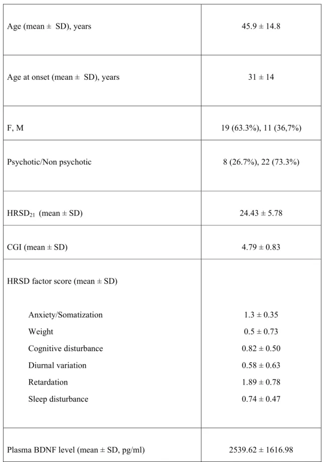
Thirty in- and outpatients of both sexes (19 women and 11 men, age between 22 and 65 years, mean ± SD: 45.9 ± 14.8 years) were recruited at the Dipartimento di Psichiatria, Neurobiologia, Farmacologia e Biotecnologie, University of Pisa, between September 2006 and December 2008, and consecutively enrolled in the study. All patients were suffering from a current major depressive episode, single episode (6) or recurrent (24), diagnosed according to the Diagnostic and Statistical Manual for Mental Disorders criteria (APA, 2000). The diagnosis was confirmed by means of the Mini International Neuropsychiatric Interview (MINI) (Sheehan et al., 1998). Exclusion criteria included the presence of organic brain disorder, substance abuse, pregnancy or any severe or chronic physical illness. Patients were either medication-naïve (n=6)
or medication-free approximately for 1 year (n=19) or at least for 2 weeks (n=5).
No woman took contraceptive drugs; 6 women were in menopause. Blood from all the fertile women was drawn in the mid-follicular phase.
Fifteen healthy subjects (3 men and 12 women, mean age±SD: 46.9±9.2 years), with no history of past or current chronic physical or mental disorders and not taking regular medications, were recruited as the control group.
All patients provided informed written consent to the study which was approved by the Ethics Committee of the University of Pisa in accordance with the Declaration of Helsinki (1996) and with the guidelines of the Good Clinical Practice (1995).
Clinical assessment
The diagnosis of major depression was confirmed by the administration of M.I.N.I. version 5.0.1. (Sheehan et al., 1998). The MINI is a brief structured interview designed to identify the major Axis I psychiatric disorders in the DSM-IV and ICD-10. In validation and reliability studies, the MINI was compared with the Structured Clinical Interview for DSM-III-R, Patient Version, and found to have acceptably high validation and reliability scores (Lecrubier et al 1997). The MINI, however, can be administered in a much shorter period and can be used by clinicians after a brief training session. The interview is divided into modules, each corresponding to a diagnostic category. At the end of each module, clinicians are able to determine whether the diagnostic criteria are met for a particular category.
The severity of depression was assessed by means of the 21-item Hamilton Rating Scale for Depression (HRSD21) (Hamilton M, 1960)
(CGI-S) (Guy W., 1976). Patients had a HRSD total score of 24.4 ± 5.7 and a CGI score of 4.8 ± 0.8.
BDNF Assay
Ten ml of venous blood were drawn in the morning, between 8:00 and 9:00 a.m., following an overnight fast, into EDTA-coated tubes that were kept on ice, centrifugated at 2000xg for 10 minutes at 4°C and refrigerated at -20°C. To measure the amount of total BDNF, acidification and subsequent neutralization of the samples were followed before proceeding with the enzyme-linked immunosorbent assay (ELISA) protocol, according to manufacturer’s instruction (Promega, Wallisellen, Switzerland). Ninety-six-well plates were coated with anti-BDNF monoclonal antibody and incubated at 4°C for 18 hours. The plates were incubated in a blocking buffer for 1 hour at room temperature, then samples were added. The samples and BDNF standards were maintained at room temperature under
shaking for 2 hours, followed by washing with the appropriate buffer. The plates were successively incubated with anti-human BDNF polyclonal antibody at room temperature for 2 hours, washed and incubated with anti-IgG antibody conjugated to horseradish peroxidase for 1 hour at room temperature. The plates were incubated in peroxidase substrate and tetramethylbenzidine solution to produce a colour reaction. The reaction was stopped with 1 M HCl. The absorbance at 450 nm was measured with a microplate reader (Model 550, Bio Rad Laboratories) to determine BDNF values that are expressed as pg/ml.
Statistical analyses
Since BDNF levels and HRSD scores were not normally distributed, non-parametric tests were used. To compare these variables amongst multiple or two independent samples, the Kruskal-Wallis and the Mann-Whitney tests were used, respectively. To analyze the
relationhips between variables the Spearman’s coefficient was computed. A p-value of <.05 was judged as statistically significant. All analyses were carried out using the SPSS version 14.0, by means of personal computer programmes.
RESULTS
Table 1 shows the socio-demographic, clinical variables and plasma BDNF values of the sample.
No significant difference was detected in age, plasma BDNF levels, HRSD total and factors scores between men and women. Only the difference in Retardation factor scores with men reporting higher values approached significance (p= 0.062). Compared to healthy control subjects, plasma BDNF concentrations were significantly reduced in depressed patients (p= .003). Significant and negative correlations were observed between BDNF levels (mean ± SD, pg/ml) and the HRSD total or Retardation factor scores (r=-0.483, p=0.007 and r=-0.397, p=0.030, respectively) (fig. 1 and 2). Moreover, after categorizing the sample on the basis of 75° percentile of the HRSD Sleep disturbances factor score (equal to 1), significant lower BDNF levels were observed in those patients (n=6)
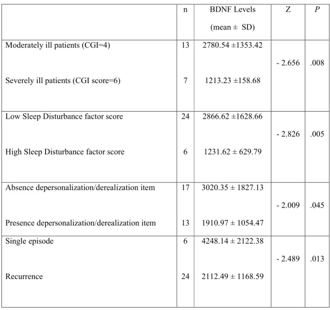
with the highest HRSD Sleep disturbances factor scores (1231.62±629.79 vs 2866.62±1628.66, Z=-2.826, p=0.005) (table 2). When the patients were distinguished in those with and those without dissociative symptoms, patients (n=13) of the first group showed significantly lower plasma BDNF levels than those of the second (1910.97±1054.47 vs 3020.35±1827.13; Z=-2.009, p=0.045).
As for as the CGI is concerned, BDNF levels differed significantly amongst groups [“moderately ill” (score=4 of the CGI “severity of illness” item) (n=13): 2780.54±1353.42; “markedly ill” (score=5) (n=10): 2573.86±2155.80; “severely ill” (score=6) (n=7): 1213.23±158.68; X2=4.350; p=0.023]. Between groups comparisons showed that the “severely ill” patients scored significantly lower than the “moderately ill” (score=4) ones (Z=-2.656, p=0.008). Furthermore, the 24 patients with recurrent episodes of depression showed significantly lower BDNF levels than those with single episode (2112.49±1168.59 vs 4248.14±2122.38, Z=-2.489, p=0.013)
(tab. 2). Finally, no correlation was observed between plasma BDNF levels and presence of psychosis or age of onset.
Given that our sample was composed by some patients with a shorter period of drug washout (at least for 2 weeks, n=5), we further replicated our analyses while eliminating these subjects from the total sample. In this case, too, HRSD total and Retardation factor scores showed the same significant negative correlations with BDNF levels (r=-0.509, p=0.007 and r=-0.384, p=0.048, respectively). Moreover, highest HRSD Sleep disturbances factor scores were associated with lower BDNF levels (1187.62±306.57 vs 3402.75±1697.90, Z=-3.723, p=0.000). In addition, patients with dissociative symptoms (n=12) showed significantly lower plasma BDNF levels than those without them (1737.67±887.19 vs 3065.63±2020.19; Z=-1.986, p=0.047). Finally, BDNF levels differed significantly amongst CGI scoring groups [“moderately ill” (n=8): 3030.90±1309.31; “markedly ill” (n=10): 2573.86±2155.80; “severely ill” (n=7): 1213.23±158.68; X2=7.096; p=0.029];
difference in BDNF levels between patients with recurrent episodes of depression (n=20) vs. those with a single episode were close to significance (Z=-1.835; p=0.067).
DISCUSSION AND CONCLUSIONS
The results of the present study led to different findings. First, we observed significant negative correlations between the severity of illness, as rated by the HRSD total score, and plasma BDNF levels, that is to say, the more severe the patient, the lower the biological parameter. This finding is consistent with previous data gathered in drug-free depressed patients (Shimizu et al., 2003; Gonul et al., 2005), but at variance with others (Lee et al., 2007). The link between BDNF and depression is hence supported by our observation that the plasma levels of the “severely ill” patients resulted significantly lower than those of the “moderately ill” ones.
Although several evidences suggest an important role of BDNF in the pathophysiology of mood disorders and response to antidepressant medications (Shimizu et al., 2003; Aydemir et al., 2005; Gervasoni et al., 2005; Gonul et al., 2005; Lee et al., 2007;
Marano et al., 2007; Huang et al., 2008; Piccinni et al., 2008; Matrisciano et al., 2009; Piccinni et al., 2009), conflicting results have been gathered on this topic. For example, a recent study reported no significant difference in peripheral BDNF concentrations comparing BDNF levels in depressed patients before and after pharmacological antidepressant treatment (Yoshimura et al., 2007). Moreover, there is little information about the source of BDNF in the blood and studies about the relationship between brain and blood BDNF levels have led to conflicting findings (Karege et al., 2002; Elfving et al., 2009). Studies of BDNF uptake or transport across the blood brain barrier have been mixed and additional data are needed to support the existence of an active transport system for uptake of BDNF into the brain (Sen et al., 2008).
Second, even the HRSD “Retardation” factor scores correlated negatively with BDNF levels. According to us, this is an intriguing finding, because psychomotor retardation is considered a core symptom of depression and has been linked generally to the severity
of illness (Benazzi F., 2002), or to melancholia (Parker G., 2000). From a neurobiological point of view, it has been shown that some phenotypes of depressive patients, including the melancholic, are characterized by a more marked disturbance of the hypothalamic-pituitary adrenal (HPA) axis, as compared with the others ( Pintor et al., 2007; Pintor et al., 2009). The influence of corticosteroid hormones on BDNF expression has been showed in preclinical studies which reported how cortisol may exert a downregulation on BDNF synthesis in the central nervous system (Schaff et al., 2000).
Third, lower BDNF plasma levels were found in those patients with the most severe sleep disturbance. Links between sleep disturbances and depression are known all along: about three quarters of depressed patients complain of sleeplessness that provokes subjective distress, impairment of quality of life, and represents a strong risk factor for suicide and recurrence (Nutt et al., 2008). Moreover a HPA overdrive, as observed especially in depressed patients with melancholic features, appears to be related to
dysregulations of sleep (Antonijevic I., 2008). Interestingly, a recent study in rats suggests a relationship between the synaptic plasticity and the homeostasis of sleep, while identifying BDNF as a major mediator of this link at the molecular level (Huber et al., 2007).
Fourth, we found lower BDNF concentrations in patients with depersonalization/derealization symptoms: this is consistent with a similar observation gathered in patients affected by burn-out, a syndrome characterized by chronic stress (Onen Sertoz et al., 2008). This finding may represent another indicator of the relationships between BDNF and stress systems, as dysregulations of the HPA axis has been reported in patients with depersonalization (Simeon et al., 2001).
Fifth, in agreement with a previous study (Lee et al., 2007), patients who were suffering from a recurrent episode had significantly lower levels of BDNF. It should, however, be underlined that the patients with single episode of our sample were
not as numerous as those with recurrent episodes, so that the results of statistical analysis may not be considered totally reliable.
This study suffers several limitations that should be acknowledged. The most important was that the sample size was small, included mainly women and some underwent a short wash-out period. Another problem is related to the extent to which blood BDNF levels may reflect brain BDNF concentrations. We chose to investigate BDNF in plasma, because poor-platelet plasma BDNF seems to be minimally affected by the amount of BDNF stored in platelets and, therefore, may represent a more reliable and sensitive marker of BDNF variations occurring in the brain and periphery(Lommatzsch et al., 2005). Nevertheless, plasma BDNF has shown high inter-individual variability. It should also be mentioned that our absolute plasma BDNF values were higher than those observed in recent publications (Lommatzsch et al., 2005; Begliuomini et al., 2008). However, as previously reported (Piccinni et al., 2008), we assayed total BDNF in plasma by the acidification
and neutralization procedures, while others might have measured the amount of the free mature form. Therefore, according to us, different methodological procedure might contribute to explain the controversial data present in literature (Karege et al., 2005; Palomino et al., 2006). The mature form and the precursor form of BDNF play different roles in cell survival, synaptic plasticity and cognitive functions in health and pathology (Martinowich et al., 2007). In animals, recently, we showed that the mature, but not the precursor, form of BDNF was significantly increased in the prefrontal cortex of rats treated for 14 days with duloxetine at 30 mg/kg/day (Mannari et al., 2008).
The use of medications has been always considered a confounding factor in the measurements of BDNF levels in mood disorders (de Oliveira et al., 2009). In our study, however, we found no difference in a further replication of the analyses by excluding patients with 2-weeks washout. Interestingly, decreased BDNF levels
have been reported in both treated and untreated euthymic patients with unipolar and bipolar (type I and II) (Monteleone et al., 2008).
In conclusion, our results showed that lower BDNF levels may be associated to both recurrence and severity of depression, as well as to symptoms suggesting dysregulations of the HPA axis. Further studies are needed to elucidate more thoroughly the involvement of BDNF in the pathophysiology of symptom patterns or dimensions that may well be present in a broad range of neuropsychiatric conditions.
REFERENCES
Akiskal HS, Walker P, Puzantian VR, King D, Rosenthal TL, Dranon M. Bipolar outcome in the course of depressive illness. Phenomenologic, familial, and pharmacologic predictors. J Affect Disord 1983; 5:115–28.
Akiskal HS. Developmental pathways to bipolarity: Are juvenile-onset depression pre-bipolar? J Am Acad Child Adolesc Psychiatry 1995; 34:754–763.
Akiskal HS. Searching for behavioral indicators of bipolar II in patients presenting with major depressive episodes: the “red sign,” the “rule of three” and other biographic signs of temperamental extravagance, activation and hypomania. J Affect Disord 2005; 84:279–90.
Alexopoulos GS, Katz IR, Reynolds CF, Carpenter D, Docherty JP, Ross RW. Pharmacotherapy of depression in older patients: A summary of the expert consensus guidelines. J Psychiatr Pract 2001; 7:361–376.
Altman J, Das GD. Autoradiographic and histological evidence of postnatal hippocampal neurogenesis in rats. J Comp Neurol, 1965; 124:319-335.
Altshuler LL, Casanova MF, Goldberg TE, Kleinman JE. The hippocampus and parahippocampus in schizophrenia, suicide, and control brains. Arch Gen Psychiatry. 1990;47:1029–1034.
Altshuler LL, Post RM, Leverich GS, Mikalauskas K, Rosoff A, Ackerman L: Antidepressant-induced mania and cycle acceleration: a controversy revisited. Am J Psychiatry 1995; 152:1130–1138.
American Psychiatric Association. Diagnostic and statistical manual of mental disorders, Text Revision, 4th ed. American Psychiatric Press, Washington DC, 2000.
Antonijevic I: HPA axis and sleep: identifying subtypes of major depression. Stress 2008;11:15-27.
Arborelius L, Owens MJ, Plotsky PM, Nemeroff CB. The role of corticotropinreleasing factor in depression and anxiety disorders. J Endocrinol. 1999 Jan;160(1):1-12.
Aydemir O, Deveci A, Taneli F. The effect of chronic antidepressant treatment on serum brain-derived neurotrophic factor levels in depressed patients: a preliminary study. Prog Neuropsychopharmacol Biol Psychiatry. 2005;29:261–265.
Barbany G, Persson H. Regulation of Neurotrophin mRNA Expression in the Rat Brain by Glucocorticoids. Eur J Neurosci. 1992;4:396–403.
Begliuomini S, Lenzi E, Ninni F, Casarosa E, Merlini S, Pluchino N, Valentino V, Luisi S, Luisi M, Genazzani AR: Plasma brain-derived neurotrophic factor daily variations in men: correlation with cortisol circadian rhythm. J Endocrinol 2008;197:429–435.
Benazzi F, Koukopoulos A, Akiskal HS: Toward a validation of a new definition of agitated depression as a bipolar mixed state (mixed depression). Eur Psychiatry 2004; 19:85–90.
Benazzi F: Clinical differences between bipolar II depression and unipolar major depressive disorder: lack of an effect of age. J Affect Disord 2003; 75:191–195.
Benazzi F: Diagnosis of bipolar II disorder: a comparison of structured versus semistructured interviews. Prog Neuropsychopharmacol Biol Psychiatry 2003; 27:985–991.
Benazzi F: Prevalence of bipolar II disorder in atypical depression. Eur Arch Psychiatry Clin Neurosci 1999; 249:62–65.
Benazzi F: Psychomotor changes in melancholic and atypical depression: 1976 unipolar and bipolar-II subtypes. Psychiatry Res 2002;112:211-20.
Bland RC. Epidemiology of affective disorders: a review. Can J Psychiatry 1997; 42:367-77.
Blazer DG. Mood disorders: epidemiology, in Comprehensive Textbook of Psychiatry, Sadock BJ, Sadock VA, editor Lippincott. Williams & Wilkins: New York; 2000. p. 1298–1308.
Bocchio-Chiavetto L, Zanardini R, Bortolomasi M, et al. Electroconvulsive Therapy (ECT) increases serum Brain Derived Neurotrophic Factor (BDNF) in drug resistant depressed patients. Eur Neuropsychopharmacol. 2006;16:620–624.
Bostwick JM, Pankratz VS. Affective disorders and suicide risk: a reexamination. Am J Psychiatry. 2000;157:1925–1932.
Bowden CL, Krishnan AA. Pharmacotherapy for bipolar depression: An economic assessment. Expert Opin Pharmacother 2004; 5:1101– 1107.
Brunoni AR, Lopes M, Fregni F. A systematic review and meta-analysis of clinical studies on depression and BDNF levels: implications for the role of neuroplasticity in depression. Int J Neuropsychopharmacol. 2008;11:1169–1180.
Chao HM, Sakai RR, Ma LY, McEwen BS. Adrenal steroid regulation of neurotrophic factor expression in the rat hippocampus. Endocrinology. 1998;139:3112–3118.
Charney DS, Nelson JC. Delusional and nondelusional unipolar depression: Further evidence for distinct subtypes. Am J Psychiatry 1981; 138:328–333.
Chen B, Dowlatshahi D, MacQueen GM, Wang JF, Young LT. Increased hippocampal BDNF immunoreactivity in subjects treated with antidepressant medication. Biol Psychiatry. 2001;50:260–265.
Chun BJ, Dunner DL. A review of antidepressant-induced hypomania in major depression: suggestions for DSM-V. Bipolar Disord 2004; 6:32–42.
Coffey CE, Wilkinson WE, Weiner RD, Parashos IA, Djang WT, Webb MC, Figiel GS, Spritzer CE. Quantitative cerebral anatomy in depression. A controlled magnetic resonance imaging study. Arch Gen Psychiatry 1993; 50:7-16.
Cohen A, Houck P, Szanto K, Dew M, Gilman S, Reynolds C. Social inequalities in response to antidepressant treatment in older adults. Arch Gen Psychiatry 2006; 63:50-56.
Cotter D, Mackay D, Chana G, Beasley C, Landau S, Everall IP. Reduced neuronal size and glial cell density in area 9 of the dorsolateral prefrontal cortex in subjects with major depressive disorder. Cereb Cortex. 2002;12:386–394.
Cotter D, Mackay D, Landau S, Kerwin R, Everall I. Reduced glial cell density and neuronal size in the anterior cingulate cortex in major depressive disorder. Arch Gen Psychiatry. 2001;58:545–553.
de Oliveira GS, Ceresér KM, Fernandes BS, Kauer-Sant'anna M, Fries GR, Stertz L, Aguiar B, Pfaffenseller B, Kapczinski F: Decreased brain-derived neurotrophic factor in medicated and drug-free bipolar patients. J Psychiatr Res 2009;43(14):1171-4.
DeBattista C, Mueller K. Is electroconvulsive therapy effective for the depressed patient with comorbid borderline personality disorder? J ECT 2001; 17:91–98.
Deckersbach T, Perlis RH, Frankle WG, Gray SM, Grandin L, Dougherty DD, Nierenberg AA, Sachs GS: Presence of irritability during depressive episodes in bipolar disorder. CNS Spectr 2004; 9:227–231.
Dell’Osso L, Bianchi C, Del Debbio A, Roncaglia I, Veltri A, Carlini M, Catena Dell’Osso M, Origlia N, Domenici L, Marazziti D, Piccinni A. Plasma brain-derived neurotrophic factor in bipolar and unipolar depression. Submitted to Giornale Italiano di Psicopatologia
Dell'Osso L, Carmassi C, Del Debbio A, Catena Dell'Osso M, Bianchi C, Da Pozzo E, Origlia N, Domenici L, Massimetti G, Marazziti D, Piccinni A. Brain-derived neurotrophic factor plasma levels in patients suffering from post-traumatic stress disorder.
Progress in Neuro-Psychopharmacology & Biological Psychiatry 2009; 33: 899–902
Drevets WC, Price JL, Simpson JR Jr, Todd RD, Reich T, Vannier M, Raichle ME. Subgenual prefrontal cortex abnormalities in mood disorders. Nature. 1997 Apr 24;386(6627):824-7.
Drevets WC. Functional anatomical abnormalities in limbic and prefrontal cortical structures in major depression. Prog Brain Res. 2000;126:413-31.
D'Sa C, Duman RS. Antidepressants and neuroplasticity. Bipolar Disord. 2002 Jun;4(3):183-94.
Duman RS, Heninger GR, Nestler EJ: A molecular and cellular theory of depression. Arch Gen Psychiatry 1997;54:597-605.
Duman RS, Malberg J, Nakagawa S, D’Sa C. Neuronal plasticity and survival in mood disorders. Biol Psychiatry. 2000;48:732–739.
Duman RS. Neural plasticity: consequences of stress and actions of antidepressant treatment. Dialogues Clin Neurosci 2004; 6:157-169.
Duman RS. Pathophysiology of depression: the concept of synaptic plasticity. Eur Psychiatry. 2002;3:306–310
Duman RS: Role of neurotrophic factors in the etiology and treatment of mood disorders. Neuromol Med 2004;5:11–25.
Dwivedi Y., Brain-derived neurotrophic factor: role in depression and suicide. Neuropsychiatric Disease and Treatment 2009; 5:433-449.
Elfving B, Plougmann PH, Müller HK, Mathé AA, Rosenberg R, Wegener G: Inverse correlation of brain and blood BDNF levels in a genetic rat model of depression. Int J Neuropsychopharmacol. 2009 Oct 2:1-10.
Eriksson PS, Perfilieva E, Bjork-Eriksson T, Alborn AM, Nordborg C, Peterson DA, Gage FH. Neurogenesis in the adult human hippocampus. Nat Med 1998; 4:1313-7.
Frank E, Cyranowski JM, Rucci P, Shear MK, Fagiolini A, Thase ME, Cassano GB, Grochocinski VJ, Kostelnik B, Kupfer DJ. Clinical significance of lifetime panic spectrum symptoms in the treatment of bipolar I disorder: A preliminary report. Arch Gen Psychiatry 2002; 59:905–911.
Frank E, Shear MK, Rucci P, Cyranowski J, Endicott J, Fagiolini A, Grochocinski VJ, Houck P, Kupfer DJ, Maser JD, Cassano GB. Influence of panic-agoraphobic spectrum symptoms on treatment response in patients with recurrent major depression. Am J Psychiatry 2000; 157:1101–1107.
Fujimura H, Altar CA, Chen R, Nakamura T, Nakahashi T, Kambayashi J, Sun B, Tandon NN: Brain-derived neurotrophic factor is stored in human platelets and released by agonist stimulation. Thromb Haemost. 2002;87:728–734.
Gervasoni N, Aubry JM, Bondolfi G, Osiek C, Schwald M, Bertschy G, Karege F: Partial normalization of serum brain-derived neurotrophic factor in remitted patients after a major depressive episode. Neuropsychobiology 2005;51:234–238.
Ghaemi SN, Boiman EE, Goodwin FK: Diagnosing bipolar disorder and the effect of antidepressants: a naturalistic study. J Clin Psychiatry 2000; 61:804–808.
Ghaemi SN, Ko JY, Goodwin FK. ‘‘Cade’s Disease’’ and beyond: misdiagnosis, antidepressant use, and a proposed definition for bipolar spectrum disorder. Can J Psychiatry 2002; 47:125– 134.
Ghaemi SN, Sachs GS, Chiou AM, Pandurangi AK, Goodwin K: Is bipolar disorder still underdiagnosed? are antidepressants overutilized? J Affect Disord 1999; 52:135–144.
Goggi J, Pullar IA, Carney SL, Bradford HF. Modulation of neurotransmitter release induced by brain-derived neurotrophic factor in rat brain striatal slices in vitro. Brain Res. 2002 Jun 21;941(1-2):34-42.