University of Pisa
PhD COURSE in
“Molecular, metabolic and functional exploration of the nervous system and the sense organs”
Effects on human transcriptome of two
BRCA1-BRCT mutations: M1775R and
A1789T
Candidate Supervisor
Abstract
BRCA1 (breast cancer 1, early onset) mutations
confer a high risk of breast and ovarian cancer. Most of BRCA1 cancer-predisposing mutations originate truncated proteins, but missense mutations have been also detected in familial breast and ovarian
cancer patients. These variants are rare and their role in cancer predisposition is often difficult to ascertain. In the present work I studied the molecular
mechanisms affected in human cells by two BRCA1 missense variants, M1775R and A1789T, both located in the second BRCT domain. These variants have
been isolated from familial breast cancer patients and their effect on cell transcriptome has been previously investigated in yeast cells. Here I compared the
expression profiles of HeLa cells transfected with one or the other variant and HeLa cells transfected with
BRCA1 wild-type.
Microarray data analysis was performed by three comparisons: M1775R versus wild-type
(M1775RvsWT-contrast), A1789T versus wild-type (A1789TvsWT-contrast) and the mutated BRCT domain versus wild-type (MutvsWT-contrast),
obtained by considering the two variants as a whole. I found 201 differentially expressed genes in
M1775RvsWT-contrast, 313 in A1789TvsWT-contrast and 173 in MutvsWT-contrast. Most of these genes participate in cell processes that are often
deregulated in cancer, such as cell cycle progression and DNA damage response and repair. These results represent the first molecular evidence of the
pathogenetic role of M1775R, already known by
functional studies, and give support to a similar role for A1789T, first hypothesized based on yeast cell experiments.
1. Introduction
1.1 BRCA1 and breast cancer
BRCA1 (breast cancer 1, early onset) has been the
first breast cancer susceptibility gene to be cloned (Hall, et al. 1990; Miki, et al. 1994). Along with
BRCA2 (breast cancer 2, early onset) it is the most
important disease-associated gene for inherited breast cancer. It acts as a tumor suppressor gene whose truncating or inactivating germline mutations confer a cumulative risk of breast cancer of up to 80%. BRCA1 germline mutations also cause a risk of 30-40% of developing ovarian cancer (O'Donovan and Livingston 2010). From an histopathological point of view, BRCA1 germline mutation carriers usually
develop high grade Invasive Ductal Carcinomas of No Special Type, negative for Estrogen receptor,
Progesterone receptor and Receptor tyrosine-protein kinase erbB-2 (triple negative). In addition, in
approximately 15% of sporadic breast cancers there is loss of nuclear BRCA1 expression, mainly due to
epigenetic inactivation or post-transcriptional downregulation (Vargas, et al. 2011).
1.2 BRCA1 in the cell cycle control
In eukaryotic cells, following DNA damage, cell cycle checkpoints are activated to arrest cells at certain stage during cell cycle, giving them enough time to repair damaged DNA before resuming cell cycle progression (Latif, et al. 2001). Cell cycle checkpoints are critical for the maintenance of genomic integrity as they prevent cells from
duplication of damaged DNA and passage of it to daughter cells. Consistently, dysfunction of proteins involved in cell cycle checkpoints often results in genomic instability and tumorigenesis (Dasika, et al. 1999). According to the different stages in which they function, cell cycle checkpoints can be categorized into G1/S, S-phase and G2/M checkpoints. BRCA1 is involved in all of these checkpoints activations.
1.2.1 BRCA1 in the G1/S checkpoint
The G1/S checkpoint is defective in BRCA1-depleted cells. BRCA1 is indeed required for TP53 (tumor protein p53) phosphorylation mediated by ATM/ATR (ataxia telangiectasia mutated and ataxia telangiectasia and Rad3 related) in response to DNA damage by ionizing or ultraviolet irradiation (Fabbro, et al. 2004). This phosphorylation of TP53 leads to cell cycle arrest through the activation of the
promoter of CDKN1A (cyclin-dependent kinase
inhibitor 1A) (el-Deiry, et al. 1994). BRCA1 has also been shown to increase TP53-mediated activation of
CDKN1A promoter (Chai, et al. 1999; Zhang, et al.
1998)
1.2.2 BRCA1 in the S-phase checkpoint
BRCA1 is specifically required for the S-phase checkpoint mediated by ATM in response to ionizing irradiation (Xu, et al. 2002). In addition, BRCA1 may also regulate ATM activation following DNA damage in S phase, as it interacts and colocalize with the MRN complex (MRE11A/RAD50/NBN: meiotic
recombination 11 homolog A (S. cerevisiae), nibrin, RAD50 homolog (S. cerevisiae)) (Zhong, et al. 1999), which directly activates ATM (Lee and Paull 2004). Evidence has also been presented that BRCA1 is involved in the ATR-mediated S-phase checkpoint activated by stalled replication forks, which can be induced by treatment of cells with UV or hydroxyurea (Tibbetts, et al. 2000). Both ATM and BRCA1-ATR interactions produce the phosphorylation of
BRCA1 on specific Ser/Thr residues (Tibbetts, et al. 2000; Xu, et al. 2002).
1.2.3 BRCA1 in the G2/M checkpoint
The phosphorylation of BRCA1 by ATM is required for cell cycle arrest in G2 induced by ionizing
irradiation (Xu, et al. 2001). In addition, loss of
BRCA1 abolishes the G2/M checkpoint activation, as BRCA1 regulates CHEK1 (CHK1 checkpoint homolog (S. pombe)) kinase activity during G2/M checkpoint activation (Yu and Chen 2004).
1.3 BRCA1 in DNA repair
In response to DNA damage, different DNA repair processes utilize different repair machineries.
However, the two predominant repair pathways are homologous recombination and non-homologous end-joining. Non-homologous end-joining is the most
common form of DNA repair in cells and constitutes a relatively error-prone type of repair as it functions without a template. Homologous recombination faithfully repairs damaged DNA as it occurs only during S and G2 phase of the cell cycle when sister chromatids are present and therefore can be used as a template (Jackson 2002).
BRCA1, along with the MRN complex and other tumor suppressors and DNA damage repair proteins, forms a super complex called BASC (BRCA1 Brca1 Associated genome Surveillance Complex). The BASC coordinates multiple activities related to the
maintenance of genomic integrity, having an
important function in DNA damage sensing as well as in DNA repair (Wang, et al. 2000). In particular,
binding protein 8) form a complex that directly partecipates in DNA double-strand break repair
mediated by homologous recombination (Chen, et al. 2008).
In addition, BRCA1 has been reported to stimulate the base excision repair mechanism by inducing the activity of key enzymes of this pathway (Saha, et al. 2010).
1.4 BRCA1 and global heterocromatin
state
BRCA1 deficiency leads to global DNA
hypomethylation and chromatin abnormalities related to cancer. Consistently, it has been shown that
DNMT1 (DNA (cytosine-5-)-methyltransferase 1), the
methylation maintenance enzyme, is a transcriptional target of BRCA1 (Shukla, et al. 2010).
The most recent hypothesis on BRCA1 concerns a role in maintaining global heterochromatin integrity. It has been proved that BRCA1 deficiency disrupts gene silencing at the tandemly repeated DNA regions,
probably through the loss of ubiquitylation of histone H2A (Zhu, et al. 2011).
1.5 BRCA1 protein structure
BRCA1 consists of different functional domains: a N-terminal RING finger domain, two nuclear
localization signals, a “SQ” cluster, a branched DNA binding domain and a C-terminal domain containing two BRCT (BRCA1 C-Terminus) repeats (Linger and Kruk 2010). The RING domain of BRCA1 has E3 ubiquitin ligase activity and facilitates protein ubiquitination, which occurs when BRCA1
heterodimerizes with BARD1 (BRCA1 associated RING domain 1) (Baer and Ludwig 2002). BRCT repeats have been found in many other proteins that regulate DNA damage response and have a crucial role for
their function (Callebaut and Mornon 1997). BRCT repeats have been also described as phosphopeptide-interacting motifs, facilitating the assembly of DNA damage signaling complexes following checkpoint kinases activation (Rodriguez, et al. 2003).
Consistently, the BRCT repeats of BRCA1 have been found to be essential for its targeting to sites of DNA damage, due to the BRCT-mediated interaction of BRCA1 with other proteins involved in DNA repair (Wang, et al. 2007). BRCT domains are also involved in the transcriptional activity of BRCA1 and the
second BRCT repeat (aa 1760-1863) is critical for the activation of the CDKN1A promoter (Chai, et al.
1999). Finally, a recent paper reported that BRCA1 tumor suppression depends on BRCT phosphoprotein binding (Shakya, et al. 2011).
Due to the relevance of this region for BRCA1
function, the study of mutations located in this region appears of particular interest.
1.6 BRCA1 mutations
A complete list of the mutations detected in
BRCA1 is reported in the BIC (Breast Cancer
Information Core) database
(http://research.nhgri.nih.gov/bic/). Most of the risk-associated BRCA1 variants are frameshift or nonsense
mutations that result in a truncated protein. However, missense mutations associated with breast cancer risk have also been reported. Some of them have not
been studied or even completely characterized because of their rarity and/or the difficulty of attributing functional significance to this type of mutation.
1.7 Aim of the present work
Aim of this work was to investigate the effects on human cell transcriptome of two BRCA1 missense variants, M1775R and A1789T, both located within the second BRCA1 BRCT domain and isolated from familial breast cancers. In a previous work we
examined the expression profiles induced by these two mutations in yeast cells (Di Cecco, et al. 2009). We found alterations of molecular mechanisms critical for cell proliferation control and genome integrity, suggestive of a putative role of these two variants in breast cancer pathogenesis. Here, I compared the expression profiles of HeLa cells transfected with one
or the other BRCA1 variant with that of HeLa cells transfected with BRCA1 wild-type.
2. Materials and methods
2.1 BRCA1 missense variants
Both BRCA1 variants are located within the second BRCT domain and, while M1775R has widely been
described as deleterious (Kawai, et al. 2002; Miki, et al. 1994; Nikolopoulos, et al. 2007; Olopade, et al. 2003; Varma, et al. 2005; Williams and Glover 2003), A1789T has been studied only by our group. In yeast cells both these mutations reverted the growth
suppression (small colony) phenotype, but only
M1775R induced homologous recombination (Caligo, et al. 2009). In HeLa cells A1789T significantly
altered the non-homologous end-joining activity as compared to BRCA1 wild-type (Guidugli, et al. 2011).
2.2 HeLa cells transfection
Five aliquots of the same clone of HeLa G1 cells were transiently transfected with the
pcDNA3-BRCA1wild-type vector, five with the BRCA1-M1775R derivative vector and five with the pcDNA3-BRCA1-A1789T derivative vector as described by Guidugli et al. [Guidugli et al., 2011].
Twenty-four hours after transfection, cells were washed twice in PBS 1X, pelleted and immediately used to extract RNA or proteins. The increased
expression of BRCA1 was assessed by Western Blot analysis as indicated below.
2.3 Microarray
Gene expression was investigated by Whole Human Genome Oligo Microarrays 4x44k G4112F (Agilent Technologies, Palo Alto, CA, USA) containing 4 arrays with 45220 60-mer oligonucleotide probes representing 34392 known and 6608 unknown human
transcripts. A reference design was adopted using as reference a pool of all the RNA samples from the wild-type clones. Non-reference samples were labeled with Cy3 and the reference sample with Cy5.
Total RNA was extracted and DNase purified with PerfectPure RNA Cultured Cell Kit (5 PRIME)
(Eppendorf, Hamburg, Germany). All RNAs, measured by NanoDrop ND-1000 Spectrophotometer (NanoDrop Technologies, Inc. Wilmington, Del, USA), displayed a 260/280 OD ratio > 1.9. The RNA integrity was
verified by 1.2% agarose-formaldehyde gel electrophoresis.
Total RNA samples were amplified and labeled with the Quick-Amp Labeling kit (Agilent
Technologies, Palo Alto, CA, USA). One hundred µl of In Situ Hybridisation Kit Plus mix (Agilent
Technologies, Palo Alto, CA, USA) containing 825 ng of Cy3 -labelled aRNA (ranging from 11 to 14 Cy3 pmoles) and 825 ng of Cy5-labelled aRNA (18 Cy5 pmoles) were hybridized to each array at 65°C for 17 h under constant rotation. The arrays were then
washed 1 min at RT in 6X SSPE, 0.005% TritonX-102; 1 min at 37°C in 0.06X SSPE, 0.005% Triton X-102; 30 sec at RT in Acetonitrile solution (Agilent
Technologies, Palo Alto, CA, USA) and 30 sec at RT in Stabilization and Drying solution (Agilent
Technologies, Palo Alto, CA, USA).
Microarray images were acquired by the Agilent scanner G2565BA and intensity raw data were
extracted by the software Feature Extraction V10.5 (Agilent Technologies, Palo Alto, CA, USA). Data
preprocessing and statistical analysis were performed by LIMMA (LInear Model of Microarray Analysis)
(Smyth 2005) tool. The intensity raw data were
background-subtracted by the normexp method and normalized within-arrays with the LOESS and, for each non-reference sample classes, between-arrays with the scale methods.
The contrast matrix was set to evaluate three comparisons: M1775R versus wild-type
(M1775RvsWT-contrast), A1789T versus wild-type (A1789TvsWT-contrast) and Mutated BRCT domain
versus wild-type (MutvsWT-contrast), considering the
two variants as a whole in the latter case. Statistical significance to each gene in each comparison was
assigned by B-statistic (Lonnstedt and Speed 2002) and only genes with B-statistic>0 were included.
The pathway analysis was done by Pathway-Express ((Draghici, et al. 2007)
http://vortex.cs.wayne.edu/projects.htm). The Gene Ontology terms were collected by Onto-Express
(Khatri, et al. 2002);
http://vortex.cs.wayne.edu/projects.htm). The
network of biological interactions among differentially expressed genes and relevant biological terms was observed by Coremine
(http://www.coremine.com/medical).
2.4 RT-qPCR
RT-qPCR was performed by the iCycler iQ
instrument (Biorad, Hercules, CA, USA) and the iQ SYBR Green Supermix (Biorad, Hercules, CA, USA). Total RNAs were reverse transcribed by the
QuantiTect Reverse Transcription kit (Qiagen,
Valencia, CA, USA). PCR primers (listed in Table 1) were designed by Beacon Designer 4.0 (Premier
Biosoft. International, Palo Alto, CA, USA). RT-qPCR experiments were performed according to MIQE
guidelines (Bustin, et al. 2009). Primer amplification efficiency (90%-110%) and correlation coefficient (>0.98) were tested. Four housekeeping genes, ACTB (actin, beta), HPRT1 (hypoxanthine
phosphoribosyltransferase 1), TBP (TATA box binding protein) and GAPDH (glyceraldehyde-3-phosphate dehydrogenase), tested for stability by geNorm
(M<1.5 and V3/4<0.07) (Vandesompele, et al. 2002), were used to normalize the differential expression values of target genes. The analysis was performed considering the variants separately for the
M1775RvsWT- and the A1789TvsWT- contrasts, but as a whole for the MutvsWT-contrast. One-tailed
Wilcoxon signed rank test was applied to evaluate the statistical significance of results adopting a threshold of 0.05.
2.5 Western Blot
Western Blot was performed as previously reported (Guidugli, et al. 2011).
The expression of BRCA1 protein was assessed using the anti-BRCA1 monoclonal antibody Ab4 diluted 1:100 (Calbiochem-Novabiochem Corp.,
Gibbstown, NJ, USA) that recognizes aa 1005–1313 in the exon 11 of BRCA1.
The level of protein expression was analyzed for: GPR56 (anti-GPR56 rabbit polyclonal antibody H-93: sc-99089, Santa Cruz Biotechnology, Inc., Santa Cruz, CA, USA, dilution 1:1000 ), MRE11A
(anti-MRE11A mouse monoclonal antibody 18: sc-135992, Santa Cruz Biotechnology, Inc., Santa Cruz, CA, USA, dilution 1:500); NFKB1 (anti-NFKB1 mouse
monoclonal antibody E-10: sc-8414, Santa Cruz Biotechnology, Inc., Santa Cruz, CA, USA, dilution 1:100 ) and PML (anti-PML mouse monoclonal IgG2b clone 36.1-104, Upstate Biotechnology, Inc.,
Gene
Symbol Gene Name Primer Sequences
Housekeepin
g genes
ACTB actin, beta F: 5'-AACTGGAACGGTGAAGGTGAC-3' R: 5'-GACTTCCTGTAACAACGCATCTC-3'
HPRT1 hypoxanthine phosphoribosyltransferas e 1 F: 5'-ACATCTGGAGTCCTATTGACATCG-3' R: 5'-TTAAACAACAATCCGCCCAAAGG-3'
GAPDH glyceraldehyde-3-phosphate dehydrogenase
F: 5'-GTGAAGGTCGGAGTCAACG-3' R: 5'-GGTGAAGACGCCAGTGGACTC-3'
TBP TATA box binding protein F: 5'-GGTGTTGTGAGAAGATGGATGTTG-3' R: 5'-CCAGATAGCAGCACGGTATGAG-3'
Target genes
CDKN1A cyclin-dependent kinase inhibitor 1A (p21, Cip1) F: 5'-ACTAGGCGGTTGAATGAGAGGTTC-3' R: 5'-CAGGTCTGAGTGTCCAGGAAAGG-3' EDN1 endothelin 1 F: 5'-CCAACCATCTTCACTGGCTTCC-3' R: 5'-GTCAGACACAAACACTCCCTTAGG-3'
EEF1E1 eukaryotic translation elongation factor 1 epsilon 1 F: 5'-TGCGGGAGGTTCTTGTTCTG-3' R: 5'-CTGTTAGACTTGGACCATTGTTTG-3' GPR56 G protein-coupled receptor 56 F: 5'-CTACAGCCGAAGAATGTGACTC-3' R: 5'-GCAGAAGCAGGATGTTTGGG-3' MRE11A MRE11 meiotic recombination 11 homolog A (S. cerevisiae) F: 5'-GATGATGAAGTCCGTGAGGCTATG-3' R: 5'-TGTTGGTTGCTGCTGAGATGC-3'
NFKB1 nuclear factor of kappa light polypeptide gene enhancer in B-cells 1
F: 5'-CCGTTGGGAATGGTGAGGTC-3' R: 5'-TTGAGAATGAAGGTGGATGATTGC-3'
OBFC2B oligonucleotide/oligosaccharide-binding fold containing 2B
F: 5'-GACGATGTTGGCAATCTG-3' R: 5'-TGGCTCACTGAAGTTAGG-3'
PML promyelocytic leukemia F: 5'-CCAAGGCAGTCTCACCAC-3' R: 5'-TTCGGCATCTGAGTCTTCC-3'
SOD2 superoxide dismutase 2, mitochondrial F: 5'-GGTGTCCAAGGCTCAGGTTG-3' R: 5'-GTGCTCCCACACATCAATCCC-3'
3. Results
3.1 Microarray results
The M1775RvsWT-contrast showed 201
differentially expressed genes, 129 downregulated and 72 upregulated, while the A1789TvsWT-contrast showed 313 differentially expressed genes, 128
downregulated and 185 upregulated and the MutvsWT-contrast showed 173 differentially expressed genes, 100 downregulated and 73 upregulated. Twenty-four of these genes were differentially expressed with the same fold change direction in all the comparisons (Fig. 1).
Complete information about the microarray experiments and results can be retrieved from the ArrayExpress database at the European
Bioinformatics Institute (EBI)
(http://www.ebi.ac.uk/arrayexpress/) by using the following accession number: E-MTAB-761.
Fig. 1: Venn diagram showing the numbers of
differentially expressed genes shared by the three comparisons.
Pathway analysis mapped 40 genes in 58 KEGG pathways for M1775RvsWT-contrast, 52 genes in in 62 KEGG pathways for A1789TvsWT-contrast and 27 genes in 37 KEGG pathways for MutvsWT-contrast. In
all the three comparisons many pathways with high impact factor were involved in cancer.
Twenty-eight pathways were in common among the three comparisons as indicated in Fig. 2.
Fig. 2: Venn diagram showing the numbers of pathways shared by the three comparisons.
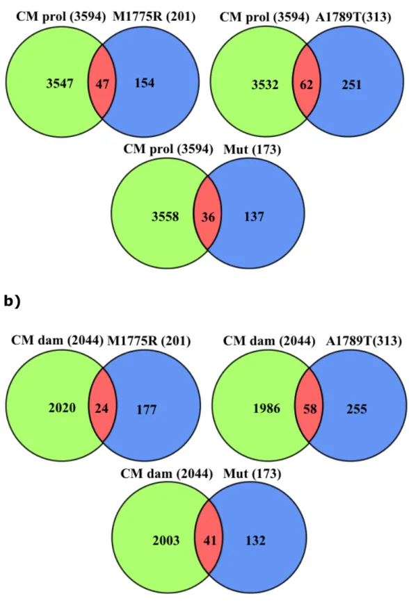
Coremine identified 3594 and 2045 genes linked to biological terms concerning “Cell Proliferation” and “DNA damage and repair” processes, respectively.
Intersections among these two lists and the three lists of differentially expressed genes are shown in Fig. 3.
a)
b)
Fig. 3: Intersections among the lists of differentially expressed genes and “Cell Proliferation” (a) and “DNA damage and repair” (b).
3.2 Microarray data validation
The differential expression of nine transcripts (Table 1) among those identified by microarray
analysis was validated by RT-qPCR: CDKN1A (cyclin-dependent kinase inhibitor 1A (p21, Cip1)), EDN1 (endothelin 1), EEF1E1 (eukaryotic translation elongation factor 1 epsilon 1), GPR56 (G protein-coupled receptor 56), MRE11A (MRE11 meiotic
recombination 11 homolog A (S. cerevisiae)), NFKB1 (nuclear factor of kappa light polypeptide gene
enhancer in B-cells 1), OBFC2B
(oligonucleotide/oligosaccharide-binding fold
containing 2B), PML (promyelocytic leukemia) and
SOD2 (superoxide dismutase 2, mitochondrial) (Fig.
4).
The differential expression was consistently confirmed for all the thirteen validations.
The differential expression of GPR56, MRE11A, NFKB1 and PML proteins was also confirmed by Western Blot analysis.
Fig. 4: Microarray and RT-qPCR log2-Fold changes for the 9 validated genes. All the log2-Fold changes are statistically significant (p-value<0.05).
4. Discussion
Aim of this study was to analyze the effects on human cell transcriptome of two BRCA1 missense variants located in the BRCT domain of the protein, M1775R and A1789T. Specifically, the gene
expression profiles of HeLa cells transfected with one or the other variant were compared with that of HeLa cells transfected with BRCA1 wild-type. We also
analyzed the combined outcome of the two BRCT mutations in comparison to the wild-type. To this purpose, three different statistical contrasts were performed: M1775R versus wild-type (M1775RvsWT-contrast), A1789T versus wild-type (A1789TvsWT-contrast) and Mutated BRCT domain versus wild-type (MutvsWT-contrast) (see Materials and methods). Pathway analysis retrieved many pathways involved in cancer onset and progression as well as linked to specific tumors. Ontological and data-mining analyses highlighted three functional categories: cell cycle
regulation, apoptosis and DNA damage response and repair, typically deregulated in cancer cells. Cell cycle and apoptosis deregulation leads to aberrant cell
proliferation, while an impaired DNA damage
response and repair causes genomic instability. All these processes are closely connected, as apoptosis, constituting a defense from anomalous proliferation, is linked to cell cycle block and is activated in
response to DNA damage.
4.1 Aberrant cell proliferation
Cancer cells abnormally proliferate. In these cells occurs overexpression of mitogenic factors, such as cell cycle positive regulators, as well as impairment of mechanisms ensuring correct cell division, including apoptosis, as reviewed by Strobl et al., Zafonte et al. and Vermeulen et al., among many (Strobl, et al. 1995; Vermeulen, et al. 2003; Zafonte, et al. 2000).
In our data, a considerable number of
differentially expressed genes is strictly linked to cell proliferation (Table 2).
Biological
Process Symbol Gene Gene Name Contrast log2 (Fold Change)
Cell cycle arrest impairment
CDKN1A cyclin-dependent kinase inhibitor 1A (p21, Cip1) M1775RvsWT -0.3066647
CEBPA CCAAT/enhancer binding protein (C/EBP), alpha M1775RvsWT MutvsWT -0.3728651 -0.3190284 SMAD3 SMAD family member 3 M1775RvsWT A1789TvsWT
MutvsWT -0.2675322 -0.4286813 -0.3196246 CCND1 cyclin D1 A1789TvsWT 0.3622112 PML promyelocytic leukemia M1775RvsWT -0.3045759
RUVBL1 RuvB-like 1 (E. coli) M1775RvsWT -0.3028029
TXNIP thioredoxin interacting
protein A1789TvsWT -0.3985633
RASSF1 Ras association (RalGDS/AF-6) domain family member 1 A1789TvsWT -0.2766158
Cell proliferation enhancement
FOS FBJ murine osteosarcoma
viral oncogene homolog
A1789TvsWT M1775RvsWT MutvsWT 0.4515777 0.4020256 0.4365775
DUSP1 dual specificity phosphatase 1
A1789TvsWT M1775RvsWT MutvsWT 0.3844494 0.7606655 0.5060076
DUSP2 dual specificity phosphatase
2 MutvsWT 0.5408689
EDN1 endothelin 1 M1775RvsWT MutvsWT 0.4442705 0.3212824
SKP1 S-phase kinase-associated protein 1 A1789TvsWT 0.3353208
ZWILCH Zwilch, kinetochore associated, homolog
(Drosophila) A1789TvsWT 0.2508541
GPR56 G protein-coupled receptor 56 M1775RvsWT A1789TvsWT MutvsWT -0.3453577 -0.3310188 -0.3407359 Apoptosis blocking NFKB1
nuclear factor of kappa light polypeptide gene enhancer in
B-cells 1 M1775RvsWT -0.2522979
TNFRSF10B tumor necrosis factor receptor superfamily, member 10b M1775RvsWT -0.247568 DYRK2 dual-specificity tyrosine-(Y)-phosphorylation regulated kinase 2 M1775RvsWT -0.282513
PLEKHF1 pleckstrin homology domain containing, family F (with FYVE domain) member 1
MutvsWT -0.2374774
4.1.1 Cell cycle arrest impairment
CDKN1A, downregulated by M1775R, is a main
effector of cell cycle arrest in response to DNA
damage and a promoter of apoptosis (Cazzalini, et al. 2010). Its expression is usually activated by BRCA1 (Chai, et al. 1999).
Cell cycle can be also arrested by the cooperation of CDKN1A with CEBPA (Harris, et al. 2001) that is in turn downregulated by M1775R.
CDKN1A expression is normally activated also by
SMAD3, a known transcription factor that acts as an effector of the TGF-beta pathway (Moustakas and Kardassis 1998; Pardali, et al. 2005), downregulated in all the three comparisons. The overexpression of SMAD3 in a breast cancer cell line causes cell cycle arrest (Tian, et al. 2003), while in SMAD3-/-
mammary epithelial cells, both TGF-beta-induced growth inhibition and apoptosis are lost (Kohn, et al. 2010).
SMAD3 also contributes to the 3-indole-induced G1 arrest in cancer cells (Huang, et al. 2011) and its inhibition depends on CCND1-CDK4 (cyclin-dependent
kinase 4) action in breast cancer cells overexpressing
CCND1 (Zelivianski, et al. 2010), upregulated by
A1789T. The loss or reduction of BRCA1 expression, moreover, significantly reduces the TGF-beta induced activation of SMAD3 in breast cancer cells (Li, et al. 2009).
Considering this, the downregulation of CDKN1A,
CEBPA and SMAD3 might constitute a key element in
carcinogenesis predisposition caused by the
considered variants. The downregulation of other four genes further supports the hypothesis of a deficiency in cell cycle arrest: PML (in M1775RvsWT) which
codifies for a phosphoprotein localized in nuclear bodies, involved in TP53 (tumor protein p53)
-mediated cell cycle arrest in G1 (Chan, et al. 1997) as well as in recognition and/or processing of DNA
breaks by recruiting TP53 and MRE11A (Carbone, et al. 2002); RUVBL1 (in M1775RvsWT) that encodes a highly conserved ATP-dependent DNA helicase which is part of chromatin-remodeling complexes and plays a role in apoptosis and DNA repair (Ikura, et al. 2000; Jha, et al. 2008; Makino, et al. 1998); TXNIP (in
A1789TvsWT) that encodes a transcriptional repressor acting as a tumor suppressor, as its transfection
induces cell-cycle arrest in G0/G1 phase and is
downregulated in human tumors (Han, et al. 2003) and RASSF1 (in A1789TvsWT), a tumor suppressor that blocks cell cycle progression by inhibiting CCND1 accumulation. It is epigenetically inactivated at high frequency in a variety of tumors, including breast cancer (Burbee, et al. 2001; Shivakumar, et al. 2002).
4.1.2 Cell proliferation enhancement
The transcription factor FOS, upregulated in all the three comparisons, is a well known protooncogene that positively regulates cell cycle progression
(Shaulian and Karin 2001) and is induced in human breast cancer cell cultures, being part of the
mitogenic signal transduction typical of these cells, as reviewed by Strobl et al. (Strobl, et al. 1995).
DUSP1, upregulated in all the three comparisons,
and DUSP2, upregulated in MutvsWT, belong to a subfamily of tyrosine phosphatases that regulate the activity of Mitogen-Activated Protein Kinases
(MAPKs). MAPKs are key effectors for cell growth control and survival in physiological and pathological conditions, including cancer. DUSPs have therefore
been proposed as potential targets for anticancer drugs, as reviewed by Nunes-Xavier et al. (Nunes-Xavier, et al. 2011). DUSP1 inhibits apoptosis in human mammary epithelial and breast carcinoma cells (Small, et al. 2004). Moreover, DUSP1
expression was found upregulated in many breast cancers (Wang, et al. 2003). The overexpression of DUSP2 in ovarian cancers has been correlated with poor outcome (Givant-Horwitz, et al. 2004).
EDN1, upregulated by M1775R and in MutvsWT, is a vasoconstrictor that has also co-mitogenic activity, potentiating the growth factors effects. Altered EDN1 signalling is involved in carcinogenesis by modulating cell survival and promoting invasiveness (Bagnato and Rosanò 2008).
SKP1, upregulated by A1789T, is a component of the SCF complex that mediates the ubiquitination of cell cycle proteins promoting cell cycle progression (Bassermann and Pagano 2010).
ZWILCH, upregulated by A1789T, is an essential component of the mitotic checkpoint that prevents cells from exiting mitosis prematurely (Kops, et al. 2005; Williams and Glover 2003).
GPR56, downregulated in all the three contrasts,
is a G protein-coupled receptor involved in adhesion processes that participates in cytoskeletal signaling, cellular adhesion and tumor invasion. It is
downregulated in melanoma cell lines, while its overexpression suppresses tumor growth and metastasis (Xu, et al. 2006).
4.1.3 Apoptosis blocking
NFKB1, downregulated by M1775R, is a pleiotropic transcription factor involved in many biological
processes like inflammation, immunity,
differentiation, cell growth, tumorigenesis and
apoptosis. Whether NFKB activation contributes or not to cancer is controversial, as reviewed by Shishodia and Aggarwal (Shishodia and Aggarwal 2004), as it regulates the expression of both antiapoptotic (Rayet and Gélinas 1999) and proapoptotic genes (Kühnel, et al. 2000; Shetty, et al. 2005).
Interestingly, TNFRSF10B, that was in turn
downregulated by M1775R, is one of the proapoptotic genes upregulated by NFKB (Shetty, et al. 2005). TNFRSF10B is one of the two apoptosis-activating receptors binding the receptor TNFSF10 (tumor
necrosis factor (ligand) superfamily, member 10) (Sheridan, et al. 1997). Upon binding, an adaptor protein called FADD (Fas(TNFRSF6)-Associated via Death Domain) is recruited to the death receptors, forming a signaling complex that leads to apoptosis through caspases activation (Suliman, et al. 2001). The downregulation of both NFKB1 and TNFRSF10B in our data suggests an inhibition of TNF-mediated
apoptosis.
Two other proapoptotic genes, DYRK2 and
PLEKHF1, resulted as downregulated in our data.
DYRK2, downregulated by M1775R, is a protein kinase that regulates TP53 to induce apoptosis in response to DNA damage downstream of ATM (Taira, et al. 2007), and PLEKHF1, downregulated in
MutvsWT, is a recently discovered
lysosome-associated protein that activates caspase-independent apoptosis (Chen, et al. 2005) by interacting with the TP53 transactivation domain (Li, et al. 2007).
4.2 Genomic instability
An improper reaction to genotoxic stress causes genomic instability, leading to tumorigenesis.
Deficiencies in DNA damage signaling and repair pathways are thus fundamental to the etiology of cancer (Khanna and Jackson 2001).
A number of differentially expressed genes takes part in genotoxic stress response. Among these
genes, some were downregulated causing an increase in genomic instability, while others were upregulated (Table 3). Many tumors, including BRCA1-deficient breast cancers, show an overexpression of genes linked to DNA repair that correlates with
chemoresistance and poor prognosis (Martin, et al. 2007; Saviozzi, et al. 2009). Moreover, an increased nuclear staining of DNA repair proteins has been
recently observed in tissue sections of breast cancers carrying the M1775R mutation, suggesting a new mechanism of tumorigenesis involving an enhance of homologous recombination (Dever, et al. 2011).
Biological
Process Symbol Gene Gene Name Contrast log2 (Fold Change)
DNA damage response and
repair downregulation
EEF1E1 eukaryotic translation elongation factor 1 epsilon 1 A1789TvsWT -0.4309041
SMC1A structural maintenance of chromosomes 1A A1789TvsWT MutvsWT -0.2754507 -0.2640263 PPP1CC protein phosphatase 1, catalytic
subunit, gamma isozyme A1789TvsWT -0.4286825
AHNAK AHNAK nucleoprotein M1775RvsWT A1789TvsWT MutvsWT
-0.3988113 -0.3103867 -0.3940570
SOD2 superoxide dismutase 2, mitochondrial M1775RvsWT MutvsWT -0.3376169 -0.2502831
DNA damage response and
repair upregulation
MRE11A MRE11 meiotic recombination
11 homolog A (S. cerevisiae) A1789TvsWT 0.3293561
TERF1 telomeric repeat binding factor (NIMA-interacting) 1 MutvsWT 0.2790907
OBFC2A oligonucleotide/oligosaccharide-binding fold containing 2A M1775RvsWT 0.3666172
OBFC2B
oligonucleotide/oligosaccharide-binding fold containing 2B
A1789TvsWT MutvsWT
0.4070777 0.3417360
Table 3: Genes linked to genomic instability.
4.2.1 DNA damage response and repair downregulation
EEF1E1, downregulated by A1789T, first
discovered as associated with a macromolecular tRNA synthetase complex, is a key factor for
ATM/ATR-mediated TP53 activation in response to DNA damage (Park, et al. 2005).
SMC1A, downregulated by A1789T, encodes an
evolutionarily conserved chromosomal protein, component of the cohesin complex (Sumara, et al. 2000). It is involved in the ATM/NBN-dependent
S-phase checkpoint pathway, associating with BRCA1 and being phosphorylated in response to ionizing radiations (Yazdi, et al. 2002).
PPP1CC, downregulated by A1789T, is the
catalytic subunit of the gamma isoform of PP1 which is a component of a signaling complex,
PPP1R1A/PPP1R15A/PPP1CC (protein phosphatase 1, regulatory (inhibitor) subunit 1A, protein phosphatase 1, regulatory subunit 15A, protein phosphatase 1, catalytic subunit, gamma isozyme) that positively regulates apoptosis in response to various stresses, including growth arrest and DNA damage (Connor, et al. 2001).
AHNAK, downregulated by A1789T, encodes a
protein typically repressed in human neuroblastoma cell lines and in several other types of tumors
(Shtivelman, et al. 1992). It firmly binds the LIG4-XRCC4 (ligase IV, DNA, ATP-dependent and X-ray repair complementing defective repair in Chinese hamster cells 4) complex on DNA stimulating its double-stranded ligation activity (Stiff, et al. 2004).
SOD2, downregulated by M1775R and in MutvsWT, is a member of the iron/manganese
superoxide dismutase family that acts as a free radical scavenger, thus protecting from oxidative damage. It is a candidate tumor suppressor gene as the loss of heterozygosity of its region on
chromosome 6 has been found in about 40% of human malignant melanomas (Oberley and Oberley 1997) and the deletion of chromosome 6 long arm has been identified in SV40 transformed human fibroblast (Bravard, et al. 1992). In addition, SOD2 overexpression suppresses the tumorigenicity of breast cancer cells (Li, et al. 1995).
4.2.2 DNA damage response and repair upregulation
MRE11A, upregulated by A1789T, encodes a
component of the BASC that specifically promotes non-homologous end-joining (see Introduction) (Wang, et al. 2000; Zhang and Powell 2005). The nuclease activity of MRE11A is required for non-homologous end-joining mediated by the MRN
complex (see Introduction), and, interestingly, BRCA1 suppresses this activity in vitro (Paull, et al. 2001). Interestingly, the A1789T variant altered the
non-homologous end-joining activity in a functional assay (Guidugli, et al. 2011).
TERF1, upregulated in MutvsWT, is a telomere-associated protein, member of the telomere
nucleoprotein complex that interacts with various macromolecular complexes including MRN (Kuimov 2004).
OBFC2A, upregulated by M1775R, and OBFC2B,
upregulate by A1789T and in MutvsWT, encode
single-stranded DNA-binding proteins essential for a variety of DNA metabolic processes, including
replication, recombination and damage detection and repair. OBFC2B, in particular, as an early participant in DNA damage response, is critical for genomic
5. Concluding remarks
This work was focused on two BRCA1 BRCT
missense variants, M1775R and A1789T, both isolated from familial breast cancers. M1775R has widely been described as deleterious (Kawai, et al. 2002; Miki, et al. 1994; Nikolopoulos, et al. 2007; Olopade, et al. 2003; Varma, et al. 2005; Williams and Glover 2003), while A1789T has been studied only by our group. In yeast cells both of these mutations reverted the
growth suppression (small colony) phenotype (Caligo, et al. 2009), showing a characteristic behaviour of cancer-predisposing missense BRCA1 BRCT mutations (Coyne, et al. 2004). M1775R also induced
homologous recombination in yeast cells (Caligo, et al. 2009). In HeLa cells A1789T significantly altered the non-homologous end-joining activity as compared to BRCA1 wild-type (Guidugli, et al. 2011).
As previously observed in yeast cells (Di Cecco, et al. 2009), also in human cells the BRCA1 variants
M1775R and A1789T affect the expression of many genes critical for cell proliferation and genome
molecular confirm of the pathogenetic role of M1775R and give support to a similar role of A1789T that we first hypothesized on the basis of the experiments in yeast cells.
References
Baer R, Ludwig T. 2002. The BRCA1/BARD1 heterodimer, a tumor suppressor complex with ubiquitin E3 ligase activity. Curr Opin Genet Dev 12(1):86-91.
Bagnato A, Rosanò L. 2008. The endothelin axis in cancer. Int J Biochem Cell Biol 40(8):1443-1451.
Bassermann F, Pagano M. 2010. Dissecting the role of ubiquitylation in the DNA damage response
checkpoint in G2. Cell Death Differ 17(1):78-85.
Bravard A, Hoffschir F, Sabatier L, Ricoul M, Pinton A, Cassingena R, Estrade S, Luccioni C, Dutrillaux B. 1992. Early superoxide dismutase alterations during SV40-transformation of human fibroblasts. Int J
Cancer 52(5):797-801.
Burbee D, Forgacs E, Zöchbauer-Müller S,
Shivakumar L, Fong K, Gao B, Randle D, Kondo M, Virmani A, Bader S, Sekido Y, Latif F, Milchgrub S, Toyooka S, Gazdar A, Lerman M, Zabarovsky E, White M, Minna J. 2001. Epigenetic inactivation of RASSF1A
in lung and breast cancers and malignant phenotype suppression. J Natl Cancer Inst 93(9):691-699.
Bustin SA, Benes V, Garson JA, Hellemans J, Huggett J, Kubista M, Mueller R, Nolan T, Pfaffl MW, Shipley GL, Vandesompele J, Wittwer CT. 2009. The MIQE guidelines: minimum information for publication of quantitative real-time PCR experiments. Clin Chem 55(4):611-622.
Caligo MA, Bonatti F, Guidugli L, Aretini P, Galli A. 2009. A yeast recombination assay to characterize human BRCA1 missense variants of unknown
pathological significance. Hum Mutat 30(1):123-133. Callebaut I, Mornon JP. 1997. From BRCA1 to RAP1: a widespread BRCT module closely associated with DNA repair. FEBS Lett 400(1):25-30.
Carbone R, Pearson M, Minucci S, Pelicci PG. 2002. PML NBs associate with the hMre11 complex and p53 at sites of irradiation induced DNA damage. Oncogene 21(11):1633-1640.
Cazzalini O, Scovassi AI, Savio M, Stivala LA, Prosperi E. 2010. Multiple roles of the cell cycle inhibitor
p21(CDKN1A) in the DNA damage response. Mutat Res 704(1-3):12-20.
Chai YL, Cui J, Shao N, Shyam E, Reddy P, Rao VN. 1999. The second BRCT domain of BRCA1 proteins interacts with p53 and stimulates transcription from the p21WAF1/CIP1 promoter. Oncogene 18(1):263-268.
Chan JY, Li L, Fan YH, Mu ZM, Zhang WW, Chang KS. 1997. Cell-cycle regulation of DNA damage-induced expression of the suppressor gene PML. Biochem Biophys Res Commun 240(3):640-646.
Chen L, Nievera CJ, Lee AY, Wu X. 2008. Cell cycle-dependent complex formation of BRCA1.CtIP.MRN is important for DNA double-strand break repair. J Biol Chem 283(12):7713-7720.
Chen W, Li N, Chen T, Han Y, Li C, Wang Y, He W, Zhang L, Wan T, Cao X. 2005. The
lysosome-associated apoptosis-inducing protein containing the pleckstrin homology (PH) and FYVE domains (LAPF), representative of a novel family of PH and FYVE
domain-containing proteins, induces caspase-independent apoptosis via the
lysosomal-mitochondrial pathway. J Biol Chem 280(49):40985-40995.
Connor J, Weiser D, Li S, Hallenbeck J, Shenolikar S. 2001. Growth arrest and DNA damage-inducible
protein GADD34 assembles a novel signaling complex containing protein phosphatase 1 and inhibitor 1. Mol Cell Biol 21(20):6841-6850.
Coyne RS, McDonald HB, Edgemon K, Brody LC.
2004. Functional characterization of BRCA1 sequence variants using a yeast small colony phenotype assay. Cancer Biol Ther 3(5):453-457.
Dasika GK, Lin SC, Zhao S, Sung P, Tomkinson A, Lee EY. 1999. DNA damage-induced cell cycle checkpoints and DNA strand break repair in development and
tumorigenesis. Oncogene 18(55):7883-7899. Dever SM, Golding SE, Rosenberg E, Adams BR, Idowu MO, Quillin JM, Valerie N, Xu B, Povirk LF,
Valerie K. 2011. Mutations in the BRCT binding site of BRCA1 result in hyper-recombination. Aging (Albany NY) 3(5):515-532.
Di Cecco L, Melissari E, Mariotti V, Iofrida C, Galli A, Guidugli L, Lombardi G, Caligo MA, Iacopetti P,
Pellegrini S. 2009. Characterisation of gene
expression profiles of yeast cells expressing BRCA1 missense variants. Eur J Cancer 45(12):2187-2196. Draghici S, Khatri P, Tarca AL, Amin K, Done A,
Voichita C, Georgescu C, Romero R. 2007. A systems biology approach for pathway level analysis. Genome Res 17(10):1537-1545.
el-Deiry WS, Harper JW, O'Connor PM, Velculescu VE, Canman CE, Jackman J, Pietenpol JA, Burrell M, Hill DE, Wang Y. 1994. WAF1/CIP1 is induced in p53-mediated G1 arrest and apoptosis. Cancer Res 54(5):1169-1174.
Fabbro M, Savage K, Hobson K, Deans AJ, Powell SN, McArthur GA, Khanna KK. 2004. BRCA1-BARD1
complexes are required for p53Ser-15
phosphorylation and a G1/S arrest following ionizing radiation-induced DNA damage. J Biol Chem
279(30):31251-31258.
Givant-Horwitz V, Davidson B, Goderstad JM, Nesland JM, Tropé CG, Reich R. 2004. The PAC-1 dual
serous ovarian carcinoma. Gynecol Oncol 93(2):517-523.
Guidugli L, Rugani C, Lombardi G, Aretini P, Galli A, Caligo MA. 2011. A recombination-based method to characterize human BRCA1 missense variants. Breast Cancer Res Treat 125(1):265-272.
Hall JM, Lee MK, Newman B, Morrow JE, Anderson LA, Huey B, King MC. 1990. Linkage of early-onset
familial breast cancer to chromosome 17q21. Science 250(4988):1684-1689.
Han SH, Jeon JH, Ju HR, Jung U, Kim KY, Yoo HS, Lee YH, Song KS, Hwang HM, Na YS, Yang Y, Lee KN,
Choi I. 2003. VDUP1 upregulated by TGF-beta1 and 1,25-dihydorxyvitamin D3 inhibits tumor cell growth by blocking cell-cycle progression. Oncogene
22(26):4035-4046.
Harris TE, Albrecht JH, Nakanishi M, Darlington GJ. 2001. CCAAT/enhancer-binding protein-alpha
cooperates with p21 to inhibit cyclin-dependent kinase-2 activity and induces growth arrest
independent of DNA binding. J Biol Chem 276(31):29200-29209.
Huang SM, Lu KT, Wang YC. 2011. ATM/ATR and SMAD3 pathways contribute to 3-indole-induced G₁ arrest in cancer cells and xenograft models.
Anticancer Res 31(1):203-208.
Ikura T, Ogryzko VV, Grigoriev M, Groisman R, Wang J, Horikoshi M, Scully R, Qin J, Nakatani Y. 2000.
Involvement of the TIP60 histone acetylase complex in DNA repair and apoptosis. Cell 102(4):463-473. Jackson SP. 2002. Sensing and repairing DNA double-strand breaks. Carcinogenesis 23(5):687-696.
Jha S, Shibata E, Dutta A. 2008. Human Rvb1/Tip49 is required for the histone acetyltransferase activity of Tip60/NuA4 and for the downregulation of
phosphorylation on H2AX after DNA damage. Mol Cell Biol 28(8):2690-2700.
Kawai H, Li H, Chun P, Avraham S, Avraham HK. 2002. Direct interaction between BRCA1 and the estrogen receptor regulates vascular endothelial growth factor (VEGF) transcription and secretion in breast cancer cells. Oncogene 21(50):7730-7739.
Khanna KK, Jackson SP. 2001. DNA double-strand breaks: signaling, repair and the cancer connection. Nat Genet 27(3):247-254.
Khatri P, Draghici S, Ostermeier GC, Krawetz SA. 2002. Profiling gene expression using onto-express. Genomics 79(2):266-270.
Kohn EA, Du Z, Sato M, Van Schyndle CM, Welsh MA, Yang YA, Stuelten CH, Tang B, Ju W, Bottinger EP, Wakefield LM. 2010. A novel approach for the
generation of genetically modified mammary
epithelial cell cultures yields new insights into TGFβ signaling in the mammary gland. Breast Cancer Res 12(5):R83.
Kops GJ, Kim Y, Weaver BA, Mao Y, McLeod I, Yates JR, Tagaya M, Cleveland DW. 2005. ZW10 links
mitotic checkpoint signaling to the structural kinetochore. J Cell Biol 169(1):49-60.
Kuimov AN. 2004. Polypeptide components of
telomere nucleoprotein complex. Biochemistry (Mosc) 69(2):117-129.
Kühnel F, Zender L, Paul Y, Tietze MK, Trautwein C, Manns M, Kubicka S. 2000. NFkappaB mediates
apoptosis through transcriptional activation of Fas (CD95) in adenoviral hepatitis. J Biol Chem
275(9):6421-6427.
Latif C, Harvey SH, O'Connell MJ. 2001. Ensuring the stability of the genome: DNA damage checkpoints. ScientificWorldJournal 1:684-702.
Lee JH, Paull TT. 2004. Direct activation of the ATM protein kinase by the Mre11/Rad50/Nbs1 complex. Science 304(5667):93-96.
Li H, Sekine M, Seng S, Avraham S, Avraham HK. 2009. BRCA1 interacts with Smad3 and regulates Smad3-mediated TGF-beta signaling during oxidative stress responses. PLoS One 4(9):e7091.
Li JJ, Oberley LW, St Clair DK, Ridnour LA, Oberley TD. 1995. Phenotypic changes induced in human breast cancer cells by overexpression of manganese-containing superoxide dismutase. Oncogene
10(10):1989-2000.
Li N, Zheng Y, Chen W, Wang C, Liu X, He W, Xu H, Cao X. 2007. Adaptor protein LAPF recruits
lysosomal destabilization in apoptosis. Cancer Res 67(23):11176-11185.
Linger RJ, Kruk PA. 2010. BRCA1 16 years later: risk-associated BRCA1 mutations and their functional
implications. FEBS J 277(15):3086-3096.
Lonnstedt I, Speed T. 2002. Replicated microarray data. Stat Sinica 12:31–46.
Makino Y, Mimori T, Koike C, Kanemaki M, Kurokawa Y, Inoue S, Kishimoto T, Tamura T. 1998. TIP49, homologous to the bacterial DNA helicase RuvB, acts as an autoantigen in human. Biochem Biophys Res Commun 245(3):819-823.
Martin RW, Orelli BJ, Yamazoe M, Minn AJ, Takeda S, Bishop DK. 2007. RAD51 up-regulation bypasses
BRCA1 function and is a common feature of BRCA1-deficient breast tumors. Cancer Res 67(20):9658-9665.
Miki Y, Swensen J, Shattuck-Eidens D, Futreal PA, Harshman K, Tavtigian S, Liu Q, Cochran C, Bennett LM, Ding W. 1994. A strong candidate for the breast and ovarian cancer susceptibility gene BRCA1.
Moustakas A, Kardassis D. 1998. Regulation of the human p21/WAF1/Cip1 promoter in hepatic cells by functional interactions between Sp1 and Smad family members. Proc Natl Acad Sci U S A
95(12):6733-6738.
Nikolopoulos G, Pyrpassopoulos S, Thanassoulas A, Klimentzou P, Zikos C, Vlassi M, Vorgias C,
Yannoukakos D, Nounesis G. 2007. Thermal unfolding of human BRCA1 BRCT-domain variants. Biochim
Biophys Acta 1774(6):772-780.
Nunes-Xavier C, Romá-Mateo C, Ríos P, Tárrega C, Cejudo-Marín R, Tabernero L, Pulido R. 2011. Dual-Specificity MAP Kinase Phosphatases as Targets of Cancer Treatment. Anticancer Agents Med Chem. O'Donovan PJ, Livingston DM. 2010. BRCA1 and BRCA2: breast/ovarian cancer susceptibility gene
products and participants in DNA double-strand break repair. Carcinogenesis 31(6):961-967.
Oberley TD, Oberley LW. 1997. Antioxidant enzyme levels in cancer. Histol Histopathol 12(2):525-535. Olopade OI, Fackenthal JD, Dunston G, Tainsky MA, Collins F, Whitfield-Broome C. 2003. Breast cancer
genetics in African Americans. Cancer 97(1 Suppl):236-245.
Pardali K, Kowanetz M, Heldin CH, Moustakas A. 2005. Smad pathway-specific transcriptional
regulation of the cell cycle inhibitor p21(WAF1/Cip1). J Cell Physiol 204(1):260-272.
Park BJ, Kang JW, Lee SW, Choi SJ, Shin YK, Ahn YH, Choi YH, Choi D, Lee KS, Kim S. 2005. The
haploinsufficient tumor suppressor p18 upregulates p53 via interactions with ATM/ATR. Cell 120(2):209-221.
Paull TT, Cortez D, Bowers B, Elledge SJ, Gellert M. 2001. Direct DNA binding by Brca1. Proc Natl Acad Sci U S A 98(11):6086-6091.
Rayet B, Gélinas C. 1999. Aberrant rel/nfkb genes and activity in human cancer. Oncogene
18(49):6938-6947.
Richard DJ, Bolderson E, Cubeddu L, Wadsworth RI, Savage K, Sharma GG, Nicolette ML, Tsvetanov S, McIlwraith MJ, Pandita RK, Takeda S, Hay RT, Gautier J, West SC, Paull TT, Pandita TK, White MF, Khanna KK. 2008. Single-stranded DNA-binding protein
hSSB1 is critical for genomic stability. Nature 453(7195):677-681.
Rodriguez M, Yu X, Chen J, Songyang Z. 2003.
Phosphopeptide binding specificities of BRCA1 COOH-terminal (BRCT) domains. J Biol Chem
278(52):52914-52918.
Saha T, Rih JK, Roy R, Ballal R, Rosen EM. 2010. Transcriptional regulation of the base excision repair pathway by BRCA1. J Biol Chem
285(25):19092-19105.
Saviozzi S, Ceppi P, Novello S, Ghio P, Lo Iacono M, Borasio P, Cambieri A, Volante M, Papotti M, Calogero RA, Scagliotti GV. 2009. Non-small cell lung cancer exhibits transcript overexpression of genes associated with homologous recombination and DNA replication pathways. Cancer Res 69(8):3390-3396.
Shakya R, Reid LJ, Reczek CR, Cole F, Egli D, Lin CS, deRooij DG, Hirsch S, Ravi K, Hicks JB, Szabolcs M, Jasin M, Baer R, Ludwig T. 2011. BRCA1 tumor
suppression depends on BRCT phosphoprotein binding, but not its E3 ligase activity. Science 334(6055):525-528.
Shaulian E, Karin M. 2001. AP-1 in cell proliferation and survival. Oncogene 20(19):2390-2400.
Sheridan JP, Marsters SA, Pitti RM, Gurney A,
Skubatch M, Baldwin D, Ramakrishnan L, Gray CL, Baker K, Wood WI, Goddard AD, Godowski P,
Ashkenazi A. 1997. Control of TRAIL-induced apoptosis by a family of signaling and decoy receptors. Science 277(5327):818-821.
Shetty S, Graham BA, Brown JG, Hu X, Vegh-Yarema N, Harding G, Paul JT, Gibson SB. 2005. Transcription factor NF-kappaB differentially regulates death
receptor 5 expression involving histone deacetylase 1. Mol Cell Biol 25(13):5404-5416.
Shishodia S, Aggarwal BB. 2004. Nuclear factor-kappaB: a friend or a foe in cancer? Biochem Pharmacol 68(6):1071-1080.
Shivakumar L, Minna J, Sakamaki T, Pestell R, White M. 2002. The RASSF1A tumor suppressor blocks cell cycle progression and inhibits cyclin D1 accumulation. Mol Cell Biol 22(12):4309-4318.
Shtivelman E, Cohen FE, Bishop JM. 1992. A human gene (AHNAK) encoding an unusually large protein
with a 1.2-microns polyionic rod structure. Proc Natl Acad Sci U S A 89(12):5472-5476.
Shukla V, Coumoul X, Lahusen T, Wang RH, Xu X, Vassilopoulos A, Xiao C, Lee MH, Man YG, Ouchi M, Ouchi T, Deng CX. 2010. BRCA1 affects global DNA methylation through regulation of DNMT1. Cell Res 20(11):1201-1215.
Small G, Shi Y, Edmund N, Somasundaram S, Moore D, Orlowski R. 2004. Evidence that mitogen-activated protein kinase phosphatase-1 induction by
proteasome inhibitors plays an antiapoptotic role. Mol Pharmacol 66(6):1478-1490.
Smyth, GK. 2005. Linear models for microarray data. In: Gentleman R, Carey V, Dudoit S, Irizarry R, W H, editors. Bioinformatics and computational biology solutions using R and Bioconductor New York: Springer. p 397–420.
Stiff T, Shtivelman E, Jeggo P, Kysela B. 2004. AHNAK interacts with the DNA ligase IV-XRCC4 complex and stimulates DNA ligase IV-mediated double-stranded ligation. DNA Repair (Amst) 3(3):245-256.
Strobl J, Wonderlin W, Flynn D. 1995. Mitogenic
signal transduction in human breast cancer cells. Gen Pharmacol 26(8):1643-1649.
Suliman A, Lam A, Datta R, Srivastava RK. 2001.
Intracellular mechanisms of TRAIL: apoptosis through mitochondrial-dependent and -independent pathways. Oncogene 20(17):2122-2133.
Sumara I, Vorlaufer E, Gieffers C, Peters BH, Peters JM. 2000. Characterization of vertebrate cohesin
complexes and their regulation in prophase. J Cell Biol 151(4):749-762.
Taira N, Nihira K, Yamaguchi T, Miki Y, Yoshida K. 2007. DYRK2 is targeted to the nucleus and controls p53 via Ser46 phosphorylation in the apoptotic
response to DNA damage. Mol Cell 25(5):725-738. Tian F, DaCosta Byfield S, Parks WT, Yoo S, Felici A, Tang B, Piek E, Wakefield LM, Roberts AB. 2003. Reduction in Smad2/3 signaling enhances
tumorigenesis but suppresses metastasis of breast cancer cell lines. Cancer Res 63(23):8284-8292. Tibbetts RS, Cortez D, Brumbaugh KM, Scully R, Livingston D, Elledge SJ, Abraham RT. 2000.
Functional interactions between BRCA1 and the
checkpoint kinase ATR during genotoxic stress. Genes Dev 14(23):2989-3002.
Vandesompele J, De Preter K, Pattyn F, Poppe B, Van Roy N, De Paepe A, Speleman F. 2002. Accurate
normalization of real-time quantitative RT-PCR data by geometric averaging of multiple internal control genes. Genome Biol 3(7):RESEARCH0034.
Vargas AC, Reis-Filho JS, Lakhani SR. 2011.
Phenotype-genotype correlation in familial breast cancer. J Mammary Gland Biol Neoplasia 16(1):27-40.
Varma AK, Brown RS, Birrane G, Ladias JA. 2005. Structural basis for cell cycle checkpoint control by the BRCA1-CtIP complex. Biochemistry
44(33):10941-10946.
Vermeulen K, Van Bockstaele DR, Berneman ZN. 2003. The cell cycle: a review of regulation,
deregulation and therapeutic targets in cancer. Cell Prolif 36(3):131-149.
Wang B, Matsuoka S, Ballif BA, Zhang D,
and RAP80 form a BRCA1 protein complex required for the DNA damage response. Science
316(5828):1194-1198.
Wang HY, Cheng Z, Malbon CC. 2003. Overexpression of mitogen-activated protein kinase phosphatases
MKP1, MKP2 in human breast cancer. Cancer Lett 191(2):229-237.
Wang Y, Cortez D, Yazdi P, Neff N, Elledge SJ, Qin J. 2000. BASC, a super complex of BRCA1-associated proteins involved in the recognition and repair of aberrant DNA structures. Genes Dev 14(8):927-939. Williams RS, Glover JN. 2003. Structural
consequences of a cancer-causing BRCA1-BRCT
missense mutation. J Biol Chem 278(4):2630-2635. Xu B, Kim St, Kastan MB. 2001. Involvement of Brca1 in S-phase and G(2)-phase checkpoints after ionizing irradiation. Mol Cell Biol 21(10):3445-3450.
Xu B, O'Donnell AH, Kim ST, Kastan MB. 2002.
Phosphorylation of serine 1387 in Brca1 is specifically required for the Atm-mediated S-phase checkpoint after ionizing irradiation. Cancer Res 62(16):4588-4591.
Xu L, Begum S, Hearn JD, Hynes RO. 2006. GPR56, an atypical G protein-coupled receptor, binds tissue transglutaminase, TG2, and inhibits melanoma tumor growth and metastasis. Proc Natl Acad Sci U S A
103(24):9023-9028.
Yazdi P, Wang Y, Zhao S, Patel N, Lee E, Qin J. 2002. SMC1 is a downstream effector in the ATM/NBS1
branch of the human S-phase checkpoint. Genes Dev 16(5):571-582.
Yu X, Chen J. 2004. DNA damage-induced cell cycle checkpoint control requires CtIP, a phosphorylation-dependent binding partner of BRCA1 C-terminal domains. Mol Cell Biol 24(21):9478-9486.
Zafonte BT, Hulit J, Amanatullah DF, Albanese C,
Wang C, Rosen E, Reutens A, Sparano JA, Lisanti MP, Pestell RG. 2000. Cell-cycle dysregulation in breast cancer: breast cancer therapies targeting the cell cycle. Front Biosci 5:D938-961.
Zelivianski S, Cooley A, Kall R, Jeruss JS. 2010.
Cyclin-dependent kinase 4-mediated phosphorylation inhibits Smad3 activity in cyclin d-overexpressing
Zhang H, Somasundaram K, Peng Y, Tian H, Bi D, Weber BL, El-Deiry WS. 1998. BRCA1 physically
associates with p53 and stimulates its transcriptional activity. Oncogene 16(13):1713-1721.
Zhang J, Powell S. 2005. The role of the BRCA1 tumor suppressor in DNA double-strand break repair. Mol Cancer Res 3(10):531-539.
Zhong Q, Chen CF, Li S, Chen Y, Wang CC, Xiao J, Chen PL, Sharp ZD, Lee WH. 1999. Association of
BRCA1 with the hRad50-hMre11-p95 complex and the DNA damage response. Science 285(5428):747-750. Zhu Q, Pao GM, Huynh AM, Suh H, Tonnu N, Nederlof PM, Gage FH, Verma IM. 2011. BRCA1 tumour
suppression occurs via heterochromatin-mediated silencing. Nature 477(7363):179-184.
Summary
Abstract ... 1
1. Introduction ... 3
1.1 BRCA1 and breast cancer ... 3
1.2 BRCA1 in the cell cycle control ... 4
1.2.1 BRCA1 in the G1/S checkpoint ... 5
1.2.2 BRCA1 in the S-phase checkpoint ... 5
1.2.3 BRCA1 in the G2/M checkpoint ... 6
1.3 BRCA1 in DNA repair ... 7
1.4 BRCA1 and global heterocromatin state ... 8
1.5 BRCA1 protein structure ... 9
1.6 BRCA1 mutations ... 10
1.7 Aim of the present work ... 11
2. Materials and methods ... 13
2.1 BRCA1 missense variants ... 13
2.2 HeLa cells transfection ... 14
2.3 Microarray ... 14
2.4 RT-qPCR ... 17
3. Results ... 21
3.1 Microarray results ... 21
3.2 Microarray data validation ... 26
4. Discussion ... 28
4.1 Aberrant cell proliferation ... 29
4.1.1 Cell cycle arrest impairment ... 31
4.2 Genomic instability ... 37
4.2.1 DNA damage response and repair downregulation ... 38
5. Concluding remarks ... 42
References ... 44