Alma Mater Studiorum – Università di Bologna
DOTTORATO DI RICERCA IN
Biologia Cellulare e Molecolare
Ciclo XXVII
Settore Concorsuale di afferenza: 05/I1 Settore Scientifico disciplinare: BIO/18
TITOLO TESI
Epigenetic role of N-Myc in Neuroblastoma
Presentata da: Giorgio Milazzo
Coordinatore Dottorato
Relatore
Prof. Davide Zannoni
Prof.Giovanni Perini
Esame finale: Bologna, Aprile 2015
Abstract
Childhood neuroblastoma is the most common solid tumour of infancy and highly refractory to therapy. One of the most powerful prognostic indicators for this disease is the N-Myc gene amplification, which occurs in approximately 25% of all neuroblastomas.
N-Myc is a member of transcription factors belonging to a subclass of the larger group of proteins sharing Basic-Region/Helix–Loop–Helix/Leucin-Zipper (BR/HLH/LZ) motif. N-Myc oncoproteins may determine activation or repression of several genes thanks to different protein-protein interactions that may modulate its transcriptional regulatory ability and therefore its potential for oncogenicity. Chromatin modifications, including histone methylation, have a crucial role in transcription de-regulation of many cancer-related genes. Here, it was investigated whether N-Myc can functionally and/or physically interact with two different factors involved in methyl histone modification: WDR5 (core member of the MLL/Set1 methyltransferase complex) and the de-methylase LSD1.
Co-IP assays have demonstrated the presence of both N-Myc-WDR5 and N-Myc-LSD1 complexes in two neuroblastoma cell lines. Human N-Myc amplified cell lines were used as a model system to investigate on transcription activation and/or repression mechanisms carried out by N-Myc-LSD1 and N-Myc-WDR5 protein complexes. qRT-PCR and immunoblot assays underlined the ability of both complexes to positively (N-Myc-WDR5) and negatively (N-Myc-LSD1) influence transcriptional regulation of crititical neuroblastoma N-Myc-related genes, MDM2, p21 and Clusterin.
Ch-IP experiments have revealed the binding of the N-Myc complexes above mentioned to the gene promoters analysed. Finally, pharmacological treatment pointed to abolish N-Myc and LSD1 activity were performed to test cellular alterations, such as cell viability and cell cycle progression. Overall, the results presented in this work suggest that N-Myc can interact with two distinct histone methyl modifiers to positively and negatively affect gene transcription in neuroblastoma.
Index
INTRODUCTION 1
NEUROBLASTOMA, AN OVERVIEW 3
GENETIC ABNORMALITIES IN NEUROBLASTOMA 6
MYC FAMILY 7
THE MYC/MXD/MNT/MAX NETWORK AND THE TRANSCRIPTIONAL CONTROL OF CELL BEHAVIOR 9
BIOLOGICAL FUNCTIONS OF MYC 11
MYC AS AN ACTIVATOR 12
MYC AS A REPRESSOR 16
EPIGENETICS, AN OVERVIEW 20
DNA METHYLATION AND HISTONE MODIFICATIONS 22
MYC FACTORS AND HISTONE MODIFIERS IN CO-‐ACTIVATOR AND CO-‐REPRESSIVE COMPLEXES 27
WDR5 METHYLTRANSFERASE 28
DE-‐METHYLASES AND LSD1 31
RESULTS 35
NEUROBLASTOMA AND WDR5 37
N-‐MYC POSITIVELY REGULATES WDR5 EXPRESSION BY DIRECLY BINDING E-‐BOXES 39
WDR5 POSITIVELY REGULATE MDM2 EXPRESSION 42
WDR5 IS ESSENTIAL IN N-‐MYC-‐WDR5 COMPLEX IN BINDING MDM2 PROMOTER 44
N-‐MYC AND WDR5 FORM A PROTEIN COMPLEX 46
WDR5 SILENCING DECREASE N-‐MYC TRANSACTIVATING POTENTIAL ON MDM2 PROMOTER 47
N-‐MYC AND LSD1 48
LSD1 INTERACTS WITH N-‐MYC 49
LSD1 INHIBITION RELEASES N-‐MYC-‐MEDIATED REPRESSION OF CDKN1A 52
N-‐MYC AND LSD1 CO-‐LOCALIZE AT CDKN1A(P21) PROMOTER 54
LSD1 AND N-‐MYC COOPERATIVELY REPRESS CLUSTERIN EXPRESSION 57
SYNERGISTIC INHIBITION OF NB CELL GROWTH BY OF LSD1 AND N-‐MYC INHIBITORS 60
DISCUSSION AND FINAL REMARKS 63
MATERIALS AND METHODS 69
CELL CULTURE 70
FLOW CYTOMETRY ANALYSIS 70
TOTAL RNA EXTRACTION 70
DNASE I TREATMENT 71
REVERSE TRANSCRIPTASE REACTION 71
SYBR GREEN SUPERMIX FOR REAL TIME QUANTITATIVE-‐PCR 72
STATISTICAL ANALYSIS 72
CH-‐IP CHROMATIN IMMUNOPRECIPITATION 73
DUAL-‐STEP CHROMATIN IMMUNOPRECIPITATION 74
DUAL-‐LUCIFERASE ASSAY 76
CO-‐IMMUNOPRECIPITATION AND GST-‐PULL DOWN ASSAYS 77
TOTAL PROTEIN EXCRACTS PREPARATION AND WESTERN BLOT 78
SIRNA TREATMENTS SH-‐RNA PRODUCTION AND SILENCING ASSAYS. 79
PRODUCTION OF TRC VIRAL SUPERNATANT 80
INFECTION USING TRC VIRAL SUPERNATANT 81
PLASMID CONSTRUCTION 82
TABLE OF PRIMERS USED FOR QRT-‐PCR, CH-‐IP AND CLONING 83
NEUROBLASTOMA, AN OVERVIEW
Neuroblastoma is one of the most common, enigmatic and heterogeneous tumors it is characterized by different phenotypes ranging from spontaneous regression to metastatic disease (1, 2).
Nerve cells of the sympathetic nervous system normally develop in the neural crest and neuroblastomas are neuro-ectodermal tumors arising from these pluripotent precursor cells (2). The transient migratory potential of pluripotent Neural Crest Cells (NCCs) is probably the cause behind the complicated and fine-regulated journey across the dynamic landscape of the developing embryo. This migration, finally, determines a remarkable variety of differentiated cell types, including sensory, autonomic, and enteric ganglia in the peripheral nervous system, the adrenal medulla, melanocytes and a range of skeletal, connective, adipose and endocrine cells (3).
Neuroblastomas may arise anywhere along the sympathetic ganglia because of their neural crest origin. Indeed, most primary tumors (65%) occur within the abdomen, with at least half of these arising in the adrenal medulla. Other frequent sites of disease include the neck, chest, and pelvis. The disease is notable for its extensive spectrum of clinical behaviour. Although substantial recovery in outcome of some well-defined subgroup of patients has been registered during the past few decades, the outcome for children with a high-risk clinical phenotype has improved only modestly, with long-term survival still less than 40% (4).
Because of its neuro-ectodermal origin, neuroblastoma can be resolved into three main clinical scenarios:
• Localized tumors • Metastatic disease • 4S disease
D'Angio and colleagues (5) first described the striking clinical phenotype of stage 4S (S=special) disease (about 5% of cases). Infants with this disease have small localized primary tumors with metastases in liver, skin, or bone marrow that almost always spontaneously regress. Neuroblastoma is characterized by the highest percentage of spontaneous regression or differentiation (i.e. into a benign ganglioneuroma) observed in human cancers: the current frequency of neuroblastoma tumors that are detected clinically and subsequently regress without pharmacological treatment is 5–10% (6). However, the amount of authentic asymptomatic neuroblastoma patients in which the tumor regress spontaneously is probably much higher, and might be equal to the number detected clinically. These clinical information arouse considerable interest in discerning the mechanisms
underlying spontaneous regression or differentiation, which in turn may advantage to therapeutic approaches to stimulate these phenomena (6).
Figure 1: Onset sites of Neuroblastoma (4).
From a histological point of view, neuroblastomas can be classified into:
• immature, consisting of a large population of small neuroblasts, highly undifferentiated, with little cytoplasm (neuroblastoma, malignant).
• partially mature, consisting of ganglion cells (ganglioneuroblastoma, with reduced malignancy but capable of metastasizing)
• fully mature ganglion cells in clusters surrounded by a dense stroma of Schwann cells (ganglioneuroma, benign)
The differentiation state of the tumor has some prognostic significance, but a more sophisticated histopathological classification has been developed to help predict outcome and select therapy. The generally accepted method is the International Neuroblastoma Staging System (INSS) (7) which subdivide Neuroblastoma into 5 different stages as follows:
Stage 1 Localized tumor with grossly complete resection with or without microscopic residual disease; negative ipsilateral lymph nodes.
Stage 2A Localized tumor with grossly incomplete resection; negative ipsilateral non adherent lymph nodes.
Stage 2B Localized tumor with or without grossly complete resection with positive ipsilateral nonadherent lymph nodes; negative contralateral lymph nodes.
Stage 3 Unresectable unilateral tumor infiltrating across the midline with or without regional lymph node involvement, or localized unilateral tumor with contralateral regional lymph node involvement, or Midline tumor with bilateral extension by infiltration (unresectable) or by lymph node involvement.
Stage 4 Any primary tumor with dissemination to distant lymph nodes, bone, bone marrow, liver, skin or other organs (except as defined for stage 4S).
Stage 4S Localized primary tumor (as defined for stages 1, 2A or 2B) with dissemination limited to skin, liver and bone marrow (limited to infants <1 year age).
In a recent study, diagnostic biopsies from 240 neuroblastomas were analysed for genome sequencing revealing a low mutation rate in a small number of individual genes. The median frequency of mutation rate was 0.60 mutations per Mb, which is markedly lower than that found in adult solid tumors (8, 9).
Constitutional chromosome abnormalities have already been reported in some neuroblastoma patients, but no consistent pattern has emerged as yet. Because of the majority of neuroblastomas occurs spontaneously, many genetic changes are already associated with these sporadic tumors. Among all, de-regulation of oncogenes expression, gain and/or loss of alleles and changes in cell ploidy have been shown to be critical in the development of sporadic neuroblastomas (7).
Taken together, the multiplicity of several and heterogeneous initiating events proposes that neuroblastoma is a complicated genetic disease in which interconnection between different effects from multiple genetic alterations might be needed for tumourigenesis.
GENETIC ABNORMALITIES IN NEUROBLASTOMA
Subsets of patients show a genetic predisposition to develop neuroblastoma following an autosomal-dominant pattern of inheritance.
The literature data suggest that almost 22% of all neuroblastomas could be the result of a germinal mutation (10). This hypothesis is fortified by several clinical observations showing that the median age at diagnosis of patients with familial neuroblastoma is reduced from 18 months to 9 months (7). Although some patients have a predisposition to develop the disease, most neuroblastomas occur sporadically (2).
In spite of the fact that most tumors have a diploid karyotypes, low stages tumors are often hyperploid. Unfortunately, this aspect is not easy to assess since cells karyotyping assays are mostly unsuccessful (11).
Another important and significant abnormality is amplification of DNA loci, which in neuroblastoma involves at 2p24 (N-Myc gene’s locus ) and also at 2p22, 12q13 (MDM2 gene), 2p13, and 1p32 (MYCL gene) (12-15).
Trisomy of 17q is one of the most recurrent genetic abnormalities in neuroblastoma (16). How genes mapping in this region are responsible for selective advantage is still largely unknown, though they have convincingly been proposed to have an anti-apoptotic role, with consequences for the survival rate (17).
Activating mutations of RAS proto-oncogene are rare in neuroblastoma, however some studies have shown a possible correlation between high expression of HRAS with a lower stage disease and good prognosis. Activation of RAS proteins may result from activation of tyrosine kinase receptor, such as TRKA (18-20). Deletions of some chromosomes are common in neuroblastoma and generally correlate with different clinical stages (4). Loss of Heterozygosity 1p (LOH 1p, 30-35% of neuroblastomas) is strictly associated with N-Myc amplification and aggressive stage of tumor. Normally, and in case of N-Myc amplification, LOH 1p cause chromosomic deletions in DNA regions encoding for several important oncosoppresor genes like: CDH5, miR34a and KIF1β (21, 22).
On the other hand, deletions in 11q and 14q have not ever been found together with 1p and N-Myc genetic status (23). Notably, Loss of heterozygosity in 11q has been linked with event-free survival but only in patients that lack N-Myc amplification. Apparently the cause is that only few of these tumors have 11q loss and N-Myc amplification and the prognostic N-Myc value is dominant (6).
Among all genetic abnormalities, N-Myc amplification (20% of all neuroblastomas) is the most important biological feature of aggressive neuroblastomas. The average number of N-Myc genomic amplification is between 50 and 500 and because of the length of genomic region amplified (from 100kb to 1Mb) other important genes and/or genomic elements are co-amplified together with N-Myc gene (6).
MYC FAMILY
In 1980s a viral oncogene directly responsible in transformation induced by Rous Sarcoma Virus (RSV) and the human homologue c-Myc were discovered (24). This new human oncogene was thoroughly investigated and two other homologous genes were discovered: N-Myc and L-Myc (25, 26). These three genes are characterized by a good degree of homology and are members of the Myc family.
It is known that all the Myc family members are differentially expressed in distinct temporal patterns during embryonic development (27). c-Myc is highly expressed in most proliferating cells and is generally low or absent during quiescence. N-Myc, although present at low levels in many neonatal tissues, is highly expressed in pre-B cells, the kidney, the hindbrain and the intestine. In other tissues such as the telencephalon, retina, and intestine, N-Myc expression has been detected throughout differentiation stages whereas c-Myc is downregulated (28-30).
During gastrulation, c-Myc is expressed at high level in extra embryonic tissues, whereas N-Myc expression is mostly detected in the expanding primitive streak and in other regions of the embryonic mesoderm; during the differentiation to epithelium N-Myc expression has been shown to be down-regulated (29).
The L-Myc genic expression is detected in the developing kidney, lung and in both proliferative and differentiative areas of the brain and neural tube (31).
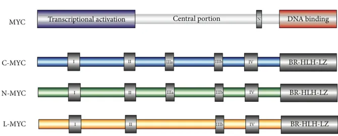
The three Myc family members are transcription factors belonging to a subclass of the larger group of proteins sharing Basic-Region/Helix–Loop–Helix/Leucin-Zipper (BR/HLH/LZ) motif. Molecular phylogenetic studies on MYC family members have revealed large segments of moderate conservation that are marked by six regions of straight homology: five MYC-boxes and one BR/HLH/LZ (32, 33).
The general structural organization of proteins belonging to the MYC family (Figure 2) is similar for all members and consist of:
- A large N-terminal portion including MYC-box I and II, involved in positive transcription regulation (TAD domain).
- An internal segment including proline rich residues (PEST) as well as two conserved regions MYC-box III and IV.
- A C-terminal portion comprising the basic-Helix-Loop-Helix leucine zipper domain (BR/HLH/LZ).
Figure 2: Structure of MYC family members (34).
The N-terminal MYC TAD domain, comprising MYC-box I and II, fused to heterologous DNA Binding Domain (DBD), is sufficient for transcriptional activation and is also chiefly responsible for MYC Ubiquitin-mediated degradation (23).
Several studies have demonstrated that MYC-box II is essential to promote, both vitro and in-vivo, cellular transformation and to positive and/or negative regulating transcriptional events. The main importance of MYC-box II in transcriptional regulation of several targets genes is surely attributable to its role in binding of co-activators like: TRRAP, GCN5, BAF53, SL1, BIN1 and PML(35-37).
In 2005 Herbst A. and collegues focused their attention on the “little studied” MYC-box III. Thanks to conditional expression of delta ER_c-MYC mutants in Rat1a mouse cell line, the critical role of MYC-box III in transcriptional repression activity of tumor related genes like P21, P15 and GADD45 was underlined (36).
Figure 3: Heterodimer Myc-Max bound to consensus E-box DNA sequence (34).
C-terminal segment of MYC factors is essential for heterodimerization with another small bHLH-LZ protein named MAX. MYC-MAX heterodimer acts as a “core DNA-binding module” (Figure 3) recognizing consensus DNA sequence “CACGTG” also called “Enhancer-BOX” (E-box) (32, 34).
THE MYC/MXD/MNT/MAX NETWORK AND THE TRANSCRIPTIONAL CONTROL OF CELL BEHAVIOR
MYC members are incapables of forming homodimers and binds to specific DNA sequences. The primary partner for MYC transcriptional regulation activity is the small bHLH-LZ protein MAX (38).
Unlike that of Myc genes, Max expression is ubiquitous and constitutive, and this 160 aminoacid protein is stable, resulting in Max levels that far exceed those of Myc (39).
MAX is a bHLH-LZ transcription factor that lack all conserved MYC box domains and can both form homodimers and heterodimers capable of directly bind to E-box DNA consensus sequences. As well as with MYC factors, MAX can heterodimerize with other bHLH-LZ proteins of the MXD family (MXD1-4), MNT and MGA (37, 38).
MXD1 and MXD4 are generally expressed in differentiated cells, whereas Mxi1 (MXD2), Mad3 (MXD3) and Mnt, like all Myc genes, are also expressed in proliferating cells. These findings give rise to the hypothesis by which the MYC/MXD/MNT/ MAX constitute a fine tuned homo and hetero dimerization network surrounding the small ubiquitously expressed bHLHLZ protein MAX. Overexpression experiments have suggested that MAX interacting proteins can antagonize the transcription regulatory activity of MYC family members. MAX homo- and/or hetero-dimerization with MXD and MNT result in repression of MYC-MAX activated gene targets. Transcriptional repression is establish by both MXDs and MNT by recruiting co-repressor complexes like N-CoR, Sin3a/Sin3b and the histone deacetylases 1 and 2 (Figure 4). The ability of MXD and MNT to physically interacts with Sin3a/Sin3b is allowed by an internal Sin3 Interaction Domain (SID) (18, 19, 39, 40).
Recent studies carried out using a negative Myc mutant called “MadMyc”, in which DNA binding and dimerization domain of Myc were fused with SID of MAD, have shown inhibition of cell proliferation and cell cycle arrest (41).
Figure 4: Differences between transcriptional regulation by MYC-MAX heterodimer and MAD-MAX heterodimer at level of E-box DNA elements (19).
Our understanding of the MYC/MXD/MNT/MAX network grew out of research on the MYC oncogene family. The first compelling idea about MYC was that its function drive cell growth and proliferation in response to a wide range of signals. Indeed, MYC genes are widely expressed during embryogenesis, and targeted deletions of c-MYC or N-Myc genes in mice lead to lethality in mid-gestation embryos (37, 38). Moreover, there is a strong correlation between MYC expression and
proliferation (9, 18, 19, 39-41). In cells with activated MYC, G1 phase is often shortened as cells enter the cell cycle, and MYC is essential for G0/G1 to S phase progression (23, 42, 43).
BIOLOGICAL FUNCTIONS OF MYC
It is now clear that a wide range of growth factors, cytokines, and mitogens induce MYC expression in many cellular backgrounds (11, 33). Both transcriptional and/or post-transcriptional regulation can determine an increase in endogenous MYC and appears to occur as an immediate early response (about 2 hours) to most mitogenic factors (44).
On the other hand, anti-proliferative signals must down regulate MYC expression, constituting a signal for cells to exit the cell cycle and undergo differentiation. Moreover, the induction of MXD family members, in response to different cues, is another important point of regulation to allow cell differentiation (20, 39, 42).
In the case of specific lineage commitment, an increase of MYC, determining boost of proliferation, also constitutes physiological event that is essential cell differentiation. Clearly, these data strongly suggest that MYC is a nexus for multiple growth signal response pathways. Therefore both MYC expression and activity are tightly regulated in non-transformed cells and finely tuned to quickly respond to proliferative cues from the extracellular milieu (14, 45).
The ability of MYC overexpressing cells to facilitate proliferation and inhibit terminal differentiation perfectly fits with different genetic rearrangements involving MYC family genes in several types of cancer, such as genomic amplification of N-Myc in almost 25% of neuroblastoma tumors (6). Indeed, many of the genomic alterations in the MYC gene result in increased MYC mRNA levels through increased transcription initiation, decreased transcription attenuation, and augmented stability of the MYC mRNA (20, 41). Moreover, many tumor-related mutations in Myc result in significant protein stabilization (23, 43).
One of the most striking findings of the past years was the discovery of the important role of enhanced expression of Myc proteins in almost every aspect of tumor cell biology (33). Whereas the ability of Myc to drive unrestricted cell proliferation and to inhibit cell differentiation has long been recognized, many studies have already underlined that deregulated Myc expression can drive cell growth and vasculogenesis, reduce cell adhesion and promote metastasis and genomic instability. Conversely, the loss of Myc proteins inhibits cell proliferation and cell growth and also accelerate differentiation, increases cell adhesion and leads to an excessive DNA damage response (33).
In the last 15 years, in several neuroblastoma cell lines, there were analysed possible interconnections between N-Myc level and miRNA expression profile, stressing that N-Myc can both activate and repress many of non coding RNAs like mir 17-92 cluster, Mir 9 and Mir-421 (46).
This findings reflects the surprisingly high number of target genes regulated by Myc, as emerged in large-scale analyses of MYC-regulated genes. Indeed, in normal cells, Myc protein appear to integrate environmental signals in order to modulate a wide, and sometimes opposite, group of biological functions including proliferation, growth, apoptosis, energy metabolism and differentiation (45).
MYC AS AN ACTIVATOR
MYC factors, as already indicated, must heterodimerize with the small b-HLH-BZ protein MAX to directly bind DNA. MYC-MAX complex have relatively weak transactivation activity both at endogenous level and in transient assays (47). Recently published transcriptomic analysis have underlined the weak ability of Myc proteins to activate the majority of target genes (generally ranging from 3- to 10-fold transactivation) (48).
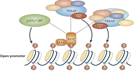
In general, the transactivation domain of Myc (TAD) recruits the basal transcription machinery either directly or indirectly, thanks to different protein complexes formed with several accessory factors. The most relevant model of MYC-mediated transcription activation postulates that MYC increases local histone acetylation in the promoter regions (33). In this connection, MYC binds to histone acetyltransferase complexes including TRRAP (transformation/transcription-domain- associated protein) and either general control of amino-acid-synthesis protein-5 (GCN5) or TIP60, which preferentially acetylate histones H3 or H4, respectively (49, 50). Myc can also binds to the p300/CBP (CREB-binding protein) acetyltransferase (51, 52).
Figure 5: Mechanisms of MYC-induced transcription. Myc recruits histone acetyltransferases, which promote localized modification of chromatin through nucleosomes acetylation (52).
The action of acetyltransferase complexes, recruited by MYC factors, determine positive signal for transcription activation. The acetylated and more relaxed chromatin status provides docking sites for acetyl-histone-binding proteins, including GCN5 and the SWI/SNF chromatin-remodelling complex, both correlating with increased transcription levels (Figure 5). Moreover, acetylated euchromatic DNA regions would permit subsequent binding of constitutive and general transcription factors that allow RNA polymerase II promoter docking (52, 53).
The recruitment of histone modifiers by transcription regulators is accepted to be a major mechanism of transactivation, shared by many other transcription factors, like: TCF (T-cell factor), E2F, the tumor suppressor TP53 and Gal4 (54).
In the last decade genome-wide expression analysis, performed by many groups and in many cellular backgrounds, has revealed a staggering number of MYC target genes, around 10-22% of all genes in most models. Chip-seq analysis have shown higher Myc affinity for cell cycle related promoters than through all Myc-related promoters (41).
Among others, target genes include the Cyclin-Dependent Kinase-4 (CDK4), the Cdc25A phosphatase which activates CDKs, cyclin D2, CKN1A(p21), p27 and the E2F family. E2F gene family encodes for transcription factors critical for G1-S progression and in quiescent cells E2F -/-Myc fails to induce G1-S progression (41, 55). Recently, -/-Myc has been shown to promote oxidative phosphorylation as well as glycolysis through coordinate transcriptional control of the mitochondrial metabolic network (56).
In addition to cell cycle control and metabolic target genes, Myc has been found to activate several essential genes involved in many biological functions like control of cell size and growth, including those encoding ribosomal proteins, translation factors, and metabolic enzymes (57-60). These findings stress the role of MYC factors in recruitment of co-activator complexes into regulatory regions contacted by RNA polymerase I and III (61-64).
MYC regulation can also occurs at the level of transcriptional elongation and not just at transcriptional initiation. The C-terminal domain (CTD) of RNA pol II undergoes to cycling phosphorylation and de-phosphorylation during transcription. Hypo-phosphorylated form of CTD determines RNA pol II recruitment to promoters, while high level of phosphorylation occurs during initiation and elongation steps. Sub-sequent de-phosphorylation allow RNA pol II recycling for another round of transcription (65). RNA pol II has been found to pause on most promoters after transcribing approximately 20–40 bases. This model fits well with the finding that Myc stimulates the release of paused RNA pol II from the promoter and stimulates subsequent transcriptional elongation (66). Myc transactivation domain (TAD) binds directly to CTD kinases determining an increase in RNA pol II phosphorylation and elongation. Myc induction occurs globally throughout the nucleus and it can be detected in the total cellular pool of RNA pol II rather than simply at MYC target-gene promoters (66, 67).
Moreover, Myc factors are also involved in control of mRNA stability, by promoting 5' methylation of guanine or 'cap', which is an essential step for protein-coding gene expression. This transcription-independent activity underlines the critical role of MYC in transcription and post-transcription regulation in both normal and tumor cells (68, 69).
Along with transcription, the most important nuclear process is DNA replication. The genome must be faithfully replicated each cell cycle and the chromosomes must be segregated to the daughter cells. Disruption of any step in this process, such as a stalled replication fork or DNA damage occurring during S phase, activates checkpoints that halting the cell cycle until the lesion can be repaired. Failure to correct this damage leads to a mutation and/or genomic instability. In fact, it has been hypothesized that high MYC expression correlate with genomic instability because of the indirect consequence of MYC mediated de-regulation in normal transcriptional activity (70, 71).
A recent study have described a direct, non-transcriptional, role for MYC in the initiation of DNA replication. Myc has been found to bind numerous components of the pre-replicative complex, and localize to early sites of DNA replication. These observations have suggested that MYC might directly control the initiation of S phase and this effect on genomic instability might not depend on the transcriptional induction of S-phase-promoting genes. Furthermore much excitement has been
generated in the past few years about the role of noncoding regulatory RNAs. The first oncogenic polycistronic microRNA is shown to be regulated by MYC (42).
Taken together, these findings raise the question: are MYC factors just like traditional transcription factors or are they guardians of cell metabolism?
Surely the transcription activity is the main known function of the oncogenic MYC protein. Apparently, a disconnection seems to exist between MYC’s dramatic effects on multiple cellular functions and its biological and molecular characterization as a relatively weak transcriptional activator. Indeed, the notion that Myc is a general chromatin regulator is nonetheless consistent with several recent observations concerning MYC function. First, independent expression microarray analysis have collectively identified a large group of genes regulated by Myc. Second, chromatin-IP experiments directly assessing Myc binding to thousands of sites throughout the genome encompassing approximately 15% of genes as well as intergenic regions (48, 66, 72, 73). Potentially, therefore, Myc can regulates a significant percentage of all genes in an organism.
The number of in-vivo binding sites exceeds the number of Myc molecules in proliferating cells, indicating that each site is bound by Myc only temporarily. Most probably, therefore, transcriptional regulation by Myc occurs by a 'hit-and-run' mechanism whereby the relatively brief binding of Myc triggers longer-lasting changes in the chromatin organization at the bound loci (45).
Many recent evidences underline the role of N-Myc in the global regulation of human genome euchromatin, including that of intergenic regions. Strikingly, N-Myc maintains 90% to 95% of total H3K9 acetylated and H3K4Me marks, with enhancer-like function, in human several neuroblastoma cell lines (74). Furthermore Myc may regulate chromatin at a distance so that Myc binding at one location can influence chromatin at another site through an high order chromatin structure (75).
Intriguingly intergenic binding sites for MYC are not enriched for E-boxes. Although E-box independent binding has been reported and may be fairly widespread, such binding may be of particular importance for Myc intergenic function (70).
Furthermore, Myc has been shown to possess another feature outside the context of E-boxes: surprisingly Myc can also act as well as a transcriptional repressor at certain target promoters consistent with the wide distribution of MYC along the genome(see below) (76-78).
MYC AS A REPRESSOR
One of the first finding highlighting the idea that MYC can also act as a transcriptional repressor derived from studies published in the 1980s, suggesting that MYC participates in a negative feedback loop (79, 80). Genome-wide analyses demonstrate that MYC represses at least as many targets as it activates, further emphasizing the role of repression in MYC function, including transformation (45, 81).
No simple consensus sequences for transcriptional repression by Myc has emerged, this finding opens up the possibility that transcriptional repression is a simply indirect consequence of the altered physiological (e.g., transformed) state of a cell induced by Myc (82, 83).
In the last 10 years a lot of studies have been carried out to fully understand the mechanism of MYC repression. Many investigators have exploited a chimeric MYC-MAD protein to better define MYC transcriptional activity, but this chimera cannot fully recover the transformation potential of wild-type MYC factors in-vivo; for example, they are unable to immortalize primary mouse embryo fibroblasts and to induce apoptosis in immortalized cells and are impaired in rescuing the proliferation defect of c-myc -/- fibroblasts (84).
These important findings, together with several other illuminating researches, support the hypothesis that the oncogenic potential of MYC factors is fulfilled by both activation and repression activity. The actual model of MYC repression is based on the indirect DNA binding on cis-genomic elements, interacting directly with other transcription regulators bound to DNA. The repressed genes, like induced genes, fall into multiple functional classes. Among all, genes encoding for factors selectively expressed in quiescent cells or involved in inhibit cell proliferation. This group encompasses the cell cycle inhibitors p21 (85-88), p27kipl (89, 90), pl5ink4b (91, 92), pl8ink4c (93), and p57kip2 (94), as well as the differentiation-inducing proteins C/EBP-a (95), the growth-arrest proteins gas1 and gas2 (96), the growth growth-arrest and DNA damage proteins gadd34, gadd45, gadd153 (70, 97, 98), and the Myc-antagonist Mad4 (99). Myc can also down regulate genes encoding for proteins deeply involved in cell adhesion, including a large number of integrins: these genes include those encoding cell surface proteins such as the class I HLA molecules in melanoma cells, the α3 β1 integrin in neuroblastomas, and the LFA-1 (αL β2 integrin) cell adhesion protein in transformed B cells as well (100). Altered cell adhesion is a hallmark of many Myc-transformed cells and has been observed in different cell types (101). Metabolic pathways such as thrombospondin and H-ferritin are also affected by Myc mediated repression (102, 103). Suppression of thrombospondin plays a causative role in the induction of angiogenesis by Myc. In the last 10 years
ABCC3 has been identified as MYC repressed gene, it encode for an important multi-drug resistance protein involved in chemo-resistance and also in cell migration (104). Therefore, Myc-mediated gene repression in the control of cellular differentiation and in the response to growth arrest signals makes a significant contribute to the phenotype of MYC-transformed cells. The basic mechanism underlying MYC's activation of transcription is well understood, but the way in which MYC
negatively regulates or represses transcription is far less understood (81). A number of Myc-repressed targets contain a subclass of initiator elements (INRs; consensus,
YYCAYYYYY, where Y is a pyrimidine base), which are usually, but not invariably, present at TATA-less promoters. Inr elements are recognized by TFIID as well as by a number of regulatory proteins, such as the transcription initiation factor TFII-I, YY-1, and the Myc interacting zinc-finger protein 1 (Miz-1). Interestingly, the last three proteins have been reported to associate with the C-terminal BR/HLH/LZ region of Myc (101).
Figure 6: Mechanisms of transcriptional repression by c-Myc. (A) Inr-dependent mechanism of MYC repression. Myc–Max heterodimers bind to the Inr element and associate with Miz-1 or other TFs, thus interfering with their activities. (B) c-Myc represses target genes transcription by Sp1-dependent mechanism. c-Myc interacts with the Sp1 transcription factor (1) or with the Smad–Sp1 complex (2) via the c-Myc central region and inhibits Sp1 transcriptional activity. This mechanism does not require DNA binding or interaction with the c-Myc partner Max (105).
MYC mediated repression of p15 and CKN1A(p21) promoters has been thoroughly described and could be perceived as prototypical for MYC dependent transcriptional repression of growth arrest genes.
There are two main independent mechanisms of MYC mediates repression. The first is based on the ability of MYC-MAX heterodimer to bind the zinc-finger protein Miz-1 and this complex is now capable of binding to transcriptional initiator elements (Inr) (figure 6, A) (101). Miz1 contains 13 zinc fingers and, at its amino-terminus, carries a BTB/POZ-domain, which is a protein-protein interaction domain found in multiple zinc-finger proteins. Miz1 binds to the 'outside' of the helix– loop–helix domain of Myc, but does not interact with Max, Mad or Mnt proteins (106, 107). The second mechanism by which MYC-MAX represses transcription implies interaction with SMAD 2/3 proteins and consequent complex formation with another TF called Sp1 (figure 6, B2) (108).
In case of p15 transcriptional regulation, Miz-1 acts as positive factor but the interaction with MYC-MAX inhibit the Miz-1 mediated p300 recruitment at Inr element. Conversely, MYC-MYC-MAX can carry out p15 repression in Inr independent manner. In fact, p15 gene activation can be establish by positive transcription complex formed by several combination of SMAD2, SMAD3 and SMAD4 with Sp1. MYC-MAX can interact with SMAD2/3 to form a larger, inactive but more stable complex formed by MYC-MAX/SMAD2/3/Sp1 (101).
Several published data have revealed both c-Myc and N-Myc repression activity on CKN1A(p21) gene transcription. MYC factors do not directly bind to DNA but they form complexes with Sp1 factors determining, as already mentioned for p15 repression, the absence of transcriptional activation (86).
On the other hand, there are some genes repressed by MYC through a mechanism that does not involve the Max protein (109, 110).
In 2005 Brenner and colleagues demonstrated recruitment of DNA methyltransferase, DNMT3a, by c-Myc to CKN1A(p21) promoter in a MAX independent complex composed by c-Myc and Miz-1. This finding disclosed the important interconnection between c-Myc repression activity and DNA methylation (106, 107). Since DNMT3a is complexed with histone deacetylases enzymes, its recruitment by Myc might lead to local histone deacetylation and inhibition of transcription (107). Recruitment of DNMT3a by Myc is an attractive mechanism for repression, since it might provide an explanation of the aberrant DNA methylation of some tumor suppressor genes that is observed in some human tumors.
Another finding that reinforce the idea that there exist multiple and variegates MYC repressive pathways is the discovery of N-Myc-PRC2 (via N-Myc and EZH2 physically interaction) repressive complex on the promoter of the tumor suppressor gene CLU in neuroblastoma cellular background (111).
All these data clearly support the notion that several pathways of repression exist. Finally, the present model is that Myc interacts with transcriptional activators that are bound directly to DNA through enhancer or initiator elements either cooperating with MAX or not. These multi-protein complexes are thought to inhibit recruitment of co-activators, facilitating the negative and oncogenic activity of co-repressors like DNA methyltransferases, histone methyltransferases and deacetylases (105, 112).
EPIGENETICS, AN OVERVIEW
The definition of epigenetic, coined by Conrad Waddington, is: “An epigenetic trait is a stably heritable phenotype resulting from changes in a chromosome without alterations in the DNA sequence” (113).
Shelley L. Berger and colleagues, proposed three categories of signals that trigger different establishment of stably heritable epigenetic states (Figure 7):
- “Epigenator,’’ which emanates from the environment and triggers an intracellular pathway;
- ‘‘Epigenetic Initiator’’ signal, which responds to the Epigenator and is necessary to define the precise location of the epigenetic chromatin environment;
- ‘‘Epigenetic Maintainer’’ signal, which sustains the chromatin environment in the first and subsequent generations (114).
Figure 7: The epigenetic pathway.
The fine tuned DNA organization inside the nucleus is an essential aspect of eukaryotic cell life. Chromatin is the macromolecular complex composed by DNA, RNA and proteins, determining genomic DNA condensation inside the nucleus. Mainly, there are four chromatin packaging degrees ranging from 11nm DNA fibers to 700nm interphasic DNA domains (Figure 8) (114, 115).
The plastic, finely tuned and rapid exchange of different levels of genomic DNA condensation is a critical step for almost all the biological issues linked to DNA.
Every 147bp, DNA is wrapped around an octameric protein complex, the nucleosome. Five different proteins (Histones) compose this functional unit: H2A, H2B, H3, H4 and H1. The nucleosome structure is globular except for the histone n-terminal “tails”, which are unstructured. As mentioned above, chromatin condensation is an essential regulating “tool” of many important biological aspects like DNA transcription (114, 115).
The nucleosome is also target of several dynamic post-translational modifications of histone n-tails which determine the “fate” of transcriptional activity of all the genes encoded by genomic DNA (116). Indeed, histone modifications are crucial to dictate different genomic packaging levels inside the nucleus (Figure 8). These changes in DNA condensation ranging from heterochromatin, highly condensed and transcriptionally repressed or silenced to a more accessible status of DNA defined as euchromatin in which genomic regions are tightly packaged and are transcriptionally active (114-117).
DNA METHYLATION AND HISTONE MODIFICATIONS
Among all the chromatin modifications, we can distinguish direct DNA modification, including methylcytosine (5mC), hydroxymethylcytosine (5hmC), formylcytosine (5fC) and 5-carboxycytosine (5caC) and n-tail covalent nucleosome modifications such as: acetylation, methylation, sumoylation, ubiquitination and phosphorylation (116-118).
DNA methylation is the covalent addition of a methyl group (CH3) to the C5 position of cytosine in CpG dinucleotides. Genomic regions containing multiple stretches of CpG dinucleotides termed as “CpG islands” and they are often associated with promoters elements (119).
In mammals somatic cells, methylated cytosines account for 1% of total DNA bases, but only 10% of these are located in CpG islands (120). Unlike the dispersed CpG elements, those in CpG islands are more resistant to methylation events (119, 121).
Cytosines are methylated by the DNA methyltransferase machinery composed of two subunits: the DNA methyltransferase (DNMTs) and the methyl CpG binding protein (MBDs). Until now there no evidences have been found about activity responsible for DNA de-methylation (119).
In 1983 the clear correlation was first demonstrated between hypomethylation and genomic instability of cancer cells. In the last 20 years many studies have corroborated the hypothesis that loss of genomic methylation is an early event in many type of cancer (122).
Many genes involved in cell-cycle regulation, tumour cell invasion, DNA repair, chromatin remodelling, cell signalling, transcription and apoptosis are known to become aberrantly hypermethylated and silenced in many tumour type. Probably the hypermethylation status increases genetic instability, allowing cancer cells to acquire advantageous genetic changes and to proliferate and to metastasize (122, 123).
Ever since Allfrey's studies in the early 1960s, we have known that histones can be post-translationally modified by a large number of different histone post-translational modifications (PTMs) (124). There are at least eight distinct types of modifications found on histones (Table 1). The dynamic and heterogeneous network of histone modifications determine the transcriptional “fate” of all the genes encoded by genomic DNA (125).
Extra complexity comes partly from the fact that methylation at lysines or arginines may be one of three different forms: mono-, di-, or trimethyl for lysines and mono- or di- (asymmetric or symmetric) for arginines (116).
a protein has to be digested before such analysis can take place limits its potential. New methodology that uses a top-down proteomics approach (identify protein first and digest subsequently) gives promise that we may, in the future, look at the intact modification pattern of differ-ent histones in a given nucleosome (Macek et al., 2006).
Once global analysis of all histone modifications is done, a prediction would be that every single nucleosome would be found to be modified in some way. This picture is of course very static. The truth is that modifications on his-tones are dynamic and rapidly changing. Acetylation, methylation, phosphorylation, and deimination can appear and disappear on chromatin within minutes of stimulus arriving at the cell surface. Thus examining bulk histones under one specific set of conditions (with either antibodies or mass spectrometry) will identify only a proportion of the possible modifications.
There are also problems of detection that are specific for antibodies. Firstly, there are the obvious issues of specificity. These are difficult to avoid as there are no true controls for modifications in mammalian cells (unlike yeast) where it is impossible to mutate the residue to make sure reactivity is lost. In addition, an adjacent modification may disrupt the binding of the antibody or a protein may occlude its recognition, both of which may give a false reading. Similarly, there are problems of detection that are specific to mass spectrometry. Peptide coverage is not equivalent for all parts of the histone and this reduces the sensitivity of detection in these regions. These facts undoubtedly contribute to our underestima-tion of the extent of modificaunderestima-tions present on histones.
We assume that each individual modification on his-tones leads to a biological consequence. However proof of a consequence is not always easy to provide and is often based on a correlation: a modification appears on a gene under certain conditions (e.g., when it is tran-scribed) and disappears when that state is reversed (e.g., when the gene is silent). Proving causality for a modification involves showing that the catalytic activity of the enzyme that mediates the modification is necessary
for the biological response. However we know that many of the histone-modifying enzymes have other nonhistone substrates. So the response may be going through another unidentified protein substrate. Furthermore, there may be signaling redundancy such that more than one enzyme may be capable of modifying a specific site. In this case, the effects of inactivating one enzyme may be masked by an upregulation in the activity of a second distinct but related enzyme. Showing that mutation of the modified residue gives the same output as mutating the enzyme is a second stringent test. However, this is not possible in humans due to many histone genes present in the genome, but it is possible in yeast.
So the truth is that we have ‘‘levels of confidence’’ regarding the causative nature of different modifications depending on how far the analysis has gone to prove the issue. We also have to be realistic and accept that, how-ever far we go in proving that a histone modification is causative, we can never exclude the possibility that modification of other substrates by the same enzyme will play a parallel role in the biological response being monitored. The many other nonhistone substrates of chromatin-modifying enzymes are not covered in this Review.
Histone-Modifying Enzymes
The identification of the enzymes that direct modification has been the focus of intense activity over the last 10 years
(Table 2). Enzymes have been identified for acetylation
(Sterner and Berger, 2000), methylation (Zhang and
Rein-berg, 2006), phosphorylation (Nowak and Corces, 2004),
ubiquitination (Shilatifard, 2006), sumoylation (Nathan
et al., 2006), ADP-ribosylation (Hassa et al., 2006),
deimi-nation (Cuthbert et al., 2004; Wang et al., 2004b), and pro-line isomerization (Nelson et al., 2006).
Most modifications have been found to be dynamic, and enzymes that remove the modification have been identified. One major exception is methylation of arginines: although they are thought to be dynamic, a demethylating activity has not yet been found. Instead Table 1. Different Classes of Modifications Identified on Histones
Chromatin Modifications Residues Modified Functions Regulated
Acetylation K-ac Transcription, Repair, Replication, Condensation Methylation (lysines) K-me1 K-me2 K-me3 Transcription, Repair
Methylation (arginines) R-me1 R-me2a R-me2s Transcription
Phosphorylation S-ph T-ph Transcription, Repair, Condensation Ubiquitylation K-ub Transcription, Repair
Sumoylation K-su Transcription
ADP ribosylation E-ar Transcription Deimination R > Cit Transcription Proline Isomerization P-cis > P-trans Transcription
Overview of different classes of modification identified on histones. The functions that have been associated with each modification are shown. Each modification is discussed in detail in the text under the heading of the function it regulates.
694 Cell 128, 693–705, February 23, 2007ª2007 Elsevier Inc.
Table 1: Overview of different classes of modification identified on histones (114).
The combinatorial complexity of all the different histone modifications that can occur at the same time in the same nucleosome had always lead to several hypotheses attempting to define “the histone code” which actually is not fully understood (126). The histone code is read and construe by the nonhistone proteins and multiprotein complexes that form the transcriptionactivating and/or -repressing molecular machinery. Moreover, different chromatin binding proteins can be recruited by specific n-tail histone markers, but the simultaneous existence of two or more marks in the same nucleosome can lead to a different scenario (126-128).
Chromatin-regulating proteins can be divided into three main groups (Figure 9): - “Epigenetic writers” that directly modify specific N-tail residues;
- “Epigenetic readers” that bind specifically to a type of covalently modified amino acid; - “Epigenetic erasers” that remove and/or convert distinct N-tail covalent modifications.
In the last decade the already quirky network of histone writers and erasers has been complicated by the discovery of many modifiers able to methylate and demethylate specific residues of protein factors involved in transcription regulation. These modification also significantly affect the ability of transcription factors to form the protein complexes required to activate and/or to repress transcription of specific genes (128, 130).
N-tail histone acetylation is the most common histone marker of opened chromatin and it occurs exclusively on lysine residues of histone H3 and H4. N-Acetylation of positively charged lysines determines an electrostatic neutralization, because of its negative charge. The effect of this change in net positive charged of histone determines an important loss of electrostatic interaction between nucleosome and DNA leading to a more relaxed and accessible chromatin status (124, 125).
In 1964 Allfrey et al. first demonstrated the highly dynamic and finely regulated balance between histone acetylation and de-acetylation of chromatin. The plastic balance between histone acetylation and de-acetylation is respectively controlled by histone acetyltransferases (HATs) and histone deacetylases (HDACs). HATs, also known as K-acetyltransferases, catalyze the addition of acetyl groups to histone lysines using acetyl coenzyme A as cofactor. GCN5, p300/CBP, and MYST families composed the three main groups of HATs (51, 53). Just as HATs are a diverse set of enzymes, the multi-protein complexes in which they reside also vary in subunits composition. The considerable combinations of these accessory subunits lead to several unique features of each HAT complex. For example, some subunits have conserved domains that cooperate to recruit the HAT to the appropriate location in the genome; these include bromodomains, chromodomains, WD40 repeats, Tudor domains and PHD finger (54, 131).
Unlike positive transcriptional HATs activity, Histone DeACetylases (HDACs) led to repressed chromatin because of the increase in electrostatic interaction between nucleosomes and wrapped DNA. Until now, eighteen distinct human HDACs have discovered and grouped into four classes. Class I HDACs (HDAC1, -2, -3, and -8) are predominantly nuclear proteins and ubiquitously expressed in most tissues and cell lines. Class II HDACs can be subdivided into two subclasses, IIa (HDAC4, -5, -7 and -9 and its splice variant MITR) and IIb (HDAC6 and HDAC10), based on the protein sequence homology and domain organization. Class IIa HDACs have one catalytic domain and a long amino-terminal adaptor domain, while class IIb HDACs contain two catalytic domains. Class III HDACs, known as sirtuins, do not contain zinc and their activity requires nicotinamide adenine dinucleotide (NAD+). Class IV HDACs include only HDAC 11, a relatively newly discovered protein, which resembles class I HDACs (128, 132).
HATs and HDACs complexes have been shown to play a critical role in carcinogenesis, trough either inappropriate activation or repression of target gene activity (53, 125, 133).
As already discussed, HATs complexes are co-activators of many TFs like MYC family oncoproteins. The role of HATs complexes in cancer, especially for p300 is not well understood, probably because its activity depends on different tumor backgrounds (134). p300 has been recognized as a potential anti-cancer drug target because its gene was found altered in most colon cancer cell lines and in some primary tumors (135, 136).
p300 is also a target of viral oncoproteins, it can be fused to MLL in leukaemia and two missense mutations were found in epithelial malignancies (137). Conversely to its supposed oncosoppressor function, in prostate cancer p300 was found to have to a clearly oncogenic potential (134).
HDACs have been intensely studied for their involvement in mediating the function of oncogenic translocation products in specific forms of leukaemia and lymphoma. For example, in acute promyelocytic leukaemia (APL), PML–RARα, represses transcription by associating with a corepressor complex that contains HDAC activity. In non-Hodgkin’s lymphoma, the transcriptional repressor LAZ3/BCL6 (lymphoma-associated zinc finger-3/B cell lymphoma 6) is strongly overexpressed and associated with aberrant transcriptional repression through recruitment of HDACs, leading to lymphoid oncogenic transformation (128).
Another histone modification that leads to a change in the electrostatic balance between nucleosomes and DNA is the phosphorylation of serine, threonine and tyrosine residues on histone N-tails (138).
The exact mechanism by which histone phosphorylation affects gene expression is not well understood; it is thought that, similarly to N-acetylation, the addition of phosphate group (negatively charged) to histone N-tails may interfere in the electrostatic interaction between nucleosomes and DNA. Like N-acetylation of histone N-tails, phosphorylation probably increases the accessibility of DNA to nuclear factors (138).
Less is known regarding the roles of histone phosphatases. Certainly, given the extremely rapid turnover of specific histone phosphorylations, there must be high phosphatase activity within the nucleus. We know, e.g., that the PP1 phosphatase works antagonistically to Aurora B, the kinase that lays down genome-wide H3S10ph and H3S28ph at mitosis (124, 139).
For the majority of kinases it is not yet clear how they are accurately recruited to their sites of action on chromatin. The mammalian MAPK1 enzyme possesses an intrinsic DNA-binding domain with which it is tethered to the DNA. Alternatively, histone kinases recruitment may require association with a chromatin bound factor before it directly contacts DNA to stabilize the overall interaction.
Even though the majority of histone phosphorylation sites lie within the N-terminal tails, there are examples of phosphorilation sites within the core histone region. For example, it was determined that the non-receptor tyrosine kinase JAK2 is responsible of H3Y41 phosphorylation (124, 140). Histones H1, H2A, H2B, H3, and H4 are all phosphorylated at multiple residues, but the most studied are so far the phosphorylations of histone H3. Phosphorylation reaction is catalysed by many distinct kinases that are mostly specific for individual histone residues (139).
A huge number of studies have underlined the important role of H3S10 phosphorylation in positive gene regulation. This histone modification can be deposited by various kinases in relation to the biological context. Phosphorylation of H3S10 by mitogen and stress-activated protein kinases 1 and 2 (MSK1 and MSK2) as well as RSK2 kinase has been shown to play a role in the activation of mitogen-stimulated immediate-early response genes, such as c-fos and c-jun (139, 141, 142). Furthermore, Pim1 kinase catalyses H3S10 phosphorylation at the E-boxes in Myc target genes, contributing to their transcriptional activation after growth factor stimulation (139). While histone acetylation and phosphorylation balance can deeply change the net charge of nucleosomes, methylation is a chemical modification that does not alter electrostatic interaction between nucleosomes and DNA. As already mentioned above, mono-, di- or tri-methylation of histone N-tails can occur on lysine, arginine and histidine residues (143). Methyltransferase activity lies with the catalytic ability to add methylic groups from S-Adenosyl methionine to specific aminoacid residues K, R and H. There are three main families of methyltransferases based on protein domains homology: Set1, DOT-1 like and PRMT (144).
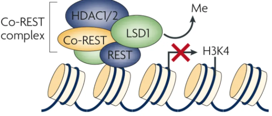
Methylation of H3K4, H3K36 and H3K79 are strongly associated with euchromatic regions, while methylation on H3K9, H3K27 and H4K20 is often found on repressed heterochromatin regions. Specifically, di- or tri- methylation of H3K4 are strictly associated with Trascriptional Start Site (TSS) DNA regions, whereas H3K4Me is closed linked with enhancer elements of active genes. While mono-methylation of H3K9 is often associated with active transcription, H3K9Me3 is a marker of transcription repression (114).
Several scientific reports have already discussed the important interconnection between nucleosome methylation pattern and carcinogenesis in many tumor backgrounds.
EZH2, together with SUZ12 and EED, forms the polycomb repressive complex 2 (PRC2), which is responsible for tri-methylation of H3K27. In cancer, EZH2 is one of the widely studied methyltransferase because of its clear relationship with many type of tumors like breast, prostate and lymphoma (130). As already highlighted for some HAT proteins several mehtyltransferases and
HDACs to cannot be absolutely classified as oncosoppresor or oncogene: it depends on the cellular background in which they act (144).
MYC FACTORS AND HISTONE MODIFIERS IN CO-ACTIVATOR AND CO-REPRESSIVE COMPLEXES
In 2014 Susanne Walz et al. performed RNA and ChIP seq analyses on U2OS and Hela cell lines respectively in a condition of doxycycline c-Myc induction and c-Myc silencing by Sh-RNA techniques. Both c-Myc activation and silencing revealed almost 30,000 MYC binding sites, and more than 200 up- and 100 down-regulated genes. Consistently, a linear support vector machine algorithm based on the set of MYC-regulated genes correctly classified 37 of 38 neuroblastomas as harbouring a single copy or amplified N-Myc gene. The most important finding that was suggested by Susanne Walz and colleagues is the correlation between high c-myc level and the occupancy of low affinity non-consensus E-box (CANNTG) sites. Conversely, change in c-myc levels did not affect occupancy in high affinity consensus E-box sequences (CACGTG) (81).
These findings strongly suggest that the dynamic role of both c-Myc and N-Myc determines a profound change in the transcription profile of different tumors. Accordingly, MYC factors are widely studied in relation with different cis-elements in the whole cell genome and with heterogeneous multiprotein complexes composed by different histone modifiers.
As already underlined before, protein-protein interactions may modulate MYC's transcriptional regulatory ability and therefore its potential for oncogenicity. A variety of proteins that interact with both c-Myc and N-Myc have been identified. Few of these have been shown to be directly recruited by MYC factors and to mediate the transactivating functions of MYC (78).
One of the most widely studied MYC co-activator “partner” is the transactivation/transformation-associated protein (TRRAP), which, together with several histone acetyltransferases (HATs), stably associated with TRRAP and the positive transcription elongation factor b (PTEFb), form large multiprotein complexes (145). Accordingly, it was reported that dominant-negative TRRAP genes or antisense TRRAP RNA can blocks MYC transformation activity (146). In 2002, Elizabeth M. Flinn et al., definitively define Myc box II as domain responsible for c-Myc interaction with GCN5 or its associated protein, TRRAP (146).
The basal transcription factor 1 (SP1), a critical zinc-finger GC binding protein, is clearly involved in N-Myc-mediated repression mechanism (147-150). The N-Myc and SP1 interaction was fully investigated (151) through both ex-vivo and in-vitro techniques, resulting in demonstrating the importance of Myc Box 2 as the domain responsible for the interaction with SP1. As already
mentioned, N-Myc-SP1 complex exerts repressive function via recruitments of chromatin modifiers such as histone deacetylases. In 2007, Marshall et al. demonstrated the role of N-Myc-SP1 complex in repression activity of the transglutaminase 2 (TG2) gene expression through recruitment of histone deacetylase 1 (HDAC1)(147). Importantly, ChIP assays have determined that MAX is not present at DNA level and is not necessary for HDAC1 recruitment. Hence, N-Myc-SP1 repressive activity can be established in absence of N-Myc “partner” and can be disrupted by the use of an HDAC1 inhibitor (trichostatin A) (148).
In 2010 Marshall et al. have also demonstrated the N-Myc-SP1 mediated inhibition of CyclinG2 gene transcription through the interaction with HDAC2. Accordingly, in 2012 Zhang et al. have revealed that both c-Myc and N-Myc can interact with paralogs of HDAC1 such as HDAC2 and HDAC3 (152).
In-vitro analyses of the N-Myc regions required for its interaction with both SP1 and MIZ-1 show that N-Myc Myc Box 2 domain can directly interact with SP1, while the basic helix-loop-helix leucine-zipper (BR/HLH/LZ) domain is required for interaction with MIZ-1. The “ternary complex” can also drives the transcriptional repression of genes such as TRKA (tyrosine kinase receptor A), P75NTR (p75 neurotrophin receptor), and CKN1A(p21) in neuroblastoma by recruitment of HDAC1 on the respective promoters (151).
Collectively, these findings highlight the complexity of N-Myc activity and suggest that many more nuclear components may be critical for N-Myc-mediated transcriptional activation/repression. The main goal of the present study is to shade light on new possible functional and physical interactions between N-Myc and the two protein factors strictly associated with histone methylation: WDR5 and LSD1.
WDR5 METHYLTRANSFERASE
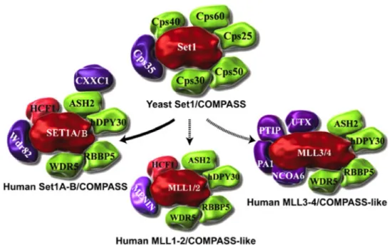
After the discovery of the COMPASS methyltransferase complex (complex of proteins associated with Set1) in yeast, in mammals too more than six COMPASS-like complexes were revealed. Although in yeast Set1 is the sole catalytic subunit of COMPASS complex, in mammals the situation is more complicated because of 2 orthologs (Set1a and Set1b) and 2 paralogs (MLLs and Ash1) (Table.2).
COMPASSes are multi subunits complexes with mono-, di- or tri-methylating activity on lysine 4 of histone H3 N-tail. Affinity pull-down experiments in mammals samples revealed the presence of WDR5 protein as one of the COMPASS subunits directly interacting with H3K4 (153, 154).
In this connection, recent studies have underlined the central role of Cps30 in yeast, and its mammals homolog WDR5, in the catalytic activity of COMPASS complexes for H3K4Me1-Me2 recognition and tri- or di-methylation (155).
Table.2: Yeast and mammalian COMPASS subunits and their functions (153).
The physical interaction between WDR5 and the conserved “Win” motifs of all the SET1 family members has also already been fully been demonstrated. Moreover, peptides that mimic both “Win” motif and H3K4 can distrupt the interaction between WDR5 and its partners. The actual mechanism hypothesized is the mutually exclusive binding to mono or di-methylated lysine 4 of H3 and the Win motif of Set1 proteins (155-157).
Figure 10: COMPASS and COMPASS-like complexes from yeast to human. COMPASS was identified in yeast as a complex of proteins associated with Set1 that can methylate H3 on Lys 4. Subsequently, six COMPASS-like complexes were identified in humans. All complexes share the core components Cps30 (WDR5), Cps50 (RBBP5), Cps25 (DPY30), and Cps60 (ASH2). COMPASS in humans also has CXXC and WDR82, which are homologous to Cps40 and Cps35 in yeast and regulate H3K4 trimethylation by COMPASS in vivoThe MLL3/4 complexes also contain the H3K27 demethylase UTX. The MLL1–4 COMPASS-like complexes function as coactivators of gene transcription in contrast to the canonical COMPASS complexes in yeast and humans. Set1s/MLLs are colored red, core components are colored green, and subunits with complex- specific functions are colored purple (157).
To better understand the mutual binding of WDR5 with MLL or Set1, in-vitro competition assays have shown that only H3K4Me1-Me2 peptides can disrupt the interaction, whereas H3K4 and H3K4Me3 cannot (156, 157). WDR5 interaction with catalytic subunits, MLL or Set1, is an essential step not only for COMPASS complex assembly but also for its core-catalytic methyltransferase activity (Figure 10)(158).
Although, most WDR5 related studies have been confined to its role in methyltransferase complexes, some recent data collected both in drosophila and in human cell lines, have revealed its possible role in structural nucleation of other protein complexes in which neither MLL or Set1 were detected (159).
There are very few data about WDR5 implication in carcinogenesis events. Some recent studies have revealed an important pattern of WDR5 over-expression in many human prostate cancer samples; ex-vivo experiments carried out on LNcaP cell line have underlined the involvement of WDR5, cooperating with H3T11P, in globally alteration of the methylation status of several AR target genes resulting in a boost of proliferation rate (154).