in cotutela con Université Claude Bernard Lyon 1
DOTTORATO DI RICERCA IN
Bioingegneria
Ciclo XXVII
Settore Concorsuale di afferenza: 09/G2 Settore Scientifico disciplinare: ING-INF/06
THE RECONSTRUCTION OF SKELETAL MOVEMENT:
THE SOFT TISSUE ARTEFACT ISSUE
PhD Thesis
Presentata da: Tecla Bonci
Coordinatore Dottorato
Relatore
Prof. Elisa Magosso
Prof. Aurelio Cappozzo
Relatore
Prof. Laurence Chèze
when you take your eyes off your goal” - Henry Ford -
In 3D human movement analysis performed using optoelectronic stereophotogrammetric systems and skin markers, bone pose estimate can only be carried out in an indirect fashion. During a motor task, the deformation of the soft tissues makes the skin markers move with respect to the underlying bone generating the so called soft tissue artefact (STA). In general, STA is caused by skin sliding associated with joint movement, soft tissue volumetric deformation due to muscular contraction, gravity and inertial effects on soft tissue masses (wobbling). This movement has a frequency content similar to the bone movement and it is, therefore, not possible to distinguish them using any filtering techniques. In addition, STA is subject-, task-, and location-specific. Therefore, STA is the main problem in optimal bone pose estimate and its compensation remains an open question.
The aim of this PhD thesis was to contribute to the solution of this crucial issue. Modelling this phenomenon based on measurable trial-specific variables is a fundamental prerequisite for its removal from skin marker trajectories. This model can be incorporated in future optimal bone pose estimators thus reducing relevant inaccuracies and significantly improve the estimate of joint kinematics.
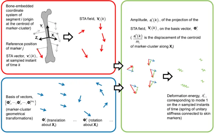
Two model architectures of STA are proposed. Initially, a model of the thigh artefact at marker-level is presented. STA was modelled as a linear combination of angular kinematics of the joints involved in the movement. This model was calibrated using both ex-vivo and in-vivo invasive measures (pin markers) of the STA. The considerable number of model parameters to be identified led to the approximation of the STA phenomenon. The STA of a body segment can be represented as a vector field composed by the displacement of each skin marker during the task with respect to its reference position in the anatomical reference frame. A modal approach was then used for the STA approximation and three STA definitions were proposed to represent the phenomenon as a series of modes: individual marker displacements, marker-cluster geometrical transformations, and skin envelope shape variations. It is important to point out that the marker-cluster geometrical transformations definition allows to separate the rigid component of the artefact from its non-rigid component. Modes obtained with the three above-mentioned definitions were selected using two different criteria: those that represent a certain percentage of the total STA energy (ranking) or those that describe the rigid component of the artefact which can be chosen a priori. Following the latter approach, it was empirically demonstrated with simulated data that only the rigid component of the artefact affects joint kinematics, regardless of the amplitude of the non-rigid component. For this reason, a model of the rigid component of the STA (at cluster-level) was then defined, using the marker-cluster geometrical transformation, for the thigh and shank segments. The selected rigid
number of the model parameters to be identified. An acceptable trade-off between STA compensation effectiveness and number of model parameters can be obtained using this STA modelling. This improves bone pose estimation and, therefore, joint kinematics accuracy.
The main potential applications of the results of this thesis are the following. First, the proposed STA models can be used to generate, at both marker- and cluster-level, realistic STAs which can be effectively used for simulation purposes when comparatively assessing skeletal kinematics estimators. Second and more importantly, by focusing only on the rigid component of the artefact, the model attains a satisfactory reconstruction of the artefact by using a reduced number of parameters. This circumstance makes incorporating the model in an optimal bone pose estimator feasible.
Quando la stima del movimento scheletrico viene condotta utilizzando un sistema non invasivo come quello stereofotogrammetrico e dei marcatori posti sulla cute, la stima della posa dell’osso può essere ottenuta utilizzando metodi indiretti.
Durante l’esecuzione di gesto motorio, la deformazione dei tessuti molli causa un movimento reale dei marcatori cutanei rispetto all’osso sottostante, generando il cosiddetto artefatto da tessuto molle (STA). In generale, lo STA è causato dallo stiramento della cute associato al movimento articolare, alla deformazione volumetrica dei tessuti dovuta alla contrazione muscolare, alla gravità ed ad effetti inerziali delle masse dei tessuti molli (il cosiddetto wobbling). Questo movimento ha un contenuto in frequenza simile a quello dell’osso sottostante e, quindi, non è possibile distinguerli usando tecniche di filtraggio. Inoltre, lo STA varia da soggetto a soggetto, varia al variare del movimento e della posizione del marcatore sul segmento in esame. Per questi motivi, lo STA è il problema principale nella stima ottima del movimento scheletrico e la sua compensazione rimane un problema, ad oggi, tuttora irrisolto.
Lo scopo di questa tesi è stato quello di dare un contributo alla soluzione di questo problema. Modellare questo fenomeno utilizzando delle variabili misurabili durante un dato esperimento è un prerequisito fondamentale per la rimozione dell’artefatto dalle traiettorie dei marcatori acquisite con un sistema stereofotogrammetrico. Questo modello può essere inserito in futuri stimatori ottimi della posa per ridurne l’inaccuratezza e per migliorare significativamente le stime di cinematica articolare.
Due architetture di modello di artefatto sono state proposte. Inizialmente, viene presentato un modello di artefatto di coscia per ogni singolo marcatore (marker-level). L’artefatto su ogni marcatore è stato modellato come combinazione lineare della cinematica articolare delle articolazioni coinvolte nel gesto motorio. Questo modello è stato calibrato utilizzando misure dirette ed invasive dell’artefatto di coscia (pin intracorticali) ottenute da dati in-vivo ed ex-vivo. Il considerevole numero di parametri del modello da identificare ha condotto a considerare soltanto un’ approssimazione dell’intero fenomeno. In particolare, l’artefatto su di un segmento corporeo può essere rappresentato come un campo vettoriale composto dallo spostamento di ogni marcatore, durante l’esecuzione del movimento, rispetto alla sua posizione di riferimento nel sistema di riferimento anatomico. Un approccio modale è stato quindi utilizzato per approssimare l’intero STA e tre definizione matematiche sono state proposte per la sua rappresentazione come una serie di modi: individual marker displacements, marker-cluster geometrical transformations, e skin envelope shape variations. È importante evidenziare che la definizione marker-cluster geometrical transformations consente di separare la componente rigida dell’artefatto da quella non-rigida. I modi ottenuti con le definizioni sopraindicate sono stati selezionati utilizzando due diversi criteri: quelli che rappresentano una certa percentuale dell’energia totale del fenomeno (ranking) o quelli che descrivono la componente rigida dell’artefatto e che possono essere definiti a priori. Utilizzando l’ultimo approccio, è stato empiricamente dimostrato utilizzando dati simulati che soltanto la componente rigida dell’artefatto influenza la posa dell’osso, e quindi la cinematica articolare, indipendentemente dall’ampiezza della componente non-rigida dello STA. Per questo motivo, è stato poi definito un modello della componente rigida dell’artefatto considerando l’intero cluster di marcatori (cluster-level), utilizzando la marker-cluster geometrical transformations, per i
dell’artefatto, che riduce i gradi di libertà del campo STA, permette di ridurre il numero di parametri del modello che devono essere identificati. Utilizzando questa modellazione per l’artefatto, si ottiene un compromesso accettabile tra compensazione del fenomeno e numero di parametri del modello. Ciò migliora la stima della posa dell’osso e, quindi, l’accuratezza della stima della cinematica articolare.
Le potenziali e principali applicazioni dei risultati presentati in questa tesi sono di seguito indicati. Prima di tutto, i modelli proposti posso essere utilizzati per generare artefatti realistici, sia a livello di singolo marcatore che per l’intero cluster di marcatori, che possono essere utilizzati in simulazione, come quando devono essere confrontati diversi stimatori della posa dell’osso. Inoltre, e principalmente, focalizzandosi soltanto sulla componente rigida dell’artefatto, il modello ottiene una ricostruzione soddisfacente dell’artefatto utilizzando un numero ridotto di parametri. Questa circostanza rende possibile l’inserimento del modello in uno stimatore ottimo della posa dell’osso.
En analyse 3D du mouvement humain, lorsqu’elle est effectuée à l’aide de systèmes stéréophotogrammétriques optoélectroniques et de marqueurs cutanés, l’estimation de la position et de l’orientation des os ne peut être effectuée que de façon indirecte. Au cours d’une tâche motrice, la déformation des tissus mous engendrent le déplacement des marqueurs cutanés par rapport à l’os sous-jacent, générant ce que l’on appelle un artefact des tissus mous (STA). En général, le STA est causé par le glissement de la peau associé aux mouvements des articulations, à la déformation volumétrique des tissus mous due à la contraction musculaire, à la gravité et aux effets inertiels sur les masses molles (oscillations). La fréquence de ces mouvements est similaire à celle du mouvement des os, rendant, de ce fait, ces mouvements impossibles à distinguer par technique de filtrage. En outre, le STA est sujet-, tâche-, et position-spécifique. Par conséquent, le STA représente le principal problème lors de l’estimation de la position et de l’orientation optimales des os, et la question de leur compensation reste ouverte.
Le but de cette thèse de doctorat a été de contribuer à solutionner ce problème crucial. Modéliser ce phénomène à partir de variables essai-spécifiques mesurables est une condition fondamentale afin d’en soustraire les effets à la trajectoire des marqueurs cutanés. Ce modèle doit être introduit dans un estimateur de position et d’orientation des os, afin de réduire de façon appropriée les inexactitudes et d’améliorer significativement l’estimation de la cinématique articulaire.
Deux architectures de modèle de STA sont proposées. Pour commencer, un modèle d’artefact de la cuisse au niveau des marqueurs est présenté. Le STA a été modélisé comme une combinaison linéaire de la cinématique angulaire de l’articulation impliquée dans le mouvement. Ce modèle a été calibré en utilisant à la fois des mesures ex-vivo et in-vivo invasives (vis intra-corticales) de STA. Le nombre considérable de paramètres du modèle à identifier a conduit à la simplification du champ de STA. Ce dernier a été défini comme le déplacement de chaque marqueur cutané pendant la tâche par rapport à sa position de référence dans le repère anatomique de référence. Une approche modale a ensuite été utilisée pour approximer le STA, pour lequel trois définitions ont été proposées afin de représenter le phénomène par une série de modes : déplacements individuels des marqueurs, transformations géométriques d’un cluster de marqueur, et variations de la forme de l’enveloppe cutanée. Il est important de souligner que la définition par transformations géométriques du cluster de marqueurs permet de dissocier la composante rigide de l’artefact de sa composante non-rigide. Les modes obtenus grâce aux trois définitions susmentionnées ont été sélectionnés selon deux critères différents : ceux représentant un certain pourcentage de l’énergie totale du STA (classement) ou ceux décrivant la composante rigide de l’artefact, qui peuvent être choisi a priori.
l’amplitude de la composante non-rigide. Pour cette raison, un modèle de la composante rigide du STA (au niveau du cluster) a été défini, en utilisant la transformation géométrique du cluster de marqueurs, pour les segments de la cuisse et de la jambe. Les modes rigides sélectionnés ont été modélisés par une combinaison linéaire des angles articulaires impliqués dans le mouvement. Cette représentation du STA, en réduisant les degrés de liberté du champ de STA, permet de réduire le nombre de paramètres du modèle à identifier. Un compromis acceptable entre l’efficacité à compenser le STA et le nombre de paramètres du modèle peut être obtenu en utilisant cette modélisation du STA. Ceci améliore l’estimation de la position et de l’orientation des os et, par conséquent, l’exactitude de la cinématique articulaire.
Les principales applications possibles des résultats de cette thèse sont les suivants. Tout d’abord, les modèles proposés permettent de générer, tant au niveau du marqueur que du cluster, des STAs réalistes qui peuvent ensuite être utilisés efficacement à des fins de simulation lors de l’évaluation comparative d’estimateurs de la cinématique du squelette. En second lieu et surtout, en se concentrant uniquement sur les composantes rigides de l’artefact, le modèle permet une reconstruction satisfaisante de l’artefact à l’aide d’un nombre réduit de paramètres. Cette caractéristique rend faisable l’introduction du modèle dans un estimateur de position et d’orientation des os.
Full length articles
• “Generalized mathematical representation of the soft tissue artefact”. R. Dumas, V. Camomilla, T. Bonci, L. Cheze, A. Cappozzo. Journal of Biomechanics, 2014, 47 (2), 476– 481.
• “A soft tissue artefact model driven by proximal and distal joint kinematics”. T. Bonci, V. Camomilla, R. Dumas, L. Cheze, A. Cappozzo. Journal of Biomechanics, 2014, 47 (10), 2354–2361.
• “A model of the soft tissue artefact rigid component”. V. Camomilla, T. Bonci, R. Dumas, L. Cheze, A. Cappozzo.
Submitted for publication (under first review in Journal of Biomechanics).
• “What portion of the soft tissue artefact requires compensation when estimating joint kinematics?”. R. Dumas, V. Camomilla, T. Bonci, L. Cheze, A. Cappozzo.
Submitted for publication (under first review in Journal of Biomechanical Engineering).
Abstract to national conference
• “Different approaches for in-vivo soft tissue artefact modelling”. T. Bonci, V. Camomilla, R. Dumas, L. Chèze, A. Cappozzo. Congress: GNB 2014 (Pavia, 2014).
Abstracts to international conferences
• “A modal approach for soft tissue artefact mathematical representation and compensation”. T. Bonci, V. Camomilla, R. Dumas, L. Chèze, A. Cappozzo. Congress: 7th World Congress of Biomechanic (Boston, 2014);
• “A modal approach for the soft tissue artefact mathematical representation in optimal joint kinematics estimators”. T. Bonci, V. Camomilla, R. Dumas, L. Cheze, A. Cappozzo. Congress: 13th International Symposium on 3D Analysis of Human Movement (Lausanne, Switzerland, 2014) Withaker-Allard Innovation Award for the oral presentation;
• “Pelvis soft tissue artefact assessment during 3-d hip movements”. V. Camomilla, T. Bonci, A. Cappozzo. Congress: 1st Clinical movement analysis world conference (Rome, 2014).
• “Generation of realistic thigh soft tissue artefacts as a function of hip and knee kinematics”. T. Bonci, V. Camomilla, R. Dumas, A. Cappozzo. Congress: ESMAC (Glasgow, 2013). Published in Gait & Posture, Vol. 39, S72–S73, June 2014.
• “A qualitative analysis of soft tissue artefact during running”. R. Dumas, V. Camomilla, T. Bonci, L. Chèze, A. Cappozzo. French speaking congress: 39ème congrès de la Société de Biomécanique (Valenciennes, 2014).
Published in Computer Methods in Biomechanics and Biomedical Engineering Vol. 17, No. S1, 124-125, 2014.
ix
Abstract i
Sommario iii
Résumé v
Scientific writing vii
1. Chapter 1: INTRODUCTION 1
1.1. Area of interest of the research 1
1.2. Context 1
1.3. Objective and structure of the thesis 3
2.Chapter 2: SOFT TISSUE ARTEFACT: STATE OF ART 6
2.1 Theoretical background 6 2.2 STA measure 8 2.2.1 Intracortical pins 9 2.2.2 External fixators 15 2.2.3 Percutaneous trackers 18 2.2.4 Imaging 22
2.3 Non-invasive statistical approach 36
2.4 Soft tissue artefact minimization and compensation 37
2.4.1 The “solidification” procedure 38 2.4.2 Multiple anatomical landmark calibration 39 2.4.3 Pliant surface modeling 40
2.4.4 Dynamic calibration 41
2.4.5 Point cluster technique 42
2.4.6 Global optimization 43
2.5 Conclusions 49
3.Chapter 3: A SOFT TISSUE ARTEFACT MODEL DRIVEN BY PROXIMAL AND DISTAL JOINT
KINEMATICS 52
3.1 Introduction 53
3.2 Methods 55
3.2.1 Model architecture and calibration 55
3.2.2 Experimental data 56
3.1.1 STAs and reconstructing hip and knee joint kinematics 57 3.1.2 Model calibration and model feasibility assessment 58 3.1.3 Generalizability of the STA estimate 58
3.1.4 Statistical analysis 59
3.2 Results 59
3.2.1 Range of joint movement 59 3.1.1 Amplitude of the soft tissue artefacts 60 3.1.2 Model calibration and architecture feasibility assessment 62 3.1.3 Assessment of model generalizability 68
3.2 Discussion 68
3.2.1 Model feasibility 69
3.2.2 Model generalizability 70
4.Chapter 4: GENERALIZED MATHEMATICAL REPRESENTATION OF THE SOFT TISSUE ARTEFACT 72
x 4.4. Experimental data
4.4.1. Ex-vivo dataset 77
4.4.2. In-vivo dataset 78
4.5. STA definition and interpretation 79
4.5.1. Individual marker displacements 79 4.5.2. Marker-cluster geometrical transformations 82 4.5.3. Skin envelope shape variations 86
4.6. STA mathematical representations: impact on knee kinematics 88
4.1. Single ranking of thigh and shank modes: impact on knee kinematics 89
4.2. Discussion 92
4.2.1. STA definitions 92
4.2.2. Ranking and selection of modes 93 4.2.3. Towards STA mathematical modelling 94 4.2.4. STA reduction and knee kinematics 95
5.Chapter 5: GEOMETRICAL TRANSFORMATION OF A MARKER-CLUSTER AND ITS IMPACT ON
BONE POSE ESTIMATION 97
5.1. Introduction 98
5.2. Material and Method 100
5.2.1. Gait data 100
5.2.2. Running data 102
5.2.3. Marker-cluster geometrical transformation 102
5.2.4. STA components 103
5.2.5. Impact of RM and NRM on bone pose estimation: reference 104 5.2.6. Impact of RM and NRM on bone pose estimation: Monte Carlo Simulation framework 104
5.3. Results 106
5.3.1. Marker-cluster geometrical transformation and number of markers 106 5.3.2. Monte Carlo Simulation: gait data 107 5.3.3. Monte Carlo Simulation: running data 110
5.4. Discussion 111
Appendix Chapter 5 113
6.Chapter 6: WHAT PORTION OF THE SOFT TISSUE ARTEFACT REQUIRES COMPENSATION WHEN
ESTIMATING JOINT KINEMATICS? 117
6.1. Introduction 118
6.2. Material and methods 119
6.2.1. Experimental data 119
6.2.2. STA approximations and compensation 120 6.2.3. Errors on knee joint kinematics estimate 122 6.2.4. Statistical analysis 122
6.3. Results 122
6.3.1. Energy and number of the selected modes 122 6.3.2. Knee joint kinematics 123
6.4. Discussion 130
7.Chapter 7: A MODEL OF THE SOFT TISSUE ARTEFACT RIGID COMPONENT 134
7.1. Introduction 135
7.2. Materials and Methods 137
7.2.1. STA rigid component model architecture 137
7.2.2. Experimental data 141
7.2.3. STA model calibration 142
7.2.4. STA compensation 142
7.2.5. Evaluation of calibration feasibility and compensation effectiveness 142
7.3. Results 144
7.3.1. STA assessment 144
7.3.2. STA modelling: selection of angle time histories 145 7.3.3. STA model calibration 148
xi
7.4.2. STA model calibration 160
7.4.3. STA compensation 160
7.5. Conclusion 161
Soft tissue artefact: from cluster-level to marker-level 163
8.Chapter 8: CONCLUSION 166
8.1. A possible model of thigh STA 166
8.2. STA degrees of freedom reduction 168
8.3. STA characterization 169
8.4. Mode selection criteria 170
8.5. STA rigid modelling 170
8.6. Future work and perspective 171
References 173
Appendix A1. 1: SOFT TISSUE ARTEFACT MODELLING USING THE SKIN ENVELOPE SHAPE
VARIATION DEFINITION: IMPACT ON HIP JOINT CENTRE ESTIMATION DURING THE STAR-ARC MOVEMENT
Introduction A1.1
Matherial and Methods A1.2
Results A1.5
Analysis of modes in terms of STA energy A1.5 Hip joint centre position estimation A1.6 Model architecture to link hip joint kinematics to modes A1.9
Discussion A1.16
References – A1 A1.19
Appendix A2. 1: PELVIS SOFT TISSUE ARTEFACT ASSESSMENT DURING 3-D HIP MOVEMENTS
Introduction A2.1
Materials and methods A2.2
Subjects and digital bone A2.2
Marker placement A2.6
Data acquisition A2.7
Data processing A2.10
Results A2.11
Discussion A2.15
1
1.
Chapter 1
“INTRODUCTION”
1.1. Area of interest of the researchExpert clinicians, physiotherapists and trainers perform a qualitative diagnosis\evaluations that can be a fast and useful instrument. However, during some instances, as occurs for comparisons performed in different periods of time to evaluate the rehabilitation strategy or to obtain an objective evaluation, the problem to perform quantitative measurements characterized by high precision arises.
The skin marker-based stereophotogrammetry is most frequently used to analyse human motions for its advantages in safety and usability. This technique consisting in gluing reflective or light-emitting markers on the skin and obtains their three dimensional positions using two or more calibrated cameras. Compared with radiographic and fluoroscopic techniques, stereophotogrammetry does not expose subjects to radiation and takes less time and effort in measuring activities. An accurate analysis of the human movement has an important role in the medical and sport field. In the medical field, the human movement analysis can be an useful support for the clinical evaluation of new prosthesis implant, for rehabilitation programs or therapeutic protocols, while in the sport field it can be useful to evaluate the motor task performed or the effect of a new training protocol.
The aim of the human movement analysis is to give the optimal estimation of the skeletal movement during the execution of different motor tasks. Nevertheless, there are some limitations due to limited awareness of the methodological fundamentals and experiments associated with the use of instruments for the study of the biological system.
1.2. Context
The application of quantitative studies of human locomotion has contributed substantially to the improvement in the treatment of injury and disease of the musculoskeletal system. In particular, the treatment of neuromuscular disorders has been improved by analysing dynamic gait characteristics of patients prior to treatment (Andriacchi and Alexander, 2000). Optoelectronic stereophotogrammetric systems and skin markers are used to analyse human movement in the three
2 dimensional (3D) space. The movement of the markers is typically used to infer the underlying relative movement between two adjacent segments (as occurs for knee joint) with the goal of precisely defining the movement of the joint. The reconstruction of the 3D human skeletal system using this method is affected by two sources of errors. The first concerns the instrumental errors due to the system used and on the methods used to cope this source of inaccuracy. The second is associated with the erroneous assumption that markers and body segments are rigidly connected. It is well known that skin markers move with respect to the underlying bones for the interposition of soft tissue. During a motor task, the deformation of the tissue surrounding the bone makes the markers move with respect to the underlying bone, causing the so called soft tissue artefact (STA). This movement generates landmark mislocation and STAs.
Its compensation is the main problem when aiming at optimal bone-pose estimate. In recent years, applied researches have become increasingly interested in the compensation of STAs: using optoelectronic stereophotogrammetry and skin markers, the inaccuracy involved with STAs still represents an unsolved problem (Leardini et al., 2005; Peters et al., 2010).
In general, an STA is caused by:
• skin sliding associated with joint movement,
• soft tissue volumetric deformation due to muscular contraction and gravity, • inertial effects on soft tissue masses (wobbling).
This phenomenon have a frequency content similar to the bone movements and it is, therefore, not possible to distinguish between them using any filtering techniques. Moreover the STA is subject-specific, task-subject-specific, location-specific. For this reasons, STA have been recognized as the major source of error in human motion analysis and several studies have been performed to better understand and measure the STA in different motor tasks. What has been done in the literature has been reviewed and reported in the Chapter 2. In order to compensate for this phenomenon, its characterization and modelling is a crucial issue. We can represent the STA as generated by the sum of two contributions: the deformation of the surface marker cluster (normally not uniform with respect to the centroid of the cluster) and the rigid displacement, translation and rotation, of the cluster with respect to the underlying body segment. Several studies have been carried out to compensate for STA, focusing on the deformation of the marker cluster (Andriacchi et al., 1998; Ehrig et al., 2006; Taylor et al., 2005). This approach showed a limited efficacy, since, as recently quantified, the most important part of the STA is produced by the rigid movement of the cluster rather than by its deformation (Andersen et al., 2012; Barré et al., 2013; Grimpampi et al., 2014).
3 1.3. Objective and structure of the thesis
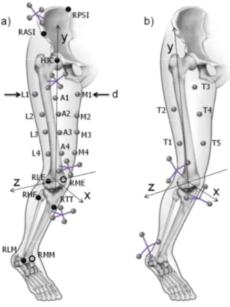
A bone pose estimator embedding an STA model that might compensate also for the rigid movement of the marker cluster remains to be assessed. Based on a quasi-linear relationship of the STA with the closest joint angles (Akbarshahi et al., 2010; Camomilla et al., 2013; Cappozzo et al., 1996), a model able to generate realistic thigh STA for both simulation and compensation purposes during multi-articular movements has been devised and assessed. Model feasibility was assessed using both skin and pin markers, in-vivo during running and from ex-vivo experimental data (Chapter 3).
In the perspective to embed an STA model in bone-pose estimator, the noticeable number of relevant parameters could cause difficulties in convergence during the optimization process, therefore this number has to be decreased. For this purpose, a modal approach (Dumas et al., 2014a) with different STA definitions (individual marker displacements; marker-cluster geometrical transformations; or skin envelope shape variations) was proposed and it was applied to the experimental data (Chapter 4). STA were represented using modes, composed by a direction and an amplitude, which can be selected or ranked according a chosen criterion. Some quantitative conclusions about that were drawn for each STA definition using information available in the literature and ex-vivo experimental data used also in the previous chapter of the thesis. In addition, the impact of different STA approximations, using the different STA definitions, was evaluated on the accuracy of knee joint kinematics estimates using running data.
Among the proposed STA definitions, the marker-cluster geometrical transformations, which addresses the rigid and non-rigid transformations of the cluster of skin markers, from the result reported in the Chapter 3, allows for the best trade-off between STA compensation effectiveness and number of modes, relative to knee kinematics accuracy and the number of parameters. Recent studies quantified these cluster STA components in different motor tasks (Andersen et al., 2012; Barré et al., 2013; de Rosario et al., 2012; Grimpampi et al.), showing that the cluster rigid motion (RM) is predominant with respect to the cluster non-rigid motion (NRM). The results obtained in these studies suggest that the cluster RM is the main component which affects the estimation of the bone pose, mostly for its amplitude with respect to its counterpart. Based on this observation, it is concluded, either explicitly or implicitly, that cluster non-rigid motion has a limited impact on bone pose estimation and that STA compensation should concentrate on the cluster rigid motion. In Chapter 5 it has been disputed the message carried by this statement and it has been demonstrated that the cluster non-rigid motion does not have a limited effect on BPE accuracy, but, rather, it has no effect whatsoever and that this is the case independently from its magnitude relative to the
4 cluster rigid motion. For this reason, the only STA component to be compensated for is the cluster rigid motion. Simulated data were used for the empirical demonstration.
Given the importance to estimate the bone pose with high accuracy, and therefore the joint kinematics, the impact of selected removal of modes representing the soft tissue artefact has been evaluated on the knee joint kinematics. In particular, it is evaluated the effect of removing a selected threshold of the STA or selected modes decided a priori (Chapter 6), as described in the previous thesis chapter.
The STA model based on the marker-cluster geometrical transformations (GT) definition has been developed for the propriety of the basis of vectors that can be defined a priori and for the numerical results shown in the thesis. Moreover, the impact of such model has been evaluated on the knee joint kinematics (Chapter 7).
The soft tissue artefacts have also an impact on the estimation of the hip joint centre (HJC) position during the star-arc movement, although an algorithm for its compensation, such as the quartic sphere fit method, proposed by Gamage and Lasenby (2002) with the correction term introduced by Halvorsen (2003), can be used. This method gave the best results compared with the symmetrical centre of rotation estimation method proposed by Ehrig et al. (2006), as shown in the study performed by Cereatti et al. (2009). Based on this results, HJC estimations were performed on ex-vivo data (Cereatti et al., 2009), selecting a distal and proximal skin marker cluster removing STA, modes by modes, using the skin envelope shape variations for the STA modal approach. In addition, the impact of STA on the HJC estimations has been evaluated modelling the thigh artefact, for its removal, as linear combination of the hip kinematics measured during the experiment (Appendix 1).
The study performed in the Chapter 7, and all the others presented here, lack of the information on the pelvis STAs, which are probably as important as the thigh STA, and their impact on hip kinematics and bone poses. To date, only one study assessed STA with reference to markers located on the pelvis. This was done by determining how the local position of anatomical landmarks (ALs) varied, as determined through manual palpation, while the hip assumed different flexion/extension angles, using a multiple anatomical calibration (Hara et al., 2014). A study has been presented using the same approach, but through a different anatomical calibration method which allowed for a better reliability (UP-CAST, Donati et al., 2008) and for different hip flexion/extension and ad-abduction angles (Appendix 2).
5 Scheme 1.1 – Structure of the thesis. Main limitations of some chapter are also shown.
6
2.
Chapter 2
“SOFT TISSUE ARTEFACT: STATE OF ART”
NomenclatureSTA Soft Tissue Artefact
RSA Roentgen Stereophotogrammetric Analysis HJC Hip Joint Centre
DOF Degree of Freedoms
AL Anatomical Landmark
CAST Calibration Anatomical System Technique
AF Anatomical Frame
CTF Cluster Technical Frames PCT Point Cluster Technique
PST Percutaneous Skeletal Trackers FHA Finite Helical Axis
CT Computed Tomography
RMS Root Mean Square
MRI Magnetic Resonance Imaging LSTAD Local STA Deformation RSTAM Rigid STA Movement BMI Body Mass Index
SVD Single Value Decomposition PSM Pliant Surface Modeling RBM Rigid Body Modelling GO Global optimization Jo in t d eg re es o f fr e ed o m FE Flexion/Extension AA Abduction/Adduction IER Internal/External Rotation
LM Medio/Lateral
AP Anterior/Posterior PD Proximal/Distal
2.1 Theoretical background
To analyze human movement in the three dimensional space, the instantaneous position and orientation of the different bones involved in the motor task have to be defined. Optoelectronic stereophotogrammetric systems and skin markers can be used for this aim.
The muscle-skeletal system reconstruction and the computation of its kinematics, using a model of the human body, exhibits a number of crucial problems. Firstly, there are issues concerning
7 instrumental errors, e.g. camera calibration and filtering or smoothing of marker position data (Chiari et al., 2005) along with the limitations introduced by the mechanical model used (as, for instance, the assumption of rigidity of the bones or the number of degrees of freedom of the joints involved in the analysis). Secondly, there are issues related to experimental errors. Actually, the muscle-skeletal system generates the most important errors in the reconstruction of the movement: the poor repeatability associated with the identification of the anatomical landmarks (Della Croce et al., 2005) and the relative movements between markers and the underlying bone (Soft Tissue Artefacts – STAs) (Andriacchi and Alexander, 2000; Leardini et al., 2005; Peters et al., 2010). The nature of this relative movement between the markers, glued on the external surface of the segment, and the underlying bone is associated with the specific marker set and experimental protocol adopted.
In recent years, applied researchers have become increasingly interested in the compensation of STAs, however the problem is still seeking for a satisfactory solution. During the execution of a motor task, the measurement of the motion of different body segments is mostly influenced by the real movement of the surrounding soft tissue leading to inaccurate estimates of the poses of the underlying bones (Leardini et al., 2005). This movement is generated by muscle contraction, skin stretching, and wobbling caused by the inertia of the limbs. Describe exactly the effect on the bone pose estimation is very difficult due to the complex nature of the human body. For example, depending on the location of the markers on the lower-limb and the analysed activity, a marker can move more than 30mm compared to its initial position (Cappozzo et al., 1996). This can affect the position and the orientation of the segments (thigh and shank) up to 31 mm and 15 deg, respectively (Sangeux et al., 2006). Then, propagated to the estimation of the tibio-femoral joint kinematics, the STA can introduce measurement error more than 18 mm and 8 deg on the estimated displacements and angles of the knee, respectively (Cappozzo et al., 1996). Moreover, movements of the body segments and the STAs have the same frequency content, so it is impossible to apply any filtering technique to eliminate this phenomenon. Then, a good knowledge and characterization of the STA is required to compensate it during different motor tasks. The measured STA depends on many factors such as the anatomical properties of the body segments and joints, the motor task performed. Hence, the acquisition protocol for the measurements is as critical as the parameters to be assessed. Their interpretation and the derived hypotheses are also dependent to the number of subjects analyzed. It is then interesting to know how the STA has been assessed, for which activity, number of subjects and what are the hypotheses formulated for the STA.
8 • the largest measurement error in in-vivo movement analysis;
• maximum in the thigh;
• both systematic and random error; • subject dependent;
• task dependent.
This chapter section addresses, first, at reviewing the studies aimed at assessing STA in the lower limb with different methods. Proposed techniques designed to minimize these effects are also reported. All the studies in the different sections are reported in a chronologic order.
2.2 STA measure
To directly measure the amplitude of soft tissue artefact on a segment of interest during experimental data acquisitions, the movement of the underling bone has to be acquired.
For this purpose the following methods can be used: bone pins, external fixator devices, skeletal trackers, or medical imaging.
9 2.2.1 Intracortical pins
The major limitation of this method is its invasiveness and the mechanical issues related to the pins (Figure 2.1). Some studies had concerns about possible bending effects on the pins (Ramsey and Wretenberg, 1999; Ramsey et al., 2003; Reinschmidt et al., 1997b) which can reduce the quality of the reference data: the pins may cause discomfort and the anaesthetics may alter the perception of the subject. To minimize the restriction of the movement of the skin marker, they should be glued not close to the bone pin insertion (Reinschmidt et al., 1997a). The worst case scenario with the use of the intracortical pins is when they break. Indeed, two studies reported the loss of data due to breakage or loosening of the pins (Benoit et al., 2006; Reinschmidt et al., 1997b). All these technical and ethical issues explain the limited number of subjects and the difficulty to recruit volunteers. Six is the maximum number of subjects analysed with intracortical pins in a study (Benoit et al., 2006).
Lafortune, 1984; Levens et al., 1948 were the pioneering using intracortical pins to analyse skeletal motion during walking. This method gave the opportunity to track directly the motion of bones with the insertion of pins equipped with reflective markers. After a local anaesthesia around the insertion site, a surgery is required to cut the skin, pass the pin around the muscles, tendons, or ligaments and then screw the pin in the bone. The advantage of this method is the employment of the same acquisition system as used for reflective markers attached on the skin. In addition, the same acquisition volume can be acquired for the reference and the skin marker system. This gives for example the possibility to analyse the STA on a full gait cycle.
The use of X-ray video-fluoroscopy and intracortical pins to quantify STA magnitude was described by Lafortune and Lake, in 1991. In this preliminary experiment using fluoroscopy, three cycles of unloaded flexion-extension of the knee were analysed. A 21 mm distal and a 23 mm posterior displacement was exhibited by a marker placed on the proximal tibia, and this displacement was found to be linearly related to knee flexion. In a second experiment, the STA magnitude was analysed at heel strike during running. Data were obtained from a marker attached to a cortical pin inserted into the tibia and from a marker glued on the skin of a volunteer over the lateral tibial condyle. The relative movement between these two markers reached 10 mm, and was Figure 2.1 – Schematic of a target cluster used by
10 dependent also upon the type of impact. The same authors, in another study (Lafortune et al., 1992) showed the tibio-femoral 3D kinematics during walking using target clusters fixed directly into the bones, without providing information on STA.
External marker devices, each consisting of a bone screw and an aluminium tripod instrumented with three reflective spherical markers, were anchored on the distal femur and on the proximal tibia (Karlsson and Lundberg, 1994). In addition, three skin markers were glued on the distal thigh and on the proximal shank. While standing, the two volunteers performed a hip internal-external rotation with extended knee. The knee joint rotations obtained with bone-anchored and skin-attached markers showed a great difference: the knee internal-external rotation when measured with the cluster of markers linked with the bone showed a range of about 20 degrees, which was observed to be about 50 degrees when measured with the skin marker cluster. Moreover, the skin displacement tracked by thigh markers was found to be higher than that by shank markers.
The impact of the STA duringthe stance phase of a level walking task on both knee and ankle was assessed by (Reinschmidt et al., 1997a). Intracortical Hofmann pins with triads of reflective markers were inserted into the lateral femoral condyle, the lateral tibial condyle and the postero-lateral aspect of the calcaneus (Figure 2.2a) in five male volunteers (age: 28.6±4.3 years, mass: 83.4±10.2 kg, height: 185.1±4.5 cm). The task was performed by each subject three times. Six skin markers were also glued on the lateral and anterior aspects of the thigh, shank and shoe. The knee and ankle joint kinematics were described using standard conventions (Cole et al., 1993; Grood and Suntay, 1983). Only the data from three subjects were valid for the measurement of the knee joint kinematics due to issues with the pins for the two other subjects. The maximal difference between bone- and skin-marker based knee rotations were 4.4 deg, 8.4 deg and 4.3 deg (obtained as the mean value over all the volunteers), in the frontal, transverse and sagittal plane, respectively. In the same anatomical planes the maximal difference between bone- and skin-marker ankle rotations were 5.4 deg, 5.1 deg and 5.9 deg. It was shown that the thigh is the most affected segment by the STA due to muscle movements during the stance phase and may be even higher during the swing phase, through segmental error analysis (the average difference between the bone- and skin marker-based motion). It was concluded that only the knee flexion/extension (FE) can be measured using skin markers, seeing as the error introduced by the STA can almost be as high in magnitude as the real joint motion for the knee abduction/adduction (AA) and internal/external rotations (IER).
11 Figure 2.2 – Experimental setup with intracortical pins implanted in lower limbs. a) Reinschmidt et al., 1997c; b)Ramsey et al., 2003; c) Houck et al., 2004; d) Benoit et al., 2006; e) Camomilla et al., 2013.
The effect of STA on 3D joint rotations was assessed also in the stance phase of five running trials by the same authors (Reinschmidt et al., 1997c). The same acquisition protocol was used for 3 subjects (age: 25.7±2.1 years; mass: 85.5±9.6 kg; height: 1.87±0.10 m). As in the previous work, the knee kinematics was defined using the Cardan angles calculated from both the external and skeletal markers. For the knee FE again a good agreement was found between skin- and bone-based knee patterns, while for the other two angular degrees of freedom (i.e., AA and IER), the difference between the two kinematics had similar amplitude with respect to the amplitude of the corresponding physiological motion. Such errors, express in percentage of the relative full range of motion were 21%, 70%, and 64%, for FE, AA and IER, respectively. Therefore, it was shown that skin markers lead to an overestimation of joint motion. When the impact of the STA was evaluated on the single segment, the error analysis for the shank did not exceed 5 degrees for all subjects and all rotations; while for the thigh the errors were consistently values higher. Not surprising the errors due to the relative movement between skin markers and the underlying bone were higher in running than in walking, due to muscle contraction that occurs during the running stance. Moreover, the
12 authors also concluded the possibility that the skin set may can move as a unit relative single unit relative to the underlying bone. Thus, a compensation method reducing only the relative movement between markers may be not enough to reduce the impact of STA on the pone pose estimation, and, therefore, joint kinematics.
Several different motor tasks were analysed using an instrumented leg with two arrays of six markers inserted directly into the tibial tubercle and the greater trochanter (Fuller et al., 1997). Twenty markers were also glued all over the thigh and shank segments. In this study two experiments were performed. One volunteer (age between 35 and 40 years; mass 91 kg; height 1.905 m) walked two times: one time only with skin markers glued on the segments and another time with also intracortical pin mounted markers. This was performed to determine the frequency of the transient oscillation, which occurs at heel strike: it was suggested that STA introduces high frequency artefact. On another volunteer (age: between 35 and 40 years; mass: 104 kg; height: 1.88 m) it was assessed the effect of the STA on the computation of the instantaneous knee helical axis during different activities: cycling, squatting, normal gait, and voluntary swing movement. It was shown that the displacement with respect to the underlying bone of the skin markers could exhibit values up to 20 mm. Moreover, STA was found to be task-dependent, showing different patterns among the tasks analysed. It was also shown that the power spectra for skin- and pin-markers cover similar frequency bands, indeed there was not a distinct soft tissue noise transient. Therefore no evidence of a distinct frequency domain for the STA: attempt to remove STA through traditional filtering techniques can result in loss of information or in introduction of spurious motion patterns. It was concluded that the skin marker trajectories are not appropriate for representing motion of the underlying bones, particularly of the femur.
In 1998, Ball and Pierrynowski modelled the STA as a time varying affine transformation of 12 degrees of freedom (three orientations, three position, three shears, three scales) between the technical frame and the anatomical frame based on the trajectories of at least 4 markers glued on a segment, moreover, 20 and 16 skin markers were glued on the surface of the thigh and the shank, respectively. The validation of such modelling was realized using intracortical pins inserted in the femur and the tibia. Three volunteers (age: 37±3 years; height: 1.81±0.07 m; mass: 82.7±4.5 kg) walked for 20 seconds on a treadmill at three velocities: slow (0.66ms-1), medium (1.10ms-1), and fast (1.54m s-1), collecting from 12 to 20 strides of gait. Bone poses and knee joint kinematics were analysed comparing the intracortical pins measurements, the traditional method using a fixed rigid transformation and the proposed method that will be described in details in the section 2.4.3. However, no numerical measurement about STA were shown in this study.
13 A new femoral tracking device was proposed by Houck et al., 2004 to measure the relative motion between the skin and the femur, and therefore, to improve the measure of the knee joint kinematics during gait trials. This device is a U shaped frame of aluminium and the femoral epicondyles were clamped with two pads. Intracortical pins were inserted into the proximal-lateral aspect of the right femur and tibia, equipped with four infrared light emitting diodes to validate this device. Three skin markers were glued along the crest of the tibia. The femoral tracking device was clamped over the femoral condyles and on its lateral extension two diodes were attached. A third diode was placed on the skin, distal with respect to the greater trochanter. Two subjects were involved in this study but only the results of one subject (age: 35 years; height: 1.73 m; mass: 80 kg) were compared in terms of knee joint kinematics obtained by the skin markers, the new device and the intracortical pins (Figure 2.2c). A ‘reasonable validity’ was claimed for the device by the authors over 85% of the stance phase of gait. Rotation error values were of few degrees, but during terminal stance and during the swing phase (i.e., when maximum knee flexion occurs) these errors increased substantially. In this study, they found absolute differences of up to 2.2 deg in the sagittal plane, 2.7 deg in the frontal plane and 1.8 deg in the transversal plane, while up to 13.9 mm of linear displacements was observed during walking.
The difference in ankle complex motion during the stance phase of walking was measured in three volunteers using skin- and bone-anchored markers in Westblad et al., 2000. Three skin markers were glued laterally on each shank, heel, and forefoot and their trajectories were acquired during a barefoot walking trial. Hoffman pins, equipped with four markers (i.e., bone-anchored markers) were inserted into the tibia, fibula, talus and calcaneus. The mean maximal differences between the skin- and bone- based joint rotations were smaller than 5 degrees. The smallest absolute difference was found for plantar/dorsiflexion. This finding was in contrast to a previous report (Reinschmidt et al., 1997a) where knee abduction/adduction showed the lowest error magnitude. This difference may be explained by the fact that subjects assessed in the latter study wore shoes.
STA was also quantified during the stance phase of gait and during cutting motion by Benoit et al., 2006 (Figure 2.2d). Eight volunteers (age: 26 years; height: 1.78 m; mass: 78.1 kg) were recruited in this study but only six of them were analysed (age: 26±4.7 years; height: 1.77±0.04 m; mass: 76.3±12.3 kg; BMI: 24.4±3.8). The information acquired from the intracortical pins were combined with a Roentgen stereophotogrammetric analysis (RSA) to remove the error due to the misplacement of the anatomical landmarks (Della Croce et al., 2005). During the foot-strike, mid-stance and toe-off of all the motor tasks performed by the volunteers the STAs were quantified in term of absolute error values in the knee kinematics, and to measure differences between both
14 systems, i.e., marker- and pin-based. The impact of the STA on the joint kinematics was between 2.4±2.2 deg to 4.4±3.2 deg for the knee angles and between 3.3±2.4 mm to 13.0±5.0 mm for the knee displacements, during the stance phase of the gait. In particular, during the toe-off, only the anterior/posterior displacement showed a high error, otherwise, all the other errors were between 3.3±2.4 mm and 8.0±5.7 mm, even for proximal/distal displacement. During the second motor task performed by the volunteers (cutting motion) the impact of the STA on knee kinematics was more marked: the absolute difference between the two knee kinematics were from 3.3±1.8 deg to 13.1±9.8 deg (the latter was measured for AA during toe-off) for the knee angles, while for the knee displacements these errors were from 5.6±5.1 mm to 16.1±8.9 mm. Moreover, the direction of the skin movement artefact was not repeatable across subjects as observed in other studies (Houck et al., 2004; Manal et al., 2000; Reinschmidt et al., 1997c).
The impact of the STA on the determination of the hip joint centre (HJC) obtained with a functional calibration using a star-arc-movement (flexion/extension-abduction/adduction of the hip in different planes followed by a half circumduction) was analysed by Cereatti et al., 2009. Intracortical pins, equipped with four reflective markers, were inserted in the pelvis and the femur, eight markers were glued on the thigh. In this study, four fresh cadaveric specimens were used. The calibration movement was repeated three times for each specimen. The HJC estimations were performed with two different approaches (constrained three-degrees of freedom or an unconstrained six-degrees of freedom: DOF) and compared. The maximum error was the same for both algorithms for the subject with largest thigh circumferences. The relative motion between the skin markers and the underlying bone was between 1.0 mm and 10.6 mm. The proximal markers, which were closer to the hip joint, were significantly (p < 0.05) more influenced by the STA. For this reason, HJC estimation errors computed using the distal marker clusters were significantly lower (p < 0.001).
Andersen et al., in 2012, analysed the components of skin markers: rigid-body (translation and rotation) and deformation components. The aim of the study was to determine which component influenced more the pose estimation of the femur and tibia, using the experimental data described in Benoit et al., 2006. Three tasks were analysed: walking, cutting, and hopping. A linear model of the STA was proposed using the principal component analysis to describe the twelve degrees of freedom of the four markers glued on the segment. The results shown that the motion of the skin marker cluster relative to the underlying bone was dominated by rigid-body motions rather than deformations.Therefore, they concluded that all the STA compensation techniques focused on the marker cluster deformation will not be effective at removing the rigid body movement.
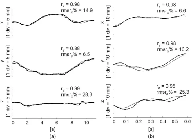
15 Recently, a model that provides the thigh STAs as a function of hip joint kinematics was identified, calibrated, and validated (Camomilla et al., 2013). This model estimates the STA which occurs on thigh markers in the anatomical reference frame, during a mono-articular movement (star-arc-movement). The proposed model was calibrated and assessed using experimental data obtained in a previous ex-vivo study (Cereatti et al., 2009): four intact fresh adult cadaver subjects positioned supine with steel pins, equipped with four-marker clusters, implanted into tibia, femur and hip-bones. In addition, 12 markers were glued on the thigh (Figure 2.2e). The model was calibrated and tested on the mono-articular movement performed for three times by an operator. The model calibration was performed for each marker using a global optimization based on the root mean square of the displacement difference between the measured skin marker and the estimated one. Different calibrations were realized to evaluate the effect of the trial-specific, subject-specific, and hip joint movement (HJM) on the estimation of the STA. The accuracy of subject-specific model estimates and the HJM independency, suggested the validity and generalizability of the model for a given subject and marker location. Besides, the large inter-subject variability of the model parameters confirmed the fact that STA is subject-dependent. The median root mean square distance values slightly increased when moving from trial- to subject-specific estimates: from 0.8 mm to 0.9 mm. Regarding HJM-dependency, these median value for all subjects was 1.0 mm. The results obtained were also different among the analysed subjects (i.e., the third subject showed moderate correlations with respect to the others in all the calibration procedures performed).
2.2.2 External fixators
To treat bone fracture, external fixators (Figure 2.3) can be used. These devices are rigidly associated with the underlying bone, giving the opportunity to directly access to bone kinematics. In some studies reflective markers were located on this device, and the same acquisition system as for the skin markers was used. However, this device was typically located on only one segment (femur or tibia) for the rarity to have fractures in both bones. Therefore, the STA of only one segment was measured. The maximum number of subjects analysed using this method was seven (Cappozzo et al., 1996). When using external fixators, as occurs for the pin insertion, the STA assessment is limited by skin sliding restrictions (Leardini et al., 2005).
16 The first study performed to analyse STA with patients wearing external devices for fracture fixation at either the femur or the tibia was done by Angeloni et al., 1992. These devices are rigidly associated with the underlying bone: a set of axes can be defined (Figure 2.4a). Markers were glued on the skin over the following four anatomical landmarks (ALs): greater trochanter, lateral epicondyle, head of the fibula, lateral malleolus. Using large elastic bands and Velcro fasteners, markers were placed on rigid plates in the proximal half of the thigh and the shank. The range of displacement of these plate-mounted markers with respect to underlying bones during walking was lower than that of the markers located on the ALs. Similar results were obtained using a semi-quantitative video-fluoroscopic analysis.
The same authors showed more detailed results using the same methods in Cappozzo et al., 1996. The STA effects were investigated on seven subjects (age: 23.3±5.7 years; height: 1.67±0.16 m; mass: 66.9±14.4 kg) while performing different motor tasks: level walking at a natural speed, cycling on an exercise bike, flexion of the lower limb while standing, repetitive isometric muscular contraction and hip external rotation while standing with the knee in full extension. Each task was repeated at least for four times. The CAST (calibration anatomical system technique) method was used to define the anatomical frames (AFs), associated with skin- and fixator- marker cluster technical frames (CTF). A stereophotogrammetric system was used to track the motion of the markers on the external fixator and the skin markers, located on the ALs, as described in the previous work (Figure 2.4a). Markers on the fixator were assumed to provide instantaneous positions and orientations (i.e., pose) of the corresponding rigidly associated bone. Other skin markers were glued on the body segment, as compatible with the presence of the fixator and camera visibility. The marker displacement with respect to the underlying bone showed remarkable magnitudes (up to 40 mm), as much as an order of magnitude larger than stereophotogrammetric errors. During the level walking task, the marker displacement due to STA was in the range of 10-30mm. The results were detailed for each subject and not averaged. However, it was clearly presented that in general, the STA associated with the markers on thigh and shank ALs showed magnitude that varies approximately linearly with respect to the joint flexion angle, irrespective of Figure 2.3 – Example of an external fixator used to
17 the motor task performed. Bone orientation affected by STA caused an error between 6 deg and 20 deg for the femur, and between 4 deg and 10 deg for the tibia. Moreover, during hip external rotation, the error in femur orientation caused by STA reached magnitudes from 6 deg to 28 deg. Therefore, estimating the knee joint kinematics using skin marker data, FE, AA and IER might be affected with inaccuracy that can be respectively as large as 10%, 20%, and 100% of the relevant expected range of motion.
Figure 2.4 – External fracture devices used in: a) Cappozzo et al., 1996; b) Cappello et al., 1997; c) Alexander and Andriacchi, 2001; d) Ryu et al., 2009.
To reduce the impact of STA on the bone pose estimation, Cappello et al., 1997 proposed a multiple anatomical landmark calibration protocol repeating such calibration in different postures. For its validation, a cycling test on a patient wearing a femoral external fixator was performed (Figure 2.4b). This technique will be described in section 2.4.2. Eight skin markers were glued on the thigh. A subset of three or more of them were used to define a CTF. In addition, four skin markers were located on the external fixator. With a CAST experimental protocol (Cappozzo et al., 1995), using a pointer, the coordinates of the femur ALs were defined. The amplitude of the STA was between 3.9 mm and 9.4 mm. Estimating the femur AF with the CAST protocol for two static postures analysed (i.e., maximal hip and knee flexion and extension) the error in the femur pose estimation (position
18 and orientation) compared with the reference obtained with the external fixator was 5.00-5.10 deg, for the orientation components, while for the position vector the error was 6.9-7.0 mm. When the double calibration procedure was used, this error decreased to 3.50 deg for the orientation and 4.4 mm for the position.
The validation of the compensation method for the STA proposed by Alexander and Andriacchi, 2001, improving the point cluster technique (PCT) (Andriacchi et al., 1998), was realized on one subject (age: 46 years; height: 1.75 m; mass: 84.1 kg). The method will be discussed in the section 1.2.5. The point cluster marker set (six markers) was glued on the shank of the subject, four markers were rigidly attached to the Ilizarov device (an external fixator), which was rigidly connected to the tibia (Figure 2.4c). The subject, which exhibited a limited range of motion, performed a 10 cm step-up onto a platform. The proposed method reduced the impact of the STA on the pose of the shank from 0.25 mm to 0.08 mm and from 0.370 deg to 0.083 deg, values obtained as average location error. However, higher errors were obtained with this method in another study (Stagni et al., 2003) tested on two subjects (age: 67 and 64 years, height: 1.55 and 1.64 m, mass: 58 and 60 kg) during a step up/down test repeated three times.
To reduce the error caused by STA on the AL positions, Ryu et al., 2009 proposed a compensation method and used it on one subject (age: 42 year.; height: 1.63 m; mass: 56 kg; BMI: 21.1) with an external fixator in the tibia for its validation (Figure 2.4d). A linear relationships between the displacement of ALs and skin markers expressed in TFs, was proposed to be used. The STA during an active knee flexion/extension had an amplitude of 4.0 mm-18.3 mm, affecting the tibia pose with errors around 3 deg-4 deg and 20 mm-65 mm on each axis of the reference frame. With the proposed compensation methods, the effect of the STA on the tibia pose was reduced of 39-83%, with error around 0.6 deg-2.4 deg and 11 mm-31 mm on each axis of the reference frame.
2.2.3 Percutaneous trackers
Percutaneous skeletal trackers (PST) were metal devices designed specifically for the fixation to the skeleton using a number of halo pins inserted into the periosteum on opposite sides, instead into the bone, as occurs using intracortical pins (Holden et al., 1997). Nevertheless, using this device, a local anaesthesia is required. The issue about the limitation of the skin displacement around the area of the insertion when this device is used to measure the STA arises (Leardini et al., 2005). Seven was the maximum number of subjects acquired with this instrument (Manal et al., 2000).
Three healthy male volunteers (age: 28, 36, and 35 year; height: 1.80, 1.91, and 1.77 m; mass: 95.0, 91.6, and 69.2 kg; BMI: 29.4, 25.24, and 22.2) participated in the study performed by Holden et al.,
19 1997. The PST was fixed in the distal part of the tibia and fibula and it was instrumented with reflective markers (Figure 2.4a). Additional markers were attached to shells mounted on the lateral surface of the mid-shank and on the dorsal aspect of the mid-foot, instead of gluing the reflective markers directly on the skin. The volunteers walked at self-selected speed for six times along a corridor, including a platform. The AF associated with the bone was defined with a static anatomical calibration with both skeletal- and surface-based markers. The impact of the STA was calculated defining the relative 3D difference between the skeletal- and the surface-based AFs. In the volunteers analysed, a peak of the rotation error had a mean magnitude of 4 deg at the 8% of the gait cycle. Additional error rotation occurred during the terminal stance and during most of the swing phase with a magnitude 8 deg in one volunteer. Maximum absolute displacements of the skin-based AF, with respect to the reference AF obtained using the skeletal data, were less than 6.0 mm in the transverse plane but reached 10.5 mm longitudinally. The displacement between skin markers and the underling bone was reproducible within subjects, but poor among subjects. Inverse dynamics were also analysed in the three volunteers during the stance phase. The impact of STA on knee moments was considered relatively small, seeing as the largest difference between two estimations was only 9 Nm. Moreover, the greatest errors occur in similar phase of the gait cycle, certainly due to the muscle activation.
20 Figure 2.5 – Percutaneous skeletal trackers used in: a) Holden et al., 1997; b) Manal et al., 2000; c) Manal et al., 2002.
In another study aimed at defining an optimal marker set to track the movement of the tibia, also further information were provided about the STA (Manal et al., 2000). Seven subjects (age: 25.6±1.9 years) performed several walking trials with eleven different configurations of markers glued on the shank, obtained by combining geometry, location (proximal/distal) and attachment (underwrap/overwrap) factors for the array of markers (Figure 2.4b). As reference, a tracker was clamped to the two malleoli and AFs were defined for the foot, shank and thigh using markers located over ALs. The differences between the PST and the marker sets were quantified using the root mean square deviation of the relative helical angle based on the three most similar trials of each subject and for each marker set. When the marker arrays over the lateral shank were placed more distal than proximal, better estimates of tibial rotation were noted. No significant difference where measured between overwrapped or underwrapped surface mounted markers, but as the trend was in favour of the underwrapped attachments. The best performance was observed for an underwrapped rigid shell with four markers located distally, as much as possible. However, even when using the

![Table 3.3 – Ex-vivo experimental data. Subject- and trial- specific model parameters ℎ , , , [mm/deg], c = x, y, z (anatomical axes: Fig](https://thumb-eu.123doks.com/thumbv2/123dokorg/8152589.126474/76.892.115.789.113.763/table-experimental-subject-trial-specific-model-parameters-anatomical.webp)
![Table 3.4 – Ex-vivo experimental data. Subject- and trial- specific model parameters , , , [mm/deg], c = x, y, z (anatomical axes: Fig](https://thumb-eu.123doks.com/thumbv2/123dokorg/8152589.126474/77.892.109.788.130.778/table-experimental-subject-trial-specific-model-parameters-anatomical.webp)