279:H2641-H2648, 2000. Am J Physiol Heart Circ Physiol
Antonio L'Abbate
Marzullo, Roberto Testa, Isabella Raugei, Micaela Papini, Mathis Schluter and
Gianmario Sambuceti, Mario Marzilli, Andrea Mari, Cecilia Marini, Paolo
control interaction
downstream from severe stenosis: pressure-flow
Clinical evidence for myocardial derecruitment
You might find this additional info useful...
40 articles, 25 of which can be accessed free at: This article cites
http://ajpheart.physiology.org/content/279/6/H2641.full.html#ref-list-1
5 other HighWire hosted articles This article has been cited by
[PDF] [Full Text]
, December 1, 2000; 279 (6): H2585-H2586. Am J Physiol Heart Circ Physiol
William M. Chilian and David D. Gutterman
Prologue: new insights into the regulation of the coronary microcirculation
[PDF] [Full Text] [Abstract]
, May 15, 2001; 103 (19): 2352-2360. Circulation
Gianmario Sambuceti, Mario Marzilli, Silvio Fedele, Cecilia Marini and Antonio L’Abbate a Severe Stenosis in Patients With Coronary Artery Disease : Reversal by Angioplasty Paradoxical Increase in Microvascular Resistance During Tachycardia Downstream From
[PDF] [Full Text] [Abstract]
, January 4, 2005; 111 (1): 76-82. Circulation
Robbert J. de Winter, Karel T. Koch, Jan G.P. Tijssen, Jos A.E. Spaan and Jan J. Piek Bart-Jan Verhoeff, Maria Siebes, Martijn Meuwissen, Bektas Atasever, Michiel Voskuil, Index
Influence of Percutaneous Coronary Intervention on Coronary Microvascular Resistance
[PDF] [Full Text] [Abstract]
, May , 2005; 288 (5): H2298-H2305. Am J Physiol Heart Circ Physiol
L'Abbate
Testa, Michaela Papini, Paolo Marraccini, Giuseppe Ciriello, Paolo Marzullo and Antonio Gianmario Sambuceti, Mario Marzilli, Andrea Mari, Cecilia Marini, Mathis Schluter, Roberto ischemic myocardium
Coronary microcirculatory vasoconstriction is heterogeneously distributed in acutely
[PDF] [Full Text] [Abstract]
2006; 291 (4): H1814-H1821. Am J Physiol Heart Circ Physiol
Gianmario Sambuceti
Claudia Kusmic, Guido Lazzerini, Flavio Coceani, Renata Barsacchi, Antonio L'Abbate and for nitric oxide and endothelin
Paradoxical coronary microcirculatory constriction during ischemia: a synergic function
including high resolution figures, can be found at: Updated information and services
http://ajpheart.physiology.org/content/279/6/H2641.full.html
can be found at: AJP - Heart and Circulatory Physiology
about Additional material and information
http://www.the-aps.org/publications/ajpheart
This information is current as of May 5, 2012.
ISSN: 0363-6135, ESSN: 1522-1539. Visit our website at http://www.the-aps.org/.
Physiological Society, 9650 Rockville Pike, Bethesda MD 20814-3991. Copyright © 2000 by the American Physiological Society. intact animal to the cellular, subcellular, and molecular levels. It is published 12 times a year (monthly) by the American
lymphatics, including experimental and theoretical studies of cardiovascular function at all levels of organization ranging from the publishes original investigations on the physiology of the heart, blood vessels, and AJP - Heart and Circulatory Physiology
on May 5, 2012
ajpheart.physiology.org
Clinical evidence for myocardial derecruitment downstream
from severe stenosis: pressure-flow control interaction
GIANMARIO SAMBUCETI,1MARIO MARZILLI,1ANDREA MARI,2CECILIA MARINI,1
PAOLO MARZULLO,1ROBERTO TESTA,1 ISABELLA RAUGEI,1MICAELA PAPINI,1
MATHIS SCHLUTER,1AND ANTONIO L’ABBATE1
1Consiglio Nazionale delle Ricerche Institute of Clinical Physiology, Pisa 56100;
and2Institute of Systems Science and Biomedical Engineering, Padua 35100, Italy
Received 6 June 2000; accepted in final form 23 August 2000
Sambuceti, Gianmario, Mario Marzilli, Andrea Mari, Cecilia Marini, Paolo Marzullo, Roberto Testa, Isa-bella Raugei, Micaela Papini, Mathis Schluter, and Antonio L’Abbate. Clinical evidence for myocardial
dere-cruitment downstream from severe stenosis: pressure-flow control interaction. Am J Physiol Heart Circ Physiol 279: H2641–H2648, 2000.—To verify the interaction between cor-onary pressure (CP) and blood flow (CBF) control, we studied nine candidates for angioplasty of an isolated lesion of the left anterior descending coronary artery [i.e., percutaneous transluminal coronary angioplasty (PTCA)]. CBF (i.e., flow velocity⫻ coronary cross-sectional area at the Doppler tip) and CP were monitored during washout of 2–5 mCi of133Xe
after bolus injection into the left main artery before and after PTCA. Xe mean transit time (MTT) was calculated as the area under the time-activity curve, acquired by a gamma camera, divided by the dose obtained from a model fit of the Xe curve in the anterior wall. CBF response to intracoronary adenosine (2 mg) was also assessed. PTCA increased baseline CBF (from 14.5⫾ 9.4 to 20 ⫾ 8 ml/min, P ⬍ 0.01), coronary flow reserve (from 1.52⫾ 0.24 to 2.33 ⫾ 0.8, P ⬍ 0.01), and CP (from 64 ⫾ 9 to 100 ⫾ 10 mmHg, P ⬍ 0.05). MTT decreased from 89⫾ 32 to 70 ⫾ 19 s (P ⬍ 0.05) after PTCA; however, MTT and CBF changes were not correlated (r ⫽ ⫺0.09, not significant). Inasmuch as MTT is the ratio of distribution volume to CBF, MTT ⫻ CBF was used as an index of perfused myocardial volume. Volume increased after PTCA from 23⫾ 18 to 56 ⫾ 30 ml. A direct correlation was observed between the percent increase in distal CP and percent increase in perfused volume (r ⫽ 0.91, P ⬍ 0.01). Thus low CP was not associated with exhaustion of flow reserve but, rather, with reduction of perfused myocardial volume. These data suggest that, in the presence of a severe coronary stenosis, derecruitment of vascular units occurs that is proportional to the decrease in driving pressure. Residual perfused units maintain a vasomotor tone, thus explaining the paradoxical persistence of coronary reserve. coronary circulation; microcirculation; coronary angioplasty; autoregulation; coronary artery disease
MYOCARDIAL METABOLISM IS STRICTLY aerobic; because of the high oxygen extraction under baseline conditions, increases in oxygen consumption can only be met by
corresponding increases in coronary blood flow (6, 37). According to this concept, it is generally assumed that myocardial metabolism is the primary factor control-ling microvascular tone and that ischemia implies a maximal vasodilation of coronary microvasculature or exhaustion of coronary flow reserve. However, several experimental studies reported the persistence of a sig-nificant vasodilator reserve in moderately ischemic myocardium (2, 7, 8, 32) and, even more, the occur-rence of a microvascular constriction during acute se-vere ischemia (17, 20). The mechanisms underlying these phenomena have not been conclusively identi-fied, although they have been attributed to anesthesia (9), an active downregulation of myocardial metabo-lism below the actual flow availability (7), or a primary microvascular constriction (10). Although this behavior seems paradoxical when the autoregulation of coro-nary circulation and myogenic reflex are considered, it agrees with observations obtained in peripheral and coronary microcirculation during hemorrhagic shock (21, 31) and suggests that further mechanisms, besides
oxygen demand, affect vasomotor tone in
is-chemic myocardium.
The most frequent cause of resting hypoperfusion in humans is the presence of a coronary stenosis. In this setting, a direct correlation exists between coronary blood flow and transstenotic pressure gradient; thus, since resting aortic pressure can be considered rela-tively constant, an inverse correlation exists between coronary blood flow and distal coronary pressure (18, 19). Several data indicate that the vascular tree, and in particular the microvascular network, is controlled to maintain an adequate pressure at the capillary level to provide correct exchanges between blood and intersti-tium (42). In the presence of a very severe stenosis, the flow demand might imply an excessive pressure gradi-ent and, thus, an excessively low coronary pressure. Such a vasoconstrictor response to reduced pressure might represent a mechanism able to maintain a cor-rect pressure in the perfused vascular units, provided
Address for reprint requests and other correspondence: G. Sam-buceti, CNR Institute of Clinical Physiology, Via P. Savi 8, 56100 Pisa, Italy (E-mail: [email protected]).
The costs of publication of this article were defrayed in part by the payment of page charges. The article must therefore be hereby marked ‘‘advertisement’’ in accordance with 18 U.S.C. Section 1734 solely to indicate this fact.
0363-6135/00 $5.00 Copyright©2000 the American Physiological Society
http://www.ajpheart.org H2641
on May 5, 2012
ajpheart.physiology.org
that it might act to exclude myocardial units as paral-lel resistors from perfusion.
The recent introduction of manometer-tipped wires, together with Doppler monitoring of blood flow, allows the measurement of microvascular coronary resistance in patients with coronary artery disease. When coupled with the analysis of the washout of a freely diffusible tracer and applied to the clinical model of coronary angioplasty, this technology permits the study of coro-nary blood flow and perfused myocardial volume at different coronary pressures under stable systemic he-modynamics. This possibility is crucial, since the sta-bility of aortic pressure minimizes the triggering of reflexes that might directly affect coronary vasomotor tone. We used this experimental setting to estimate the interrelationship between coronary pressure, blood flow, and myocardial volume.
MATERIALS AND METHODS
Study population. Nine candidates for coronary angio-plasty (mean age 61⫾ 3 yr) were included according to the following criteria: 1) history of stable angina, 2) electrocar-diographic (ECG) evidence of exercise-induced ischemia, 3) single-vessel disease of the left anterior descending coronary artery, 4) no previous myocardial infarction, 5) left main stem longer than 2 cm, 6) no angiographic evidence of collat-eral circulation, 7) absence of arterial hypertension and/or left ventricular hypertrophy, and 8) no diabetes.
Study protocol. Patients were studied after overnight fast, under active treatment with oral diltiazem (60 mg 3 times/ day), isosorbide mononitrate (20 mg 3 times/day), and aspi-rin. An 8.0-F guiding catheter was advanced into the left main coronary artery, a control angiography was performed, and the best projection was selected. Heparin (10,000 IU) was injected intravenously; isosorbide dinitrate (0.6 mg) was administered into the heart. A 0.014-in. fiber-optic pressure-monitoring guide wire (Radi Medical, Uppsala, Sweden) was calibrated and positioned distal to the stenosis (34). Finally, a 2.5-F Doppler-tipped catheter (Millar Instruments, Hous-ton, TX) was placed in the prestenotic segment, and a coro-nary angiography was again obtained to measure cross-sec-tional area at the tip of the Doppler catheter. Care was taken to avoid side branching between the catheter tip and the stenosis and to maintain the catheter in the center of the lumen to obtain a stable flow-velocity signal.
A small field-of-view mobile gamma camera (model F1, Elscint, Haifa, Israel) was brought to the catheterization room. The system was equipped with a low-energy, high-sensitivity parallel-hole collimator and was oriented on the patient’s chest in a 70° left anterior oblique projection.
Stable blood flow and hemodynamics were verified forⱖ5 min before baseline recordings. Thereafter, a bolus of 2–4 mCi of133Xe was rapidly injected through the guiding
cath-eter soon after initiation of a dynamic acquisition set accord-ing to the followaccord-ing parameters: energy window centered on the photo peak of133Xe and one frame every 2 s for 180 s. At
the end of acquisition, a bolus of adenosine (2 mg) was selectively injected into the left anterior descending coronary artery through the Doppler catheter.
The following signals were continuously monitored: 1) four ECG leads (I, II, III, and V4), 2) phasic and mean aortic pressure, 3) phasic and mean distal coronary pressure, and 4) phasic and mean coronary blood flow velocity. Paper record-ings (2.5 cm/s) were obtained at baseline and 30 s after
intracoronary adenosine. Distal coronary pressure was also measured during balloon coronary occlusion.
After completion of this protocol, the Doppler catheter was removed, and all patients underwent coronary angioplasty. After revascularization, the Doppler catheter was advanced in the treated vessel, and the protocol was repeated as before angioplasty.
Data analysis. Stenosis severity (percent lumen area re-duction) and vessel diameter at the tip of the Doppler cath-eter were measured using an automated edge detection sys-tem (Mipron, Kontron). Coronary blood flow index was obtained by multiplying mean blood flow velocity by cross-sectional area at the site of the Doppler transducer, as pre-viously described (16).
Mean transit time of Xe was calculated according to the residue detection method. Original images were grouped into 20-s frames to allow a better definition of the left ventricular walls. Two regions of interest were drawn on the anterior and posterolateral wall, and frames were displayed in cine mode to verify a stable geometry. Thereafter, the computer was asked to plot the time-activity curves in stenotic-anterior and control-posterolateral regions using the original 2-s frames.
The mean transit time was calculated, according to the stochastic analysis, from the tracer residue curves as the ratio of the area under the curve from zero to infinity to the dose entering the region (44). These two values were esti-mated using the model described below.
The tracer mass balance in the region of interest is q⫽ fin⫺ fout (1)
where q is the tracer mass in the region, which is propor-tional to the Xe radioactivity, finis influx, and foutis efflux.
For the influx, the following representation was used fin共t兲 ⫽ D⌫共兲共/兲t⫺1e⫺共/兲t (2)
where D is the tracer dose entering the region (the integral from zero to infinity of fin),⌫ is the gamma function, is the
mean transit time of fin, which is related to the delay of the
tracer appearance into the region, and quantifies the dis-persion of the dose around its mean transit time. Equation 2, with only three parameters, ensures a flexible description of the input function, which starts from zero, rapidly reaches a peak, and returns to zero. Equation 2 is actually equivalent to fin⫽ atbe⫺ct, where a, b, and c are the parameters, but the
more complex expression is used because the parameters have a clear meaning.
The efflux is represented as the convolution of the influx and the transit time density function of the region. The transit time density function, p(t), was modeled as the con-volution of a single-exponential function with a two-exponen-tial function. This gives p(t) the characteristics of a typical efflux curve, which rapidly rises to a peak and decays with a biphasic pattern. The expression used for p(t) is
p共t兲 ⫽ e⫺t 䊟 关w␣1e⫺␣1t⫹ 共1 ⫺ w兲␣2e⫺␣2t兴 (3)
where V is the convolution operator. The parameter deter-mines the initial rising of p(t),␣1and␣2are the exponents of
the biphasic decay, and w represents the relative contribu-tion of the first exponential term of the decay.
By combining Eqs. 1–3, the differential equation describ-ing the tracer residue curves is obtained. This equation was solved using inversion of Laplace transforms, inasmuch as Eqs. 1–3 can be conveniently handled in the domain of Laplace transforms. Inversion of Laplace transforms was performed with the algorithm of de Hoog et al. (12). The parameters of Eqs. 1 and 2 were estimated by least-squares
H2642 SEVERE STENOSIS CAUSES MYOCARDIAL TISSUE DERECRUITMENT
on May 5, 2012
ajpheart.physiology.org
fit of the tracer residue curves. Calculations were performed using the language of technical computing MatLab (the rou-tine for the inversion of Laplace transforms was kindly pro-vided by Karl Hollenbeck, Dept. of Hydrodynamics and Wa-ter Resources, Technical University of Denmark, Lyngby, Denmark).
The mean transit time in the region (calculated as area/ dose) is equal to the mean transit time of p(t), which is obtained from the parameters of Eq. 3. Thus, although the model provides an estimate of the input function (Eq. 2) and of the transit time density function of the region (Eq. 3), it is in practice only used for estimating the dose and for extrap-olating the tail of the residue curve beyond the observation window.
Mean transit time of a diffusible tracer is the ratio of the distribution volume of the tracer to the flow crossing that volume. When133Xe is injected into the left main coronary
artery, such a formulation can be written as follows MTT⫽ V/CBF (4) where MTT is the mean transit time, V is the distribution volume of Xe, and CBF is the blood flow in the labeled volume. Accordingly, the distribution volume of Xe, which is an index of the perfused myocardium, was computed by rearranging Eq. 4 as follows
V⫽ MTT ⴱ CBF (5) Statistical analysis. Values are means ⫾ SD. ANOVA, followed by Newman-Keuls procedure for multiple compari-sons and repeated measures, was used in each population to identify significant changes in blood flow indexes at the various stages of the protocol before or after angioplasty. Linear regression analysis was performed by least-squares method. P⬍ 0.05 was considered significant.
RESULTS
Clinical and hemodynamic findings. No serious side effects occurred during the study. Left ventriculogra-phy showed mild to moderate anterior hypokinesis in three of nine patients; left ventricular ejection fraction
was 0.56 ⫾ 0.04. Left ventricular end-diastolic
pres-sure was 9 ⫾ 4 mmHg. Coronary angioplasty was
successful in all patients and was optimized by a stent deployment in seven of nine patients. Percent arterial
area reduction decreased from 96⫾ 4 to 14 ⫾ 6% (P ⬍
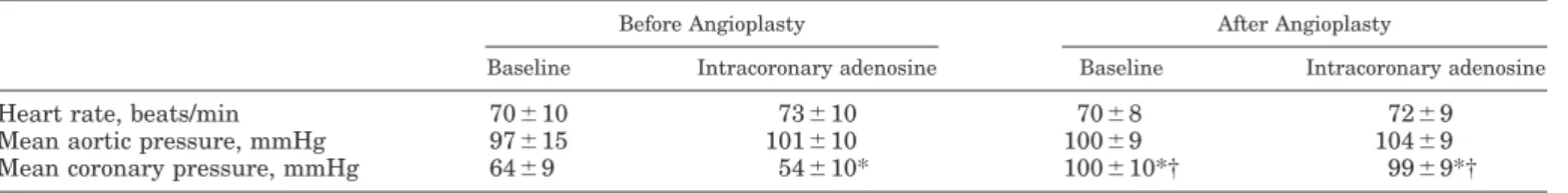
0.01). After revascularization, there were no signifi-cant changes in heart rate or systolic or diastolic aortic pressure (Table 1).
Coronary pressure and Doppler blood flow index.
Baseline blood flow index was 14.5⫾ 9.4 ml/min and
increased in all patients to 152 ⫾ 24% (range 115–
200%, P⬍ 0.02) after adenosine. Transstenotic
pres-sure gradient was 27 ⫾ 14 mmHg at baseline,
in-creased to 37⫾ 15 mmHg after adenosine (P ⬍ 0.01),
and virtually disappeared after revascularization at
rest (2⫾ 2 mmHg) and after adenosine (5 ⫾ 4 mmHg,
P ⬍ 0.01 vs. corresponding values before
revascular-ization). Mean distal pressure was 64 ⫾ 9 mmHg at
baseline, decreased to 54⫾ 10 mmHg after adenosine
(P ⬍ 0.01 vs. baseline), and markedly (P ⬍ 0.01)
in-creased after angioplasty at rest (100⫾ 10 mmHg) and
after adenosine (99 ⫾ 9 mmHg; Table 1). During
bal-loon coronary occlusion, coronary wedge pressure was
19⫾ 7 mmHg.
Angioplasty increased (P⬍ 0.01) blood flow index to
20⫾ 8 ml/min, corresponding to 180 ⫾ 93% of baseline
(Figs. 1 and 2). Similarly, a large increase was ob-served in maximal flow capacity, which increased from
152⫾ 24 to 324 ⫾ 242% (P ⬍ 0.01) of preangioplasty
baseline flow, and coronary flow reserve, which
in-creased from 1.52⫾ 0.24 to 2.33 ⫾ 0.8 (P ⬍ 0.01).
Xe washout analysis. Background activity, before postpercutaneous transluminal coronary angioplasty study, was virtually absent in all cases, resulting in ⬍1% of peak activity after tracer injection. Similarly,
heart-to-background ratio was ⬎4 at the end of all
studies. Fitting of the Xe washout curve was possible in all cases. The model residuals, i.e., the differences between the measured and model-predicted Xe radio-activity, indicated that the model fit was acceptable. In the majority of the time points from 0 to 180 s (81% of the points), the mean residuals were within 2 SE for the point, indicating no bias. Although in a significant proportion of the time points (19%) the mean residuals
differed from zero by⬎2 SE, the value was ⬍1% of the
peak value, and the mean residuals exhibited small oscillations but not a tendency to under- or overesti-mate the slope of the tracer curves. These results indicate that the model extrapolation of the tail of the tracer curve was adequate. The model-estimated dose
entering the regions of interest was 113⫾ 1.3% (SE) of
the measured Xe peak (all 18 curves pooled). This result is consistent with the notion that a small frac-tion of the tracer dose (13% in our calculafrac-tion) leaves the region of interest before the peak value is reached.
Mean transit time of Xe washout was 89 ⫾ 32 s at
baseline and decreased to 70 ⫾ 19 s (76 ⫾ 21%, P ⬍
0.05) after angioplasty. In the contralateral region, this
variable similarly decreased from 94⫾ 34 to 68 ⫾ 24 s
(P⬍ 0.05). Thus mean transit time was similar in the
stenotic and remote myocardium before and after
re-Table 1. Hemodynamic data before and after revascularization
Before Angioplasty After Angioplasty
Baseline Intracoronary adenosine Baseline Intracoronary adenosine
Heart rate, beats/min 70⫾10 73⫾10 70⫾8 72⫾9
Mean aortic pressure, mmHg 97⫾15 101⫾10 100⫾9 104⫾9
Mean coronary pressure, mmHg 64⫾9 54⫾10* 100⫾10*† 99⫾9*†
Values are means⫾ SD. Heart rate and aortic pressure did not change throughout the study. *P ⬍ 0.05 vs. baseline before percutaneous transluminal coronary angioplasty (PTCA); † P⬍ 0.05 vs. intracoronary adenosine before PTCA.
on May 5, 2012
ajpheart.physiology.org
vascularization. No correlation was observed between the increase in coronary blood flow and the decrease in mean transit time of Xe induced by revascularization
(r⫽ 0.19, not significant; Fig. 3).
The volume of distribution of Xe, derived by Doppler
coronary blood flow ⫻ mean transit time, increased
from 23 ⫾ 18 to 56 ⫾ 30 ml (P ⬍ 0.05) after
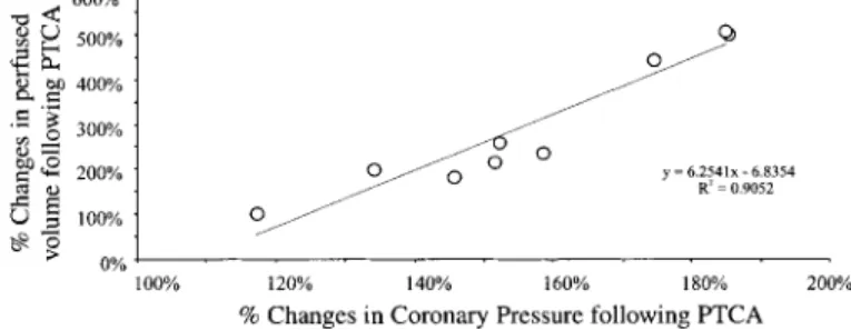
revascu-larization; this phenomenon showed a large variation between patients (Fig. 4); however, a close direct cor-relation was observed between the increase in driving coronary pressure and the increase in volume of
distri-bution of the tracer (r ⫽ 0.91, P ⬍ 0.01; Fig. 5). No
correlation was observed between changes in perfused myocardial volume and coronary wedge pressure
mea-sured during balloon coronary occlusion (r⫽ 0.13, not
significant). DISCUSSION
In the present study, angioplasty of severe coronary artery stenosis was associated with a marked increase
in absolute coronary blood flow and coronary flow re-serve assessed by intracoronary Doppler catheter. However, the increase in resting coronary blood flow was markedly larger than (and not correlated with) the
decrease in mean transit time of 133Xe, indicating a
significant increase in the volume of distribution of the tracer and, thus, in the mass of perfused myocardium after revascularization. Moreover, a significant corre-lation was observed between the increase in the per-fused volume and the increase in distal coronary pres-sure. These findings suggest that, in the presence of a very severe stenosis and transstenotic pressure gradi-ent, observed resting coronary blood flow is distributed to only a portion of the vascular units that are perfused in the absence of the stenosis, i.e., after revasculariza-tion. The spatial and temporal distribution of the per-fused units, within the jeopardized region, cannot be identified from the present study.
Coronary angioplasty and coronary blood flow. An-gioplasty markedly increased Doppler coronary blood
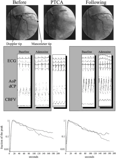
Fig. 1. Coronary angiography (top), pressure and flow velocity traces (middle), and Xe washout curves (bottom) at rest and before and after percutaneous transluminal coronary angioplasty (PTCA). PTCA abolished epicardial stenosis and increased distal coronary pressure (dCP) to values similar to aortic pressure (AoP) at rest and after intracoronary aden-osine. Adenosine significantly increased coronary blood flow velocity (CBFV) before and after PTCA. Balloon coronary occlusion markedly decreased dCP. PTCA markedly increased coronary blood flow at rest and after adenosine. Nevertheless, Xe wash-out curve did not change after revascularization in the jeopardized area (solid line) and remained sim-ilar to the washout curve observed in the remote region (dashed line). Thus the increase in flow in-duced by PTCA was not associated with a marked increase in Xe mean transit time. ECG, electrocar-diogram.
H2644 SEVERE STENOSIS CAUSES MYOCARDIAL TISSUE DERECRUITMENT
on May 5, 2012
ajpheart.physiology.org
flow index in all patients. Such a phenomenon might reflect the occurrence of postocclusion reactive hyper-emia (39) or the presence of a significant hypoperfusion before vessel recanalization. After revascularization, adenosine markedly increased coronary blood flow, re-vealing the presence of a significant coronary flow reserve. In agreement with previous studies (4, 33, 40), this observation strongly suggests a chronic hypoper-fusion in myocardial regions exposed to a reduced driv-ing pressure due to the presence of a severe stenosis. However, Xe mean transit time was remarkably simi-lar in the myocardium supplied by the stenotic artery and in the remote region before and after angioplasty, despite the marked increase in flow to the revascular-ized myocardium. Since, according to tracer kinetic theory, Xe mean transit time is the ratio of flow to tracer distribution volume (23, 30, 44), this observation would imply a similar increase in blood flow to both regions or an increase in the distribution volume in the
revascularized myocardium. Coronary blood flow in the nonstenotic coronary artery was not measured. How-ever, heart rate and systolic aortic pressure remained unchanged, and thus, in agreement with previous studies, an increase in blood flow in this region, beyond the modest increase in flow compatible with the in-crease in Xe washout rate, seems unlikely (33). By contrast, at least two observations suggest that the volume of distribution of Xe in the revascularized area was actually increased by angioplasty: the increase in flow was higher than (and not correlated with) the decrease in mean transit time, and the increase in tracer distribution volume was directly correlated with the increase in driving coronary pressure. The volume of distribution of Xe reflects two main factors: the so-called partition coefficient (related to the chemical tissue constituents) (11) and the perfused myocardial mass (related to the mass of the myocardium reached by the tracer and thus by flow) (23, 30). Since dramatic changes in the chemical components of the
revascular-Fig. 3. Relationship between changes in absolute CBF (blood flow velocity⫻ cross-sectional area of the coronary artery) and changes in specific myocardial blood flow [inverse of Xe mean transit time (MTT)]. In a homogeneously perfused system with a constant vol-ume, a linear direct correlation should be observed between these 2 variables. The lack of this observation suggests an effect of revascu-larization on the distribution volume of the tracer.
Fig. 4. Effect of PTCA on perfused myocardial volume (CBF⫻ Xe MTT) before and after PTCA.
Fig. 2. A: changes in absolute coronary blood flow (CBF) (blood flow velocity⫻ cross-sectional area of the coronary artery) induced by PTCA. B: effect of PTCA on specific flow (flow per unit mass) as measured by the inverse of Xe mean transit time (MTT). The increase in absolute flow was higher than the increase in specific myocardial flow.
on May 5, 2012
ajpheart.physiology.org
ized myocardium are unlikely to occur in such a short period of time, it seems reasonable to hypothesize an increase in perfused myocardial mass. Thus these find-ings strongly suggest a relationship between driving coronary pressure and amount of the perfused myocar-dium in patients with coronary artery disease.
Relationship between metabolic control of blood flow and vascular control of pressure. An increase in flow heterogeneity during ischemia has been described pre-viously (13, 31, 41, 43, 45). This phenomenon has been frequently explained by a heterogeneous distribution of extravascular compressing forces or by the structure of the coronary tree (3, 24). Actually, it is well known that such a difference might explain the relative reduc-tion in subendocardial flow during maximal vasodila-tion, which has been documented in regions supplied by a severely stenotic vessel or at low pressure (22, 24). However, in the present study, resting hypoperfusion was not associated with an exhausted vasodilator re-serve, inasmuch as adenosine increased flow across the coronary microcirculation, despite a reduced distal cor-onary pressure, indicating the persistence of a residual microcirculatory vasomotor tone. In line with this find-ing, several authors documented vasodilator reserve in the hypoperfused myocardium (2, 7, 8, 19, 32), and, more recently, we observed even an active, severe va-soconstriction of coronary microvasculature during un-stable angina (29) and ischemia induced by atrial
pac-ing tachycardia (38). This mismatch between
myocardial metabolism and microvascular resistance behavior might appear paradoxical when myocardial metabolism is assumed to be the primary determinant of vasomotor tone regulation. However, one should also consider the relevance of pressure-driven control, which avoids the notion that changes in aortic pressure are directly brought at the capillary level with the obvious dramatic consequence of altered plasma-inter-stitial water and solute exchanges (26, 42). The myo-genic reflex, which is the most acknowledged mecha-nism in coronary autoregulation, could operate such a control (5, 15). In patients with coronary artery dis-ease, it seems conceivable that the putative vasodila-tion required to match oxygen demand with blood flow would imply an excessive pressure drop across a severe stenosis (19). Since resting aortic pressure can be
con-sidered relatively stable, the pressure gradient would result in an excessively low capillary pressure, thus hampering the Starling forces (26, 42). Under these conditions, the only mechanisms able to preserve cap-illary pressure would be a venous constriction or the exclusion of perfused units in a parallel circuit model. In the present study, perfused myocardial volume in-creased after revascularization, and this increase was directly correlated with the increase in driving coro-nary pressure. This observation suggests that the transstenotic pressure gradient caused a reduction in perfused myocardium and a “derecruitment” of vascu-lar units from perfusion. These data do not elucidate whether this is an all-or-none phenomenon nor do they verify whether the exclusion of any given vascular unit is continuous or intermittent. However, in agreement with data by Chilian and Layne (10), these findings suggest that the coronary microvasculature reacts to a markedly reduced pressure by an active vasoconstric-tion. Such a response leads to exclusion of myocardial tissue from perfusion and thus to restoration of ade-quate flow rates and microvascular pressures in the perfused units. Such a mechanism might be even more powerful than the effects of myocardial metabolism on overall microvascular resistance. This concept closely agrees with the experimental evidence of a recruitment in perfused myocardial tissue in response to increases in coronary pressure (25) under autoregulation and during maximal vasodilation. Similarly, this model might also help explain the marked heterogeneity of blood flow, coronary reserve, and metabolic indexes of ischemia, which has been observed in the experimental setting (1, 13, 21, 27, 31, 41, 45).
Clinical implications. In patients with coronary ar-tery disease, myocardial dysfunction is often observed in regions subtended by severely stenotic coronary arteries. In several instances, a contractile recovery can be observed after revascularization (4). Since this phenomenon has important prognostic implications, its mechanisms have been extensively investigated. It has not been fully defined whether regional dysfunc-tion is the effect of chronic hypoperfusion (myocardial hibernation) (35) or prolonged dysfunction after repet-itive ischemia (myocardial stunning) (28). On the one hand, studies evaluating myocardial perfusion by the delivery or uptake of tracers, such as microspheres,
201
Tl-Sestamibi, or [13N]ammonia, documented resting
perfusion defects in viable myocardial regions (4). In contrast, this finding has been rarely reported by positron emission tomography studies of myocardial blood flow with radioactive water as a flow tracer (28). Similarly, although the former techniques often re-ported an increase in blood flow to revascularized ar-eas, water studies of blood flow showed a smaller effect of revascularization on resting myocardial blood flow. These puzzling discordances might reflect the different kinetics between diffusible tracers (which estimate blood flow washout rates) and deposit tracers (which estimate blood flow by the amount of tracer delivered) (14, 23, 43). In the present study, mean transit time of Xe was not markedly reduced in the “ischemic” region,
Fig. 5. Relationship between changes in coronary pressure and changes in perfused myocardial volume (CBF⫻ Xe MTT) induced by revascularization (PTCA). A close direct correlation was observed between these 2 variables, indicating an effect of driving coronary pressure on the mass of myocardium perfused.
H2646 SEVERE STENOSIS CAUSES MYOCARDIAL TISSUE DERECRUITMENT
on May 5, 2012
ajpheart.physiology.org
indicating a near-to-normal specific flow in the per-fused myocardium. In agreement with this finding, Xe mean transit time did not markedly decrease after revascularization However, the mass of the perfused myocardium was reduced, since it markedly increased after the restoration of a normal driving pressure. This finding indicates that a significant fraction of the ische-mic myocardium was excluded from perfusion and thus from tracer handling before revascularization. Accord-ingly, it seems conceivable that, at least in some in-stances, diffusible tracers might overestimate myocardial blood flow because of underestimation of myocardial mass, whereas they could underestimate the improve-ment in blood flow induced by revascularization be-cause of the increase in perfused mass induced by the increase in driving coronary pressure.
Finally, these data point out the relevance of perfu-sion pressure as a primary determinant of the regula-tion of myocardial blood flow in the myocardium sup-plied by a severely stenotic coronary artery under resting conditions. The early beneficial effect of revas-cularization on blood flow regulation might reflect the increase in driving coronary pressure. Actually, this mechanism might also retain a potential relevance in the precipitation of acute ischemia, thus justifying the finding of a paradoxical vasoconstrictor response to increasing myocardial metabolic demand (38). Meth-ods able to verify such a possibility might help more accurately characterize the pathogenesis of ischemia in patients with coronary artery disease.
Limitations. The increase in coronary blood flow ob-served after angioplasty might reflect the disappear-ance of collateral circulation. In this instdisappear-ance, actual perfusion rate to the myocardium might have been less modified by coronary angioplasty, thus causing the relative reduction in mean transit time. However, pa-tients with angiographic evidence of collaterals were excluded from the present study. Moreover, coronary wedge pressure was remarkably low in all patients, and no correlation was observed between this variable and changes in coronary blood flow and perfused vol-ume. Coronary pressure measured during balloon oc-clusion is considered the most accurate clinical descrip-tor of collateral feeding to the ischemic vascular bed (36). Thus these observations imply that collaterals were not extensively developed in these patients and that the observed increase in coronary blood flow was not closely related to their presence.
Xe washout was not monitored after adenosine ad-ministration because of the problems in maintaining stable levels of coronary blood flow, coronary pressure, and heart rate during adenosine infusion for the dura-tion of Xe washout. Accordingly, the present study does not elucidate the behavior of perfused myocardial vol-ume under pharmacological vasodilation and at low perfusion pressure. An increase in perfused mass, un-der vasodilation, might have further confirmed the hypothesis of the active nature of the heterogeneous vasoconstriction.
Coronary blood flow was not monitored in the con-tralateral region. This evaluation might have allowed a
more precise definition of perfused volume in a myo-cardium not subjected to changes in perfusion pres-sure, at least in the large arterial system. However, placing a further instrument in an untreated coronary artery was considered unethical in patients with nary artery diseases underlying a study during coro-nary angioplasty.
Utilization of the gamma-camera allowed accurate myocardial imaging, with clear separation between regions supplied by the stenotic or nonstenotic vessels. However, an exact definition of possible regional dif-ferences within the region supplied by the left anterior descending coronary artery could not be identified with the planar imaging method adopted. Moreover, be-cause of the limited spatial resolution of the method, no conclusion can be drawn about the distribution of flow heterogeneity or the individual size of hypoperfused or normally perfused myocardial units. These limitations, together with the limited patient population, prevent an accurate definition of the shape of the relationship between coronary pressure and perfused myocardial mass as well as the temporal variability of myocardial perfusion.
REFERENCES
1. Austin RE, Aldea GS, Coggins DL, Flynn AE, and Hoffman
JIE. Profound spatial heterogeneity of coronary reserve. Circ
Res 67: 319–331, 1990.
2. Aversano T and Becker LC. Persistence of coronary vasodila-tor reserve despite functionally significant flow reduction. Am J Physiol Heart Circ Physiol 248: H403–H411, 1985.
3. Bassingthwaighte JB, King RB, and Roger SA. Fractal nature of regional blood flow heterogeneity. Circ Res 65: 578– 590, 1989.
4. Bax JJ, Wijns W, Cornel JH, Visser FC, Boersma E, and
Fioretti PM. Accuracy of currently available techniques for
prediction of functional recovery after revascularization in pa-tients with left ventricular dysfunction due to chronic coronary artery disease: comparison of pooled data. J Am Coll Cardiol 30: 1451–1460, 1997.
5. Bayliss WM. On the local reaction of the arterial wall to changes of internal pressure. J Physiol (Lond) 28: 220–231, 1902. 6. Belloni FL and Sparks HV. Dynamics of myocardial oxygen
consumption and coronary vascular resistance. Am J Physiol Heart Circ Physiol 233: H34–H43, 1977.
7. Bristow JD, Arai AE, Anselone CG, and Pantely GA. Re-sponse to myocardial ischemia as a regulated process. Circula-tion 84: 2580–2587, 1991.
8. Canty JM and Klocke FJ. Reduced regional myocardial per-fusion in the presence of pharmacologic vasodilator reserve. Circulation 71: 370–371, 1985.
9. Canty JM and Smith TP. Adenosine-recruitable flow reserve is absent during myocardial ischemia in unanaesthetised dogs studied in the basal state. Circ Res 76: 1079–1087, 1995. 10. Chilian WM and Layne SM. Coronary microvascular
re-sponses to reductions in perfusion pressure: evidence for persis-tent arteriolar vasomotor tone during coronary hypoperfusion. Circ Res 66: 1227–1238, 1990.
11. Conn HL. Equilibrium distribution of radioxenon in tissue. Xenon-hemoglobin association curve. J Appl Physiol 16: 1065– 1070, 1961.
12. De Hoog FR, Knight JH, and Stokes AN. An improved method for numerical inversion of Laplace transforms. SIAM J Sci Stat Comput 3: 357–366, 1982.
13. Factor SM, Sonnenblick EH, and Kirk ES. The histologic border zone of acute myocardial infarction—islands or peninsu-las? Am J Pathol 92: 111–124, 1978.
14. Fluck DS, Etherington PJE, O’Hare D, Winlove CP, and
Sheridan DJ. Myocardial tissue perfusion determined by
on May 5, 2012
ajpheart.physiology.org
ticulate and diffusible tracers during ischemia: what is mea-sured? Cardiovasc Res 32: 869–878, 1996.
15. Folkow B. Description of the myogenic hypothesis. Circ Res 15, Suppl 1: 279–287, 1964.
16. Golino P, Piscione F, Willerson JT, Cappelli-Bigazzi M,
Focaccio A, Villari B, Indolfi C, Russolillo E, Condorelli M, and Chiariello M. Divergent effects of serotonin on
coronary-artery dimensions and blood flow in patients with coronary atherosclerosis and control patients. N Engl J Med 324: 641– 648, 1991.
17. Gorman MW and Sparks HV. Progressive coronary vasocon-striction during relative ischemia in canine myocardium. Circ Res 51: 411–420, 1982.
18. Gould KL. Pressure-flow characteristics of coronary stenoses in unsedated dogs at rest and during coronary vasodilation. Circ Res 43: 242–253, 1978.
19. Gould KL, Lipscomb K, and Calvert C. Compensatory changes of the distal coronary vascular bed during progressive coronary vasoconstriction. Circulation 51: 1085–1094, 1975. 20. Guyton RA, McClenathan JH, Newman GE, and Michaelis
LL. Evolution of regional ischemia distal to a proximal coronary
stenosis: self-propagation of ischemia. Am J Cardiol 40: 381– 392, 1977.
21. Haljamae H. Microcirculation and hemorrhagic shock. Am J Emerg Med 2: 100–107, 1984.
22. Hoffmann JIE. Determinants and prediction of transmural myocardial perfusion. Circulation 58: 381–391, 1978.
23. Klocke FJ. Coronary blood flow in man. Prog Cardiovasc Dis 19: 117–166, 1976.
24. L’Abbate A, Marzilli M, Ballestra AM, Camici P, Trivella
MG, Pelosi G, and Klassen GA. Opposite transmural
gradi-ents of coronary resistance and extravascular pressure in the working dog’s heart. Cardiovasc Res 14: 21–29, 1980.
25. L’Abbate A, Mildenberger RR, Zborowska-Sluis DT, and
Klassen GA. Myocardial tissue recruitment in the dog as
deter-mined by double tracer dilution method. Circ Res 39: 276–281, 1976.
26. Landis EM and Pappenheimer JR. Exchange of substances through capillary walls. In: Handbook of Physiology. Circulation. Bethesda, MD: Am. Physiol. Soc., 1972, sect. 2, vol. II, chapt. 29, p. 961–1034.
27. Loncar R, Flsche CW, and Deussen A. Coronary reserve of high and low flow regions in the dog heart left ventricle. Circu-lation 98: 262–270, 1998.
28. Marinho NVS, Keogh BE, Costa DC, Lammerstma AA, Ell
PJ, and Camici PG. Pathophysiology of chronic left ventricular
dysfunction. New insights from the measurement of absolute myocardial blood flow and glucose utilization. Circulation 93: 737–744, 1996.
29. Marzilli M, Sambuceti G, Fedele S, and L’Abbate A. Coro-nary microcirculatory vasoconstriction during ischemia in pa-tients with unstable angina. J Am Coll Cardiol 35: 327–334, 2000.
30. Maseri A, L’Abbate A, Michelassi C, Pesola A, Pisani P,
Marzilli M, De Nes M, and Mancini P. Possibilities,
limita-tions and technique for the study of regional myocardial perfu-sion in man by xenon-133. Cardiovasc Res 11: 277–290, 1977. 31. Miyazaki K, Hori S, Inoue S, Adachi T, Bessho M,
Kuwa-hira I, Mori H, Nakazawa H, Aikawa N, and Ogawa S.
Characterization of energy metabolism and blood flow distribu-tion in myocardial ischemia in hemorrhagic shock. Am J Physiol Heart Circ Physiol 273: H600–H607, 1997.
32. Pantely GA, Bristow JD, Swenson LJ, Ladley HD,
John-son WB, and Anselone CG. Incomplete coronary vasodilation
during myocardial ischemia in swine. Am J Physiol Heart Circ Physiol 249: H638–H647, 1985.
33. Parodi O, Sambuceti G, Roghi A, Testa R, Inglese E, Pirelli
S, Spinelli F, Campolo L, and L’Abbate A. Residual coronary
reserve despite decreased resting blood flow in patients with critical coronary lesions. A study by technetium-99m human albumin microsphere myocardial scintigraphy. Circulation 87: 330–344, 1993.
34. Pijls NHJ, De Bruyne B, Peels K, Van Der Voort PH,
Bonnier HJ, Bartunek J, Koolen JJ, and Koolen JJ.
Mea-surement of fractional flow reserve to assess the functional severity of coronary artery stenoses. N Engl J Med 334: 1703– 1708, 1996.
35. Rahimtoola SH. The hibernating myocardium. Am Heart J 117: 211–221, 1989.
36. Ronnow Sand NP, Rehling M, Bagger JP, Thuesen L, Flo
C, and Nielsen TT. Functional significance of recruitable
col-laterals during temporary coronary occlusion evaluated by 99mTc-Sestamibi single photon emission computed tomography. J Am Coll Cardiol 35: 624–632, 2000.
37. Rubio R and Berne RM. Regulation of coronary blood flow. Prog Cardiovasc Dis 18: 105–136, 1975.
38. Sambuceti G, Marzilli M, Marraccini P, Schneider-Eicke
J, Gliozheni E, Parodi O, and L’Abbate A. Coronary
vaso-constriction during myocardial ischemia induced by rises in metabolic demand in patients with coronary artery disease. Circulation 95: 2652–2659, 1997.
39. Serruys PW, Di Mario C, Meneveau N, de Jaegere P,
Strikwerda S, de Feyter PJ, and Emanuelsson H.
Intra-coronary pressure and flow velocity with sensor-tip guide wires: a new methodologic approach for assessment of coronary hemo-dynamics before and after coronary interventions. Am J Cardiol 71: 41D–53D, 1993.
40. Smith SC Jr, Gorlin R, Herman MV, Taylor WJ, and
Col-lins JJ Jr. Myocardial blood flow in man: effects of coronary
collateral circulation and coronary artery bypass surgery. J Clin Invest 51: 2556–2565, 1972.
41. Steenbergen C, Deleeuw G, Barlow C, Chance B, and
Williamson JR. Heterogeneity of the hypoxic state in perfused
rat heart. Circ Res 41: 606–615, 1977.
42. Taylor AE. Capillary fluid filtration. Starling forces and lymph flow. Circ Res 49: 557–575, 1981.
43. Yoshida S, Akizuki S, Gowski D, and Downey JM. Discrep-ancy between microsphere and diffusible tracer estimates of perfusion to ischemic myocardium. Am J Physiol Heart Circ Physiol 249: H255–H264, 1985.
44. Zierler K. Equations for measuring blood flow by external monitoring of radioisotopes. Circ Res 16: 309–321, 1965. 45. Zuurbier CJ, Van Iterson M, and Ince C. Functional
heter-ogeneity of oxygen supply-consumption ratio in the heart. Car-diovasc Res 44: 488–497, 1999.
H2648 SEVERE STENOSIS CAUSES MYOCARDIAL TISSUE DERECRUITMENT
on May 5, 2012
ajpheart.physiology.org
![Fig. 3. Relationship between changes in absolute CBF (blood flow velocity ⫻ cross-sectional area of the coronary artery) and changes in specific myocardial blood flow [inverse of Xe mean transit time (MTT)]](https://thumb-eu.123doks.com/thumbv2/123dokorg/8288402.131232/6.907.147.773.89.410/relationship-changes-absolute-velocity-sectional-coronary-specific-myocardial.webp)