RATIONAL TESTING
Tests for diagnosing and monitoring non-alcoholic
fatty liver disease in adults
Christopher D Byrne professor of endocrinology and metabolism
1 2, Janisha Patel consultant
hepatologist
3, Eleonora Scorletti clinical research fellow
1 2, Giovanni Targher associate professor
endocrinology and metabolism
41Nutrition and Metabolism, Faculty of Medicine, University of Southampton, UK; 2Southampton National Institute for Health Research Biomedical Research Centre, University Hospital Southampton, Southampton General Hospital, Southampton, UK; 3Department of Hepatology, Southampton General Hospital, Southampton, UK; 4Department of Medicine, Section of Endocrinology, Diabetes and Metabolism, Azienda Ospedaliera Universitaria Integrata di Verona, Verona, Italy
This series of occasional articles provides an update on the best use of key diagnostic tests in the initial investigation of common or important clinical presentations. The series advisers are Steve Atkin, professor of medicine, Weill Cornell Medical College Qatar; and Eric Kilpatrick, division chief, clinical chemistry, Sidra Medical and Research Center, Qatar; honorary professor, department of clinical biochemistry, Hull Royal Infirmary, Hull York Medical School. To suggest a topic for this series, please email us at [email protected].
What you need to know
• Liver ultrasonography is a pragmatic first line test to diagnose hepatic steatosis and exclude other liver pathology in those with non-alcoholic fatty liver disease (NAFLD)
• In patients with confirmed hepatic steatosis, use simple non-invasive markers of fibrosis (such as an enhanced liver fibrosis blood test (ELF)) and/or FibroScan to investigate for liver fibrosis
• Offer patients with hepatic fibrosis referral for specialist opinion, as hepatic fibrosis is the strongest predictor of overall and liver related mortality in those with NAFLD
At a routine work health check, a 52 year old sedentary computer programmer was found to have a serum alanine aminotransferase (ALT) concentration of 68 IU/L (normal 0-40 IU/L), and a triglyceride concentration of 1.9 mmol/L. His fasting plasma glucose level was 5.8 mmol/L and other basic liver, renal, and lipid blood tests were normal. He had an unremarkable medical history and took no regular medications, did not smoke, and consumed <7 units of alcohol/week. Clinical examination was unremarkable. His body mass index was 29 kg/m2; waist circumference 102 cm, and blood pressure 134/88 mmHg. A repeat serum ALT measurement remained raised some months later, at 62 IU/L.
Non-alcoholic fatty liver disease (NAFLD) is a metabolic liver disease that encompasses a spectrum of progressive pathological conditions, ranging from non-alcoholic fatty liver (NAFL) to steatohepatitis (NASH), fibrosis, and cirrhosis. When hepatic steatosis occurs in the absence of excessive alcohol consumption and other recognised causes of liver fat, and with
cardiometabolic risk factors, it is likely that the diagnosis is NAFLD as NAFLD is principally a diagnosis of exclusion. NAFLD is the commonest liver disease in high income countries, and is estimated to affect at least 25%-30% of adults in the general population and up to 70%-90% of persons with obesity or type 2 diabetes.1NAFLD is associated not only with
liver related morbidity and mortality, but also with an increased risk of developing cardiovascular disease and type 2 diabetes.2 3
Liver biopsy remains the reference method for diagnosing NAFLD, as it provides the most accurate assessment of disease grade and stage.4 5 However, undertaking a liver biopsy is costly,
risky, and potentially painful. Moreover, interpretation of NAFLD severity can be compromised by sampling errors in what can be a patchy disease.6 7
In this article, we discuss the diagnosis of NAFLD, testing for liver fibrosis in those with NAFLD, and monitoring of those most likely to develop advanced liver disease. We examine the evidence and guidelines from Europe, the United States, and the UK’s National Institute for Health and Care Excellence (NICE)8-10 for and against the use of specific diagnostic tests.
Our approach to the use of liver ultrasound in establishing a diagnosis of hepatic steatosis differs from the recent NICE guidelines,10 but complements British Society of
Gastroenterology guidelines.11 Treatment options are beyond
the scope of this article.
Correspondence to C Byrne [email protected]
What are the next investigations?
After performing a history and examination, the next investigations to establish whether the patient has NAFLD or another liver condition are:• A non-invasive liver screen, which includes tests such as serology for hepatitis B and C viruses, and measurement of liver auto-antibodies, immunoglobulins, caeruloplasmin, alpha 1 anti-trypsin, and ferritin concentrations
• A liver ultrasound to look for features suggestive of NAFLD (hepatic steatosis) and to rule in or out other pathology.
An ultrasound scan of the liver is the most typical imaging test requested by non-specialists in patients in whom NAFLD is suspected. This is because it can help to confirm and exclude other causes of liver disease such as gall stones or metastasis, as well as confirm hepatic steatosis. Although the NICE guidelines state that ultrasonography is “not cost effective,”3 it
is important to understand why such a statement was made, despite widespread acceptance that the most useful imaging technique to detect steatosis is ultrasonography.1 2 12 In estimating
the cost effectiveness of any test or intervention, what is considered is not only the cost, but also the relationship between the measured factor (ie, hepatic steatosis) and the outcome. Ultrasonography enables an accurate diagnosis of hepatic steatosis, but the presence of steatosis does not predict risk of end-stage liver disease. Rather, it is advanced fibrosis (which is not accurately detected by ultrasonography) that more accurately predicts the risk of developing end-stage liver disease and hepatocellular carcinoma.13-16 Ultrasonography is, however,
a pragmatic approach to investigating the possibility of NAFLD because it also allows the exclusion of other liver conditions and so is central to the approach laid out in this article. A validated, widely accepted procedure for the diagnosis and monitoring of NAFLD does not yet exist. We propose a potential approach (infographic) including “red flags” and when to seek specialist advice. The infographic gives an overview of testing for NAFLD and excluding other pathologies in a person with abnormal liver function test results. The graphic includes features of abnormal liver function tests and factors that are useful to note in the history and examination. It provides information about further testing in those identified with NAFLD to diagnose liver fibrosis, and about how to monitor those found to have advanced fibrosis.
Is it NAFLD or something else?
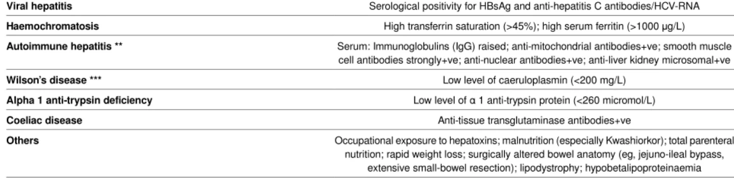
When hepatic steatosis occurs in the absence of recognised causes of liver fat (table 1), and with cardiometabolic risk factors (table 2), it is likely that the diagnosis is NAFLD.
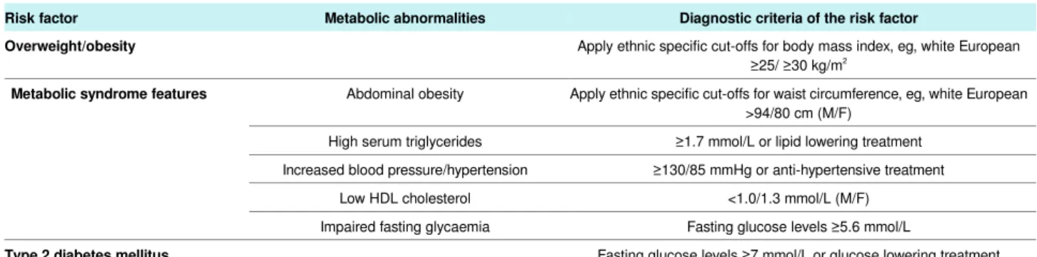
NAFLD might be suspected because the patient is overweight or obese, has type 2 diabetes, or has other metabolic syndrome features.17 More often, a diagnosis of NAFLD is suspected when
liver blood tests show mild to moderate elevations of serum aminotransferase levels. However, serum aminotransferase levels are not sensitive or specific to make or rule out a diagnosis of NAFLD. Table 2 describes these risk factors. Be aware that NAFLD can also occur in non-obese or lean individuals (termed “lean NAFLD”).
An alternative pathology might be more likely if a non-invasive liver screen of other factors in the history, such as high alcohol intake, suggests another cause (table 1).
What techniques can be used to test for
hepatic steatosis?
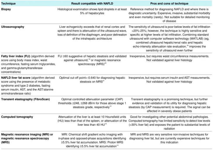
The presence of hepatic steatosis, and therefore NAFLD, can be diagnosed by various methods (table 3).
Ultrasonography is the first line imaging technique for diagnosing hepatic steatosis. Compared with histology, it has a good sensitivity (~85%) and specificity (~95%) for detecting moderate steatosis,15 16 and traditionally its sensitivity is thought
to be poor when <20%-30% of hepatocytes are steatotic.22
Combining standard ultrasonography with computer software technology (MATLAB) (eg, combined ultrasound hepatic/renal ratio and hepatic echo-intensity attenuation rate evaluation)18
improves the sensitivity of ultrasonography further. In this methodology, the ultrasound hepatic/renal echo-intensity ratio and ultrasound hepatic echo-intensity attenuation rate were obtained from ordinary ultrasound images using the MATLAB program. Compared with proton magnetic resonance
spectroscopy (ie, the gold standard for detecting low levels of liver fat content) (see table 3), at levels of <15% liver fat content, the sensitivity and specificity of the ultrasound quantitative model was 81.4% and 100%.
Computed tomography, magnetic resonance imaging, and magnetic resonance spectroscopy can be used, but such imaging techniques are more expensive and less readily available.8 9
Some non-invasive biomarkers of steatosis (eg, fatty liver index) have been proposed, but they have limited clinical utility, as they often do not accurately quantify steatosis as assessed histologically. Controlled attenuation parameter (CAP, assessed by transient elastography) can also be used, although it remains uncertain what CAP thresholds should be adopted to diagnose steatosis.23
Liver biopsy remains the reference method for diagnosing and staging NAFLD, but is not a practical first line investigation. Undertaking serial liver biopsies over time is fraught with difficulties, and is unacceptable to monitor disease. Nevertheless, biopsy is the only method for diagnosing inflammation in NAFLD (ie, NASH), and should also be also considered when other chronic liver diseases cannot be definitively excluded.
For those with NAFLD, what further
investigations are offered?
Previously thought to be a harmless condition, hepatic steatosis is now increasingly being recognised as a cause of progressive and advanced liver disease. Recent follow-up studies showed that, contrary to conventional paradigm, patients with NAFL (ie, simple steatosis on histology) can develop progressive liver fibrosis.24 Hepatic steatosis (detected by ultrasonography) is
also strongly associated with an increased risk of fatal and non-fatal cardiovascular disease, type 2 diabetes, and chronic kidney disease.2 3 After a diagnosis of hepatic steatosis has been
established, strong evidence8-10 now indicates that it is clinically
more important to stage liver fibrosis than to ascertain the presence of NASH.
Characterise the severity of NAFLD
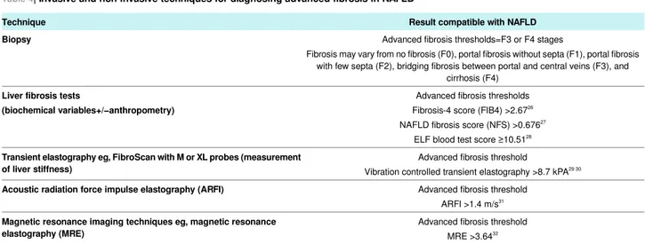
Once steatosis has been diagnosed, the presence and severity of liver fibrosis should be assessed using combined non-invasive tests to identify those individuals with advanced fibrosis who should be referred to specialists in hepatology for further investigations. Staging of liver fibrosis can be undertaken with the use of biopsy or various non-invasive tests9 25 (table 4).
Choice of test will depend on local availability. The infographic outlines two possible approaches. Tests such as Fibrosis-4 score
(FIB4), NAFLD fibrosis score (NFS), and enhanced liver fibrosis (ELF) can be conducted by non-specialists. How they are calculated is outlined at the foot of table 4.
The ELF test (a commercial blood test using three direct fibrosis biomarkers) has good performance for diagnosing significant and advanced fibrosis, and it is now strongly recommended by the NICE guidelines, although it is not used worldwide. Other “biochemical” score systems (eg, the NFS and FIB4 scores, which are both cost effective and highly sensitive tools to exclude patients with advanced fibrosis) and second line “physical” techniques (liver stiffness measurements assessed with transient elastography [FibroScan] or with newer imaging techniques) are frequently used to assess the severity of liver fibrosis. The combination of FibroScan with FIB4/NFS measurements has shown excellent accuracy in distinguishing advanced fibrosis.25
All non-invasive tests for liver fibrosis are better at excluding advanced fibrosis than diagnosing it. They have only modest positive predictive value for advanced fibrosis, but a much stronger negative predictive value. Furthermore, none is good at detecting intermediate stages of fibrosis. As such, no test can fully replace liver biopsy. For example, the NAFLD fibrosis score, the most widely validated non-invasive test, has good performance for identifying patients without fibrosis, but poorer performance for diagnosing clinically significant and advanced fibrosis. As recommended by the European8 and American9
practice guidelines, current non-invasive tests of fibrosis should be used in a staged approach, utilising their high negative predictive value to rule out patients who are unlikely to have advanced fibrosis, and so reserving liver biopsy for patients who are most likely to have substantial (clinically significant) fibrosis or when there is diagnostic uncertainty.
Outcome
The man’s general practitioner requested a liver ultrasound scan (confirming the presence of hepatic steatosis) together with a repeat serum ALT measurement of 62 IU/L. Other blood tests (including serology for hepatitis B and C viruses, liver auto-antibodies, immunoglobulins, caeruloplasmin, alpha-1 antitrypsin and ferritin levels) excluded other causes of liver disease.
The patient is likely to have NAFLD. How this article was made
We searched PubMed for original articles and reviews using the keywords “nonalcoholic fatty liver disease,” or “fatty liver” combined with “diagnosis,” “prognosis,” or “mortality” published between 1990 and 2018. Articles published in languages other than English were excluded from the analysis.
How patients were involved in the creation of this article Several of our patients have told us that doctors are inconsistent in their approaches to investigating their liver disease. Two patient representatives (Irene McGill, who has NAFLD, and Jane Putsey, who has cared for her father with NAFLD) participated in the NICE NAFLD NG 49 Guideline Development Group and contributed to the guideline. Ms McGill and Ms Putsey advised Professor Byrne of what they thought was important for patients with NAFLD, which influenced the writing of this manuscript. Both representatives commented on the article and gave helpful suggestions to drafts of the manuscript to improve its clarity. For example, they asked for clear information on how NAFLD could be diagnosed.
Acknowledgments: The authors thank Ms Irene McGill and Ms Jane Putsey for their helpful comments and advice during the writing of this article.
Contributors: CDB and GT wrote the first draft and all authors reviewed and contributed to the writing of the article.
Provenance and peer review: commissioned; externally peer reviewed. We have read and understood The BMJ policy on declaration of interests and declare that we have no competing interests.
1 Younossi ZM, Koenig AB, Abdelatif D, Fazel Y, Henry L, Wymer M. Global epidemiology of nonalcoholic fatty liver disease-Meta-analytic assessment of prevalence, incidence, and outcomes. Hepatology 2016;64:73-84. 10.1002/hep.28431 26707365
2 Byrne CD, Targher G. NAFLD: a multisystem disease. J Hepatol 2015;62(Suppl):S47-64. 10.1016/j.jhep.2014.12.012 25920090
3 Targher G, Byrne CD. Non-alcoholic fatty liver disease: an emerging driving force in chronic kidney disease. Nat Rev Nephrol 2017;13:297-310.
10.1038/nrneph.2017.16 28218263
4 Bravo AA, Sheth SG, Chopra S. Liver biopsy. N Engl J Med 2001;344:495-500. 10.1056/NEJM200102153440706 11172192
5 Tapper EB, Lok AS. Use of liver imaging and biopsy in clinical practice. N Engl J Med 2017;377:756-68. 10.1056/NEJMra1610570 28834467
6 Byrne CD, Targher G. Time to replace assessment of liver histology with MR-based imaging tests to assess efficacy of interventions for nonalcoholic fatty liver disease.
Gastroenterology 2016;150:7-10. 10.1053/j.gastro.2015.11.016 26602219
7 Rockey DC, Caldwell SH, Goodman ZD, Nelson RC, Smith ADAmerican Association for the Study of Liver Diseases. Liver biopsy. Hepatology 2009;49:1017-44.
10.1002/hep.22742 19243014
8 European Association for the Study of the Liver (EASL)European Association for the Study of Diabetes (EASD)European Association for the Study of Obesity (EASO). EASL-EASD-EASO Clinical Practice Guidelines for the management of non-alcoholic fatty liver disease. Diabetologia 2016;59:1121-40. 10.1007/s00125-016-3902-y 27053230 9 Chalasani N, Younossi Z, Lavine JE, etal . The diagnosis and management of nonalcoholic fatty liver disease: Practice guidance from the American Association for the Study of Liver Diseases. Hepatology 2018;67:328-57. 10.1002/hep.29367 28714183
10 Glen J, Floros L, Day C, Pryke RGuideline Development Group. Non-alcoholic fatty liver disease (NAFLD): summary of NICE guidance. BMJ 2016;354:i4428.
10.1136/bmj.i4428 27605111
11 Newsome PN, Cramb R, Davison SM, etal . Guidelines on the management of abnormal liver blood tests. Gut 2018;67:6-19.
12 Karlas T, Petroff D, Sasso M, etal . Individual patient data meta-analysis of controlled attenuation parameter (CAP) technology for assessing steatosis. J Hepatol 2017;66:1022-30. 10.1016/j.jhep.2016.12.022 28039099
13 Schwenzer NF, Springer F, Schraml C, Stefan N, Machann J, Schick F. Non-invasive assessment and quantification of liver steatosis by ultrasound, computed tomography and magnetic resonance. J Hepatol 2009;51:433-45. 10.1016/j.jhep.2009.05.023 19604596 14 Szczepaniak LS, Nurenberg P, Leonard D, etal . Magnetic resonance spectroscopy to
measure hepatic triglyceride content: prevalence of hepatic steatosis in the general population. Am J Physiol Endocrinol Metab 2005;288:E462-8.
10.1152/ajpendo.00064.2004 15339742
15 Hernaez R, Lazo M, Bonekamp S, etal . Diagnostic accuracy and reliability of ultrasonography for the detection of fatty liver: a meta-analysis. Hepatology 2011;54:1082-90. 10.1002/hep.24452 21618575
16 Ballestri S, Nascimbeni F, Baldelli E, etal . Ultrasonographic fatty liver indicator detects mild steatosis and correlates with metabolic/histological parameters in various liver diseases. Metabolism 2017;72:57-65. 10.1016/j.metabol.2017.04.003 28641784 17 Alberti KG, Eckel RH, Grundy SM, etal. International Diabetes Federation Task Force on
Epidemiology and PreventionNational Heart, Lung, and Blood InstituteAmerican Heart AssociationWorld Heart FederationInternational Atherosclerosis SocietyInternational Association for the Study of Obesity. Harmonizing the metabolic syndrome: a joint interim statement of the International Diabetes Federation Task Force on Epidemiology and Prevention; National Heart, Lung, and Blood Institute; American Heart Association; World Heart Federation; International Atherosclerosis Society; and International Association for the Study of Obesity. Circulation 2009;120:1640-5.
10.1161/CIRCULATIONAHA.109.192644 19805654
18 Zhang B, Ding F, Chen T, Xia LH, Qian J, Lv GY. Ultrasound hepatic/renal ratio and hepatic attenuation rate for quantifying liver fat content. World J Gastroenterol 2014;20:17985-92. 10.3748/wjg.v20.i47.17985 25548498
19 Bedogni G, Bellentani S, Miglioli L, etal . The fatty liver index: a simple and accurate predictor of hepatic steatosis in the general population. BMC Gastroenterol 2006;6:33. 10.1186/1471-230X-6-33 17081293
20 Cuthbertson DJ, Weickert MO, Lythgoe D, etal . External validation of the fatty liver index and lipid accumulation product indices, using 1H-magnetic resonance spectroscopy, to identify hepatic steatosis in healthy controls and obese, insulin-resistant individuals. Eur
J Endocrinol 2014;171:561-9. 10.1530/EJE-14-0112 25298375
21 Kotronen A, Peltonen M, Hakkarainen A, etal . Prediction of non-alcoholic fatty liver disease and liver fat using metabolic and genetic factors. Gastroenterology 2009;137:865-72. 10.1053/j.gastro.2009.06.005 19524579
22 Saadeh S, Younossi ZM, Remer EM, etal . The utility of radiological imaging in nonalcoholic fatty liver disease. Gastroenterology 2002;123:745-50. 10.1053/gast.2002.35354 12198701 23 Vuppalanchi R, Siddiqui MS, Van Natta ML, etal. NASH Clinical Research Network.
Performance characteristics of vibration-controlled transient elastography for evaluation of nonalcoholic fatty liver disease. Hepatology 2018;67:134-44.
10.1002/hep.29489 28859228
24 Singh S, Allen AM, Wang Z, Prokop LJ, Murad MH, Loomba R. Fibrosis progression in nonalcoholic fatty liver vs nonalcoholic steatohepatitis: a systematic review and meta-analysis of paired-biopsy studies. Clin Gastroenterol Hepatol 2015;13:643-54.e1, 9, quiz e39-40. 10.1016/j.cgh.2014.04.014 24768810
25 Petta S, Wong VW, Cammà C, etal . Serial combination of non-invasive tools improves the diagnostic accuracy of severe liver fibrosis in patients with NAFLD. Aliment Pharmacol
Ther 2017;46:617-27. 10.1111/apt.14219 28752524
26 Shah AG, Lydecker A, Murray K, Tetri BN, Contos MJ, Sanyal AJNash Clinical Research Network. Comparison of noninvasive markers of fibrosis in patients with nonalcoholic fatty liver disease. Clin Gastroenterol Hepatol 2009;7:1104-12.
27 Angulo P, Hui JM, Marchesini G, etal The NAFLD fibrosis score: a noninvasive system that identifies liver fibrosis in patients with NAFLD.Hepatology 2007;45:846-54. 28 Guha IN, Parkes J, Roderick P, etal . Noninvasive markers of fibrosis in nonalcoholic fatty
liver disease: Validating the European Liver Fibrosis Panel and exploring simple markers.
Hepatology 2008;47:455-60. 10.1002/hep.21984 18038452
29 Tapper EB, Challies T, Nasser I, Afdhal NH, Lai M. The performance of vibration controlled transient elastography in a US cohort of patients with nonalcoholic fatty liver disease. Am
J Gastroenterol 2016;111:677-84.
30 Xiao G, Zhu S, Xiao X, Yan L, Yang J, Wu G. Comparison of laboratory tests, ultrasound, or magnetic resonance elastography to detect fibrosis in patients with nonalcoholic fatty liver disease: A meta-analysis. Hepatology 2017;66:1486-501.
10.1002/hep.29302 28586172
31 Cassinotto C, Boursier J, de Lédinghen V, etal . Liver stiffness in nonalcoholic fatty liver disease: A comparison of supersonic shear imaging, FibroScan, and ARFI with liver biopsy. Hepatology 2016;63:1817-27. 10.1002/hep.28394 26659452
32 Loomba R, Wolfson T, Ang B, etal . Magnetic resonance elastography predicts advanced fibrosis in patients with nonalcoholic fatty liver disease: a prospective study. Hepatology 2014;60:1920-8. 10.1002/hep.27362 25103310
33 Xie Q, Zhou X, Huang P, Wei J, Wang W, Zheng S. The performance of enhanced liver fibrosis (ELF) test for the staging of liver fibrosis: a meta-analysis. PLoS One 2014;9:e92772.
Published by the BMJ Publishing Group Limited. For permission to use (where not already granted under a licence) please go to http://group.bmj.com/group/rights-licensing/ permissions
Tables
Table 1| Tests and factors which do not suggest NAFLD
Factors that do not suggest NAFLD Risk factors for liver disease
History of excessive alcohol consumption
Alcohol
>21 standard drinks per week in men and >14 standard drinks per week in women *
History of drug exposure
Drugs
Valproic acid, oestrogens, tamoxifen, corticosteroids, tetracycline, amiodarone, perhexiline maleate, methotrexate,
4,4′-diethylaminoethoxyhexesterol, chloroquine, L-asparaginase
Serological positivity for HBsAg and anti-hepatitis C antibodies/HCV-RNA
Viral hepatitis
High transferrin saturation (>45%); high serum ferritin (>1000 μg/L)
Haemochromatosis
Serum: Immunoglobulins (IgG) raised; anti-mitochondrial antibodies+ve; smooth muscle cell antibodies strongly+ve; anti-nuclear antibodies+ve; anti-liver kidney microsomal+ve
Autoimmune hepatitis **
Low level of caeruloplasmin (<200 mg/L)
Wilson’s disease ***
Low level of α 1 anti-trypsin protein (<260 micromol/L)
Alpha 1 anti-trypsin deficiency
Anti-tissue transglutaminase antibodies+ve
Coeliac disease
Occupational exposure to hepatoxins; malnutrition (especially Kwashiorkor); total parenteral nutrition; rapid weight loss; surgically altered bowel anatomy (eg, jejuno-ileal bypass,
extensive small-bowel resection); lipodystrophy; hypobetalipoproteinaemia
Others
*The alcohol thresholds for liver disease reported in table 1 are not entirely congruent with the UK current thresholds for safe alcohol consumption, which are >14 units (standard drinks) per week in both men and women. ** Low titres of anti-nuclear, anti-smooth muscle, and anti-mitochondrial antibodies can be noted in patients with NAFLD (in the absence of autoimmune hepatitis) ***Slightly lower caeruloplasmin levels can also be found
Table 2| Common cardiometabolic risk factors for NAFLD
Diagnostic criteria of the risk factor Metabolic abnormalities
Risk factor
Apply ethnic specific cut-offs for body mass index, eg, white European ≥25/ ≥30 kg/m2
Overweight/obesity
Apply ethnic specific cut-offs for waist circumference, eg, white European >94/80 cm (M/F)
Abdominal obesity
Metabolic syndrome features
≥1.7 mmol/L or lipid lowering treatment High serum triglycerides
≥130/85 mmHg or anti-hypertensive treatment Increased blood pressure/hypertension
<1.0/1.3 mmol/L (M/F) Low HDL cholesterol
Fasting glucose levels ≥5.6 mmol/L Impaired fasting glycaemia
Fasting glucose levels ≥7 mmol/L or glucose lowering treatment
Type 2 diabetes mellitus
Other modifiable risk factors for NAFLD are cigarette smoking (due to its pro-fibrotic hepatic effect), excessive dietary intakes of fructose, carbohydrates, and saturated fatty acids
Table 3| Invasive and non-invasive techniques for diagnosing hepatic steatosis in NAFLD
Pros and cons of technique Result compatible with NAFLD
Technique
Reference method for diagnosing NAFLD and where there is diagnostic uncertainty. Expensive, invasive, substantial morbidity
and even mortality (rarely). Not suitable for detailed monitoring of disease
Histological examination shows lipid droplets in at least 5% of hepatocytes
Biopsy
The sensitivity of ultrasound is poor below levels of fat infiltration <20%-25%; however, the technique is highly sensitive and specific at higher levels of fat infiltration. Combining standard ultrasound with computer software technology (MATLAB) (eg,
combined ultrasound hepatic/renal ratio and hepatic echo-intensity attenuation rate evaluation,18
improves the sensitivity of ultrasound even further Liver echogenicity exceeds that of renal cortex and
spleen and there is attenuation of the ultrasound wave, loss of definition of the diaphragm, and poor delineation
of the intrahepatic architecture
Ultrasonography
Inexpensive, but requires waist circumference measurements. Not validated against liver histology
FLI ≥60 suggestive of hepatic steatosis and validated against ultrasound,19 or magnetic resonance
spectroscopy (MRS)20
Fatty liver index (FLI) (algorithm derived
score using body mass index, waist circumference, fasting serum triglycerides, and gamma-glutamyltransferase concentrations)
Inexpensive, but requires serum insulin and AST measurements. Not validated against liver histology
Optimal cut-off point=-0.640 for diagnosing hepatic steatosis on MRS21
NAFLD liver fat score (algorithm derived
score using the presence of metabolic syndrome and type 2 diabetes, fasting serum insulin, AST, and the AST/alanine aminotransferase ratio)
Transient elastography is a promising technique, but further evidence and validation of its utility for diagnosing hepatic steatosis (by CAP measurement) is required. The signal can be
affected in severely obese patients Optimal controlled attenuation parameter (CAP)
thresholds ≥248, ≥268 dB/m for those above stage 1 steatosis grade, respectively12
Transient elastography (FibroScan)
Good for investigating other potential abdominal pathologies. Computed tomography has limited sensitivity to detect low levels (<30% liver fat) and exposes the patient to substantial levels of
radiation Attenuation of the liver is at least 10 Hounsfield units
(HU) less than that of the spleen, or attenuation of the liver less than 40 HU13
Computed tomography
MRI and MRS are very sensitive non-invasive techniques for diagnosing liver fat, but are currently expensive techniques for
this indication MRI: Chemical shift gradient echo imaging with
in-phase and opposed-phase acquisitions identifying ≥5.5% liver fat accumulation. MRS: Proton MRS
identifying ≥5.5% liver fat accumulation14
Magnetic resonance imaging (MRI) or magnetic resonance spectroscopy (MRS)
Combining standard ultrasonography with computer software technology (MATLAB) (eg, combined ultrasound hepatic/renal ratio and hepatic echo-intensity attenuation rate evaluation)18 improves the sensitivity of ultrasonography. Compared with proton-magnetic resonance spectroscopy (ie, the gold standard for detecting low levels of liver fat content), at levels of <15% liver fat content, the sensitivity and specificity of the ultrasound quantitative model was 81.4% and 100%.
Table 4| Invasive and non-invasive techniques for diagnosing advanced fibrosis in NAFLD
Result compatible with NAFLD Technique
Advanced fibrosis thresholds=F3 or F4 stages
Fibrosis may vary from no fibrosis (F0), portal fibrosis without septa (F1), portal fibrosis with few septa (F2), bridging fibrosis between portal and central veins (F3), and
cirrhosis (F4)
Biopsy
Advanced fibrosis thresholds Fibrosis-4 score (FIB4) >2.6726
NAFLD fibrosis score (NFS) >0.67627
ELF blood test score ≥10.5128
Liver fibrosis tests
(biochemical variables+/−anthropometry)
Advanced fibrosis threshold
Vibration controlled transient elastography >8.7 kPA29 30
Transient elastography eg, FibroScan with M or XL probes (measurement of liver stiffness)
Advanced fibrosis threshold ARFI >1.4 m/s31
Acoustic radiation force impulse elastography (ARFI)
Advanced fibrosis threshold MRE >3.6432
Magnetic resonance imaging techniques eg, magnetic resonance elastography (MRE)
1The FIB4 score is calculated as (age×AST)÷(platelet count×√ALT) 2. The NFS is calculated as follows:−1.675+0.037×age+0.094×BMI+1.13×IFG or diabetes (yes=1, no=0)+0.99×AST/ALT ratio−0.013×platelet count−0.66×serum albumin 3. The ELF score is a commercial blood test that combines quantitative measurements of three serum direct fibrosis biomarkers (ie, tissue inhibitor of metalloproteinase 1, procollagen III N-terminal peptide, and hyaluronic acid) to a single value. In a recent meta-analysis, the summary sensitivities and specificities of ELF score for detecting significant fibrosis were 83% and 73%, respectively; those for detecting advanced fibrosis were 78% and 76%, whereas those for detecting cirrhosis were 80% and 71%, respectively.33 4. In a recent meta-analysis, the summary sensitivities and specificities of FibroScan with the M probe (threshold of 8.7-9.0 kPA) for detecting advanced fibrosis were 87% and 79%, respectively.30
A Fibroscan with the XL probe has also been validated for severely obese patients, and has a diagnostic accuracy substantially comparable with that of the standard M probe 5. Magnetic resonance elastography has the highest diagnostic accuracy for staging fibrosis in NAFLD. Patients with NASH might or might not have substantial liver fibrosis. The “gold standard” for diagnosis of NASH is only liver biopsy, with evidence of hepatocellular ballooning and Mallory bodies.