I
U
NIVERSITÀ DEGLIS
TUDI DIN
APOLIF
EDERICOII
DEPARTMENT OF CHEMICAL SCIENCES
PhD IN CHEMICAL SCIENCES
XXXI CYCLE
S
TRUCTURE OF POTENTIAL
LPS
AGONIST AND
ANTAGONISTS FROM DIFFERENT BACTERIAL
SOURCES
Mateusz Pallach
Tutor Supervisor
Prof. Alba Silipo Prof. Gerardino D’Errico
Co-tutor
Prof. Antonio Molinaro
II
Summary
Gram-negative bacteria can be found in various habitats, they interact with humans, different animals, plants or other organisms as symbionts or parasites, can be used for industrial purpose goods production like bioethanol or drugs upon application of genetic engineering, for production of food and beverages. Moreover, they can be found in unhaltable niches for higher organisms, comprising tremendous concentrations of salt, extremely low/high pH or temperature, different organic compounds. Nearly all Gram-negative bacteria possess an outer membrane (OM) whose outer leaflet is predominantly composed of lipopolysaccharides (LPS). These amphiphilic glycoconjugates are built of three chemically, biologically and genetically distinct domains including: the lipid A, inner glycolipid moiety anchoring the LPS to the OM; - the hydrophilic moiety comprising a core
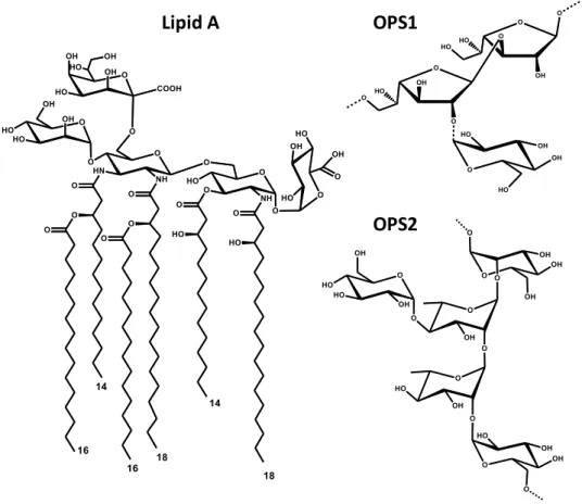
Figure 1. Structure of Acetobacter pasteurianus CIP103108 LOS and OPSs
LOS OPS1
OPS2 * Glc substitution ~75% *
III
oligosaccharide and the O-polysaccharide, whose presence defy the smooth LPS form, whereas molecules devoid the last domain are characterized as rough LPS (or Lipooligosaccharide, LOS). LPSs are crucial for bacterial survival, contributing significantly to the integrity and stability of the OM and protecting the bacterium from the external milieu stress factors. More interestingly, the LPS (or more precisely the lipid A) is classified as a PAMP (Pathogen Associated Molecular Pattern), recognized by the immune system upon binding to a receptorial complex formed by a transmembrane protein belonging to the Toll-like receptors family (TLRs) and by a small secreted protein, the MD-2. The event activates the defence systems and stimulates production of cytokines, being generally beneficial for the host, as it helps to combat invading microorganisms. Unfortunately, overstimulation of the immune system may lead to fatal effects as life-threading septic shock. Nonetheless modifications in the lipid A region influence significantly the immunostimulant properties of the whole molecule, diminishing or even deactivating the immunological potential, and therefore can even lead to inhibition of signaling triggered by the “agonist” LPS. Thus, the search of LPS exhibiting inhibitory activity is a high important and interesting topic and is the main aim of the presented work.
IV
The LPS here characterized were isolated from non- human pathogen bacteria derived from different environments. The work includes structural elucidation of the LPS domains of different Gram-negative bacteria, basing on a combination of extensive chemical composition analysis, NMR spectroscopy, and MALDI-TOF MS and MS2.
The structural assays showed particular features of the lipid As derived from an acetic acid bacterium, Acetobacter pasteurianus CIP103108 (Figure 1), a sponge symbiont Endozoicomonas sp. HEX311 (Figure 2) and a marine bacterium, Phaeobacter gallaeciensis BS107 (Figure 3). Moreover, interesting immunostimulatory features were shown for lipid As from A. pasteurianus and P. gallaeciensis upon assays performed with HEK298 cells and Monocytes-derived Macrophages (MoMs).
A Gram-negative bacterium, the ethanologenic Zymomonas mobilis was shown to lack LPS in the outer membrane. Interestingly, by means of DOSY-NMR and DLS analyses it was proven that Zymomonas mobilis exopolysaccharide (Figure 4) played a role in the high ethanolic environment tolerance mechanism. A part of this work is focused on interaction studies of two different lipopolysaccharides with the MD-2, a lipid binding protein, pivotal in the innate LPS recognition system and part of the receptorial complex responsible for the recognition of the LPS,
Figure 3 Structure of Phaeobacter gallaeciensis BS107 lipid A 12:1
10 10
10
10/12:1 10
V
namely the TLR4/MD2 complex. The study was performed applying number of DOSY-NMR studies and Cryo-EM.
The author of the present PhD thesis acknowledges: Prof. Alba Silipo, Prof. Antonio Molinaro, Dr. Flaviana Di Lorenzo, Dr. Roberta Marchetti, Dr. Katarzyna Anna Duda, Prof. Jesús Jiménez Barbero, Dr. Helena Coelho, Dr. Sandra Delgado, Prof. Francesco Peri, Dr. Fabio Alessandro Facchini, Prof. Gerardino D’Errico, Dr. Antonio Fabozzi, Prof. Maria-Lina Bernardini, Dr. Luigi Lembo Fazio for the scientific support.
VI
Abbreviations
AA Acetylated Alditols Ac2O Acetic Anhydride
AI Allergy Immunotherapy AP-1 Activator protein 1 BMDM Bone Marrow-Derived Macrophages
BSTFA
O-bis-(trimethylsilyl)trifluoroacetamide CARS Anti-Inflammatory Response Syndrome
CD14 Cluster Differentiation Antigen 14
CMC Critical Micellization Concentration
COSY Correlation Spectroscopy CPS Capsular Polysaccharide CXCL-8 Interleukin 8
DAMP Damage Associated Molecular Patterns
DHB 2,5-dihydroxybenzoic acid DLS Dynamic Light Scattering DMEM Dulbecco's Modified Eagle's medium
DMSO Dimethyl Sulfoxide
DMSP Dimethylsulfoniopropionate DOC-PAGE Sodium Deoxycholate-Polyacrylamide Gel Electrophoresis DOSY Diffusion-Ordered Spectroscopy
DQF-COSY Double Quantum Filter Correlation Spectroscopy EI-MS Electron Impact Mass Spectrometry
EPS Exopolysaccharide ESI Electrospray Ionization FA Fatty Acid
FAB Fast Atom Bombardment FAME Fatty Acid Methyl Ester FBS Fetal Bovine Serum FCB Flexibacter-Cytophaga-Bacteroides
GC Gas Chromatography
GSL Glycosphingolipid HEK Human Embryonic Kidney [cells]
HMBC Heteronuclear Multiple Bond Correlation spectra HSQC Heteronuclear Single Quantum Coherence IgE Immunoglobulin E IgG Immunoglobulin G IL Interleukin IMAC Metal Affinity Chromatography
Kdo 3-deoxy-D -manno-oct-2-ulopyranosonic acid Ko D-glycero-D -talo-oct-2-ulopyranosonic acid LBP LPS Binding Protein LC Liquid Chromatography LOS Lipooligosaccharide LPS Lipopolysaccharide LTA Lipoteichoic acid
MALDI MS Matrix Associated Laser Desorption Ionisation Mass Spectrometry
MD-2 Myeloid Differentiation Protein-2
MeOH Methanol
MGA Acetylated O-methyl Glycosides
MHC Major Histocompability Complex
MoMs Monocytes-Derived Macrophages
MPEG Macrophage Expressed Protein
MPL® Monophosphoryl Lipid A® MS Mass Spectrometry
MS2 Tandem Mass Spectrometry
MyD88 Myeloid Differentiation Factor NF-kB Nuclear Factor Kappa-Light-Chain-Enhancer Of Activated B Cells NMR Nuclear Magnetic Resonance Spectroscopy
NOE Nuclear Overhauser effect NOESY Nuclear Overhauser Enhancement Spectroscopy
VII OM Outer Membrane
OPS O-polysaccharide OS Oligosaccharide PAMP Pathogen Associated Molecular Pattern
PCP Petroleum/Chloroform/Phenol Extraction
PFG Pulsed Field Gradient PGN Peptidoglycan
PMAA Partially Methylated Alditol Acetates pNPP para-Nitrophenylphosphate PRR Pathogen Recognition Receptor PS Polysaccharide Pyr Pyridine R-LPS Rough Type Lipopolysaccharide
ROESY Rotating Frame Overhauser Enhancement Spectroscopy
SDS-PAGE Sodium Dodecyl Sulfate –Polyacrylamide Gel Electrophoresis
SEAP Secreted Embryotic Alkaline Phosphatase
SEC Size Exclusion Chromatography S-LPS Smooth Type Lipopolysaccharide
SLS Static Light Scattering
STD Saturation Transfer Difference TDA Tropodithietic Acid
TEM Transmission Electron Microscopy
TFA Trifluoroacetic Acid Th T-Helper Cell TLR Toll-Like Receptor TNF-α Tumor Necrosis Factor α TOCSY Total Correlation Spectroscopy TOF Time Of Flight
VLCFA Very Long Chain Fatty Acids WT Wild Type
VIII
INDEX
SECTION I: Introduction
Chapter I Gram negative bacteria ... 2
1.1. The prokaryotes ... 3
1.2. The cell envelope ... 4
1.3. The Lipopolysaccharides (LPS) ... 6
1.3.1. Lipid A, structure and functions ... 8
1.3.2. Core oligosaccharide structure and functions ... 12
1.3.3. O-antigen structure and functions ... 14
1.4 Exopolysaccharides ... 14
1.5. Other glycolipids ... 16
1.6. Innate immunity and adaptive immunity ... 17
1.7. Basis of TLR4/MD-2 recognition of LPS ... 19
1.8. Modulation of TLR4/MD-2 signalling ... 21
1.9 LPS in sepsis and as vaccine adjuvant ... 23
Aims ... 26
References: ... 27
Chapter II Elucidation of LPS and LOS structure ... 33
2.1. Isolation and purification ... 34
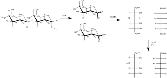
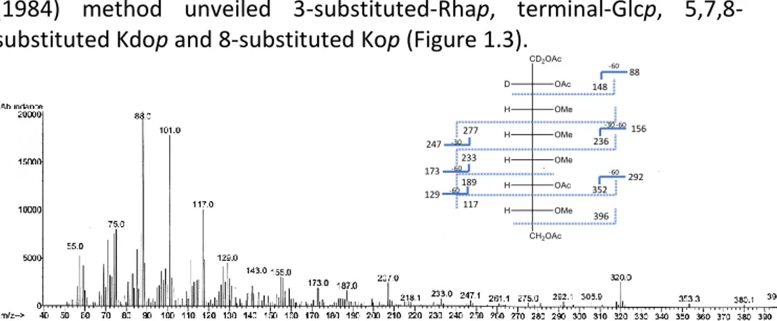
2.2 Degradation and derivatization techniques ... 35
2.2.1 Degradation techniques ... 35
2.2.2. Derivatization techniques ... 36
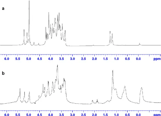
2.3 Spectral techniques in LPS structural elucidation ... 40
2.3.1 Nuclear magnetic resonance ... 40
2.3.2. Mass spectrometry ... 42
IX
SECTION II: Structural, immunological and functional
characterization of bacterial cell wall components
Chapter III Acetobacter pasteurianus CIP103108 ... 48
Premise... 49
3.1 Isolation of the cell wall components ... 50
3.2. Chemical characterization of Acetobacter pasteurianus cell wall components ... 51
3.3 Isolation and purification of the OPS and lipid A. ... 53
3.4 Structural characterization of Acetobacter pasteurianus CIP103108 OPS ... 54
3.4.1. Structural characterization of PS1 ... 54
3.4.2. Structural characterization of OPS2 ... 57
3.5. Structural characterization of the lipid A ... 60
3.6. Structure of the core region ... 64
3.7. Immunological assays ... 71
3.8. Discussion ... 72
3.9. Interaction studies with MD-2 ... 75
3.9.1. Isolation of ligands ... 76
3.9.2. Preparation and purification of recombinant hMD-2 ... 77
3.9.3. Effect of MD-2 on Acetobacter pasteurianus CIP103108 LPS aggregation ... 78
3.9.4. Effect of MD-2 on Bradyrhizobium BTAi-1 Δshc LPS aggregation .. 80
3.9.5 Discussion ... 82
References: ... 84
Chapter IV Endozoicomonas sp. HEX 311 ... 91
Premise... 92
4.1 Isolation ... 93
4.2 Compositional analysis ... 94
X
4.4. Discussion ... 101
References: ... 103
Chapter V Phaeobacter gallaeciensis BS107 SA/WT ... 107
Premise... 108
5.1 Isolation and purification of the LPS ... 108
5.2 Compositional analysis ... 109
5.3 MALDI-TOF and MS2 analysis of the lipid A ... 110
5.4 Immunological studies on Phaeobacter gallaeciensis BS107 LPS ... 115
5.5. Discussion ... 116
References: ... 118
Chapter VI Zymomonas mobilis ... 121
Premise... 122
6.1. Isolation steps of Zymomnas mobilis cell wall components ... 123
6.2 Isolation and compositional analysis of Zymomonas mobilis EPS ... 124
6.3 NMR spectroscopy structural characterization of PS1 and PS2 ... 125
6.4 PFG-NMR and DLS analysis ... 130
6.5. Calculation of the average molecular mass ... 135
6.5.1. Molecular Weight estimation by DOSY ... 135
6.5.2. Molecular Weight estimation by SLS ... 137
6.6. Discussion ... 138
References: ... 141
SECTION III: Experimental section
Chapter VII Materials and methods... 1477.1. Acetobacter pasteurianus CIP103108 ... 148
7.2 Endozoicomonas sp. HEX 311 ... 152
7.3 Phaeobacter gallaeciensis BS107 SA and WT ... 154
7.4 Zymomonas mobilis ... 156
7.5. LPS/MD-2 interaction studies ... 159
XI
Conclusion ... 164 Summary ... 166
1
SECTION I
2
Chapter I
3
1.1. The prokaryotes
The prokaryotes are a group of simple, unicellular organisms, divided into two distinct domains, namely Archea and Bacteria. The term Prokaryota originates from two Greek words, πρό (pro) “before" and κάρυον (karyon) "nut or kernel", as they were the first organisms found on Earth, present billions of years prior any organism of the third, Eucaryota domain (Campbell, 2000). This fact leads to the logical conclusion that the Prokaryotes are rather simple organisms, with cells organised in a different fashion in comparison to Eucarya (Figure 1.1). The first noticeable difference is lack of a membrane enclosed nucleus in case of Prokaryotes. Therefore, unlike Eucarya, the genetic material is kept inside a nucleoid. Moreover, they lack numerous membrane-locked organelles. Therefore, Prokaryotes are known to form cells of a small size in range 1 -10 μm, around ten-fold smaller than Eukaryotes (10-100 μm).
Figure 1.1 Prokaryotic cell scheme
Despite the simplicity, the Prokaryotes create a highly diverse group of microorganisms, widespread on Earth and present in all types of environment, including rivers, oceans, soils, also in conditions considered as “extreme” like hydrothermal vents, hypersaline, highly acidic or basic environments. Furthermore, those organisms are being found interacting with all kinds of organisms, including plants and animals, involved in both, symbiotic and pathogenic interactions and are important decomposers, being crucial element of different elements cycle (Whitman, 1998; Di Lorenzo, 2017a).
4
Bacteria are a group of prokaryotic unicellular microorganisms that differ from Eukaryotes for their simple cellular organization. The bacteria domain includes a heterogenious group of organisms, diverse in terms of cell structure, size or shape (Yang, 2016). Most of them are single-celled but some are organized in multicellular forms consisting of numerous cells that constitute distinctive cell morphologies (coccus, bacillus, spirillum, filamentous) and create distinct colonies visible by light microscopy when grown on petri plates (Raven & Johnson, 2001). Further division can be made basing on the cell wall structure, in particular, Gram-positive bacteria, possessing a thick cell wall between which the dye-iodine complex is trapped, respond to the crystal-violet forming a purple stain, whereas the Gram-negative bacteria possess a thinner, less layered envelope and the dye can be washed out easily (Staley, 2007).
As for the Archea domain, it represents a group of mostly extremophilic and non-human pathogen organism, phylogenetically different form Bacteria.
1.2. The cell envelope
Bacterial cell is covered by a multi-layered and complex structure referred to as cell envelope. Unlike many higher organisms, in order to survive, bacteria had to evolve this type of structures to protect themselves from unfavourable conditions of the outer milieu. As stated previously, basing on fundamental differences in the cell wall structure, two main groups can be distinguished- positive bacteria and
5
Figure 1.2 Structure of Gram-positive and Gram-negative cell envelope
Despite numerous differences in the envelope architecture, common features are evident (Figure 1.2), as the presence of an inner cytoplasmatic membrane, possessing integral proteins and surrounded by the cell envelope. Thus, a layer of peptidoglycan (or so-called murein) is present, whose main function is providing the structural strength to the cell wall. The murein is built of repeating units of β-(1→4) N-acetylglucosamine (GlcNAc) and N-acetylmuramic acid cross linked at position 3 by short peptide chains containing three to five amino acids with alternating L and D-amino acids. The peptide chain contributes to the formation of a 3D mesh-like structure of the polymer (Tytgat & Lebeer, 2014). Although peptidoglycan is present in both, Gram-positive and negative bacteria, it possesses several structural differences among both groups (Figure 1.3). Firstly, the third position of the oligopeptide in case of Gram-positive is occupied by lysine while in case of Gram-negative species by meso-diaminopimelic acid is present. Furthermore, the peptidoglycan layer in Gram positive bacteria is significantly thicker (30-100 nm) than the one found in Gram-negative (couple nanometres). Gram-positive bacterial murein layer possess specific immersed glycolipids, namely lipoteichoic acid (LTA) and wall teichoic acid (WTA) (Vollmer, 2008, Tytgat & Lebeer, 2014).
Phospholipid Transmembrane proteins MraY, MurG, FtsW-RodA
MurNAc GlcNAc Peptide stem monomer
Cross-linked peptide stem
Peptidoglycan synthesis And remodeling enzymes
Lipopolysaccharide Phospholipid
Wall teichoic acid Lipoteichoic acid
Glycoproteins Lipoproteins Surface proteins
Peptido-glycan Inner-membrane Outer-membrane Surface proteins 20 –35 nm 2 –8 nm 35 -4 0 nm 40 -60 nm Envelope of Gram-positive bacteria Envelope of Gram-negative bacteria
6
In addition to the PGN layer, the Gram-negative cell wall also contains an asymmetric bilayer called the outer membrane (OM) which “coats “ the bacterium and faces into the external environment. The OM is constituted from the inner part mainly of phospholipids, whereas the external leaflet is covered up to 75% by a glycolipid known as the lipopolysaccharide (LPS) (Alexander & Rietschel, 2001).
Figure 1.3 Structure of Gram-negative and Gram-positive peptidoglycan (Staley, 2007).
As the LPS are highly-charged, the Gram-negative cell wall has an overall negative charge. The chemical structure of these macromolecules is unique to specific bacterial strain (e.g. sub-species) and is responsible for many of their biological properties. Bacteria can be further covered by glycans, namely capsular polysaccharides (CPS) or exopolysaccharides (EPS).The complexity of all these macromolecules leads to infinite possibilities of structural and functional diversity (Tytgat & Lebeer, 2014).
1.3. The Lipopolysaccharides (LPS)
The lipopolysaccharides (Figure 1.4) are glycoconjugates found on the external leaflet of the outer membrane of Gram-negative bacteria, covering around 75% of the cell surface and greatly contributing to the structural integrity and to the protection of the bacterial cell envelope. These amphiphilic macromolecules play significant role in bacterial survival. They contribute to the membrane integrity, fluidity and permeability upon electrostatic interactions of negatively charged groups with divalent metal ions (Ca2+ or Mg2+). They help to resist hydrophobic
7
components, play a key role in mediating host-bacterium interactions like recognition, colonization or adhesion, virulence, but also tolerance for commensal bacteria and symbiosis (Raetz, 1990; Silipo, 2010, Molinaro, 2015).
Figure 1.4 Scheme of the structure of LPS
All known lipopolysaccharides possess highly conserved architecture, characterized by presence of three chemically, biologically, biosynthetically and genetically distinct domains including the lipid A, core oligosaccharide and O-polysaccharide. The glycolipid portion, named the
lipid A is covalently linked to a hydrophilic portion formed by the core
oligosaccharide (core OS) and a hydrophilic hetero-polysaccharide called
O-polysaccharide chain or O-chain (Holst, 1996; Alexander & Rietschel,
2001). The presence of a complete LPS, the smooth-type LPS (S-LPS) provides a smooth aspect to the bacterial colonies; bacteria can also synthesize lipooligosaccharide (LOS) if the O-chain is absent and are also termed rough type-LPS (R-LPS) because of a rough morphology of the bacterial colonies. The LPSs structures present a significant variability, where composition of all domains may change among species or even bacterial strains. The LPS by itself is not present as single conserved compound, but as a mixture of molecules varying in terms of molecular mass (e.g. presence of different number of repeating units of O-antigen) and slight compositional variations (e.g. different acylation pattern of the lipid A) (Raetz, 1990; Raetz & Whitfield, 2002).
The LPSs were discovered in the late nineteenth century, by demonstrating that heat killed cholera (Vibrio cholerae) bacteria were still toxic, therefore the molecule was named endotoxin (Holst, 1996). During years the LPS was widely studied and its functions and impact on the human body well defined. Nowadays it is well known that the lipopolysaccharides, with their most potent immunologically active site,
O-antigen Core oligosaccharide Lipid A R-LPS S-LPS
8
the lipid A, are considered as pathogen associated molecular patterns (PAMPs), recognized by specific pathogen recognition receptor (PRR), the toll-like receptor 4 (TLR4) upon binding the LPS with the key protein, myeloid differentiation 2 (MD-2) (Park & Lee, 2013). This binding event is crucial to activate the innate immune response and will be widely discussed in further part of the work (see 1.3.7).
1.3.1. Lipid A, structure and functions
The lipid A is the most inner portion of the LPS, represents the most conserved part of the lipopolysaccharide and anchors the whole molecule to the external leaflet of the OM through hydrophobic and electrostatic interactions with the phospholipid layer. Its general structure consists of a disaccharide backbone formed by a β-(1→6) D-glucosamine backbone, phosphorylated at position 1 of the proximal α-D-GlcN (GlcN I) and position 4 of distal β-D-GlcN (GlcN II) and acylated at position 2 and 3 by so called “primary” fatty acids, in turn further acylated at hydroxyl position with “secondary” fatty acids, usually not hydroxylated and with different length (Molinaro, 2015).
The first complete chemical structure of the lipid A derived from E. coli and S. enterica were elucidated in 1983. The lipid A from E. coli is
9
composed by a disaccharide backbone [P→4-β-D-GlcpN-(1→6)-β-D -GlcpN-1→P] acylated at position 2 and 3 of both GlcpNs and carrying at position 2 and 3 of both GlcpNs as primary fatty acids four 14:0 (3-OH) (Figure 1.5). The primary fatty acids located on the distal GlcpN (GlcNII) were both esterified at their hydroxy group by two secondary fatty acids; the amine linked 14:0 (3-OH) was esterified by a 12:0; the ester linked 14:0 (3-OH) by a 14:0 (Figure 1.5). This hexa-acylated lipid A possesses a symmetric (4+2) distribution of the acyl chains.
Even though the lipid A remains the most conserved domain of the LPS, different modifications can be present among different bacterial species, possibly in every region (Figure 1.6) (Silipo & Molinaro, 2011). The saccharidic skeleton of the lipid A is the less variable part of the moiety. The most common modification is the presence of 2,3-dideoxy-2,3-diamino-glucose (GlcN3N or DAG) in place of glucosamine residues. This particularity is found among a number of strains including members of Rhizobiaceae and Bradyrhizobiaceae, Rhodopseudomonas (Di Lorenzo, 2017b) or Bradyrhizobium strains (Komaniecka, 2010; Silipo, 2014). Interestingly, the lipid A isolated from Camphylobacter jejuni is found to possess both, GlcN and DAG in the backbone (van Mourik, 2010). Furthermore, some of bacteria are found to carry an “extended” sugar backbone, comprising of more than two sugar residues. The lipid As among Rhizobiaceae and Bradyrhizobiaceae families are reported to possess a skeleton composed of GlcN or DAG with decoration of mannose and uronic acids like glucosamine (GlcA) or galactosamine (GalA). A GlcpN3N disaccharide backbone has been also identified in Pseudomonas diminuta, Aquifex pyrophilus, Leptospira interrogans (Plötz, 2000; Silipo & Molinaro, 2011). A unique and exceptional lipid A structure identified so far is that of Rizhobium leguminosarum and Rizhobium etli, characterised by an unusual trisaccharide backbone containing a distal galacturonic acid in β-1,4 linkage to GlcNII which is 1,6 linked to a proximal 2-aminogluconic acid residue.
Further modifications are found in the polar decorations. Despite most of known lipid As are bis-phosphorylated, different substitutions are found, as pyrophosphate (PP), phospho- or pyrophospho-etanolamine (P-EtN and PP-(P-EtN), phospho-4-amino-4-deoxy-L-arabinopyranose (P-L -Ara4N, as in Burkholderia (De Soyza, 2008) or Pseudomonas aeruginosa.) Moreover, phospho-glucosamine methyl phosphate, phospho-L,D
-10
glycero-D-manno-heptose has been found linked to both glycoside and non-glycoside phosphates isolated from several species of lipid A.
The acyl chains constitute, together with the polar heads, the most variable component. The heterogeneity is found in terms of fatty acid composition and substitution. Lipid A is often constituted by a mixture of species whose carbohydrate backbone is frequently differently acylated. The acyl chains are generally saturated and usually possess an even number of carbon atoms between 10 and 28, nevertheless also 2-deoxy or oxo-fatty acids were reported. Some bacteria, including Bradyrhizobium, Sinorhizobium or Rodopseudomonas are found to possess Very Long Chain Fatty Acids (VLCFA) of number of carbon atoms up to 32. Secondary fatty acids rarely possess functional groups , while odd numbered secondary acyl chains can be present (Haag, 2011; Silipo, 2014; Di Lorenzo, 2017b). Moreover, a fascinating example of variability is given by the lipid A isolated from Bradyrhizobium strains which is found to possess a sterol-like hopanoid domain linked to the VLCFA (Silipo, 2014). Thus, the term lipid A defines more correctly not one molecule but a family of species structurally related but expressing differences in the acylation and in the phoshorylation pattern. Bacteria can dynamically modulate their acylation pattern in response to environmental changes. An example is given by Yersinia pestis, which synthetizes hexa- or tetra-acylated lipid A dependently from the bacterial growth temperature. (Montminy, 2006; Telepnev, 2009).
The acyl chain composition and distribution are strictly connected with the 3D shape of the lipid A (Figure 1.7.). Asymmetric hexaacylated lipid A species are known to adopt a cone-like structure with a tilt up to 50°. In contrast, hypo-acylated and/or symmetric molecules tend to adopt a cylindrical shape with a tilt up to 15° (Seydel, 1993, 2000).
11
Figure 1.6 Variability of lipid A structures
The lipid A is highly functionalized domain and plays a key role in bacterial survival. The polar anionic substituents are significantly involved in the stabilization of the membrane, by forming electrostatic interactions with divalent cations (Ca2+ and Mg2+) which connect the LPS molecules,
reducing the membrane permeability and forming an efficient protective barrier (Silipo & Molinaro, 2011). Moreover, many structural variations strongly contribute to bacterial resistance and protection. As an example, the presence of Ara4N in the lipid A increases significantly the resistance to cationic antimicrobial peptides by shielding the negatively charged residues (Hamad, 2012). Other examples may be found in the fascinating world of extremophiles, where structure of lipid A is strictly connected to harsh conditions of external milieu (Di Lorenzo, 2017a).
Finally, the lipid A plays a pivotal role in inflammation provoked by Gram-negative bacteria. The domain is recognized by the human innate immune system by binding to a specific TLR4/MD-2 receptorial complex. Immunostimulant activities of the lipid A are described widely further in Chapter 1.7. and 1.8.
12
Figure 1.7 Intrinsic conformations of asymmetric hexa acylated lipid A (A), less active
monophosporyl lipid A ® (B), and showing low activity, inactive or antagonistic penta- (C) and tetra acylated lipid A (D) and symmetric hexa-acylated lipid A (E) (Seydel, 2000).
1.3.2. Core oligosaccharide structure and functions
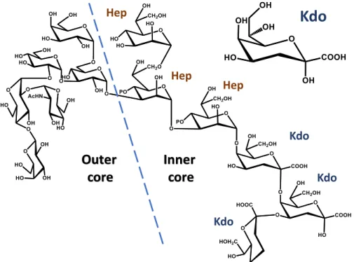
The lipid A is covalently connected to the core oligosaccharide. This part is composed by different monosaccharide units, up to 15, in either linear or branched architecture, frequently possessing non-carbohydrate components, which can be present in a non-stoichiometric fashion (Holst, 2011). The core can be further divided into two regions, namely inner core and outer core (Figure 1.8).
The inner core, which is proximal to the lipid A, is structurally less variable and contains peculiar monosaccharides like heptose (L-glycero-D -manno-heptose and D-glycero-D-manno-heptose) and 3-deoxy-D -manno-oct-2-ulopyranosonic acid (Kdo), a monose marker for all Gram-negative bacteria that connects the core oligosaccharide to lipid A backbone trhough the non-reducing GlcNII (Silipo & Molinaro, 2010). Furthermore, other ulosonic acids can be also found, like like D-glycero-D -talo-oct-2-ulopyranosonic acid (Ko) in Burkholderia, Acinetobacter, Yersinia or Serratia species (Isshiki, 2003; Holst, 2011) or 8-amino-3,8-dideoxy-α-D -manno-oct-2-ulopyranosonic acid (Kdo8N) like in case of Shewanella strains (Vinogradov, 2003, 2004). Negatively charged substituents are
13
often present, as they are involved in the formation of electrostatic interactions with divalent cations, significantly reducing the permeability of the membrane, enhancing the stability and providing resistance to antibiotic compounds and antimicrobial peptides. The most common negative substituents include phosphate (P), pyrophosphate (PP), pyrophosphoryl-2-amino-ethanol (PPEtN), phospho-arabinosamine (PAra4N), uronic acids (often GalpA). On the other side, positively charged groups also might be present, including 2-amino-ethanol (EtN), Ara4N, GlcN, which can substitute cations and interact with the phosphate groups bringing the LPS molecules together and blocking the entrance of positively charged molecules. The outer core, that in case of R-LPS is the most exposed part of the molecule, is characterized by higher structural variability then the inner core, and is mainly composed of common hexoses including Glcp, GlcpN, Galp and GalpN (Silipo & Molinaro, 2010). However, some particularities in this region can be also found, as Shewanella oneidensis MR-1 with a novel type of glycosidic linkage involving an open chain acetal linkage of a GlcN present as non-cyclic carbonyl form (Figure 1.9) (Vinogradov, 2003).
Figure 1.8. Structure of the core OS of E. coli. Indicated inner and outer core, Kdo and
heptoses (Hep). On the top right α-Kdop residue.
Kdo Kdo Kdo Hep Hep Hep
Inner
core
Outer
core
Kdo
14
Figure 1.9. 2-acetamido-2-deoxy-D-galactose (N-acetylgalactosamine, GalNAco) in an open-chain form, linked as cyclic acetal to O-4 and O-6 of D-galactopyranose in
Shewanella oneidensis MR-1 core OS.
1.3.3. O-antigen structure and functions
The O-antigen (also called O-polysaccharide, OPS) is the outermost domain of lipopolysaccharides. The OPS is characterized by high structural diversity, which may occur not only between species, but also between bacterial strains (Raetz & Whitfield, 2002). The domain is made of number of repeating oligosaccharide units containing two to eight monosaccharide residues, the repeating unit can be homopolymeric or heteropolymeric, linear or branched. The addition of non-carbohydrate substituents like phosphate, amino acids, acetyl or formamide groups, often present in non-stoichiometric amount, can be also present. (Knirel, 2011).
Therefore, the O-antigen structure characterizes the serological specificity of the organism. The number of the serogroup are various, while e.g. E. coli produces ~170 serotypes (Stenutz, 2006), S. enterica is known to form only 46 serogroups (Popoff, 2003).
1.4 Exopolysaccharides
The cell wall polysaccharides or exopolysaccharides are a big group of glycans, present at the surface of bacterial cell wall forming kind of a “coat” around the bacterium. They can either form a biofilm or a capsule covering the cell surface. Predominantly the exopolysaccharides are
15
represented by large polymers with average molecular weight 1 × 10⁵ to
3 × 10⁵ Da, composed of different number of monosaccharide residues in
the repeating unit (commonly up to 10), often carrying negative charge at physiological pH, nonetheless neutral macromolecules are also present. Not infrequently substituents of non-saccharidic nature are present, like ketal-linked pyruvate, acetate groups, succinic acid or inorganic residues such as phosphate or sulphate (Poli, 2010). The arrangement of monosaccharides strongly influences the physical properties of the molecule. Furthermore, the structures of EPS vary significantly and a vast number of different structures was reported. A mixture of polysaccharides (Ates, 2015; Kwon, 1994) can be also produced.
The EPSs play a pivotal role in the biofilm development, making up a matrix for this type of formation. Biofilms represent a living micro-environment for microbes, trapping water, ions and soluble products and enabling biochemical contacts between bacteria and surrounding cells. The exopolysaccharides can be attached either physically to the cell wall or remain associated with it, forming a capsule. Therefore, secreted polysaccharide remains loosely bound to the cellular surface. The EPS play highly important biological roles, are known to carry a significant protective task, shielding the bacterium against unfavourable environmental conditions as high or low temperatures, extreme acidity, UV radiation, heavy metals. In case of high salinity or variations of the pH, the polysaccharide viscosity does not change significantly, and the high stability of the polymer assures protection to the bacterial cell. The EPS can also play a key role in reduction of water loss and subsequently assist in rehydration enhancing the water uptake.
Moreover, the EPSs also are highly involved in host-microbe interaction, includin both, symbiotic and pathogenic interactions with animals and including adhesion or immunomodulation. Finally, some of the EPSs found their industrial applications. The examples can be given by xanthan, dextran, gellan and curdlan produced by Xanthomonas campestris, lactic acid bacteria, Pseudomonas elodea and Alcaligenes faecalis, respectively (Mollakhalili & Mohammadifar, 2015; Nwodo, 2012).
16
1.5. Other glycolipids
The lipopolysaccharides are strictly connected with Gram-negative bacteria, being a preserved component of their cell wall. Nonetheless it was reported that some organisms are devoid of the LPS on the outer leaflet of the cell wall. An interesting example is given by bacteria from the genus Sphingomonas, known to possess other type of glycoconjugate on their outer membrane, the Glycosphingolipids (GSL). Similarly to the LPS, GSLs possess an amphiphilic nature as they can be divided into a polar
head containing a few monosaccharide units and a hydrophobic tail (Figure 1.10.). The lipophilic part is made up of a ceramide, specific type of lipid, composed of sphingosine and amide - linked fatty acid, whose nature may vary inside and throughout different microbes. A common characteristic is the presence of a negatively charged monosaccharide, glucosamine or neuraminic acid, substituted with the ceramide. In Sphingomonas paucimobilis and Sphingomonas capsulatus the GSL varies not only in terms of ceramide fatty acids, but also the saccharidic part, therefore two molecules are clearly distinguished, the GSL-1 possessing ceramide linked to GlcpA and the GSL-4A possessing a tetrasaccharide domain linked to the ceramide (Figure 1.11.) (Kawahara, 2000; Kawasaki, 1994). Interestingly, significant conformational similarities can be noticed comparing E. coli Re-LPS and GSL-1 if three molecules are packed together. While in the LPS two Kdo units, two phosphate groups and six fatty acid chains are present, three GSL-1 comprise of three uronic acids and six fatty acid chains. Furthermore, it was shown that in the system of asymmetric lipid bilayers the GSL-1 behaves similarly to LPS for what concerns the channel function of porin, activation of complement and
17
interaction with polymyxin B. Furthermore, due to the lack of a complicated carbohydrate domain, the GSLs possess small variation of antigenicity (Kawasaki, 1994).
Figure 1.11. Structure of GSL-1 and GSL-4A.
1.6. Innate immunity and adaptive immunity
The immunity is the ability of highly developed organisms to resist infections. Mammals respond to microbe invasion with innate and adaptative immune responses. Specific or adaptative immunity is the acquired capacity to recognize and destroy pathogens or their metabolic compounds. The adaptive immune system is capable to “remember” previously encountered microbes and destroy them just when they attack again. Therefore, using this strategy, the human body can avoid most of infections. Unfortunately, sufficient amount of time is needed to give effective response against invading microbes (Janeway, 2001). Given this, a first line of defence had to be developed and this important role is played by the innate immune system.
Innate immunity relies on recognition of microbial patterns,
conserved molecules commonly found in the pathogens, but not found in host organism. Those molecules are known as pathogen associated molecular patterns (PAMPs), and are recognized by specific pattern recognition reseptors, expressed by some of immune system cells. The can be further divided into distinct families (Brubaker, 2015), including
18
predominantly toll-like receptors (TLRs) or lectins. Skin and any epithelial surface separating the inside of the body with external environment is considered as a part of innate defence and therefore constitute the first barrier protecting human and in general animal body from invading microorganisms. When this is overcame (e.g. orally or by skin damage), innate immunity relies on its specific cells. These includes several types like monocytes and macrophages, neutrophils, eosinophils, basophils, mast cells, NK cells, NKT cells, γδ T cells and dendritic cells. Among all cell players, it is possible to distinguish phagocytes, constituted by macrophages and neutrophils, which are known to express significant variety of cell surface receptors and recognize invading microorganisms. Their phagocytic capabilities rely on ability to induction of actin polymerization upon recognition by the ligand by surface receptors, which lets the membrane to surround the microbe and incorporate it forming a phagosome. The germ gets finally degraded in oxygen-dependent manner, basing on reactive oxygen species or oxygen-independent manner, utilizing lysosomes, specific organelle containing enzymes capable of braking the cell wall of invader. The monocytes and mast cells are afterwards able to release cytokines inducing further immunological responses.
Macrophages also contribute to the adaptive immune system. The adaptive immune system (or also so-called specific immune system) can be further spited into two other groups, namely humoral immunity and cell-mediated immunity. The main cellular players in adaptive immunity are two groups of lymphocytes, namely the T lymphocytes involved in expression of cell-mediated immunity and B-lymphocytes which express humoral immunity. The first step of immune response is antigen-processing by macrophages and dendritic cells followed by presentation to B and T lymphocytes which are recognized by specific receptors expressed on their membrane. Therefore, the binding of the antigens to the receptors provokes activation, proliferation and differentiation into effector cells by a process of clonal selection.
In case of cell mediated immunity, the process involves activation of phagocytes, and T-lymphocytes followed by release of different cytokines. In detail, the cytotoxic T-cells are able to attack the leukocytes presenting the antigenic determinants bounded to proteins of major histocompability complex of class I (MHCI) and combat them.
19
Furthermore, also T-helper cells play an important role by recognition of the complex formed by the antigen complexed with MHC class II, effecting in release of cytokines which later promotes the activity of macrophages. In contrast, the humoral response involves substances found in the humor (body fluids) and is mediated by molecules such as antibodies, complement proteins and antimicrobial peptides. The process consists of activation of B-cells or plasma cells which later secret antibodies circulating in the bloodstream and all fluids in general, bind to the foreign antigen causing its destruction. Therefore, some the cells undergo a phase differentiation into memory cells, which in future can encounter the new antigen, making the response fester and more effective.
1.7. Basis of TLR4/MD-2 recognition of LPS
One of the key molecules recognized by the innate immune system in case of Gram-negative infection are the lipopolysaccharides. LPS are recognized as Pathogen associated molecular patterns (PAMPs), structurally conserved molecules able to trigger innate immune response by binding to specific Pathogen Recognition Receptors (PRRs) (Medzhitov, 2001; Akira & Kiyoshi, 2004; Molinaro, 2015). In case of LPS, the innate immune response is triggered by binding of the lipid A to a receptorial complex formed by a small secreted protein, MD2 and a specific transmembrane receptor, member of the Toll-Like family, the TLR4 (Lu, 2008). Several proteins are involved in the endotoxin induced signalling pathway. Once in the body, the LPS is extracted and de-aggregated by a serum protein, the LPS binding protein (LBP) and later transferred to a lymphocyte extrinsic membrane glycoprotein, the cluster differentiation antigen 14 (CD14), whose main role is to enhance the sensitivity towards the lipid A, reducing the binging affinity even to picomolar concentrations (Wright, 1990; Tobias, 1995). It is however important to point out that too high concentrations of the LBP and CD14 (soluble and membrane CD14) were demonstrated to inhibit the immune response (Zweigner, 2001; Kitchens, 2001). The myeloid differentiation protein-2 (MD-2) is a small (17kDa) lipid binding protein which plays a pivotal role in the activation process, complexing the N-terminal segment of TLR4 ectodomain. The MD-2 protein possesses two anti-parallel β-sheets which form a hydrophobic pocket accommodating the lipid A (Shimazu, 1999; Kim, 2007; Park, 2009), or more precisely, the acyl chains of the domain.
20
Thereafter, the appropriate acylation pattern inducing conformational change in the protein is a requirement for the dimerization of the TLR4/MD-2 receptorial complex (Park, 2009; Ohto, 2012). The MD-2 itself is able to bind the LPS and discriminate different lipid As, therefore TLR4, in absence of MD-2, does not recognise the ligand (Shimazu, 1999; Kim, 2007; Park, 2009). The heterodimer formation further induces dimerization of intracellular TIR domains initiating the signaling (Fig 1.8) (Jin & Lee, 2008). TLR4 is the only TLR that is able to transduct the signal through two pathways, namely, utilizing either MyD88 or TRIF adapter. This finally leads to activation of transcription factors like NF-kB or AP-1 from MyD88 and the IFN-β (Figure 1.12.).
Figure 1.12. Model of LPS signalling
TRIF TRAM TIR MyD88 Mal/TIRAP
TRIF IRAK 1/2/4 TRAF6 TRAF6 Ub Ub Ub TRIF TBK IRF3 IKKα/β/γ IKKα/β/γ MKK JNK/p38 IkB IkB NFκB NFκB NFκB NFκB AP1 Nuclear membrane Cell membrane LBP MD2 LBP CD14 TLR4 TLR4* MD2 MD2* LPS
21
1.8. Modulation of TLR4/MD-2 signalling
The hexa-acylated, asymmetric (4+2) lipid A of E. coli LPS is considered as the most potent TLR4/MD-2 agonist. The capacity of various endotoxin to induce or antagonise the synthesis of cytokine and of other
effector molecules is strictly correlated to the primary structure. Structural variations can strongly affect the lipid A potential to induce the TLR4/MD-2 mediated immunological response (Figure 1.13.).
Many structural features including predominantly the number and nature of the acyl chains are considered to modulate the effect upon binding. Lipid A possessing a bis-phosphorylated disaccharide backbone with an asymmetric (4+2) distribution of six acyl residues, of which four saturated primary 3-hydroxylated and two secondary non-hydroxylated acyl residues generally of 12-14 carbon atoms in length (as in the case of E. coli lipid A, Figure 1.13), represents the most stimulatory agonist
Figure 1.13. Lipid A structure-activity relationship. Indicated chemical changes of the E. coli lipid A and factor by which the structure is less active. Blue indications refer to
22
structure for innate immunity in human. Structural variations respect to E. coli hexa-acylated lipid A can correspond to less or not agonist active species. Hypo-acylated lipid As or symmetrical (3+3) hexa-acylated lipid A, adopting a cylindrical shape (Chapter 1.3.1) are known to act as anatagonists or weak agonists. Furthermore, the activity depends also on the number of phosphate groups, the presence of other decorations or variation in the acylation pattern (Rietschel, 1994; Fox, 2010; Molinaro, 2015).
These strong structure-activity correlation results in the fine tuning of the “agonist properties” of the Lipid A, and results in creation of molecules possessing low immunostimulant properties. A fascinating example is given by Monophosphoryl lipid A® (MPL ®) (Figure 1.14.), which consists of Salmonella minnesota lipid A lacking (R)-3-hydroxytetradecanoyl group and the 1-phosphate which possesses low immunostimulatory activities and therefore is used as an anticancer vaccine adjuvant (Cluff, 2010) (see Chapter 1.9). Further modifications in the structure can lead to complete disappearance of the toxic properties and even inhibition of the TLR4/MD-2 activation.
A well-known TLR4/MD-2 antagonist is a tetra-acylated E. coli bio-synthetic precursor, the lipid IVa (Figure 1.15). It was shown that the lipid IVa is able to compete with the toxic LPS for the interaction with the
23
human TLR4/MD-2, can operate as “antagonist” reducing or, in a dose-dependent manner, completely inhibiting the inflammatory cascade induced by strongly endotoxic lipid A species. Importantly, the molecule acts as a weak agonist in murine TLR4/MD-2 (Saitoh, 2004). Other interesting examples of antagonist lipid A were found in Rhodobacter capsulatus (Loppnow, 1990) or Rhodobacter sphaeroides (Anwar, 2015) whose structure was an inspiration in antiseptic drug designment (Kim, 2007).
Figure 1.15. Structure of the lipid IVa
1.9 LPS in sepsis and as vaccine adjuvant
The LPS induced activation of innate immune system through the TLR4/MD-2 complex is a beneficial event for the host, which provides a successful first line of defence. However, overstimulation of the system leads to serious, life-threatening effects.
Sepsis is a condition, based on the overwhelmed response of the innate immune system. It is considered as the primary death cause sourcing from infection, especially when not properly treated or
24
recognized (Singer, 2016). Agonist LPS/lipid A species are able to trigger powerful immune response through TLR4/MD-2 receptor complex, therefore inflammation takes place. When the concentration of the LPS in the body reached high values, systemic inflammatory response syndrome (SIRS) arises. Moreover, as a response to inflammation a contradictory reaction effecting in production of anti-inflammatory cytokines, named Compensatory Anti-Inflammatory Response Syndrome (CARS) occurs. This leads to severe organ dysfunction and finally life-threatening septic shock. Sepsis remains one of the main death reasons among developed countries, furthermore, in the beginning of the century, 45% - 60% of sepsis cases were caused by Gram-negative bacteria (Bone, 1996; McCormick, 2001). Therefore, high demand on new treatment strategies leaded to development of different approaches towards combating the septic cascade. An inspiring example is given by Eritoran (Figure 1.16), antispectic molecule based on a non-toxic lipid A isolated from Rhodobacter capsulatus. The drug has shown a significant decrease of TNF-α and IL-6 levels in a randomized controlled trial on individuals challenged with endotoxin. Finally, Eritoran reached phase III of clinical trials (Peri, 2011).
A further lipid A analogue possessing notable applications is the MPL®. The MPL® is a dephosphorylated lipid A obtained from Salmonalla minnesota R595, which due to removal of the phosphate groups presents significantly lower toxicity in comparison to the native Salmonalla minnesota lipid A (Figure 1.14). This made the compound a promising vaccine adjuvant, administered to various subjects in clinical trials, including allergen immunotherapy.
Allergy is a pathology of the immune system, characterized by a hypersensivity to a substance termed as an allergen which in normal condition is harmless. When the person is sensitized the T helper type 2 cells (Th2) type response, characterized by production of IgE antibodies by B-cells is obtained, which further leads to allergy reaction. The treatment includes allergy immunotherapy (AI) which is based on building immunological tolerance by the patient. After the treatment, immunological system of the patient is modified, therefore instead of IgE release, IgG antibodies which are unable to cause allergic reaction are produced by Th1 pathway. The immunotherapy includes use of pro-Th1
25
adjuvants, which tend to act as pro-inflammatory agents, being able to trigger immune response.
The agents being possible to use are TLR4/MD-2 ligands, represented by the monophosphoryl lipid A®. Interestingly the tests of MPL® as adjuvant in murine model was successful promoting Th-1 directed responses (Wheeler, 2001; Larsen, 2016). The MPL® is nowadays used in a therapeutic pollen-allergy vaccine, Polinex Quatro®. It is important to point out, that the primary clinical development of the MPL® was concentrated on the potential use of the agent in cancer therapy. For instance, the MPL® is used as an adjuvant in a licensed anticancer vaccine, Melacine® (Fox, 2010). Furthermore, the monophosphoryl lipid A® is also found as a constituent of prophylactic vaccines, including GlaxoSmithKline’s (GSK) Fendrix® Cervarix® against hepatitis B and human papilloma virus (HPV) respectively (Fox, 2010). Finally, the MPL® was also combined with other immunostimulants in various adjuvant systems which are in clinical development. For instance, the MPL® in a combination with the saponin QS21 in a liposomal formulation is used as a component of a malaria vaccine which reached phase 3 clinical trials (Lell, 2009). Following the success of the monophosphoryl lipid A® several lipid A based agents are being evaluated for clinical use, like the glucopyranosyl lipid A (GLA), synthetic MPL® derivatives or other E. coli lipid A derivatives (Fox, 2010).
26
Aims
The TLR4/MD-2 receptorial complex is a target correlated with broad spectrum of modern-day disorders which still lack specific pharmacological treatment. These include autoimmune disorders, chronic inflammations, asthma, allergies, infectious and central nervous system diseases, and cancer. Thus, throughout last years many efforts were made to find novel, natural or synthetic TLR4/MD-2modulators.
An inspiring example is given by Eritoran, synthetic and well tolerated by humans lipid A mimetic, which acts as TLR4/MD-2 inhibitor. Successfully, the drug reached phase III clinical trials as an antisepsis agent. An inspiration to create such molecule were two natural lipid As isolated from Rhodobacter sphaeroides and R. capsulatus, phototrophic bacteria found in freshwater or marine environments, which showed agonist activity towards the TLR4/MD-2. On the other hand, compounds able to trigger low immunological response, like the lipid A analogue, MPL® found their role as used nowadays vaccine adjuvants in allergy and cancer therapy or disease prevention. In this context, structural and immunological characterization of the LPS isolated from human non-pathogenic or symbiotic bacteria became an inspiring source of novel, potential TLR4/MD-2 agonists and antagonists. The LPS derived from such bacteria are expected to not induce significant activation of human innate immune system or, even inhibit the signalling caused by agonist LPS. Thus, the main aim of presented work is structural and immunological characterization of new TLR4/MD-2 modulators. An additional goal is to characterize structures and functions of other cell wall glycoconjugates or glycans of these bacteria.
27 References:
Akira S., Kiyoshi K. (2004) Toll-like receptor signalling, Nature Reviews Immunology, 4, 499–511
Alexander C., Rietschel E.T. (2001) Bacterial lipopolysaccharides and innate immunity. J. Endotoxin Res., 7, 167-202.
Anwar M.A., Panneerselvam S., Shah M., Choi S. (2015) Insights into the species-specific TLR4 signaling mechanism in response to Rhodobacter sphaeroides lipid A detection. Sci. Rep. 5, e7657.
Ates O. (2015) Systems biology of microbial exopolysaccharides production. Front. Bioeng. Biotechnol. 3, e200.
Bone R.C. (1996) The sepsis syndrome. Definition and general approach to management. Clin. Chest. Med. 17(2), 175-181.
Brubaker S.W., Bonham K.S., Zanoni I., Kagan J.C. (2015) Innate immune pattern recognition: a cell biological perspective. Annu. Rev. Immunol. 33, 257-290. Campbell N. (2000) Biology: Concepts & Connections. 4th edition. Pearson
Education, San Francisco.
Cluff C.W. (2010) Monophosphoryl lipid A (MPL) as an adjuvant for anti-cancer vaccines: clinical results. Adv. Exp. Med. Biol. 667, 111-123.
De Soyza A., Silipo A., Lanzetta R., Govan J.R., Molinaro A. (2008) Chemical and biological features of Burkholderia cepacia complex lipopolysaccharides. Innate. Immun. 14, 127-144.
Di Lorenzo F., Billod J.-M., Martín-Santamaría S., Silipo A., Molinaro A. (2017a) Gram‐Negative Extremophile Lipopolysaccharides: Promising Source of Inspiration for a New Generation of Endotoxin Antagonists. Eur. J. Org. Chem. 4055-4073.
Di Lorenzo F., Palmigiano A., Al Bitar-Nehme S., Sturiale L., Duda K.A., Gully D., Lanzetta R., Giraud E., Garozzo D., Bernardini M.L., Molinaro A., Silipo A. (2017b) The Lipid A from Rhodopseudomonas palustris Strain BisA53 LPS Possesses a Unique Structure and Low Immunostimulant Properties. Chem. Eur. J. 23, 3637-3647.
Fox C.B., Friede M., Reed S.G., Ireton G.C. (2010) Synthetic and Natural TLR4 Agonists as Safe and Effective Vaccine Adjuvants. In Sub-cellular biochemistry 53.
28
Endotoxins: Structure, Function and Recognition (Eds. Wang X., Quinn P.J.). Springer, Dordecht, Heidelberg, London, New York, pp 303 – 322.
Haag A.F., Wehmeier S., Muszyński A., Kerscher B., Fletcher V., Berry S.H., Hold G.L., Carlson R.W., Ferguson G.P. (2011) Biochemical characterization of Sinorhizobium meliloti mutants reveals gene products involved in the biosynthesis of the unusual lipid A very long-chain fatty acid. J. Biol. Chem. 286(20), 17455-17466.
Hamad M.A., Di Lorenzo F., Molinaro A., Valvano M.A. (2012) Aminoarabinose is essential for lipopolysaccharide export and intrinsic antimicrobial peptide resistance in Burkholderia cenocepacia. Mol. Microbiol. 85(5), 962-974.
Holst O., Ulmer A.J., Brade H., Flad H.D., Rietschel E.T. (1996) Biochemistry and cell biology of bacterial endotoxins. FEMS Immunol. Med. Microbial. 16, 83-104. Holst, O. (2011) Structure of the lipopolysaccharide core region. In Bacterial Lipopolysaccharides (Eds. Knirel Y.A., Valvano M.A.). Springer-Verlag, Vienna, Austria, pp 21-39.
Isshiki Y., Zähringer U., Kawahara K. (2003) Structure of the core oligosaccharide with a characteristic D-glycero-α-D-talo-oct-2-ulosylonate-(2→4)-3-deoxy-D -manno-oct-2-ulosonate [α -Ko-(2→4)-Kdo] disaccharide in the lipopolysaccharide of Burkholderia cepacia. Carbohydr. Res. 338, 2659–2666. Janeway C.A. Jr, Travers P., Walport M., Shlomchik M.J (2001) Immunobiology 5th edition. Garland Science, New York.
Jin M.S., Lee J.O. (2008) Structures of the toll-like receptor family and its ligand complexes. Immunity 29(2), 182-191.
Kawahara K., Moll H., Knirel Y.A., Seydel U., Zähringer U. (2000) Structural analysis of two glycosphingolipids from the lipopolysaccharide-lacking bacterium Sphingomonas capsulate. Eur. J. Biochem. 267(6), 1837-1846.
Kawasaki S., Moriguchi R., Sekiya K., Nakai T., Ono E., Kume K., Kawahara K. (1994) The Cell Envelope Structure of the Lipopolysaccharide-Lacking Gram-Negative Bacterium Sphingomonas paucimobilis. J. Bacteriol. 176(2), 284-290. Kim H.M., Park B.S., Kim J.I., Kim S.E., Lee J., Oh S.C., Enkhbayar P., Matsushima N., Lee H., Yoo O.J., Lee J.O. (2007) Crystal structure of the TLR4-MD-2 complex with bound endotoxin antagonist Eritoran. Cell. 130(5), 906-917.
Kitchens R.L., Thompson P.A., Viriyakosol S., O’Keefe G.E., Munford R.S. (2001) Plasma CD14 decreases monocyte responses to LPS by transferring cell-bound LPS to plasma lipoproteins. J. Clin. Invest. 108, 485-493.
29
Knirel Y.A. (2011) Structure of O-Antigens. In Bacterial Lipopolysaccharides (Eds. Knirel Y. A., Valvano M. A.). Springer-Verlag, Vienna, Austria pp 41-116.
Kwon K. J., Park K. J., Kim J. D., Kong J. Y., Kong I. S. (1994) Isolation of two different polysaccharides from halophilic Zoogloea sp. Biotechnol. Lett. 16(8), 783–788.
Larsen J.N., Broge L., Jacobi H. (2016) Allergy immunotherapy: the future of allergy treatment. Drug Discov. Today. 21(1), 26-37.
Lell, B., Agnandji, S., von Glasenapp, I., Haertle, S., Oyakhiromen, S., Issifou, S., Vekemans, J., Leach, A., Lievens, M., Dubois, M.C., Demoitie, M.A., Carter, T., Villafana, T., Ballou, W.R., Cohen, J., Kremsner, P.G. (2009) A randomized trial assessing the safety and immunogenicity of AS01 and AS02 adjuvanted RTS,S malaria vaccine candidates in children in Gabon. PLoS ONE, 4, e7611.
Loppnow H., Libby P., Freudenberg M., Krauss J.H., Weckesser J., Mayer H. (1990) Cytokine induction by lipopolysaccharide (LPS) corresponds to lethal toxicity and is inhibited by nontoxic Rhodobacter capsulatus LPS. Infect. Immun. 58 (11), 3743–3750.
Lu Y.-C., Yeh W.-C., Ohashi P.S. (2008) LPS/TLR4 signal transduction pathway. Cytokine. 42(2), 145-151.
McCormick J.K., Yarwoo J.M., Schlievert P.M. (2001) Toxic shock syndrome and bacterial superantigens: an update. Annu. Rev. Microbiol. 55, 77–104.
Medzhitov R. (2001) Toll-like receptors and innate immunity. Nature Reviews Immunology, 1, 135–145.
Molinaro A., Holst O., Di Lorenzo F., Callaghan M., Nurisso A., D’Errico G., Zamyatina A., Peri F., Berisio R., Jerala R., Jiménez-Barbero J., Silipo A., Martín-Santamaría S. (2015) Chemistry of Lipid A: At the Heart of Innate Immunity. Chem. Eur. J. 21, 500-519.
Mollakhalili Meybodi N., Mohammadifar M. A. (2015) Microbial exopolysaccharides: A review of their function and application in food sciences. J. Food Qual. Hazards Control. 2(4), 112–117.
Montminy S.W., Khan N., McGrath S., Walkowicz M.J., Sharp F., Conlon J.E., Fukase K., Kusumoto S., Sweet C., Miyake K., Akira S., Cotter R.J., Goguen J.D., Lien E. (2006) Virulence factors of Yersinia pestis are overcome by a strong lipopolysaccharide response. Nat. Immunol. 7, 1066-1073.
Nwodo U.U., Green E., Okoh A.I. (2012) Bacterial exopolysaccharides: Functionality and prospects. Int. J. Mol. Sci. 13(11), 14002-14015.
30
Ohto U., Fukase K., Miyake K., Shimizu T. (2012) Structural basis of species-specific endotoxin sensing by innate immune receptor TLR4/MD-2. Proc. Natl. Acad. Sci. 109, 7421-7426.
Park B.S., Lee J.O. (2013) Recognition of lipopolysaccharide pattern by TLR4 complexes. Exp. Mol. Med. 45, e66.
Park B.S., Song D.H., Kim H.M., Choi B.S., Lee H., Lee J.O. (2009) The structural basis of lipopolysaccharide recognition by the TLR4-MD-2 complex. Nature. 458, 1191-1195.
Peri F, Piazza M, Calabrese V, Cighetti R. (2011) Modulation of Lipopolysaccharide Signalling Through TLR4 Agonists and Antagonists. In Bacterial Lipopolysaccharides (Eds. Knirel Y. A., Valvano M. A.). Springer Verlag, Vienna, Austria pp 389-416.
Plötz B.M., Lindner B., Stetter K.O., Holst O. (2000) Characterization of a novel lipid A containing D-galacturonic acid that replaces phosphate residues. The
structure of the lipid a of the lipopolysaccharide from the hyperthermophilic bacterium Aquifex pyrophilus. J. Biol. Chem. 275(15), 11222-11228.
Poli A., Anzelmo G., Nicolaus B. (2010) Bacterial exopolysaccharides from extreme marine habitats: production, characterization and biological activities. Mar. Drugs. 8(6), 1779-1802.
Popoff M.Y., Bockemuhl J., Gheesling L.L. (2003) Supplement 2001 (no. 45) to the Kauffmann-White scheme. Res. Microbiol. 154, 173–174.
Raetz C.R. (1990) Biochemistry of endotoxins. Annu. Rev. Biochem. 59, 129-170. Raetz C.R., Whitfield C. (2002) Lipopolysaccharide endotoxins. Annu. Rev. Biochem. 71, 635–700.
Raven P.H., Johnson G.B. (2011) Biology 9th Edition, Chapter 34 (Ed.: McGraw Hill).
Rietschel E.T., Krikae T., Schade F.U., Mamat U., Schmidt G., Loppnow H., Ulmer A.J., Zahringer U., Seydel U., Di Padova F., Schreier M., Brade H. (1994) Bacterial endotoxin: molecular relationships of structure to activity and function. FASEB J. 8, 217-225.
Saitoh S., Akashi S., Yamada T., Tanimura N., Kobayashi M., Konno K., Matsumoto F., Fukase K., Kusumoto S., Nagai Y., Kusumoto Y., Kosugi A., Miyake K. (2004) Lipid A antagonist, lipid IVa, is distinct from lipid A in interaction with Toll-like receptor 4 (TLR4)-MD-2 and ligand-induced TLR4 oligomerization. Int. Immunol. 16(7), 961-969.
31
Seydel U., Labischinski H., Kastowsky M., Brandenburg K. (1993) Phase behavior, supramolecular structure, and molecular conformation of lipopolysaccharide. Immunobiology. 187(3-5), 191–211.
Seydel U., Oikawa M., Fukase K., Kusumoto S., Brandenburg K. (2000) Intrinsic conformation of lipid A is responsible for agonistic and antagonistic activity. Eur. J. Biochem. 267, 3032-3039.
Shimazu R., Akashi S., Ogata H., Nagai Y., Fukudome K., Miyake K., Kimoto M. (1999) MD-2, a molecule that confers lipopolysaccharide responsiveness on toll-like receptor 4. J. Exp. Med. 189, 1777-1782.
Silipo A., De Castro C., Lanzetta R., Parrilli M., Molinaro M. (2010) Lipopolysaccharides. In Prokaryotic cell wall compounds structure and biochemistry (Eds. Konig, H., Herald, C., Varma, A.). Springer-Verlag, Berlin, Germany, pp 133–154.
Silipo A., Molinaro A. (2010) The Diversity of the Core Oligosaccharide. In Sub-cellular biochemistry 53. Endotoxins: Structure, Function and Recognition (Eds. Wang X., Quinn P.J.). Springer, Dordecht, Heidelberg, London, New York, pp. 69-99.
Silipo A., Molinaro A. (2011) Lipid A Structure. In Bacterial Lipopolysaccharides (Eds. Knirel Y.A., Valvano M.A). Springer-Verlag, Vienna, Austria, pp. 1-20. Silipo A., Vitiello G., Gully D., Sturiale L., Chaintreuil C., Fardoux J., Gargani D., Lee H.I., Kulkarni G., Busset N., Marchetti R., Palmigiano A., Moll H., Engel R., Lanzetta R., Paduano L., Parrilli M., Chang W.S., Holst O., Newman D.K., Garozzo D., D'Errico G., Giraud E., Molinaro A. (2014) Covalently linked hopanoid-lipid A improves outer-membrane resistance of a Bradyrhizobium symbiont of legumes. Nat. Commun. 5, e5106.
Singer M., Deutschman C.S., Seymour C.W., Shankar-Hari M., Annane D., Bauer M., Bellomo R., Bernard G.R., Chiche J.D., Coopersmith C.M., Hotchkiss R.S., Levy M.M., Marshall J.C., Martin G.S., Opal S.M., Rubenfeld G.D., van der Poll T., Vincent J.L., Angus D.C. (2016) The Third International Consensus Definitions for Sepsis and Septic Shock (Sepsis-3). JAMA. 315(8), 801-810.
Staley J.T., Gunsalus R.P., Lory S., Perry J.J. (2007) Microbial Life 2nd Edition,
chapter 4. Sinauer Associates, Inc., Sunderland.
Stenutz R., Weintraub A., Widmalm G. (2006) The structures of Escherichia coli O-polysaccharide antigens. FEMS Microbiol. Rev. 30, 382–403.
Telepnev M.V., Klimpel G.R., Haithcoat J., Knirel Y.A., Anisimov A.P., Motin V.L. (2009) Tetraacylated lipopolysaccharide of Yersinia pestis can inhibit multiple
32
Toll-like receptormediated signaling pathways in human dendritic cells. J. Infect. Dis. 200, 1694-1702.
Tobias P.S., Soldau K., Gegner J.A., Mintz D., Ulevitch R.J. (1995) Lipopolysaccharide binding protein-mediated complexation of lipopolysaccharide with soluble CD14. J. Biol. Chem. 270, 10482-10488.
Tytgat H.L., Lebeer S. (2014) The sweet tooth of bacteria: common themes in bacterial glycoconjugates. Microbiol. Mol. Biol. Rev. 78(3), 372-417.
Vinogradov, E., Korenevsky, A., Beveridge, T.J. (2003) The structure of the rough-type lipooligosaccharide from Shewanella oneidensis MR-1, containing 8-amino-8-deoxy-Kdo and an open-chain form of 2-acetamido-2-deoxy-D-galactose. Carbohydr. Res. 338, 1991–1997.
Vinogradov, E., Korenevsky, A., Beveridge, T.J. (2004) The structure of the core region of the lipopolysaccharide from Shewanella algae BrY, containing 8-amino-3,8-dideoxy-D-manno-oct-2-ulosonic acid. Carbohydr. Res. 339, 737–740.
Vollmer W., Blanot D., de Pedro M.A. (2008) Peptidoglycan structure and architecture. FEMS Microbiol. Rev. 32, 149-167.
Wheeler A.W., Marshall J.S., Ulrich J.T. (2001) A Th1-inducing adjuvant, MPL, enhances antibody profiles in experimental animals suggesting it has the potential to improve the efficacy of allergy vaccines. Int. Arch. Allergy Immunol. 126(2), 135-139.
Whitman W.B., Coleman D.C., Wiebe W.J. (1998) Prokaryotes: The unseen majority. PNAS. 95(12), 6578-6583.
Wright S.D., Ramos R.A., Tobias P.S., Ulevitch R.J., Mathison J.C. (1990) CD14, a receptor for complexes of lipopolysaccharide (LPS) and LPS binding protein. Science 249, 1431-1433.
Yang D.C., Blair K.M., Salama N.R. (2016) Staying in Shape: The Impact of Cell Shape on Bacterial Survival in Diverse Environments. Microbiol. Mol. Biol. Rev. 80 (1), 187–203.
Zweigner J., Gramm H.J., Singer O.C., Wegscheider K., Schumann R.R. (2001) High concentrations of lipopolysaccharide-binding protein in serum of patients with severe sepsis or septic shock inhibit the lipopolysaccharide response in human monocytes. Blood, 98, 3800-3808.
33