I
INDEX
Abstract I Chapter 1 1 INTRODUCTION 1.1 Introduction 1 1.2 Breast tumors 21.3 Estrogens and the classical estrogen receptor (ER) 5
1.4 The G protein-coupled estrogen receptor (GPER) 9
1.5 Tumor microenvironment 13
1.6 Environmental contaminants 16
1.7 Aryl hydrocarbon receptor (AHR) 20
1.7.1 Cytochrome P450 1B1 (CYP1B1) 24
1.8 Aim of the study 28
Chapter 2 29
MATERIALS AND METHODS 2.1 Reagents 29 2.2 Cell cultures 29 2.3 Real-time PCR 30 2.4 Western blotting 31 2.5 Transient transfections 32 2.5.1 Plasmids 32
2.5.2 Gene silencing experiments 32
2.5.3 Luciferase assays 33
2.6 Site-directed mutagenesis 34
2.7 Chromatin immunoprecipitation (ChIP) assay 34
2.8 Ethoxyresorufin-O-Deethylase activity assay 35
2.9 Immunofluorescence 36
2.10 Co-immunoprecitation assay 36
2.11 Molecular docking 37
II
2.13 In Vivo studies 38
2.14 Immunohistochemistry 39
2.15 Imaging 39
2.16 3D Cell growth assay 39
2.17 Statistical analysis 40
Chapter 3 41
RESULTS 3.1 E2 and G-1 induce CYP1B1 expression through GPER-mediated signalling 41
3.2 A half-ERE site is required for CYP1B1 transcription by E2 and G-1 43
3.3 c-Fos is involved in CYP1B1 expression by E2 and G-1 45
3.4 CYP1B1 activity is stimulated by E2 and G-1 48
3.5 GPER and CYP1B1 are involved in the up-regulation of growth 49
regulatory genes by E2 and G-1 3.6 GPER and CYP1B1 are involved in the growth effects triggered 52
by E2 and G-1 in breast cancer xenografts 3.7 3MC induces CYP1B1 expression through both AHR and GPER 55
3.8Interaction between GPER and 3MC 57
3.9 3MC, E2 and G-1 induce AHR nuclear translocation and CYP1B1 expression 60
through AHR and GPER-mediated signaling 3.10 3MC, E2 and G-1 activate the EGFR/ERK/c-Fos transduction pathway 63
toward CYP1B1 regulation 3.11 Cyclin D1 and CYP1B1 are involved in the growth effects 65
triggered by 3MC, E2 and G-1 through AHR and GPER Chapter 4 71
Discussion References 76
III
Abstract
The cytochrome P450 1B1 (CYP1B1) is a heme-thiolate monooxygenase involved in both estrogen and environmental contaminants metabolism. For instance, CYP1B1 catalyzes the hydroxylation of 17-β estradiol (E2) leading to the production of 4-hydroxyestradiol that may act as a potent carcinogenic agent. In addition, CYP1B1 is overexpressed in different tumors including breast cancer. In this scenario, it is worth mentioning that CYP1B1 expression is triggered by estrogens through the estrogen receptor (ER)α in breast cancer cells. In the present study, we evaluated whether the G protein estrogen receptor namely GPER may provide an alternate route toward the expression and function of CYP1B1 in ER-negative breast cancer cells, in cancer-associated fibroblasts (CAFs) obtained from breast cancer patients, in CAFs derived from a cutaneous metastasis of an invasive mammary ductal carcinoma and in breast tumor xenografts. Our results show that GPER along with the EGFR/ERK/c-Fos transduction pathway can lead to CYP1B1 regulation through the involvement of a half-ERE sequence located within the CYP1B1 promoter region. As a biological counterpart, we found that both GPER and CYP1B1 mediate growth effects in vitro and in vivo. Altogether, these data suggest that estrogens in ER-negative cell contexts may engage the alternate GPER signaling toward CYP1B1 regulation. CYP1B1 is a well-known target gene of the aryl hydrocarbon receptor (AHR) that may be activated by the carcinogenic pollutant 3-methylcholanthrene (3MC). Hence, we aimed to provide novel insights into the molecular mechanisms by which 3MC and E2 may activate a cross talk between AHR and GPER transduction pathways leading to the stimulation of breast cancer cells and CAFs. In particular, our results demonstrate that 3MC and E2 trigger the EGFR/ERK/c-Fos signalling through both AHR and GPER toward the up-regulation of CYP1B1 and cyclin D1 as well as the stimulation of growth responses. Altogether, the present findings suggest that a functional interaction between AHR and GPER may occur toward breast cancer progression.
1
Chapter 1
Introduction
1.1 IntroductionBreast cancer is the most frequent malignancy and the second leading cause of cancer death among females [1]. The elevated incidence of breast cancer in women has been associated with prolonged exposure to high levels of estrogens [2] and environmental contaminants [3]. Estrogens act mainly through the classical estrogen receptor (ER)α and ERβ [4], however, the identification of GPER as a further estrogen receptor has suggested new possibilities by which estrogenic compounds might cause biological effects in different normal and cancer cell types [5-6]. There is increasing alertness that estrogens may regulate certain cell functions through a network of signaling pathways. Beyond estrogens, certain metabolites of either 17β-estradiol (E2) or other compounds as dioxin, benzo(a)pyrene (BaP) and polycyclic aromatic hydrocarbons (PAHs) may influence the development of breast malignancy. Therefore, a great attention has been addressed to the mechanisms involved in the metabolism and the biological functions of both estrogens and xenobiotics [7-8]. For instance, it has been reported that diverse cytochrome P450 enzymes (CYP) contribute to key processes leading to the metabolism of pro-carcinogen compounds [9]. In particular, CYP1B1 (cytochrome P450, family 1, subfamily B, polypeptide 1), which is a heme-thiolate monooxygenase mainly expressed in endocrine-regulated tissues like breast, uterus and ovary, has been indicated as a primary enzyme involved in estrogen and xenobiotics metabolism [10]. In addition, CYP1B1 has been suggested to play an essential role in the development of various hormone-dependent tumors, including breast cancer, through the biotransformation of endogenous estrogens and environmental carcinogens [7, 11-14]. In this context, CYP1B1 is involved in the metabolism of E2 into 4-hydroxyestradiol (4OHE2) that forms DNA adducts and generates free radicals leading to DNA damage and tumorigenesis in different tissues like breast [2, 15-16]. Several compounds as dioxin, (BaP) and PAHs stimulate the transcription of CYP1B1 [8, 17] as wells as its metabolic activity [2]. It is worth noting that estrogens generate a feed-forward
2
loop triggering the transcription of CYP1B1, which in turn is primarily involved in the metabolic conversion of these steroids [17-19]. For instance, the transcription of CYP1B1 was induced in breast and endometrial cancer cells by E2 through the activation of ERα and its binding to an estrogen responsive element (ERE) located within the CYP1B1 promoter sequence [18]. These findings may underline the physiological relevance of CYP1B1 regulation by estrogens in the landscape of the estrogen homeostasis and action, in particular in hormone-sensitive tissues [2, 18-19]. The transcription of CYP1B1 is mainly regulated by the aryl hydrocarbon receptor (AHR), which acts as ligand-activated transcription factor and it is known for mediating the toxicity and tumor-promoting properties of diverse environmental contaminants [20-23]. High levels of AHR and its constitutive nuclear localization have been found in aggressive tumors and tumor cell lines, suggesting that the AHR is chronically activated in tumors and facilitates their progression [22]. The biological responses induced by environmental pollutants such as 3-methylcholanthrene (3MC) involve AHR and its functional interactions with diverse signal molecules, contributing to the development and progression of diverse types of tumours [24-26].
1.2 Breast tumors
Breast cancer is the most common malignancy and the second leading cause of cancer-related death in women worldwide. Whereas localized disease is largely curable, metastatic or recurrent disease carries an unfavourable prognosis [1]. As a greater percentage of breast cancers are being diagnosed at an earlier stage, the medical community has been challenged to develop diagnostic and treatment modalities that maximize benefit from therapy while reducing the morbidity associated with treatment [27]. The management of breast cancer has changed considerably in the last two decades with improvements in systemic therapy and advances in surgical techniques [28]. There are two main types of breast cancer:
1. Ductal carcinoma starts in the ducts that move milk from the breast to the nipple.
2. Lobular carcinoma starts in the lobules of the breast that produce milk. In rare cases, breast cancer can start in other areas of the breast.
Breast cancer may be invasive or non-invasive. Non-invasive breast cancer is also called "in situ."
3
3. Ductal carcinoma in situ (DCIS), or intraductal carcinoma, is breast cancer in the lining of the milk ducts that has not yet invaded nearby tissues. It may progress to invasive cancer if untreated.
4. Lobular carcinoma in situ (LCIS) is a marker for an increased risk of invasive cancer in the same or both breasts (Figure 1.2.1).
Fig. 1.2.1 Representation of the anatomy of the Lobular Carcinoma and Mammary Ductal Carcinoma
Many risk factors lead to breast malignancy development:
Age and gender. The risk of developing breast cancer increases with age. Most advanced breast cancer cases are found in women over age 50 [29]. Women are 100 times more likely to get breast cancer than men are.
Family history of breast cancer. You may also have a higher risk for breast cancer if you have a close relative who has had breast, uterine, ovarian, or colon cancer. About 20-30% of women with breast cancer have a family history of the disease.
Genes. Some people have genes that make them more likely to develop breast cancer. The most common gene defects are found in the BRCA1 and BRCA2 genes. These genes normally produce proteins that protect you from cancer. If a parent passes you a defective gene, you have an increased
4
risk for breast cancer. Women with one of these defects have up to an 80% chance of getting breast cancer sometime during their life [30].
Menstrual cycle. Women who got their periods early (before age 12) or went through menopause late (after age 55) have an increased risk for breast cancer [31].
Other risk factors include:
Alcohol use. Drinking more than 1-2 glasses of alcohol a day may increase your risk for breast cancer [32].
Childbirth. Women who have never had children or who had them only after age 30 have an increased risk for breast cancer. Being pregnant more than once or becoming pregnant at an early age reduces your risk of breast cancer [33].
Hormone replacement therapy (HRT). You have a higher risk for breast cancer if you have received hormone replacement therapy with estrogen for several years or more [34].
Obesity. Obesity has been linked to breast cancer, although this link is controversial. The theory is that obese women produce more estrogen, which can fuel the development of breast cancer [35].
Radiation. The radiation therapy to treat cancer of the chest area, increase higher risk to develop breast cancer [36].
Treatment is based on many factors, including: type and stage of the cancer, whether them cancer is sensitive to certain hormones, whether the cancer over-expresses a gene called HER2/neu. In general, cancer treatments may include chemotherapy medicines to kill cancer cells, radiation therapy to destroy cancerous tissue, surgery to remove cancerous tissue, lumpectomy removes the breast lump; mastectomy removes all or part of the breast; hormonal therapy. Most women receive a combination of treatments. For women with stage I, II, or III breast cancer, the main aim is to treat the cancer and prevent it from returning. For women with stage IV cancer, the objective is to improve symptoms and help them live longer. In most cases, stage IV breast cancer cannot be cured.
5
Stage 0 and DCIS. Lumpectomy plus radiation or mastectomy is the standard treatment. There is some controversy on how best to treat DCIS. Stage I and II. Lumpectomy plus radiation or mastectomy with some sort of
lymphnode removal is the standard treatment. Hormone therapy, chemotherapy, and biologic therapy may also be recommended following surgery.
Stage III. Treatment involves surgery, possibly followed by chemotherapy, hormone therapy, and biologic therapy.
Stage IV. Treatment may involve surgery, radiation, chemotherapy, hormonal therapy or a combination of these treatments.
After treatment, some women will continue to take medications such as tamoxifen for a period. All women will continue to have blood tests, mammograms, and other tests after treatment. Women who had a mastectomy may have reconstructive breast surgery, either at the same time as the mastectomy or later.
1.3 Estrogens and the classical estrogen receptor (ER)
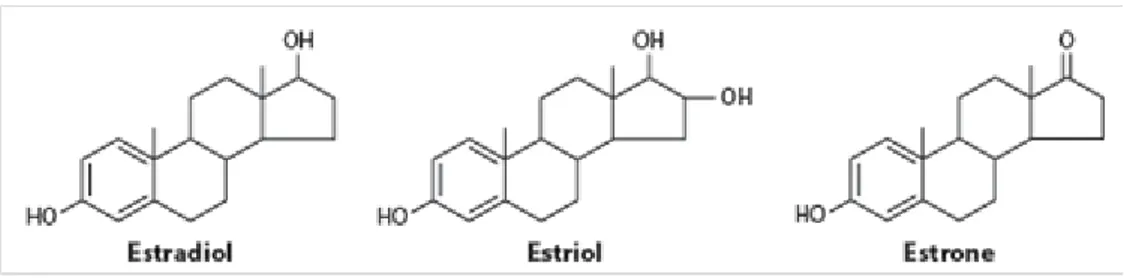
Estrogens are sex steroid hormones, which exhibit a broad spectrum of physiological functions ranging from regulation of the menstrual cycle and reproduction to modulation of bone density, brain function, and cholesterol mobilization [37]. Despite the normal and beneficial, physiological actions of endogenous estrogen in women, abnormally high estrogen levels are associated with the increased incidence of certain types of cancer, in particular breast and endometrial cancer. The predominant intracellular estrogen is 17β-estradiol (E2). Other types of estrogen include estrone (E1) and estriol (E3) (Figure 1.3.1). In premenopausal women, primarily the ovaries secrete E1 and E2 during the menstrual cycle, with minor levels derived from adipose tissue and the adrenal glands. The placenta also produces E3 during pregnancy [38].
6
Fig. 1.3.1 Chemical structures of estrogens
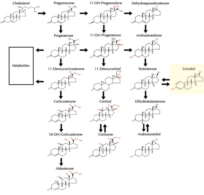
In the ovary, granulosa cells synthesize estrogen from androgen [39]. Ovarian production of estrogen is regulated by the hypothalamic-pituitary-ovarian (HPO) axis and begins by anterior pituitary release of luteinizing hormone (LH) and follicle-stimulating hormone (FSH) in response to the hypothalamic peptide gonadotropin-releasing hormone (GnRH). Acting in concert, LH stimulates androgen production, whereas FSH up-regulates aromatase, which catalyzes the rate-limiting and final step of estrogen biosynthesis: the aromatization of androgen to estrogen (Figure 1.3.2). During ovulation, E2 production rises dramatically by eight- to ten-fold. High levels of estrogen in turn act via negative feedback to dampen estrogen production to inhibit the release of GnRH, LH, and FSH [40]. The primary mediator of estrogen biosynthesis in postmenopausal women is aromatase, which is found in adipose tissue as well as in the ovaries, placenta, bone, skin, and brain [41]. After menopause, ovarian estrogen biosynthesis is minimal, and circulating estrogen is derived principally from peripheral aromatization of adrenal androgen. As such, for obese postmenopausal women, adipose tissue becomes the main source of estrogen biosynthesis; this biosynthetic route is far less significant for non-obese postmenopausal women [42].
7
Fig. 1.3.2 Biosynthesis of Estrogens
Numerous studies have demonstrated the association of estrogen with the development and/or progression of various types of cancer, including breast, endometrium, ovary, prostate, lung, and colon cancer [43-44]. Estrogen mediates its biological effects in target tissues primarily by binding to specific intracellular receptors named ERα and ERβ (Fig.1.3.3). These receptors are encoded respectively by ESR1 and ESR2 which are located on different chromosomes. Like all other members of the nuclear receptors super-family, human ERα and ERβ, are ligand-activated receptors with high degree of sequence homology and similar three-dimensional structure. The ERs are modular proteins composed of four functional domains (Fig.1.3.3):
8
The N-terminal transactivation domain, which is involved in protein protein interactions and in transcriptional activation of target-gene expression.
Fig.1.3.3. Schematic diagram showing the domain organization of human ERα and ERβ
The DNA binding domain (DBD), which plays the most important role in receptor dimerization and in the binding of specific DNA sequences (i.e.EREs).
The hinge region which is the most variable region within ERs.
The C-terminal E/F region encompassing the LBD, the AF-2 domain, the homo- and/or hetero-dimerization domain, and part of the nuclear localization region. It has been demonstrated that ERα acts by multiple mechanisms. In classical genomic mechanism, ligand-activated ERs dimerize and translocate in the nucleus where they recognize specific estrogen response elements (ERE) located in the promoter region of DNA of the target genes.
Besides, E2 can also modulate gene expression by a second indirect mechanism involving the interaction of ER with other transcription factors such as the activator protein (AP)-1, nuclear factor-kB (NF-kB), specificity protein 1 (SP1) which, in turn, binds their specific DNA elements. In addition to the classical mechanism of estrogen signal transduction, which implies the binding of the receptor to DNA, there are a number of non-genomic signaling through which estrogen may exert their biological effects. Indeed, it is now well accepted that ER function can be modulated by extra-cellular signals even in the absence of E2. These findings focus primarily on the ability of polypeptide growth factors such as epidermal growth factors (EGF) and insulin like growth factor-I/II (IGF-I/II) to activate ER and increase the expression of E2 target genes. Moreover, E2 exerts its non-genomic actions, which are too rapid to be accounted for by the activation of RNA and protein synthesis, through the activation of four main signaling cascade: phospholipase C (PLC)/protein kinase C (PKCs), Ras/Raf/MAPK, phosphatidyl inositol 3 kinase (PI3K)/AKT, and cAMP/ protein kinase A (PKA). A rapid activation of the cAMP/PKA
9
pathway has been demonstrated in many different cell types. Phospholipase C (PLC) dependent IP3 production, calcium influx, and PKC activation have also been reported in many different cultured cell types. Moreover, E2 rapidly stimulates the activation of MAPK pathways in MCF-7 cell-line, endothelial, bone and HepG2 cells. E2 can also down regulates MAPK phosphatase-1 activity, leading to the up regulation of extracellular regulated kinase (ERK) activity in breast cancer cells (Figure 1.3.4).
Fig.1.3.4. Representation that summarizes the possible mechanisms of estrogen signal transduction
The E2-induced rapid signals indicate its localization at the plasma membrane. Some authors have suggested that the non genomic actions of estrogen are mediated through a subpopulation of ERα and ERβ located to the plasma membrane. However, in the last few years, a member of the 7-transmembrane G protein-coupled receptor family, GPR30/GPER, has been implicated in mediating both rapid and transcriptional events in response to estrogen under certain circumstances.
1.4 The G protein-coupled estrogen receptor (GPER)
Estrogens regulate different physiological function such as development, reproduction and homeostasis; however, they are involved in the progression of different tumours. The biological actions of estrogens are traditionally mediated by the activation of classical estrogenic receptors ERα and ERβ, which act as transcriptional factors binding specific DNA region, named estrogen responsive elements (EREs) located within the promoter sequence of target genes. Estrogenic receptors are considered as nuclear receptors;
10
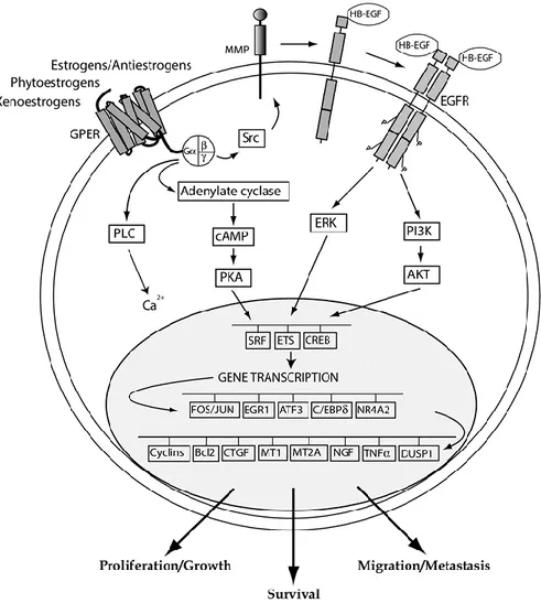
therefore, the estrogenic non-genomic actions are mediated by receptors located on the plasma membrane. In recent years, the identification of GPER as an estrogen receptor has opened a new scenario regarding a further mechanism trough which estrogenic compounds can trigger relevant biological actions in diverse cell contexts. GPER was first identified as an orphan member of the 7-transmembrane receptor family in the late 1990s [47-48]. GPER belongs to the rhodopsin-like receptor superfamily [47] and its gene is mapped to chromosome 7p22.3 [49]. Several studies have reported GPER expression on the plasma membrane, in the endoplasmic reticulum and in the Golgi apparatus as well as in the nucleus of CAFs extracted from mammary biopsies [50-52]. As it concerns signalling pathways, it has been demonstrated that GPER ligands may bind the receptor and activate heterotrimeric G proteins, which then activate Src and adenylyl cyclase (AC) resulting in intracellular cAMP production. Src is involved in matrix metalloproteinases (MMP) activation, which cleave pro-heparan-bound epidermal growth factor (pro-HB-EGF) and release free HB-EGF. The latter activates EGF receptor (EGFR), leading to multiple downstream events; for example, activation of phospholipase C (PLC), PI3K, and MAPK [53]. Activated PLC produces inositol triphosphate (IP3), which further binds to IP3 receptor and leads to intracellular calcium mobilization [54]. The downstream signal of PI3K is AKT transduction pathway. Main biological consequence of AKT activation is closely related to cancer cell growth, catalogued loosely into three aspects: survival, proliferation and growth [55]. The MAPK and PI3K cascade results in activation of numerous cytosolic pathways and nuclear proteins, which further regulate transcription factors such as SRF, CREB, and members of the E26 transformation specific (ETS) family by direct phosphorylation [4,56]. This promotes the expression of a second wave of transcription factors such as Fos, JUN, EGR1, ATF3, C/EBPδ, and NR4A2. Cells are then reprogrammed under the effect of this network of transcription factors and a series of GPER target genes, like CTGF, cyclins, EGR-1, HIF-1, VEGF, are up-regulated [4] (Figure 1.4.1). Superimposed on these responses, there may be a variety of signalling crosstalk pathways and both negative and positive feedback loops. For example, it has been demonstrated that EGF up-regulates GPER expression through the EGFR/MAPK pathway in ER-negative breast cancer cells, most likely by promoting the recruitment of the c-Fos-containing transcription factor AP-1 to the GPER promoter [49]. Considering that GPER signalling uses the EGFR/MAPK pathway, a positive feedback loop is conceivable. This mechanism is also operational for EGF and the related growth factor
11
TGFα in ERα-positive breast cancer cells [57]. GPER gene expression has been detected in at least four kinds of human tumour specimens or cell lines, including breast cancer [4, 58-61], endometrial cancer [62-65], ovarian cancer [49, 66], thyroid cancer [67], and a rat pheochromocytoma cell line PC-12 [68]. In addition, there is a growing body of evidence supporting that GPER is strongly associated with cancer proliferation [49, 62, 65, 67,69-72], migration [4, 73], invasion [62], metastasis [60-61], differentiation [62], and drug resistance [74-75]. Indeed, as estrogen stimulates the progression of breast cancer in approximately two-thirds of patients who are ER + [76-77], some selective estrogen receptor modulators (SERMs), such as tamoxifen, have been clinically used to antagonize the binding of estrogen to its classic ERs, which is an effective therapeutic strategy in attenuating the growth of ER+ breast cancers. However, there are around 25% of ER + breast cancer patients that do not respond to anti-estrogen therapy (Early Breast Cancer Trialists Collaborative Group 2005). It implies that blockade of classic ERs alone may not be enough to abolish estrogen-induced breast cancer cell growth, since estrogen may promote cell growth through other receptor besides classic ERs. Such hypothesis is further supported by the discovery of GPER as the third specific ER with different structure and function to ERα and ERβ. GPER has a high binding affinity to not only estrogen, but also some SERMs, such as tamoxifen and ICI 182,780. Estrogen and SERMs stimulate GPER action without any antagonist effects [59]. These important findings provide a new possible mechanism for the progression of estrogen-related cancers, and raise a new potential target for anti-estrogen therapy. As it concerns clinical findings, GPER overexpression was associated with lower survival rates in endometrial and ovarian cancer patients [64] as well as with a higher risk of developing metastatic disease in breast cancer patients [60]. Moreover, in a previous extensive survey, GPER was found to be highly expressed and significantly associated with tumour size (>2 cm), with the presence of distant metastases and increased human EGFR-2 (HER-2)/neu expression [60]. Likewise, in a recent study performed in the aggressive inflammatory breast cancer, the majority of tumours were GPER positive [78], suggesting that GPER expression may be considered a predictor of an aggressive disease. In addition to the aforementioned studies on the potential functions of GPER in cancer and possibly other pathological conditions, this receptor was implicated in a broad range of physiological functions regarding the reproduction, the metabolism, the bone, the cardiovascular, the nervous and immune systems [79]. Estrogen binds to GPER with a high affinity of a reported Kd 2.7 nM [59] or
12
6 nM [6], through which alternative estrogen signalling pathways are activated. Moreover, two different synthetic compounds, G-1 [80] and G15 [81], which were identified using virtual and bio-molecular screening, are respectively a specific agonist and antagonist of GPER. In addition, different studies show that ICI 182,780 [58-59], tamoxifen [58], and 4-hydroxytamoxifen (OHT) [4, 65-67] are also able to bind GPER and mimic estrogen effects. It has been reported that a variety of xenoestrogens, including bisphenol A, can bind and activate GPER leading to important biological responses [82].
Fig. 1.4.1 GPER-mediated transduction pathways
GPER was also demonstrated to mediate the stimulatory action of estrogens in cancer-associated fibroblasts (CAFs), indicating its potential to contribute to cancer progression also through these important players of the tumour microenvironment [51, 83].
13
1.5 Tumor Microenvironment
The breast cancer microenvironment is a complex combination of several different cell types and molecules and is a key contributor to malignant progression [84]. The role of tumor microenvironment is becoming more and more important in breast cancer. Several stromal cell types are implicated in promoting the ‘hallmarks’ of cancer cells [85]. Tumor microenvironment includes fibroblasts, macrophages, immune cells, adipocytes, endothelial cells, and antigenic vascular cells (Figure 1.5.1). Stromal cells surround and interact with tumor cells. Over the last years, a robust body of evidence has highlighted the importance of the crosstalk between tumor and stoma. Tumor microenvironment has been shown to play a crucial role in tumorigenesis, from initiation to progression. Stromal cells promote cancer growth and invasion through the chemokine–chemokine receptor axis [86-87]. Infiltrating immune cells energize the immune effectors and vascular cells permit nutrients and oxygen uptake by tumors. In a normal mammary duct, there are luminal epithelial cells internally and epithelial cells externally delimited by a basement membrane, which maintains the luminal cell polarity [88]. The extracellular matrix (ECM) allows communication with the surrounding stroma. Genetic and epigenetic alterations lead to luminal cell proliferation, loss of epithelial polarity and decrease of myoepithelial cells, and changes in the ECM/basal membrane, finally resulting in mammary tumor development [89].
Fig. 1.5.1 Tumour Microenvironment
Among the components of the tumour microenvironment, the cancer-associated fibroblasts (CAFs) represent a particularly important cell type, which stimulate cancer progression towards an aggressive phenotype [86-87]. Numerous studies on the origin of CAFs (Figure
14
1.5.2) have reported that the precursors could be resident tissue fibroblasts, bone marrow-derived cells (BMDCS), hematopoietic stem cells, epithelial cells (epithelial-mesenchymal transition, EMT), endothelial cells, (endothelial-mesenchymal transition; EndMT) and cells derived from adipose tissue [90-91]. During the last few years, many studies have highlighted the importance of the cross talk between the tumour and the stroma. As opposed to normal fibroblasts, CAFs [92] improve tumor growth and metastasis by producing growth factors and ECM proteins, as well as by modulating immune polarization [93]. In addition, the number of CAFs is increased during tumor progression [94]. Accordingly, growth factors, cytokines, chemokines, and matrix metalloproteinases secreted by stromal cells lead to the recruitment of macrophages, endothelial precursor cells, and regulatory lymphocytes, which sustain tumor progression [95]. It is worth noting that stroma has been correlated with clinical outcomes and response to therapy in breast cancer [96]. The expression of ECM genes, uniformly expressed in both neoplastic and adjacent stromal cells, may divide breast cancers into different subgroups with different clinical outcomes [97-98]. A study performing hierarchical clustering of the gene-expression profile of ECM-related genes classified breast cancer samples into four groups associated with different clinical outcomes [99]. Stromal signatures are highly informative for patients with breast cancer. A serum-activated gene-expression signature from activated fibroblasts was identified as a negative prognostic factor in patients with breast cancer [100]. In addition, a 26-gene signature called the stroma-derived prognostic predictor was generated by tumor-associated stroma and matched normal stroma from breast cancer samples [101]. This signature was found to be an independent prognostic factor [102]. So tumor microenvironment influences patient outcomes and stromal gene expression signatures represent a strong prognostic value recapitulating the immune, angiogenic, and hypoxic responses [100]. The stromal cells can be divided into three general classes:
Infiltrating immune cells Angiogenic vascular cells
Cancer-associated fibroblastic cells
In the process of tumor formation, the normal microenvironment ‘niche’ changes to an altered (ie, reactive or desmoplastic) stroma which is composed of non-malignant supporting cells (ie, blood vessels, infiltrating inflammatory cells and blast-like cells) [101-102]. This altered microenvironment acts as a collaborative partner in the process of
15
tumourigenesis by influencing the homeostasis of cancer cells via paracrine regulators (e.g., growth factors, cytokines and chemokines) and exosomes containing nucleic acids [101, 103-105]. CAFs are α-smooth muscle actin positive, spindle-shaped, blast-like cells. Differentiation of CAFs from other cell types, such as local fibroblasts, hepatic stellate cells, mesenchymal stem cells, endothelial and epithelial cells, is mainly mediated by transforming growth factor-β1 (TGF-β1), but other factors, such as growth hormones (ie, epidermal growth factor (EGF), fibroblast growth factor (FGF) and platelet-derived growth factor (PDGF)), chemokines, epigenetic regulators and oxidative stress also may play a role in CAFs differentiation [104, 106-107] (Figure 1.5.2). CAFs, phenotypically, closely resemble normal myofibroblasts, but they express specific markers (ie, fibroblast activation protein (FAP), fibroblast-specific protein 1, neuronglial antigen-2, vimentin, Thy-1, tenascin (TN)-C, periostin (POSTN), palladin or podoplanin (PDPN)) and display an increased proliferation and migratory behaviour in vitro [108-109]. CAFs produce and secrete various extracellular matrix (ECM) proteins (ie, collagens I, III, IV), proteoglycans (ie, fibronectin, laminin, TN), chemokines (eg, CXCL and CCL), cytokines (eg, interleukin (IL)-6 and IL-8) and other tumour-promoting factors which affect vascularization (ie, PDGF, vascular endothelial growth factor (VEGF), stromal-derived factor-1 (SDF-1), matrix metalloproteinase (MMPs)), proliferation capacity, tumour cell invasiveness and survival (ie, TGF-β, EGF, hepatocyte growth factor (HGF) or FGF) [101, 110-112]. Regarding anticancer therapy, the frequency of genetic mutations in CAFs is one of the most important issues. Cells with genetic stability may be less prone to escape or resistance to chemotherapy than those with genomic instability [113]. Several studies demonstrated that high percentage of CAFs undergo genetic alterations, such as loss of heterozygosis or mutation of tumour suppressor genes (i.e., phosphatase and tensin homolog and P53) [114-117]. The theory of genetic coevolution of CAFs and the neighbouring cells (i.e., random mutation of CAFs generated independently from neoplastic epithelial cells that may support tumour progression) is under debate due to the potential artefacts caused by the analytical methods used for the identification of these genetic alterations [118]. Other groups described that the somatic mutations of CAFs are found to be extremely rare and are unlikely to be responsible for their stable cancer-promoting attributes [119]. Interestingly, CAFs derived proteins, which may have an important role in the development of environment-mediated drug resistance [101-102], may act as powerful prognostic markers and [101] may be promising targets of anticancer therapy [118].
16
Fig. 1.5.2 CAFs origin
1.6 Environmental contaminants
Polycyclic aromatic hydrocarbons (PAHs) are a large group of organic compounds comprised of two or more fused benzene rings, belonging to this group: 2- aminoanthracene, benz[a]anthracene (B[a]A), benzo[a]pyrene (B[a]P), B[a]P-4,5-dihydroepoxide (BPE), B[a]P diol epoxide (BPDE), benzo[e]pyrene (B[e]P), benz[j]aceanthrylene (B[j]A), benzo[k]fluoranthene (B[k]F), benz[l]aceanthrylene (B[l]A), cyclopenta[c,d] pyrene (CPP), dibenzo[a,l]pyrene (DB[al]P), 7,12-dimethylbenz[a] anthracene (DMBA), DMBA-3,4-diol, fluoranthene, fluorene, indeno [1,2,3-c,d]pyrene (IND), 3-methylcholanthrene, 1-methylpyrene, 1- nitropyrene, perylene, phenanthrene, and pyrene [120] (Figure 1.6.1). They generally originate from combustion processes and are widely distributed in the natural environment as a result of atmospheric transportation, wet and dry deposition, and surface-to-air exchange processes [121-122]. Sixteen PAHs are included among the 129 priority pollutants announced by the U.S. Environmental Protection Agency [123], some of which are carcinogenic [124-125]. ePAHs and the constitutent PAHs may be taken up by living organisms and accumulate via the food chain, and are exposed to humans via inhalation and the ingestion of food; therefore, they pose potential threats to the ecosystem and human health [126-127]. ePAHs can be found in various sources, such as air, dust, smoke, sediment, water, soil and oil in the environment
17
and/or pollutants, as forms such as particulate matter and materials in oven, fuel and tar. PAHs in the air mainly originate from the incomplete combustion of carbonaceous materials such as fossil fuel and biomass including emissions from coke and coal burning in occupational settings and forest fire, the exhaust fumes of motor vehicles, flues of biomass burning, for cooking and heating in rural areas, and tobacco smoke [121, 128-130].
Fig. 1.6.1 chemical structures of polycyclic aromatic hydrocarbons (PAHs)
These chemical carcinogens in the environment are chemically inert in themselves and require metabolic activation by cytochrome P450 (CYP) enzymes to more reactive metabolites in order to exhibit carcinogenicity in experimental animals and humans. Of the
18
17 families of human CYPs identified to date, the CYP1, 2, and 3 family members play major roles in the metabolic activation of a variety of environmental carcinogens (Figure 1.6.2).
Fig. 1.6.2 Schematic representation of the cytochrome P450-CYP-mediated effects of PAHs
It has been suggested that CYP1A1 and CYP1B1 are responsible for the activation of most carcinogenic PAHs to epoxide intermediates, which are further converted to more reactive diol-epoxides with the aid of epoxide hydrolase. Many studies have reported correlations between the induction of cytochrome P450 (CYP), elevated 7-ethoxyresorufin-O-deethylase (EROD) activities, lysosomal membrane destabilization, DNA damage, and endocrine and reproductive effects in fish and invertebrates with PAH contamination in the water and sediments they inhabit. Humans may be exposed to environmental PAHs via the intake of PAH-contaminated food as well as the unintentional ingestion of soil and dust via hand-to-mouth behaviors, inhalation, and dermal contact [127, 130-133]. Epidemic studies have demonstrated that heavy exposure to PAHs from occupational environments increases the risk of developing various cancers including lung, skin, bladder, and larynx cancers [126, 134-136]. PAHs are known to stimulate AHR mediated cell signalling, and their mechanisms of actions have been examined extensively [137]. AHR is a ligand-activated transcription factor that binds to planar aromatic hydrocarbons, such as flavonoids, polyphenols, indoles, and the polyaromatic and halogenated aromatic hydrocarbons of natural and industrial origins. Previous studies described the association or crosstalk between AHR and various cell signaling pathways, such as mitogen-activated protein kinase (MAPK), NF-kB, and cell cycle/ apoptosis-related pathways [138-141]. Since AHR
19
is not a direct regulator of hormonal actions, the mechanisms underlying the endocrine-disrupting effects of AHR and its ligands require the involvement of hormone receptors and/or regulators. Since estrogen and estrogenic chemicals are an important category of endocrine modulators and sometimes act as endocrine disruptors, the involvement of estrogen signalling associated with AHR-mediated cell signalling stimulated with PAHs has been investigated in an attempt to elucidate the mechanisms responsible for their biological effects [21,142-143]. The biological activities of PAHs are categorized by specific signalling pathways (MAPK and other signalling pathways), regulatory mechanisms (chromatin/epigenetic regulation, cell cycle/DNA damage control, and cytoskeletal/adhesion regulation), and cell functions (apoptosis, autophagy, immune responses/inflammation, neurological responses, and development/differentiation), receptor-mediated pathways, growth factor/cytokine-signalling pathways, and key-regulator/modulator-dependent pathways. 3-Methylcholanthrene is involved in signalling pathways, cell functions, such as chromatin condensation in apoptosis and cell adhesion regulation during epithelial-mesenchymal transition [144] and in diseases, such as rheumatoid arthritis [145]. Many naturally occurring and anthropogenic compounds exhibit estrogenic properties in a variety of in vivo and in vitro animal bioassay systems.PAHs act in the same manner as hormonal estrogens, duplicating their physiologic effects, directly compete with and inhibit the effects of hormonal estrogens and act indirectly to modify estrogen availability or expression of hormonal effects in target tissues. Benzo[a]pyrene (BaP), is a mammary carcinogen, as are several other members of the PAH class [146-150]. For many years, 7,12-dimethylbenz[a]anthracene (DMBA) and 3-methylcholanthrene (3MC) have been used as model compounds for studying the molecular mechanisms of mammary carcinogenesis in the Sprague-Dawley rat Mammary. Tumours induced by PAH are predominantly estrogen-dependent, either directly or indirectly via release of pituitary prolactin [151-155].Only a few carcinogenic PAHs and their hydroxyl derivatives have been studied for ER binding properties. The results of ER binding studies with DMBA in calf uterine cytosol and rat mammary and uterine cytosol were conflicting [156-158]. The binding affinity of 3MC for rat uterine ER was reported to be similar to that of tamoxifen [159], although in other studies [160] 3MC binding was observed only to a non-ER cytosol protein. Neither 3MC, dibenz[a,h]anthracene, or dibenz[a,c]anthracene interfered with estradiol binding in calf uterine cytosol [161]. By altering the regulatory activity of fibroblasts, PAH may conceivably affect the estrogenic
20
control of mammary growth promotion and differentiation, and ultimately cell transformation.Since DMBA and other carcinogenic PAH, are genotoxic per se following metabolic bioactivation, it is not currently possible to assess the relative contribution, if any, of altered peripheral estrogen metabolism to the process of PAH-induced neoplastic transformation in estrogen-responsive tissues. Carcinogenic PAHs extensively localize in breast tissue of rats [162-163], are metabolized by human mammary cells to reactive products that bind to DNA [164], and elicit genotoxic effects in human and rat mammary epithelial cells [165]. In a recent clinical report, BaP-like DNA adducts were detected at significant levels in the breast tissues of 41% of a group of breast cancer patients, and were absent in all noncancerous controls [166]. These findings lend strong support to the possible involvement of carcinogenic PAHs in the etiology of human breast cancer, although they suggest no a prior reason why estrogen interactions should participate. Nevertheless, the currently available scientific literature for DMBA and 3MC provides persuasive evidence for the hypothesis that certain carcinogenic PAHs produce a unique duality of pathologic effects encompassing both genotoxic and non-genotoxic components. The genotoxic component may cause the activation of oncogenes, inactivation of tumour-suppressor genes, or amplification of growth factor genes, while the non-genotoxic component results in increased cell proliferation. A fundamental assumption in carcinogenesis is that irreversible fixation of genetic damage can only occur in replicating cells, and the greater the rate of cell division the greater will be the probability for initiated cells to progress to a malignant tumour [167]. Carcinogenic PAH such as DMBA and 3MC possess not only the capability for producing direct DNA damage. This dynamic relationship between genotoxic and mitogenic stimuli, in conjunction with dose and various host factors (e.g., age, genetic predisposition, high fat diet), may be a critical determinant of cancer risk for exposure to all carcinogenic PAHs having steroidal activity.
1.7 Aryl hydrocarbon receptor (AHR)
AHR is a cytosolic ligand-activated transcription factor, which belongs to the member of bHLH/PAS (basic helix–loop–helix/period [Per]-aryl hydrocarbon receptor nuclear translocator [ARNT]-single-minded [SIM]) family of heterodimeric transcriptional regulators [168-170]. The bHLH motif located in the amino (N)-terminal of AHR protein, has two functionally distinctive and highly conserved domains, which together make up a region of approximately 60 amino-acid residues. At the N-terminal end of this motif is the
21
basic domain, which binds AHR to DNA at the consensus regulatory sequences (5′-T/GCGTG-3′) termed AHREs (aryl hydrocarbon response elements), also XREs (xenobiotic response elements) or DREs (dioxin response elements), located in the promoter region of its target genes. At the carboxyl (C)-terminal end of this motif is the helix–loop–helix (HLH) domain, which facilitates protein–protein interactions to form heterodimeric complexes with its partner protein, the ARNT. The PAS domains, including PAS-A and PAS-B, support specific secondary interaction with the ARNT, so that the heterozygous protein complex can form. The ligand-binding site of AHR is contained within the PAS-B domain and contains several conserved residues critical for ligand binding. Finally, a Q-rich domain is located in the C-terminal region of the protein and is involved in co-activator recruitment and transactivation (Figure 1.7.1). In the absence of ligand, AHR exists as part of a cytosolic protein complex, which contains two molecules of the 90 kDa molecular chaperone heat shock protein 90 (HSP90), the HSP90-interacting protein p23 and the immunophilin-like protein XAP2 (also AIP or ARA9). HSP90 interacts with AHR via both the bHLH region and PAS B, which contains the ligand region, and this association is essential for AHR signalling.
22
Following activation by agonist (such as 2,3,7,8- tetrachlorodibenzodioxin, TCDD) binding, AHR changes its conformation that exposes the nuclear localization sequence (NLS) through alteration of XAP2 binding, and its N-terminal nuclear localization signal is activated, directing it to translocate into the nucleus, where it releases its chaperone proteins and dimerizes with its partner protein, the ARNT. This AHR/ARNT heterodimer interacts with several histone acetyltransferases, chromatin remodeling factors, and a partially characterized set of co-activators and/or co-repressors, and the resulting multiprotein complex binds to the XREs and simultaneously bridges the enhancer and the TATA box in the promoter region of its target genes, and recruits RNA polymerase II to initiate transcription [171]. Once transcriptional regulation has occurred, AHR is quickly exported to the cytosol by chromosome region maintenance 1 (CRM1), where it is degraded by the 26S ubiquitin–proteasome, hence preventing constitutive receptor activity [172]. A picture as to how the AHR/ARNT heterodimer actually mediates gene transcription is starting to emerge. The AHR/ARNT complex can alter transcription both by binding to its cognate response element and through tethering to other transcription factors [173]. In addition, AHR activity in the cell is negatively regulated by the presence of the AHR repressor protein (AHRR), whose expression is regulated by AHR. The AHRR is also an AHR-related bHLH/PAS transcription factor, which may dimerize with the ARNT and compete with AHR to bind the XREs. The result is a negative feedback mechanism that involves a down regulation of all genes regulated by AHR [174]. Furthermore, many factors are necessary for AHR-mediated modulation of target gene transcription [173, 175-181]. As a consequence of AHR activation, many detoxification genes are transcriptionally induced, including those coding for the phase I drug/xenobiotic-metabolizing cytochrome P450 enzymes (such as CYP1A1, CYP1A2, CYP1B1, and CYP2B1), the phase II enzymes (e.g. UDPglucuronosyl transferase (UGT1A6), NAD(P)H dependent quinone oxydoreductase-1 (NQO1), the aldehyde dehydrogenase (ALDH3A1), and several glutathione-S-transferases), and the phase III transporters (for example, P-glycoprotein (P-gp), multidrug resistance-associated proteins (MRPs), and organic anion transporting polypeptide 2 (OATP2)), which are expressed in many tissues, such as liver, intestine, kidney and brain, and play crucial roles in drug absorption, distribution and excretion. This drug-metabolizing enzyme system plays central roles in the metabolism, elimination and detoxification (or activation) of xenobiotics and drugs introduced into the human body [182]. In addition to the genomic pathway, several non-genomic pathways
23
have been identified recently. For instance, following exposure to TCDD and other AHR ligands, there is a rapid increase in intracellular calcium concentration (from both extracellular and endoplasmic reticulum sources). TCDD also leads to the functional activation of the tyrosine kinase Src by releasing it from the AHR complex [183]. This could be accompanied by the activation of the Focal Adhesion Kinase (FAK) and by the modification of the adhesion properties of the cell through disruption of focal adhesion points [183-184]. Src activation could be accompanied also very rapidly by the activation of MAP kinases, ERK1 and ERK2. All these processes may converge to regulate pathophysiological processes such as inflammation. Indeed, the calcium influx causes the activation of protein kinase C (PKCa) which phosphorylates a serine residue of a cytosolic enzyme, phospholipase A2 (cPLA2) with the subsequent production of arachidonic acid. The parallel activation of MAP kinases by Src leads to the transcription of cyclooxygenase 2 (COX2) which uses arachidonic acid to produce prostaglandins that can cause inflammation. Thus, these two signalling pathways, which were initially activated by AHR ligands, converge towards the stimulation of inflammation [185]. Moreover, the AHR interacts with Wnt/b-catenin, ER-alpha or NF-kB and strongly modulates their actions [186-189]. On the other hand, these transcription factors also affect AHR signalling. For example, b-catenin has been indicated as a coactivator of this receptor [190]. DNA microarray studies have established that AHR either directly or indirectly regulates a myriad of genes involved in a wide variety of biochemical pathways, including energy metabolism, lipid and cholesterol synthesis, xenobiotic metabolism and various transportation pathways. AHR, it may be considered as an important intracellular chemosensor responsive to both natural and man-made environmental compounds, it is widely expressed in a variety of animal species and humans [191-192]. For several decades, AHR has been studied largely because of its critical role in xenobiotic-induced toxicity and carcinogenesis [193-194]. This is an increasing area that covers multiple aspects of physiology, such as cell proliferation and differentiation, endogenous mechanisms of activation, gene regulation, tumour development, cell motility and migration, and others (Figure 1.7.2) [193].
24
Fig. 1.7.2. The functional relationship between the AHR ligands and the regulatory roles of this receptor in physiology and pathophysiology
1.7.1 Cytochrome P450 1B1 (CYP1B1)
Numerous studies have indicated that some 17β-estradiol (E2) metabolites and environmental contaminants may influence the development of breast cancer; therefore, great attention has been addressed to a better understanding of the biological effects induced by estrogenic and xenobiotics metabolites and the processes of their biosynthesis [7, 195]. Furthermore, it has been demonstrated that various cytochrome P450 enzymes (CYPs) participate actively in the key processes involved in the metabolism of E2 [9]. In particular, CYP1B1 (cytochrome P450, family 1, subfamily B, polypeptide 1), a heme-thiolate monooxygenase mainly expressed in hormone-sensitive tissues such as breast, uterus and ovary, has been indicated as a primary enzyme involved in estrogen and environmental carcinogens metabolism [10]. The gene encoding for CYP1B1 is placed on chromosome 2, at the level of region 2p21-22 [196-197]; the gene contains three exons (371, 1044 and 3707 bp) and two introns (390 and 3032 bp) [196, 198-199]. Although mainly located at the nuclear level, CYP1B1 was also found in the cytoplasmic compartment [200]. CYP1B1 expression has been reported increased in tumor tissues and in tumor microenvironment compared to the normal counterpart [200-203] (Figure
25
1.7.1.1). CYP1B1 over-expression is a prognostic factor in neoplastic progression and tumour metabolism and although it has been observed in diverse cancer cells (colon, pulmonary, renal, bladder and glaucoma), it appears particularly high in hormone-responsive carcinomas (prostatic, mammary, endometrial, and ovarian) [198, 202, 204].
Fig. 1.7.1.1 Role of CYP1B1 in cancer cells and cancer microenvironment
Therefore, the induction of CYP1B1 and the biotransformation of endogenous estrogens and environmental carcinogens, represents an important element in determining what is the risk associated with hormone-dependent tumours, including breast cancer [7, 11, 14]. The metabolic activity of CYP1B1 is a key to better understand the role of environmental compounds in cancer initiation and progression. Many factors are involved in genomic mutation accumulation and in cancer development, in this context CYP1B1 activates polycyclic aromatic hydrocarbons, aromatic and heterocyclic amines, aflatoxin B1 in pro-carcinogenic compounds (Figure 1.7.1.2) [205-207].
26
Fig 1.7.1.2 CYP1B1-mediated PAHs metabolism
In addition, CYP1B1 catalyzes the hydroxylation of E2, leading to the formation of 4OHE2 [196] and subsequently of oestradiol-3,4-quinone, a potent carcinogen metabolite which, by binding to the N-7 position of the guanine, leads the destabilization of the glycosidic bond and subsequent DNA purification and mutagenesis [2, 8, 19, 209] (Figure 1.7.1.3). Considering that 4OHE2 levels in hormone-sensitive tumours, such as breast cancer, are higher than normal tissues [8, 14, 202], CYP1B1 is considered a valid pharmacological target in the therapeutic treatment of hormone-dependent cancer [210].
27
Fig. 1.7.1.3. CYP1B1-mediated estrogen metabolism
The transcription of CYP1B1 is mainly regulated by AHR, which acts as a transcription factor activated by specific ligands, such as dioxin, halogenated aromatic hydrocarbons, BaP and PAHs [8, 211]. In particular, following agonist binding, the cytoplasmic complex consisting of AHR, the heat shock protein-90 and the XAP2 and p23 proteins, translocate at the nuclear level, with consequent dissociation of the AHR complex and formation of the AHR/ARNT heterodimer. This heterodimer binds specific DNA region, named dioxin-responsive-elements (DRE), located in the promoter sequences of target genes including CYP1B1 [212-214] (Figure 1.7.1.4). Growing body of evidence have demonstrated that estrogens generate a feed-forward loop triggering the transcription of CYP1B1, which in turn is primarily involved in the metabolic conversion of these steroids [17-19]. For instance, the transcription of CYP1B1 was induced in breast and endometrial cancer cells by E2 through the activation of ERα and its binding to an estrogen responsive element (ERE) located within the CYP1B1 promoter sequence [18]. These findings may underline
28
the physiological relevance of CYP1B1 regulation by estrogens in the landscape of the estrogen homeostasis and action, in particular in hormone-sensitive tissues [2, 18- 19].
Fig 1.7.1.4. Transcriptional regulation of CYP1B1
1.8 Aim of the study
The aim of this study was to ascertain the potential role elicited by estrogens on CYP1B1 expression and metabolic activity. In particular, we aimed to establish whether estrogenic GPER signalling regulates CYP1B1 expression in ER-negative and GPER-positive breast cancer cells, CAFs obtained from breast cancer patients and CAFs derived from a cutaneous metastasis of an invasive mammary ductal carcinoma (met-CAFs). Moreover, we aimed to evaluate the possible functional cross-talk between GPER and AHR in breast cancer cells and CAFs, which may affect gene expression changes and biological effects upon exposure to the cognate ligands, estrogens and 3MC, respectively. Our results further extend the molecular mechanisms by which 3MC and estrogens could affect breast cancer progression, hence providing novel biological targets for innovative treatments of breast tumor.
29
Chapter 2
Materials and Methods
2.1 Reagents17β-Estradiol (E2), salicylamide (2-hydroxybenzamide), resorufin (7-hydroxy-3H-phenoxazin-3-one), resorufin ethyl ether (7-ethoxy-3H-phenoxazin-3-one), 3-methylcholanthrene (3MC), CH223191 1-Methyl-N-[2-methyl-4-[2-(2-methylphenyl)diazenyl]phenyl-1H-pyrazole-5-carboxamide, were purchased from Sigma-Aldrich (Milan, Italy). G-1 (1-[4-(-6-bromobenzol [1,3]diodo-5-yl)-3a,4,5,9b-tetrahidro3H5cyclopenta[c]quinolin-8yl]-ethanone), G-15 (3aS,4R,9bR)-4-(6-bromo-1,3-benzodioxol-5-yl)-3a,4,5,9b-3H-cyclopenta[c]quinolone and TMS 1-[2,(3,5-dimethoxyphenyl)ethenyl]-2,4-dimethoxybenzene were obtained from Tocris Bioscience (Space, Milan, Italy). Tyrphostin AG1478 (AG) and PD98059 (PD) were obtained from Calbiochem (DBA, Milan, Italy). Mithramycin A (MTMA) was purchase from Abcam (Euroclone, Milan, Italy). All the aforementioned compounds were dissolved in dimethyl sulfoxide (DMSO), except for salicylamide that was dissolved in methanol, 3MC that was dissolved in toluene and mithramycin A that was dissolved in ethanol.
2.2 Cell cultures
SkBr3 and MDA-MB-231 breast cancer cells were obtained by ATCC (Manassas, VA, USA), used less than 6 months after resuscitation, routinely tested, and authenticated according to the ATCC suggestions. SkBr3 cells were maintained in RPMI-1640 (Life Technologies, Milan, Italy) without phenol red, supplemented with 10% fetal bovine serum (FBS) and 100μg/ml penicillin/streptomycin (Life Technologies, Milan, Italy). MDA-MB-231 cells were maintained in DMEM (Dulbecco’s modified Eagle’s medium) (Life Technologies, Milan, Italy) with phenol red, with a supplement of 5% FBS and 100 μg/ml of penicillin/streptomycin. CAFs obtained from breast malignancies and met-CAFs obtained from biopsy of cutaneous metastasis in a patient with a primary invasive mammary ductal carcinoma, who previously had undergone surgery, were characterized
30
and maintained as we have previously described [51, 215]. Briefly, specimens were cut into smaller pieces (1–2mm diameter), placed in digestion solution (400 IU collagenase, 100 IU hyaluronidase, and 10% serum, containing antibiotic and antimycotic solution) and incubated overnight at 37 °C. The cells were then separated by differential centrifugation at 90× g for 2 min. Supernatant containing fibroblasts was centrifuged at 485× g for 8 min; the pellet obtained was suspended in fibroblasts growth medium (Medium 199 and Ham’s F12 mixed 1:1 and supplemented with 10% FBS) and cultured at 37°C in 5% CO2. Primary cells cultures of metastasis-derived fibroblasts and CAFs were characterized by immunofluorescence. Briefly, cells were incubated with human anti-vimentin (V9) and human anti-cytokeratin 14 (LL001), both from Santa Cruz Biotechnology (DBA, Milan, Italy). To characterize fibroblasts activation, we used anti-fibroblast activated protein α (FAPα) antibody (H-56; Santa Cruz Biotechnology, DBA, Milan, Italy) (data not shown). CAFs and metastasis-derived CAFs were maintained in Medium 199 and Ham’s F12 (mixed 1:1) supplemented with 10% FBS and 100μg/ml penicillin/streptomycin. All cell lines were grown in a 37°C incubator with 5% CO2. All cell lines to be processed for immunoblot and RT-PCR assays were switched to medium without serum and phenol red the day before treatments. Signed informed consent from all the patients was obtained and all samples were collected, identified and used in accordance with approval by the Institutional Ethical Committee Board (Regional Hospital of Cosenza, Italy).
2.3 Real-time PCR
Total RNA was extracted from cells maintained for 24 hours in medium without serum and treated with ligand for indicated times, using Trizol commercial kit (Invitrogen, Milan, Italy) according to the manufacturer’s protocol. RNA was quantified spectrophotometrically, and cDNA was synthesized from the RNA by reverse transcription using murine leukemia virus reverse transcriptase (Invitrogen). We quantified the expression of selected genes by real-time PCR. This method is based on the use of intercalating agents, which bind to double stranded DNA. These molecules, when excited by laser beams, emit fluorescence and allow to follow in real-time the progress of the reaction and the increase of the amount of nucleic acid. In this study, we used SYBR Green as the detection method and platform Quant Studio7 Flex Real-Time PCR (Life Technologies). Gene-specific primers were designed using Primer Express version 2.0 software (Applied Biosystems). For CYP1B1, c-Fos, cyclin D1, cyclin E, cyclin A and the ribosomal protein 18S, which was used as a control gene to obtain normalized values, the
31
primers were: TGTGCCTGTCACTATTCCTCATG-3′ (CYP1B1 forward) and
5′-GGGAATGTGGTAGCCCAAGA-3′ (CYP1B1 reverse);
5′-CGAGCCCTTTGATGACTTCCT-3′ (c-Fos forward) and
5′-GGAGCGGGCTGTCTCAGA-3′ (c-Fos reverse); 5′-GTCTGTGCATTTCTGGTTGCA-3′ (cyclin D1 forward) and GCTGGAAACATGCCGGTTA-3′ (cyclin D1 reverse); 5′-GCATGTCACCGTTCCTCCTTG-3′ (cyclin A forward) and
5′-GGGCATCTTCACGCTCTATTTT-3′ (cyclin A reverse);
5′-GATGACCGGGTTTACCCAAAC-3′ (cyclin E forward) and 5′-GAGCCTCTGGATGGTGCAA-3′ (cyclin E reverse); 5′-GGCGTCCCCCAACTTCTTA-3′ (18S forward) and 5′-GGGCATCACAGACCTGTTATT-5′-GGCGTCCCCCAACTTCTTA-3′ (18S reverse). Assays were performed in triplicate and the results were normalized for 18S expression and then calculated as fold induction of RNA expression.
2.4 Western blotting
Cells were grown in 10-cm dishes, exposed to treatments and then lysed in 500 μL of 50 mmol/L NaCl, 1.5 mmol/L MgCl2, 1 mmol/L EGTA, 10% glycerol, 1% Triton X-100, 1% sodium dodecyl sulfate (SDS), and a mixture of protease inhibitors containing 1mmol/L aprotinin, 20 mmol/L phenylmethylsulfonyl fluoride and 200 mmol/L sodium orthovanadate. Protein lysates from tumour homogenates obtained from nude mice were processed as previously described [217]. Protein concentration was determined using Bradford reagent according to the manufacturer’s recommendations (Sigma-Aldrich, Milan, Italy). Equal amounts of whole protein extract were resolved on a 10% SDS-polyacrylamide gel, transferred to a nitrocellulose membrane (Amersham Biosciences, Sigma-Adrich, Milan, Italy), probed overnight at 4 °C with antibodies against CYP1B1 (TA339934), cyclin D1 (TA801655), cyclin E (TA590076), cyclin A (TA890057) (OriGene Technologies, DBA, Milan, Italy), AHR (Cell Signalling), GPER (AB137479) (ABCAM, Euroclone, Milan, Italy), c-Fos (E8), pEGFR Tyr 1173 (sc-12351), EGFR (1005), phosphorylated ERK1/2 (E-4), ERK2 (C-14), and β-actin (C-2) (Santa Cruz Biotechnology, DBA). Proteins were detected by horseradish peroxidase-linked secondary antibodies (Santa Cruz Biotechnology, DBA) and then revealed using the chemiluminescent substrate for western blotting Westar Nova 2.0 (Cyanagen, Biogenerica, Catania, Italy).
32
2.5 Transient transfections
The transfections allow inserting exogenous biological material, such as nucleic acids, into the eukaryotic cell. The transfection is defined "transient" when the inserted genetic material remains in the cell as an extrachromosomal fragment and does not integrate into the cellular genome; in this case, the features induced by transfection persist for a short time, usually disappear prior to 72 hours. The main problem in the transfer of nucleic acids is provided by the presence of negative charges, due to phosphate groups, in the skeleton of the molecules. Because of these charges, the exogenous material is not able to overcome the cell membrane, as electrostatic forces of repulsion occur. One of the methods of transfection more employed to mask the anionic groups of the DNA is represented by the use of cationic lipids. This method is included in the field of chemical techniques of transfection and requires the use of amphipathic lipid molecules, which associate to form liposomes. These, being constituted by amphipathic lipids, in contact with the aqueous environment form a phospholipid bilayer very similar to cell membranes. Moreover, the liposomes may contain within them charged molecules, such as DNA, as their polar heads are turned towards the inner of the vesicle. This complex lipid/DNA can fuse with the plasma membrane and carry the exogenous material within the cell. The cationic lipids most commonly used have characteristics such as high efficiency; low cytotoxicity, quick and simple protocol for usage and some can be used also in the presence of serum.
2.5.1 Plasmids
The plasmid DN/c-Fos, which encodes a c-Fos mutant that heterodimerizes with c-Fos dimerization partners but does not allow DNA binding, was a kind gift from Dr C. Vinson (NIH, Bethesda, MD, USA). pGL3-promoter plasmid containing the 5’-flanking region from -2299 to +25 respect to the transcription initiation site (TIS) [195] of the CYP1B1 gene and CYP1B1 promoter deletion constructs containing fragments -1652 to +25, -1243 to +25, -1022 to +25, -988 to +25, -910 to +25 respect to TIS were generated as previous described [217].
2.5.2 Gene silencing experiments
Cells were plated into 10-cm dishes and transfected using X-treme GENE 9 DNA Transfection Reagent (Roche Diagnostics, Sigma-Adrich, Milan, Italy) for 24 h before treatments with control shRNA, shRNA for GPER (shGPER) or shRNA for CYP1B1 (shCYP1B1, Santa Cruz Biotechnology, DBA, Milan, Italy). The silencing of GPER
33
expression was obtained by using the constructs, which we have previously described, and used [218].
2.5.3 Luciferase assays
To perform the luciferase assay two "reporter" enzymes are simultaneously expressed in a single system and their activities are measured. The activity of the experimental reporter is correlated to the specific conditions of treatment, while the basal cell activity is compared to that of the co-transfected control reporter (pRL-CMV). Comparing the activity of the experimental and control reporters, it is possible to normalize experimental variability that generally is caused by the differences between the number of cells and effectiveness of the transfection. In this assay in one sample are measured sequentially the activities of two luciferase: the firefly or firefly luciferase (Photius pyralis) and the Renilla luciferase (Renilla reniformis). These enzymes have different structures and requires different substrates, so that it is possible to discriminate selectively the respective bioluminescent reactions. The activity of firefly luciferase is measured initially adding the LAR II (Luciferase Assay Reagent II) to the cell lysate. This generates a light signal that is appropriately quantified using a luminometer (Lumat model LB 9507, Berthold Technology). Then, adding in the same tube the Stop & Glo reagent, the first enzymatic reaction is stopped and simultaneously start the second reaction catalyzed by Renilla, which also generates a light signal. Finally, the values of the Luciferase activity are compared with the corresponding values of Renilla and expressed as “Relative Luciferase Units”. In this study for the luciferase assays, Cells (1 x 10⁵) were plated into 24-well dishes with 500 µl/well of regular growth medium the day before transfection. Growth medium was replaced with medium lacking serum on the day of transfection, which was performed using X-tremeGene9 reagent, as recommended by the manufacturer (Roche Diagnostics), with a mixture containing 0.5 μg of each reporter plasmid and 1 ng of pRL-TK. After 8 h, the medium was replaced with fresh medium lacking serum and the cells were incubated for 18 h with treatments. Luciferase activity was then measured with the Dual Luciferase Kit (Promega, Milan, Italy) according to the manufacturer’s recommendations. Firefly luciferase activity was normalized to the internal transfection control provided by the Renilla luciferase activity. The normalized relative light unit values obtained from cells treated with vehicle (−) were defined as one fold induction, relative to which the activity induced by treatments was calculated.