UNIVERSITY OF CATANIA
INTERNATIONAL PhD PROGRAM IN NEUROSCIENCE
XXIX Cycle
V
ALENTINA
L
A
C
OGNATA
READING THROUGH THE BUILDING BLOCKS OF
THE GENOME: EXONIC VARIATION IN
PARKINSON'S DISEASE
PhD Thesis
January 2017
Supervisors:
Prof. Velia D’Agata
Dr. Sebastiano Cavallaro
Coordinator:
Prof. Salvatore Salomone
Copyright © V. La Cognata, 2017
All rights reserved. No part of this book may be reproduced, stored in a retrieval system or
transmitted in any form or by any means, without prior permission of the author.
Abstract ... 5
Chapter 1 ... 7
PARKINSON’S DISEASE ... 9
HAS EXONIC VARIATION A ROLE IN PD? ...12
AIMS OF THE PhD WORK ...13
Chapter 2 ... 15
ABSTRACT ...17
INTRODUCTION ...18
CNVs: A PREVALENT SOURCE OF GENOMIC VARIATIONS ...20
COPY NUMBER VARIATIONS IN FAMILIAR PD GENES ...24
SNCA ... 24
PARK2... 29
PINK1 ... 31
PARK7... 32
ATP13A2 ... 33
THE 22q11.2 DELETION ...33
HIGH-THROUGHPUT WHOLE-GENOME STUDIES TO MAP CNVs IN PD ...35
A SYSTEMS BIOLOGY APPROACH FOR RARE AND SINGLETON CNVs ...37
CONCLUSIONS ...45
Chapter 3 ... 59
ABSTRACT ...61
INTRODUCTION ...62
MATERIALS AND METHODS ...64
GENE SELECTION AND aCGH DESIGN STRATEGY ... 64
CLINICAL SAMPLES SELECTION ... 65
MICROARRAY EXPERIMENT AND DATA ANALYSIS ... 65
VALIDATION ... 66
RESULTS ...67
aCGH DESIGN ON A TARGETED PD GENE PANEL ... 67
CNVs OF PD-RELATED GENES DETECTED THROUGH THE NEUROARRAY PLATFORM ... 70
DISCUSSION ...75
CONCLUSIONS ...77
ABSTRACT ...87
INTRODUCTION ...88
GENETICS OF PARKINSON’S DISEASE ...90
AUTOSOMAL DOMINANT PD GENES ...91
SNCA ... 91
LRRK2 ... 94
VPS35 ... 95
AUTOSOMAL RECESSIVE PD GENES ...97
EARLY- ONSET TYPICAL PD GENES ... 97
JUVENILE ATYPICAL PD GENES ... 101
X-LINKED PARKINSONISM ... 104
OTHER PD-RELATED GENES ... 106
SNCAIP ... 106
MAPT ... 107
GBA ... 108
GENOME-WIDE RNA EXPRESSION ANALYSIS REVEALS GLOBAL ALTERNATIVE SPLICING
CHANGES IN PD ... 111
THE ROLE of miRNA AND lncRNA in PD ALTERNATIVE SPLICING MODULATION ... 112
CONCLUSIONS ... 113
Chapter 5 ... 125
ABSTRACT ... 127
INTRODUCTION ... 128
ALTERNATIVE SPLICING, BASIC CONCEPTS ... 129
ALTERNATIVE SPLICING OF PARK2 ... 131
HUMAN PARK2 ALTERNATIVE SPLICE VARIANTS ... 131
RAT Park2 ALTERNATIVE SPLICE VARIANTS ... 135
MOUSE Park2 ALTERNATIVE SPLICE VARIANTS ... 137
SPECIES-SPECIFIC ALTERNATIVE SPLICING OF PARK2 IN HUMAN, RAT AND MOUSE ... 139
ALTERNATIVE SPLICING OF PARK2 PRODUCES DIVERSITY ... 140
ALTERNATIVE SPLICING OF PARK2 IS TISSUE AND CELL SPECIFIC ... 142
CONCLUSIONS ... 147
Chapter 6 ... 153
ABSTRACT ... 155
INTRODUCTION ... 156
PARK2 ALTERNATIVE SPLICE TRANSCRIPTS PRODUCE ISOFORMS WITH DIFFERENT
STRUCTURES AND FUNCTIONS ... 162
EVIDENCES OF MULTIPLE PARKIN ISOFORMS IN BRAIN ... 163
THE DIVERSIFIED PANEL OF ANTIBODIES COMMERCIALLY AVAILABLE AGAINST PARK2 . 167
CONCLUSION ... 177
Chapter 7 ... 183
GENERAL DISCUSSION ... 185
CONCLUDING REMARKS AND FUTURE PERSPECTIVES ... 188
GENERAL LIST OF REFERENCES ... 189
ABSTRACT
Parkinson's disease (PD) is one of the most common movement disorders
worldwide, characterized by a profound and selective loss of dopaminergic neurons in
the substantia nigra pars compacta. Treatments aimed at compensating dopamine
deficit can alleviate the major motor symptoms and enhance the patients’ quality of
life, but finally are not able to halt or slow down disease progression. Therefore, there
is an urgent need to better understand the molecular mechanisms underlying the
physiopathology of PD and to identify new biomarkers and new therapeutic targets.
The hypothesis addressed in this PhD thesis aims to decipher the structural
variability of exonic regions in PD-linked genes and in their relative mRNA transcripts,
in order to investigate if these perturbations have some effects on PD pathogenesis.
Two major cellular events able to trigger exonic variations in both DNA and mRNA
molecules will be examined: copy number variations and alternative splicing. Both
mechanisms are well known to play a crucial role in PD onset and can modulate
disease severity. An improved comprehension of exonic variability at both genomic
and transcriptomic level may prompt new insights to understand the “missing
heritability” and the variety of phenotypic outcomes in PD patients.
CHAPTER
1
PARKINSON’S DISEASE
PD is the second most common progressive neurodegenerative disorder after
Alzheimer’s disease. It affects 1-2 % of all individuals above the age of 65 years old,
increasing to 4-5% by the age of 85. Old age represents the greatest risk factor; indeed
the onset is extremely rare before age 40. PD is a slowly progressive disorder, which
begins insidiously, gradually worsens in severity and usually affects one side of the
body before spreading to involve the other side. It is characterized by four cardinal
symptoms: bradykinesia, resting tremor, rigidity, and postural instability. The early
symptoms of PD are usually alleviated by the treatment with levodopa or dopamine
agonists. As PD advances from year to year, late symptoms such as flexed posture, loss
of postural reflexes and freezing phenomenon, do not respond to the treatment
anymore. Surgical interventions such as deep brain stimulation of striatal output
pathways have proven effective in some cases. While motor symptoms dominate PD
clinical features, many patients show also non-motor symptoms. These include fatigue,
depression, anxiety, sleep disturbances, constipation, decreased motivation, apathy
and a decline in cognition that can progress to dementia.
PD is due to the relatively selective loss (70-90%) of dopaminergic neurons in
the Substantia Nigra pars compacta (SNc), which leads to a profound reduction in
striatal dopamine (DA). The loss of dopaminergic neurons is asymmetric, slow and
progressive as the disease itself. With the progressive loss of dopaminergic neurons,
there is a corresponding decrease of DA content in both the Substantia Nigra and the
striatum. The loss of the nigrostriatal pathway can be detected during life using PET
(positron emission tomography) and SPECT (single-photon emission computed
tomography) scanning, showing a progressive reduction of fluoro-DOPA (FDOPA) and
DA transporter ligand binding in the striatum. The neuronal loss is accompanied by an
increase in glial cells and loss of neuromelanin, pigment normally contained in
dopaminergic neurons. Lewy Bodies and dystrophic neuritis, called Lewy Neurites
(LNs), are present in some of the remaining dopaminergic neurons and are the typical
pathological hallmark of PD. LBs are round eosinophilic inclusions composed of a halo
of radiating fibrils and a less defined core (Figure 1). Both LBs and LNs are composed
by the accumulation of cytoplasmic aggregates containing a variety of proteins, of
which α-synuclein is the major component.
Figure 1. Neuropathology of Parkinson's disease
(A) Schematic representation of the normal nigrostriatal pathway (in red). It is composed of dopaminergic neurons whose cell bodies are located in the substantia nigra pars compacta (SNpc; see arrows). These neurons project (thick solid red lines) to the basal ganglia and synapse in the striatum (i.e., putamen and caudate nucleus). The photograph demonstrates the normal pigmentation of the SNpc, produced by neuromelanin within the dopaminergic neurons.
(B) Schematic representation of the diseased nigrostriatal pathway (in red). In Parkinson's disease, the nigrostriatal pathway degenerates. There is a marked loss of dopaminergic neurons that project to the putamen (dashed line) and a much more modest loss of those that project to the caudate (thin red solid line). The photograph demonstrates depigmentation (i.e., loss of dark-brown pigment neuromelanin; arrows) of the SNpc due to the marked loss of dopaminergic neurons.
(C) Immunohistochemical labeling of intraneuronal inclusions, termed Lewy bodies, in the SNpc dopaminergic neurons. Immunostaining with an antibody against α-synuclein reveals a Lewy body (black arrow) with an intensely immunoreactive central zone surrounded by a faintly immunoreactive peripheral zone (left photograph). Conversely, immunostaining with an antibody against ubiquitin yields more diffuse immunoreactivity within the Lewy body (right photograph).
From (Dauer and Przedborski 2003)
PD is a multifactorial disease caused by both genetic and environmental factors.
The sporadic form of the disease has been suggested to spread from the interaction
with chemicals in the environment. This may be due to the larger exposure to
environmental toxins (like MPTP), herbicide (i.e. paraquat) or pesticides (i.e.
rotenone). The familial cases are about the 10% of the total number of patients and
are based on the genetic component of the disease. Patients with familial PD usually
have an early start, greater consanguinity rate and greater frequency of a similar
disease in their parents. Highly penetrant mutations producing rare, monogenic forms
of the disease have been discovered in singular genes such as SNCA, LRRK2, Parkin, DJ1
and PINK1 (Table 1). Moreover, a number of variants with incomplete penetrance have
been shown to be strong risk factors for PD in certain populations. However, only a
small portion of the genetic variance involved in PD has been identified; the remaining
substantial components remain unknown and urgently need to be addressed.
Symbol
Gene locus
Gene
Inheritance
Disorder
PARK1
4q21-22
SNCA
AD
EOPD
PARK2
6q25.2–q27
Parkin
AR
EOPD
PARK3
2p13
Unknown
AD
Classical PD
PARK4
4q21–q23
SNCA
AD
EOPD
PARK5
4p13
UCHL1
AD
Classical PD
PARK6
1p35–p36
PINK1
AR
EOPD
PARK7
1p36
DJ-1
AR
EOPD
PARK8
12q12
LRRK2
AD
Classical PD
PARK9
1p36
ATP13A2
AR
Kufor-Rakeb syndrome; atypical
PD with dementia, spasticity, and
supranuclear gaze palsy
PARK10
1p32
Unknown
Risk factor
Classical PD
PARK11
2q36-37
Unknown
AD
Late-onset PD
PARK12
Xq21–q25
Unknown
Risk factor
Classical PD
PARK13
2p12
HTRA2
AD or risk
factor
Classical PD
PARK14
22q13.1
PLA2G6
AR
Early-onset dystonia-parkinsonism
PARK15
22q12–q13
FBX07
AR
Early-onset
parkinsonian-pyramidal syndrome
PARK16
1q32
Unknown
Risk factor
Classical PD
PARK17
16q11.2
VPS35
AD
Classical PD
PARK18
3q27.1
EIF4G1
AD
Classical PD
PARK19
1p31.3
DNAJC6
AR
Juvenile onset, atypical PD
PARK20
21q22.11
SYNJ1
AR
Juvenile onset, atypical PD
PARK21
3q22.1
DNAJC13
AD
Late-onset PD
Table 1. The table lists the set of Mendelian genes currently linked to PD onset. Abbreviations: AR (autosomal recessive), AD (autosomal dominant), EOPD (early-onset PD).
From (Kalinderi, Bostantjopoulou et al. 2016).
Despite we are still far from comprehensively understand the genetic basis of
PD, investigating Mendelian forms has provided precious insights into the
pathophysiological mechanisms that underline the more common idiopathic form. The
most affected processes described until now include the ubiquitin-proteasomal
pathway, synaptic transmission, endosomal trafficking, lysosomal autophagy, energy
metabolism and mitophagy.
HAS EXONIC VARIATION A ROLE IN PD?
The human genome is a dynamic system, which constantly varies because of
sequence and structural changes. Sequence variations comprise both single nucleotide
polymorphisms (SNPs) and small insertions or deletions (indels), while structural
variations include deletions, duplications, mobile-element insertions, inversions,
balanced or unbalanced translocations, chromosomal aneuploidies and complex
genomic rearrangements. Such kind of modifications constitute a natural phenomenon
and represent major contributors to human phenotypes, leading to either benign or
pathogenic consequences. Thanks to the massive advances in our ability to map the
human genome at high resolution, it is now possible to characterize the distribution
and the role of genomic variation in both physiological and pathological states.
Human genes are an integral part of the genome, and their assembly and
regulation are the results of a slow adaptive process, which over time has become
more refined and tidy. Each of our 20,000 genes may undergo changes in the coding
areas (the exons), in the longer non-coding segments (the introns), or in one of the
regions responsible for regulation (promoter, enhancer, silencer, splice sites). These
genetic variants are able to influence the transcriptional expression and their proteins
production in diverse ways according to size, type, or location.
The exons, the building blocks of genes, constitute the most fragile sites,
harboring the higher fraction of pathogenic variants linked to human diseases. The
scientific community has spent many efforts and time to find out the cause-effect
relationship between mutations of exonic sequence and human pathologies. However,
while a number of successes have been achieved for monogenic disorders, for complex
multigenic pathologies (like PD) decoding the genetic contribution is not easy, and
several controversial inconclusive findings have been reported.
The hypothesis addressed in this PhD work focuses on deciphering the
structural variability of exonic regions in PD-related genes. Exonic variation in DNA
molecules can arise after rearrangement events (deletions or duplications of genomic
intervals) or can occur directly at transcriptomic level thanks to the alternative
splicing process. The first mechanism (copy number changes) translates into a
gene-dosage alteration of transcriptional regulation; the second one works in absence of
genomic changes and is responsible for shuffling and assembling the cassette exons in
order to produce several protein-coding mRNA transcripts and multiple protein
isoforms starting from a single-gene. Both copy number changes and alternative
splicing, therefore, contribute to enhancing exonic variation and functional diversity
by increasing the diversification of gene products (Jin, Kryukov et al. 2008).
The comprehension of exonic variability at both genomic and transcriptomic
level may prompt new clues to understanding the “missing heritability” and the wide
spectrum of phenotypic outcomes in PD patients. Indeed, it is a laborious task, which
could be extended to other complex neurodegenerative multifactorial diseases. In the
word of a metaphor, if we have so far investigated the small cracks that are able to
alter the solid wall of our genome, now we are going to address a more complex
challenge: deciphering the assembling of the bricks.
AIMS OF THE PhD WORK
The long-term goal of the present PhD thesis is to evaluate the role of exonic
variations in genes and mRNA transcripts linked to PD, and to investigate if these
alterations have some effects on PD pathogenesis. I will primarily focus on two
different mechanisms able to trigger exonic variations in both DNA and mRNA
elements: copy number variations and alternative splicing. The main aims discussed
in next pages can be summarized as follows:
Aim 1)
To evaluate the global impact of exonic Copy Number Variations in PD and
examine the effect of rare individual rearrangements observed in PD patient by a
system biology approach. Our analysis revealed that disregarded individual CNVs
functionally act in common deregulated biological processes relevant for PD
pathogenesis and therefore, potentially account for a portion of the “missing
heritability” underlying PD.
Aim 2)
To design a customized exon-centric Comparative Genomic Hybridization array
tailored to detect single/multi-exon deletions and duplications in a large panel of
PD-related genes. This ad hoc designed high-throughput platform provides a focused
evaluation of clinically relevant exonic regions at relatively low cost and enables the
exploration of new potential genetic biomarkers underlying PD pathogenic
mechanisms.
Aim 3)
To assess the potential relevance of alternative splicing mechanisms in PD
pathogenesis. We characterize the alternative splicing regulation of PD-linked genes
and discuss the globally splicing changes observed in PD patients through
genome-wide approaches.
Aim 4)
To better investigate the alternative spliced transcripts of a familiar autosomal
recessive PD gene, PARK2, and correlate them to those in rat and mouse, two common
animal models for studying human disease genes.
Aim 5)
To investigate the alternative splice protein isoforms of human, rat and mouse
PARK2 and analyze the diversified panel of commercially available antibodies
CHAPTER
2
Copy number variability in Parkinson’s disease: assembling
the puzzle through a systems biology approach
La Cognata V. et al. Human Genetics (2016); doi: 10.1007/s00439-016-1749-4
Copy number variability in Parkinson’s disease: assembling
the puzzle through a systems biology approach
Valentina La Cognata
1-2, Giovanna Morello
1, Velia D’Agata
2, Sebastiano Cavallaro
1*1
Institute of Neurological Sciences, National Research Council, Catania, Italy;
2
Section of Human Anatomy and Histology, Department of Biomedical and
Biotechnological Sciences, University of Catania, Catania, Italy.
* Corresponding author: Sebastiano Cavallaro, M.D., Ph.D., Institute of Neurological
Sciences (ISN), Italian National Research Council, Via Paolo Gaifami 18, 95125, Catania,
Italy; Phone: +39 095 7338111; Fax: +39 095 7338 110; e-mail:
[email protected]
ABSTRACT
Parkinson’s disease (PD), the second most common progressive
neurodegenerative disorder of aging, was long believed to be a non-genetic sporadic
origin syndrome. The proof that several genetic loci are responsible for rare Mendelian
forms has represented a revolutionary breakthrough, enabling to reveal molecular
mechanisms underlying this debilitating still incurable condition.
While single nucleotide polymorphisms (SNPs) and small indels constitute the
most commonly investigated DNA variations accounting for only a limited number of
PD cases, larger genomic molecular rearrangements have emerged as significant
PD-causing mutations, including submicroscopic Copy Number Variations (CNVs). CNVs
constitute a prevalent source of genomic variations and substantially participate to
each individual's genomic make-up and phenotypic outcome. However, the majority
of genetic studies have focused their attention on single candidate-gene mutations or
on common variants reaching a significant statistically level of acceptance. This
gene-centric approach is insufficient to uncover the genetic background of polygenic
multifactorial disorders like PD, and potentially masks rare individual CNVs that all
together might contribute to disease development or progression.
In this review, we will discuss literature and bioinformatic data describing the
involvement of CNVs on PD pathobiology. We will analyze the most frequent copy
number changes in familiar PD genes and provide a “systems biology” overview of rare
individual rearrangements that could functionally act on commonly deregulated
molecular pathways. Assessing the global genome-wide burden of CNVs in PD patients
may reveal new disease-related molecular mechanisms, and opens the window to a
new possible genetic scenario in the unsolved PD puzzle.
Keywords: Parkinson’s disease, Genetics, Genomics, Copy Number Variations, DNA
rearrangements, systems biology
INTRODUCTION
Parkinson’s disease (PD) is a progressive debilitating movement disorder,
affecting approximately 1% of the population over 65 [1]. The characteristic major
motor symptoms derive from the profound and selective loss of dopaminergic neurons
from substantia nigra pars compacta, coupled with an accumulation of round
cytoplasmic inclusions (Lewy bodies) and dystrophic neurites (Lewy neurites) in
surviving neurons [1]. In more advanced stages, patients can also develop a range of
non-motor symptoms, including rapid eye movement, sleep behavior disorder,
constipation, depression and cognitive decline. Treatments aimed at compensating
dopamine deficit (such as levodopa and deep brain stimulation) can alleviate the
motor symptoms but finally are not effective to halt or slow down disease progression
[2]. Despite the molecular mechanisms underlying PD are still far from being
understood, the progressive deterioration of vulnerable dopaminergic neurons seems
to arise from several cellular disturbances including protein misfolding and
aggregation [3], synaptic damages, apoptosis, mitochondrial dysfunctions [4],
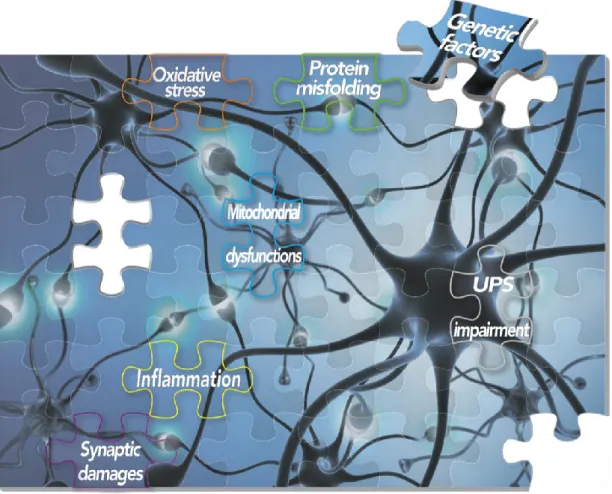
oxidative stress [5], impairment of the Ubiquitin/Proteasome System (UPS) [6] and
neuro-inflammation [7] (Figure 1).
PD was for a long time believed to be a typical non-genetic disorder. When in
1997 Polymeropoulos and colleagues reported the first SNCA pathogenic mutation in
the Italian Contursi kindred [8], they revolutionized this view opening the way to new
interesting perspectives about the genetic contribution to this still incurable condition
[1]. From that moment, an increasing number of genetic loci and numerous risk factors
have been discovered [9,10], starting from the familiar genes responsible of the
Mendelian inherited forms, such as the autosomal dominant genes (SNCA, LRRK2,
VPS35, GBA), the typical recessive (PARK2, PINK1, PARK7) and the atypical recessive
ones (ATP13A2, PLA2G6, FBXO7) [11]. Despite the existence of these rare monogenic
forms, it is now clear that PD is a genetically heterogeneous and most likely complex
disorder, often complicated by incomplete penetrant traits and variable expressivity.
The list of candidate genes is continuously updated [10,12,13], mainly thanks to the
massive advancement in genomic biotechnologies that have allowed to detect
hundreds of pathogenic or susceptibility variants at the single nucleotide
Figure 1. Schematic representation of molecular elements and common altered pathways underlying the complex PD puzzle.
polymorphism (SNP) level. However, a lot of work still has to be done to identify
additional sources of missing heritability or to assign a precise causal mechanism to
the growing number of discovered loci [14].
While single nucleotide polymorphisms (SNPs) and small indels constitute the
most commonly investigated DNA variations, submicroscopic chromosomal
rearrangements, also known as Copy Number Variations (CNVs), are emerging as
crucial players in the individual’s genomic architecture and in modeling complex
human diseases, including PD. However, the majority of CNVs association studies have
been conducted by using the traditional candidate-gene approach that, although
provides valuable information on common variants, is inadequate to completely
dissect the genetic background of polygenic multifactorial disorders like PD. The
search for single-gene mutations has to be changed, turning into the need to assess the
collective effect of common and rare variants that together may converge on PD
pathology. In this context, the “systems biology” approach represents a worthwhile
instrument to analyze complex biological systems, moving beyond the conventional
gene-centric scheme, and finally generating a more defined molecular picture of PD.
Herein, we will review the most common CNV-altered genes and detail the
current knowledge about their pathogenic or susceptibility impact on PD
pathobiology. Moreover, we will collect the set of rare individual CNVs reported so far
in PD patients and analyze them by a “systems biology” approach. This new perspective
reveals these private CNVs cluster in common deregulated biological processes that
could contribute to disease onset or progression, and open the window to a new
possible genetic scenario in the unsolved PD puzzle.
CNVs: A PREVALENT SOURCE OF GENOMIC VARIATIONS
The DNA sequence of human genome is constantly changing and this process allows
humans to evolve and adapt. The scientific community has long been aware of genetic
variations of extreme size (i.e. cytogenetically recognizable elements and SNPs) [15].
However, about 10 years ago, scientists began to recognize abundant variations of an
intermediate size class known as structural variations. Within this class, Copy Number
variations represent the largest component by far. CNVs are defined as genomic
segments showing copy number variability among individuals compared to a
reference genome. The size of CNVs ranges from 50 bp to several Mb, with a significant
drop of variant numbers in 50 bp to 1 kb range [16]. These structural variants can
include either a single gene or a contiguous set of genes, encompassing more
polymorphic base pairs than SNPs and finally resulting in an altered DNA diploid
status (i.e. gain or loss of genomic region).
Depending on their size, CNVs can be measured by a multitude of laboratory testing
methods, either targeting the whole genome (genome-wide level) or restricted to
certain locations on chromosomes (locus-specific levels) (Figure 2) [17]. While
targeted approaches such as FISH or quantitative PCR-based strategies have been long
used in the past, the most advanced current screenings rely on whole-genome
applications, such as array Comparative Genomic Hybridization or Next Generation
Sequencing experiments. Both these biotechnologies have dramatically improved and
catalyzed the detection and characterization of multiple CNVs, offering the
simultaneous testing of thousands of loci with high reproducibility, high resolution,
and scalability for complete mapping of imbalances [18-22]. However, these
whole-genome strategies still need post-experimental validations and therefore, a
gold-standard analysis has not been defined yet.
CNVs are very common and arise in presence of specific architectural genomic
elements that render DNA regions very susceptible to rearrangements. Depending on
whether the same rearrangement is identified in unrelated individuals, CNVs can be
grouped as recurrent or non-recurrent events [23]. The most common cause of
recurrent genomic rearrangements is the non-allelic homologous recombination
(NAHR), that occurs between two DNA blocks of high homology, like the
region-specific low-copy repeats sequences (LCRs) (Figure 3, panel A). On the contrary,
non-recurrent CNVs can result from non-homologous end joining (NHEJ) or fork stalling
and template switching (FoSTeS) mechanisms. NHEJ represents the major cellular
mechanism for double-strand break repair: upon a double-strand break, NHEJ
reconnects chromosome ends leaving random nucleotides at the site of the breakage
to facilitate the strands’ alignment and ligation (Figure 3, panel B) [24]. FoSTeS occurs
when the DNA replication machinery pauses, and the template is switched with
another region in physical proximity to the original replication fork (Figure 3, panel C)
[25]. Such template switching may occur several times before the replication process
gets back to its original template, resulting in complex rearrangements [24].
Figure 2. CNVs can be measured by a spectrum of laboratory-methods targeting specific locations on chromosomes (locus-specific levels), or the whole genome (genome-wide level). These numerous methodologies are characterized by different levels of resolutions. The locus-specific techniques encompass i) PCR-based strategies, such as quantitative real-time PCR (qPCR), Multiplex Ligand Probe Amplification (MLPA) or multiplex amplifiable probe hybridization (MAPH); ii) the Fluorescence in situ Hybridization (FISH) assays and iii) the RFLP (restriction fragment length polymorphism)– Southern blot analysis. The whole-genome methodologies include i) the classical chromosomal G-bandage (karyotyping); ii) the aCGH (Comparative Genomic Hybridization array) platforms and iii) the NGS (Next Generation sequencing) technology. These two latter are increasingly replacing both the classical detections methods and the locus-specific techniques.
CNVs can control phenotype in several ways: they can affect gene expression through
the simple gene-dosage effects, or through more intricate mechanisms as, for example,
insertions and deletions of regulatory regions and alterations of chromatin
architecture [14]. To this regard, CNVs can interfere with a form of regulatory scaffold
of the chromatin (the so-called Topologically Associating Domains or TADs) by
disrupting or repositioning boundaries, and therefore, constraining the enhancer or
silencer activity with their target genes [26]. Similarly, CNVs in other non-coding
regions may alter the normal rate and tissue specific transcription pattern of the
neighboring, otherwise intact, genes by changing, for example, the affinity for
transcription factors. This cis-acting effect of non-coding variations has been recently
demonstrated for a SNP in a distal enhancer element regulating the expression of SNCA
[27]. Some representative pictures about the mechanisms of non-coding variants and
their implication in human genetics are reported in a number of excellent reviews
[28-30], whose the reader is referred for a better understanding.
Figure 3. Schematic illustration of the three most common events causing genomic rearrangements. Panel A: NAHR generates CNVs when genomic segments with high sequence similarity (direct low-copy repeats sequences, green arrows) recombine. This recombination can generate a duplication of the similar locus (red arrow) on one chromosome, while removing the copy from the other. Panel B: Double stranded breaks (DBS) in DNA sequence recruit NHEJ associated proteins to repair and ligate DNA strands together. First, end-repair protein replaces lost nucleotides on the double strand break and DNA ligase associates broken DNA fragments together. If fragments from different chromosomes ligate together, duplications or deletions of sequence can occur. Panel C: After the original stalling of the replication fork (black lines), the lagging strand disengages and anneals to a second fork (blue lines), followed by extension of the now 'primed' second fork and DNA synthesis. After the fork disengages, the tethered original fork with its lagging strand (black and blue lines) could invade a third fork (green lines). Serial replication fork disengaging and lagging strand invasion could occur several times (e.g. FoSTeS x 2, FoSTeS x 3, etc.) before resumption of replication on the original template. It should be noted that the CNVs created through FoSTeS are difficult to be distinguished from those generated by micro-homology-mediated breakpoint-induced repair (MMBIR), a mechanism of end-joining that relies on small-scale homology of DNA sequence at the ends of DSBs.
All together, CNV alterations may account for adaptive or behavioral traits, may have
no phenotypic effects or can underlie diseases. For this reason, determining the clinical
significance of CNVs is very challenging and comprehensively relies on frequency
information from healthy control cohorts, hereditability, size, gene content, type (copy
number state) and location on chromosome (interstitial, centromeric or
repeat-regions) [31].
Notwithstanding the difficulties in interpreting quantitative data, specific large
CNVs and single-gene dosage alterations have emerged as critical elements for the
development and maintenance of the nervous system [32] and have appeared to
contribute to hereditable or sporadic neurological diseases, such as neuropathies,
epilepsy forms, autistic syndromes, psychiatric illnesses and also neurodegenerative
diseases, including PD [23,33-37].
In the next paragraphs, we will focus on the current evidence about the
occurrence of CNVs in familiar PD genes by highlighting strengths and weaknesses of
interpretations for diagnosis and biomarkers usefulness. Moreover, we will collect
from published literature the currently known set of rare CNVs observed in PD
patients and analyze them through a systems biology point of view, in order to assess
their biological role, their interactions and the possible functional impact on PD
pathobiology.
COPY NUMBER VARIATIONS IN FAMILIAR PD GENES
SNCA
SNCA (alpha-synuclein) represents the most convincing locus causing both
familiar and sporadic PD. This gene encodes a small natively unfolded presynaptic
protein that aggregate in Lewy bodies and Lewy neurites, the pathological hallmark
lesions of PD [38]. As we will discuss here below, SNCA is the best example of
dosage-dependent toxicity: the more alpha-synuclein you have, the worse will be PD.
The first genomic triplication of SNCA was observed within the Spellman–
Muenter family (better known as Iowa Kindred) a large family with autosomal
dominant inheritance transmission of PD and dementia [39]. Later, several families
with different ethnic background have been described, including members carrying
four copies (triplication) or three copies (duplication) of SNCA (Table 1)
[40-54,37,55-59]. In general, triplication generates very high expression of mRNA and protein
molecules and influence the clinical manifestations of PD, causing severe forms of
Parkinsonism similar to dementia with Lewy Body. In contrast, the clinical phenotype
of patients with duplicated SNCA resembles idiopathic PD, mainly with late age at
onset, good efficacy for levodopa therapy, slower disease progression and without
early development of dementia.
An interesting familiar pedigree, the “Lister family”, present both duplicated
and triplicated SNCA carriers within different branches of the pedigree (branches J and
I), suggesting a primary duplication event followed later by another one and resulting
in the triplication [60,61]. Similarly, the Ikeuchi family has both heterozygous and
homozygous duplication carriers born from a consanguineous marriage (producing a
pseudo-triplication) [62]. The clinical features of individuals with the SNCA
homozygous duplication showed severe parkinsonism similar to that of triplication
carriers.
Along with the familiar forms, a good percentage of sporadic PD patients carry
de novo duplication of SNCA (Table 1) [47,63-67]. Generally, their clinical course is
similar to typical sporadic PD without severe progression or cognitive decline.
The breakpoint of SNCA multiplications is not the same in each patient. The
largest multiplication detected so far is about 41.2 Mb, containing 150 genes and
defined a partial trisomy 4q [63], while the smallest one counts about 0.2 Mb [48]. The
size and gene make-up of each multiplicated region does not seem to severely
influence the clinical presentation of the carriers.
Interesting insights derive from the mosaicism condition of SNCA
rearrangements. To this regard, two interesting PD cases have been described,
resulted negative to exon dosage test in peripheral blood, and positive for SNCA copy
number changes on oral mucosa cells [68]. Both patients displayed a parkinsonian
clinical phenotype of SNCA copy number carriers. Starting from this evidence, authors
suggest to take into consideration the possibility to examine cells from both peripheral
lymphocytes and other tissues in order to detect low-grade mosaicism.
CNVs in α-synuclein gene in PD
CNV type Size Ethnicity Phenotype Methodology F-S-D Reference Triplication (Spellman–Muenter family
or Iowa Kindred)
1,61 – 2,04
Mb Iowa PD and dementia with LBs qPCR, FISH F [39]
Duplication (Lister Family, branch J)
0,7987 – 0,9359 Mb
Sweden, United States
Late-onset parkinsonism and early dysautonomia qPCR; Microsatellite markers analysis; Affymetrix 250K microarray F [60, 61] Triplication (Lister Family,
Swedish-America, Branch I)
Early-onset parkinsonism with dementia and dysautonomia Duplication (Ikeuchi family)
5 Mb Japan Progressive parkinsonism with
dementia with LBs
Microsatellite markers
analysis, qPCR F
[62]
Homozygous duplication (Ikeuchi family - consanguineous marriage)
Duplication (Uchiyama family) 0,5 – 1,6
Mb Japan Parkinsonism with dementia with LBs qPCR F [46]
Duplication n.a. Korea
Early onset parkinsonism with rapidly progressive course, cognitive impairment, and dysautonomia (Ahn
family)
Semi-quantitative multiplex PCR, FISH
F [47]
Typical PD S
Duplication n.a. Germany Early-onset parkinsonism qPCR, MLPA D [66]
Duplication n.a. European and
North African Early onset PD
MLPA , Microsatellite
markers analysis S [65]
Duplication (Family A) 0,6 Mb
Japan Parkinsonism with or without dementia
Microsatellite markers analysis, qPCR, FISH, aCGH
(BACS and Affymetrix)
F [48, 49, 56, 58] Duplication (Family B) 0,4 Mb Duplication (Family C) 0,4 Mb Duplication (Family D) 0,4 Mb Duplication (Family E) 0,2 Mb Duplication (Family F) 0,6 Mb Duplication (Family G) 0,6 Mb Triplication (FPD-014) (Pat 011) 2,61 – 2,64 Mb France. Italy
Atypical autosomal dominant parkinsonism Semi-quantitative Multiplex PCR, Microsatellite analysis, Affymetrix GeneChip Human Mapping 250K microarray (just for P59
F [50 -52, 57-59] Duplication (FPD-131 o P59) (Pat
024-022-026) 4,928 Mb
Typical autosomal dominant PD
Duplication (FPD-321) (Pat 021) 3,47 – 3,58
Duplication (FPD-410) (Pat 001) 0,63 – 0,65 Mb family: FISH, 44k CGH arrays Agilent) Duplication (FPD-437) (Pat 010-012) 0.42 - 0.43 Mb
Duplication (Sironi family) 3,65 Mb Italy PD with progression to dementia MLPA, Agilent 105A chip F [53]
Duplication n.a. Belgium Parkinsonian syndrome Multiplex amplicon
quantification, qPCR S [67]
Triplication (Keyser family) n.a.
South African (French-Italian origin)
PD with dementia MLPA, qPCR F [40]
Duplication n.a. Korea PD with cognitive dysfunction Semi-quantitative
multiplex PCR S [64]
Triplication n.a. Asian Early-onset and severe clinical features
of parkinsonism
qPCR, MLPA, microsatellite
analysis F [41]
Duplication 3 Mb n.a. PD Illumina370Duo arrays F [54]
Homozygous duplication 0,928 Mb Pakistan Young-Onset Parkinsonism MLPA, Nimblegen 135 K
array CGH F [42]
Duplication (Partial Trisomy 4q) 41,2 MB Belgium Young onset, dopa-responsive
parkinsonism Karyotype, aCGH, MLPA D [63]
Duplication n.a. Non-Hispanic
Caucasian Autosomal Dominant Early-onset PD
Customized 4 × 72 k format CGH microarrays by
NimbleGen; Taqman qPCR
F [37]
Duplication (family Elia A) 773 Kb Northern
Argentina
Early onset PD that was variably associated with nonmotor features,
such as dysautonomia, cognitive deficits, and psychiatric disturbances
MLPA, qPCR, Affymetrix high-resolution single nucleotide
polymorphism-array analysis
F [55]
Duplication (Family Elia B) 4820 Kb Italian
Early onset PD dementia with psychiatric disturbances to late onset
PD with mild cognitive impairment
Duplication 6,4 Mb Caucasian
English
Atypical clinical presentation strongly reminiscent of frontotemporal
dementia and late-onset pallidopyramidal syndromes
MLPA, aCGH Agilent 8x60K F [58]
Duplication
n.a. Iranian PD typical clinical features MLPA, qPCR F-S [43]
Duplication (mosaicism) n.a. American mitochondrial haplogroup and European autosomal markers Early-onset Parkinsonism
MLPA (no dosage alteration in buccal swab),
FISH (no rearrangements in peripheral leukocytes; duplication - triplication in
oral mucosa)
F-S [68]
Duplication n.a. American Parkinsonism with LBs and Lewy
neurites n.a. n.a. [56]
Triplication 1,3 Mb Italian
Early-onset parkinsonism combined with depression, behavior disturbances,
sleep disorders, and cognitive decline
Genome-wide SNP
microarrays, FISH, MLPA F [44]
Triplication 351 Kb Italian
Severe parkinsonism featuring early onset dyskinesia, psychiatric symptoms,
and cognitive deterioration
CGH-Array, MLPA, qPCR F [45]
Table 1 lists all the currently studies describing SNCA copy number changes in PD. The CNVs mutation type, the size of the mutation, the ethnicity of patients, the phenotype and the methodological approaches to measure quantitative genomic variations are reported. The column F-S-D reports if described cases are familial, sporadic or de novo.
PARK2
Although SNCA story suggests a gain of function, several early-onset forms of
PD have demonstrated the role of loss of function genes in the etiology of the disease.
The most common loss-of-function mutations belong to Parkin (or PARK2) gene, one
of the largest in our genome harbored in the long arm of chromosome 6 (6q25.2-q27)
and encoding an E3 ubiquitin ligase. Mutations of PARK2 are particularly frequent in
individuals with familiar recessive inheritance and account for 50% of the cases with
autosomal recessive juvenile PD. Parkin mutations also explain ~15% of the sporadic
cases with onset before 45 [69,70] and act as susceptibility alleles for late-onset forms
of PD (2% of cases) [71].
PARK2 gene has a high mutation rate because it is located in the core of FRA6E
site, one of the most mutation-susceptible common fragile site of human genome [24].
For this reason, more than 200 putative pathogenic mutations have been reported so
far, affecting numerous ethnic populations [72-75,67,76,77,40,78-80,37,81-85]. The
PARK2 mutation spectrum includes homozygous or compound heterozygous missense
and nonsense point mutations, as well as several exon rearrangements (both
duplications and deletions) involving all the originally cloned 12 exons and the
promoter region. Recently, our research group has outlined a complex alternative
splicing mechanism regulating the expression of PARK2 [86-88]. These data suggest
that 5 additional exons exist, that however have never been considered for mutational
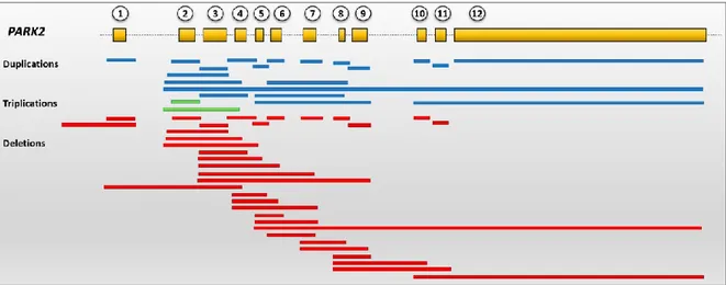
or dosage screening. Overall, currently known Parkin CNVs are summarized in Figure
4 and are collected in the Parkinson Disease Mutation database
(http://www.molgen.vib-ua.be/PDMutDB), whose the reader is referred for more
details.
CNV rearrangements involving PARK2 exons accounts for 50–60% of all
pathogenic anomalies, rendering gene-dosage assays essential in parkin mutational
screening [89]. However, the hot-spot nature of this gene makes its quantitative
analysis a particular challenge, and several issues need to be pointed out in this regard.
Firstly, the determination of mutational phase of the rearrangements, meaning the
assessment that amplified or deleted exons are really contiguous. Phase determination
seems to be a fundamental requisite for PARK2 molecular diagnosis: by phase
determination, several patients with apparent contiguous multi-exon deletions were
re-diagnosed as compound heterozygotes [89]. A second important point refers to
breakpoint mapping which can be useful to compare exon rearrangements between
patients and families and to study the possible causing event mechanism [90]. Just a
few papers have addressed this issue so far, but mostly report rearrangements into
the region between PARK2 exons 2 and 8 [90,24]. In the majority of mapped cases,
micro-homologies at breakpoint junctions were present, thus supporting NHEJ and
FoSTeS as the major mechanisms responsible for PARK2 genomic rearrangements
[24]. Moreover, some data underpin the possible effects of ancient common founder
in minor ethnic groups [91]. For example, microsatellite markers analysis in four
families from The Netherlands have shown that a common haplotype of 1.2 Mb could
be distinguished for the exon 7 duplication and a common haplotype of 6.3 Mb for the
deletion of exon 4, suggesting common founder effects for distinct large
rearrangements in parkin [90].
A relevant matter of ongoing debates is the pathogenic role of single
heterozygous PARK2 CNVs. Several studies have sought to address this issue, but the
findings published so far are controversy and conflicting. Some reports indicate that
CNVs heterozygous mutations in PARK2 associate with increased PD risk [76,54,92],
while others found no differences for association [77,37]. Also, examinations of family
Figure 4. Schematic representation of PARK2 genetic structure and currently identified CNVs in PD patients. All the canonical PARK2 exons are involved in exons rearrangements. Red bars correspond to exons deletions, blue bars to duplications and green bars to triplications. All depicted CNVs can be found at the PDMutDB.
pedigrees revealed heterozygous members with mild late-onset PD [93,94], or without
typical clinical signs of the disease [37].
PINK1
Pathogenic mutations in PINK1 (PTEN-induced kinase gene) are a less common
cause of early-onset PD with a frequency variable from 1 to 9% depending on the
ethnic background [95]. The encoded protein is a putative serine/threonine kinase of
581 amino acids involved in mitochondrial quality control and oxidative stress [96].
Homozygous and compound heterozygous mutations deletions involving
different combinations of exons 4-8 have been described in both familial and sporadic
early-onset cases coming from Japan, Brazil, Sudan and Iran (Table 2) [97-100,43]. A
breakpoint analysis has been performed just in one of these patients, revealing a
complex rearrangement involving the neighboring DDOST gene and maybe resulting
from FoSTeS mechanism [100]. Moreover, single heterozygous cases have been
described, albeit these mutations do not completely explain the recessive inheritance
pattern. The largest heterozygous deletion known so far (56 kb) includes the entire
PINK1 genetic region, two neighboring genes, and two highly similar AluJo repeat
sequences, which have been suggested as responsible of an unequal crossing-over
[101]. Further heterozygous deletions involving exons 1, 3-8 and exon 7 have been
described in familial or sporadic cases of early-onset PD (Table 2) [102,103,80].
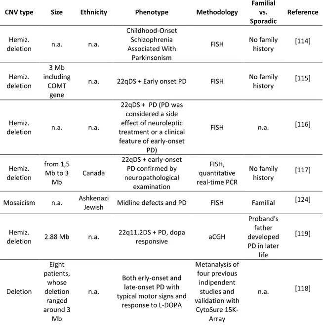
CNVs overlapping PINK1 gene in PD
CNV type Size Ethnicity Phenotype Methodology F-S Reference Homoz. deletion Exons 6-8 Japan Early onset PD + dementia n.a. F [98] Homoz. deletion ~4600
bp Japan Early onset PD n.a. n.a. [97]
Heteroz.
deletion ~56kb Italy Definite PD
qPCR, FISH, Microsatellite markers analysis
S [101]
Homoz.
deletion Exon 7 Brazil Early onset PD Sequencing, qPCR S [99]
Homoz. deletion 8,669 bp (Exons 4-8)
Sudan Early onset PD MLPA, sequencing F
[100] Heteroz.
deletion
Exons
3-8 China Early onset PD qPCR S [102]
Heteroz.
deletion Exon 7 Spanish
Early onset PD with
Compound heteroz. deletion Exon 2 + exons 2-4 Iran Typical clinical features MLPA, qPCR S [43] Homoz. deletion Exon 5 and exon 4 Typical clinical features and PD with dementia F Heteroz.
deletion Exon 1 Brazil Early onset PD MLPA, qPCR n.a. [80]
Table 2 lists all the currently studies describing PINK1 copy number changes in PD. The CNVs mutation type, the size of the mutation, the ethnicity of patients, the phenotype and the methodological approaches to measure quantitative genomic variations are reported. The column F-S reports if described cases are familial or sporadic.
PARK7
PARK7 was the third gene identified in 2001 as responsible of early-onset PD
[104,105]. It encodes a conserved multifunctional protein belonging to the peptidase
C56 family (also called DJ1) which acts as positive regulator of transcription,
redox-sensitive chaperone, sensor for oxidative stress, and apparently protects neurons from
ROS-induced apoptosis [106-108].
The proof that PARK7 was a gene-causing disease came from a study on a Dutch
family where members carried a 14 kb homozygous deletion involving the first five of
seven exons [104]. Later, three siblings of Iranian origins born from consanguineous
parents and carriers of a homozygous deletion of exon 5 have been reported (Table
3) [43]. Further heterozygous CNVs (both deletions and duplication) involving the
exons of DJ-1 gene have been published so far [102,109-111], although they do not
completely explain the recessive pattern of the PD phenotype.
CNVs overlapping PARK7 gene in PD
CNV type Size Ethnicity Phenotype Methodology F vs.
S Reference Homoz. Deletion Exons 1-5 (14.082 bp) Dutch Autosomal Recessive Early-Onset PD Microsatellite markers analysis, cloning, PCR, sequencing F [104] Heteroz. Deletion Exons 5-7 Caucasian (Tyrol, Austria) Early-onset PD Quantitative duplex PCR S [109] Heteroz.
Heteroz.
Duplication Exons 1-5 Dutch Early-onset PD MLPA, sequencing S
[111] Heteroz.
Deletion Exon 2 China Early onset PD qPCR S
[102]
Homoz. Deletion Exon 5 Iran Typical clinical
features MLPA, qPCR F
[43]
Table 3 lists all the currently studies describing PARK7 copy number changes in PD. The CNVs mutation type, the size of the mutation, the ethnicity of patients, the phenotype and the methodological approaches to measure quantitative genomic variations are reported. The column F-S reports if described cases are familial or sporadic.
ATP13A2
ATP13A2 mutations are associated with Kufor-Rakeb syndrome (KRS), a form of
recessively levodopa-responsive inherited atypical Parkinsonism [112]. It encodes a
large protein belonging to the ATPase transmembrane transporters, and recently it
has been identified as a potent modifier of the toxicity induced by alpha-synuclein
[113]. To our knowledge, just one family from Iran with deletion of ATP13A2 has been
reported, including three affected siblings born from consanguineous parents and
carriers of a homozygous deletion of exon 2 [43]. All three individuals presented
moderate mental retardation, aggressive behaviors, visual hallucinations,
supranuclear vertical gaze paresis, slow vertical saccades and dystonia. Cognitive
function deteriorated rapidly, and all of them developed dementia by age 10. Further
clinical and genetic follow-up of KRS patients will increase the knowledge of the
natural history and clinical features of this syndrome.
THE 22q11.2 DELETION
A separate speech deserves the 22q11.2 deletion that lately is receiving more and
more attention in PD field. Deletions at 22q11.2 are classically associated with a
heterogeneous range of clinical syndromes, overall named 22q deletion syndrome
(22qDS). The clinical phenotype of 22q deletion carriers varies widely, with multiple
system involvement, including cleft palate, dysmorphic facial features, cardiac defects,
skeletal deformities, developmental delays, learning disabilities and increased risk of
developing schizophrenia and other mental disorders. Despite the multiple system
involvement, the association between 22q11.2 deletion and PD was not suspected
until the publication of independent case reports of co-occurrence of parkinsonism in
patients with 22q11.2 deletion syndrome (Table 4) [114-116].
The interest in this possible link increased after Butcher and colleagues reported
four patients with early-onset PD in their study of 159 adults with 22q11.2 deletion
syndrome, founding that the use of antipsychotics in these patients delayed diagnosis
of PD, and assessing after autopsy examination the presence of typical Lewy bodies
and Lewy neurite formations too [117]. A couple of months ago, Mok et al. [118]
performed the reverse experiment, namely pooling data from previous large PD
case-control studies and assessing the frequency of 22q11.2 deletion carriers. Eight
patients with PD and none of the controls had the deletion, providing a statistical
significant association between the 22q deletion and an increased risk of developing
the disease (Table 4). In according with this result, a single case-report from Virginia
describes a 37-years-old early-onset PD patient carrying the 22q11.2 deletion but
without any features of typical 22qDS [119]. All together, this evidence suggests
22q11.2 deletion might underlie early-onset PD, warning clinicians to take into
consideration this genetic test as part of their evaluation for patients with early-onset
PD.
The chromosome 22q11.2 region contains some excellent candidate genes for
PD: COMT (or Catechol-O-Methyltransferase), a key regulator of synaptic dopamine
levels and a target of inhibitory drugs for the treatment of wearing-off phenomena in
PD patients [120]; SEPT5, a vescicle- and membrane-associated protein playing a
significant role in inhibiting exocytosis, as well as a parkin substrate [121,122]; DGCR8
that encodes a complex-subunit involved in the biogenesis of microRNAs, including
miR-185 which is predicted to target LRRK2 [123].
Interestingly, Perandones et al. [124] reported a case of mosaicism of a patient
from the Ashkenazi Jewish ethnic group with a history of midline defects and PD onset
at 46 years (Table 4). In this patient, FISH test detected a mosaicism of the 22q
deletion in 24% of the analyzed blood cells, highlighting the relevance of performing
individual cell-by-cell analysis.
CNVs involving the 22q11.2 region
CNV type Size Ethnicity Phenotype Methodology
Familial vs. Sporadic
Reference
Hemiz.
deletion n.a. n.a.
Childhood-Onset Schizophrenia Associated With Parkinsonism FISH No family history [114] Hemiz. deletion 3 Mb including COMT gene
n.a. 22qDS + Early onset PD FISH No family
history [115]
Hemiz.
deletion n.a. n.a.
22qDS + PD (PD was considered a side effect of neuroleptic treatment or a clinical feature of early-onset PD) FISH n.a. [116] Hemiz. deletion from 1,5 Mb to 3 Mb Canada 22qDS + early-onset PD confirmed by neuropathological examination FISH, quantitative real-time PCR No family history [117]
Mosaicism n.a. Ashkenazi
Jewish Midline defects and PD FISH Familial
[124] Hemiz. deletion 2.88 Mb n.a. 22q11.2DS + PD, dopa responsive aCGH Proband's father developed PD in later life [119] Deletion Eight patients, whose deletion ranged around 3 Mb n.a.
Both erly-onset and late-onset PD with typical motor signs and
response to L-DOPA Metanalysis of four previous indipendent studies and validation with CytoSure 15K-Array n.a. [118]
Table 4 lists all the currently studies describing 22q11.2 deletions in PD patients. The CNVs mutation type, the size of the mutation, the ethnicity of patients, the phenotype and the methodological approaches to measure quantitative genomic variations are reported. The column Familial vs. Sporadic reports if described cases are familial or sporadic PD.