CHAPTER 2
General Introduction
Part of this chapter will be published under the title:
“The Chromosomal Instability Pathway in Colon Cancer”,
Pino MS and Chung DC; Gastroenterology, May 2010.
2
Introduction
Colorectal cancer (CRC) is the second to the fourth most common cancer in industrialized countries, responsible for an estimated 52,000 deaths per year in the United States and 146,000 per year in the European Union [1,2]. CRCs develop through an ordered series of events beginning with the transformation of normal colonic epithelium to an adenomatous intermediate and then ultimately adenocarcinoma, the so-called “adenoma-carcinoma sequence” [3]. Multiple genetic events are required for tumor progression and mutations in specific oncogenes and tumor suppressor genes are associated with discrete steps of this tumorigenic process. Genomic instability is now recognized as an essential cellular feature that accompanies the acquisition of these mutations. In colon cancer, at least 3 distinct pathways of genomic instability have been described, the chromosomal instability (CIN), microsatellite instability (MSI), and the CpG island methylator phenotype (CIMP) pathways, each with distinctive tumor genotypes and phenotypes (Figure 1).
In 1990, Fearon and Vogelstein first proposed a multistep genetic model of colorectal carcinogenesis that has come to serve as a paradigm for solid tumor progression [4]. Inactivation of the Adenomatous Polyposis Coli (APC) tumor-suppressor gene occurs first, followed by activating mutations of KRAS. Subsequent malignant transformation is driven by additional mutations in the TGF-β, PIK3CA, and TP53 pathways [5-8]. This model predicts that at least 7 distinct mutations are required. Recent genome-wide sequencing efforts have calculated as many as 80 mutated genes per colorectal tumor, but a smaller group of mutations (< 15) were considered to be the true “drivers” of tumorigenesis (Table 1) [9,10].
Table 1. Genes frequently involved in colorectal cancer development.
Gene Chromosome location Function
APC 5q21-22 Tumor suppressor gene
TGFBR2 3p22 Cell signaling
hMSH2 2p16 DNA mismatch repair
hMLH1 3p21 DNA mismatch repair
hMSH6 2p16 DNA mismatch repair
KRAS 12p12·1 Oncogene
TP53 17p13 Tumor suppressor
SMAD2/4 18q21·1 Tumor suppressor
p16INK4A 9p21·3 Cell cycle control COX2 1q25·2-3 Cell proliferation
DCC 18q21·3 Tumor suppressor gene
Bcl-2 18q21·3 Apoptosis
BAX 19q13·3-4 Apoptosis
MGMT 10q26 DNA repair gene
Genomic Instability and CRC
Baseline mutation rates are insufficient to account for the multiple mutations that are required for cancer to develop. The rate of mutations per nucleotide base pair is estimated to be as low as 10-9 per cellular generation. As proposed by Loeb et al., cancer cells must acquire some form of intrinsic genomic instability, a “mutator phenotype”, that increases their rate of new mutations [11]. One type of genomic instability occurs at the chromosomal level and is designated chromosomal instability (CIN). CIN is observed in 65-70% of sporadic colorectal cancers; the term refers to an accelerated rate of gains or losses of whole or large portions of chromosomes that results in karyotypic variability from cell to cell [12]. The consequence of CIN is an imbalance in chromosome number (aneuploidy) and a high frequency of loss of heterozygosity (LOH). The second pathway, involving about 15% of sporadic colorectal cancers, is referred to as the microsatellite instability (MSI) pathway. Such tumors display frameshift mutations and base pair substitutions that are commonly found in short, tandemly repeated, nucleotide sequences known as microsatellites. This form of genetic destabilization is commonly caused by loss of the DNA mismatch repair (MMR) system, which normally recognizes and repairs mismatched nucleotides and insertion/deletion loops caused by slippage of DNA polymerase. In addition to deletions and inactivating mutations, epigenetic events have now been recognized as an important mechanism of gene silencing. A CpG island methylator phenotype (CIMP) leads to gene silencing by hypermethylation of CpG islands, GC-rich regions present in almost half of all human genes in the 5’ area, often encompassing the promoter and transcriptional start site of the associated gene [13]. When methylation takes place, the transcription factors cannot link at the promoter area, thereby inhibiting
transcription, and consequently the gene is silenced. CIMP has some overlap with MSI, because hypermethylation of hMLH1, a gene of the MMR system, is commonly observed in both pathways [14]. Because the definitions of these 3 pathways are not mutually exclusive, a tumor can occasionally exhibit features of multiple pathways. For example, up to 25% of MSI colorectal cancers can exhibit chromosomal abnormalities [15]. In addition, whereas the CIMP phenotype can account for most of the MSI-positive/CIN-negative CRCs, up to 33% of CIMP-positive tumors can exhibit a high degree of chromosomal aberrations [16]. Conversely, as many as 12% of CIN-positive tumors exhibit high levels of MSI [17]. The significance and implications of these overlapping features are not yet fully defined.
Microsatellites and Genomic Instability
Microsatellites are stretches of DNA in which a short motif (usually one to five nucleotides long) is repeated several times. The most common microsatellite in humans is a dinucleotide repeat of cytosine and adenine, (CA)n, which occurs in tens of thousands of
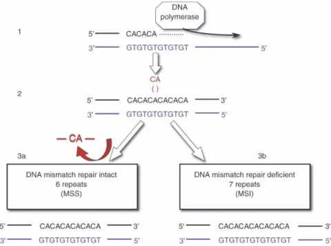
locations in our germ line. They are inherently hypermutable because of their propensity for strand slippage during DNA replication. Microsatellite sequences are usually located in the non-coding regions of the genome, but they are also found within the coding regions of a select number of genes involved in tumor initiation and progression. Microsatellite instability (MSI) is a situation in which a germline microsatellite allele has gained or lost repeated units and has thus undergone a somatic change in length (Figure 2). Because this type of alteration can be detected only if many cells are affected by the same change, it is an indicator of the clonal expansion that is typical of a neoplasm.
Figure 2. Mechanism of MSI. (1) Replication of DNA. (2) A CA repeat erroneously built into the replication strand. The error is repaired by mismatch repair enzymes (3a) or the error is not repaired causing MSI (3b).
DNA is replicated accurately because of the intrinsic ‘‘proofreading’’ capacity of the DNA polymerases. Replicative errors still occur, albeit with low frequency; the estimated substitution error rate is 1/106 to 107 base pair. The mismatch repair (MMR) system is the checkpoint responsible for repairing replication errors. A proficient MMR system lowers the net error rate to 1/1010 base pair, that is, enhances replication accuracy 1,000 to 10,000-fold. The types of errors the MMR system corrects are base-base mismatches and insertion-deletion loops that both typically involve microsatellite foci. When the errors involve coding regions, single-base mismatches contribute to point mutations (missense or nonsense), whereas uncorrected insertion-deletion loops lead to frameshift mutations. Defective mismatch repair presumably facilitates malignant transformation by allowing the rapid accumulation of mutations that inactivate genes that ordinarily have key functions in the cell. These genes include receptors for growth factors, such as transforming growth factor-β receptor II (TGFBR2) and insulin-like growth factor II receptor, cell cycle regulators (E2F4) and regulators of apoptosis (BAX), and some of the MMR genes themselves. The most commonly mutated gene in tumors with MSI is the
TGFBR2 gene, which harbors an (A)10 repeat that undergoes a frame shift. This mutation
leads to a disruption in the function of TGF-β, a tumor suppressor of prime importance in colorectal cancer [5]. The BAX gene has a (G)8 microsatellite which lose one or two
guanines, resulting in a frame shift that inactivates the gene and disrupts the apoptosis pathway mediated by Bcl-2, in about 35% of all tumors with defective mismatch repair
The Mismatch Repair System
The DNA MMR system performs a proofreading, or “housekeeping,” function, therefore, cells lacking effective DNA MMR accumulate mutations at a very high rate. The MMR system is a DNA repair pathway conserved from prokaryotes through eukaryotes including yeast and human. The terminology used to describe the MMR system in eukaryotes is largely based on the analogous system in prokaryotes, best characterized in
Escherichia coli. Several ‘‘Mut’’ (from ‘‘mutational’’) genes, when inactivated, cause
hypermutable strains. Bacterial MMR proteins are designated as MutS, MutL, and MutH. Eukaryotic MutS homologs are designated as MSH2, MSH3, and MSH6, and are responsible for recognizing the mismatch and recruiting MutL to the mismatch location, thus starting the downstream activity. MutL homologs include MLH1 and a subgroup of Post-Meiotic Segregation proteins PMS1 and PMS2. Human chromosomal loci containing genes encoding MMR proteins are outlined in Table 2.
Repair is initiated when complexes of MutS homologs, either MSH2-MSH6 (MutSα) or MSH2-MSH3 (MutSβ), bind to a mismatch (Figure 3). The MSH2-MSH6 complex represents up to 90% of the cellular level of MSH2 and its function is to recognize the mismatch of base-base insertions and deletions containing one or two unpaired
Table 2. Human chromosomal loci carrying MMR genes.
Gene Chromosome location
hMSH2 2p22-21 hMSH3 5q11-13 hMSH6 2p16-15 hMLH1 3p23-21 hPMS1 2q31-33 hPMS2 7p22
nucleotides [19]. The MSH2-MSH3 complex is primarily responsible for recognizing and repairing insertions and deletions containing up to 16 extra nucleotides in one strand, with some overlap in the specificity of these two complexes and hence redundancy in activity. Eukaryotes also encode multiple MutL homologs that form different heterodimers: MLH1-PMS2 (MutLα), MLH1-PMS1 (MutLβ) and MLH1-MLH3 (MutLγ). MutLα is the most active of these complexes in humans and is involved in repairing a wide variety of mismatches. The function of human MutLβ and MutLγ is currently unknown. Eukaryotes have no known homolog of Escherichia coli MutH, so the origins and identities of the entry point(s) for strand excision in vivo are currently less certain in eukaryotes than in
Microsatellite Instability in CRC
Microsatellite instability occurs in hereditary as well as sporadic colorectal cancer through two different mechanisms. In Lynch syndrome (LS), a germline mutation in a MMR gene accounts for more than 90% of cases, whereas in 10-15% of sporadic colorectal cancers MSI is due to loss of expression of a mismatch repair gene (most commonly
hMLH1) caused by epigenetic silencing. After the recognition of MSI cancers in 1993, LS
and sporadic MSI cancers came to be regarded as the familial and sporadic counterparts of the same pathway of tumorigenesis.
Family history is considered the most useful indicator of Lynch in individual patients in the clinical setting, whereas no association between MSI status and a family history of CRCs is seen at the population level. MSI is observed more frequently in women and in CRCs that occur proximal to the splenic flexure. CRCs exhibiting MSI are associated with deep tumor invasion, poor histological differentiation, a mucinous or ring cells histology, frequently peritumoral lymphocytic infiltration (“Crohn’s like inflammation”), and have a higher incidence of synchronous and metachronous tumors. It may therefore be difficult to distinguish between MSI cancers occurring as part of the LS and sporadic MSI cancers, especially when the patient is of intermediate age and is uncertain of family history details.
CRCs with MSI have longer overall and cancer specific survival than stage-matched patients with cancers exhibiting CIN [20]. The important contrast in survival between the two types of CRC remains unexplained. The pronounced genetic instability of cells with MSI may increase susceptibility to apoptosis because of an accumulation of mutations in genes that are required for cell growth. Mutations of the TP53 gene, associated with poor
prognosis, are less common in tumors that exhibit MSI than in those that develop via the CIN pathway. The same holds true for allelic imbalance at other genetic loci, such as 18q. Adjuvant chemotherapy with fluorouracil benefits patients with tumors exhibiting CIN, but apparently not those with tumors exhibiting MSI [21]. However, an overall reduced benefit from adjuvant therapy in patients with MSI and CRC could not be demonstrated in a recent systematic review and meta-analysis, but overall survival with MSI-positive colorectal tumors was better [22]. Thus, the mechanism by which MSI renders cancers less aggressive clinically remains interesting but unresolved. Real differences in adjuvant chemotherapy response between tumors exhibiting MSI and CIN remain to be demonstrated.
References
1. Evaluation of Genomic Applications in Practice and Prevention (EGAPP) Working Group. Recommendations from the EGAPP working group: Genetic testing strategies in newly diagnosed individuals with colorectal cancer aimed at reducing morbidity and mortality from Lynch syndrome in relatives. Genet Med 11, 35-41 (2009).
2. Teutsch S, Bradley L, Palomaki G, et al. The evaluation of genomic applications in practice and prevention (EGAPP) initiative: Methods of the EGAPP working group.
Genet Med 11, 3-14 (2009).
3. Morson B. President's address: The polyp-cancer sequence in the large bowel. Proc
R Soc Med 67, 451-457 (1974).
4. Fearon E, Vogelstein B. A genetic model for colorectal tumorigenesis. Cell 61, 759-767 (1990).
5. Markowitz S, Wang J, Myeroff L, et al. Inactivation of the type II TGF-β receptor in colon cancer cells with microsatellite instability. Science 268, 1336-1338 (1995). 6. Thiagalingam S, Lengauer C, Leach F, et al. Evaluation of candidate tumor
suppressor genes on chromosome 18 in colorectal cancers. Nat Genet 13, 343-346 (1996).
7. Samuels Y, Velculescu V. Oncogenic mutations of PIK3CA in human cancers. Cell
Cycle 3, 1221-1224 (2004).
8. Baker S, Fearon E, Nigro J, et al. Chromosome 17 deletions and p53 gene mutations in colorectal carcinomas. Science 244, 217-221 (1989).
9. Wood L, Parsons D, Jones S, et al. The genomic landscapes of human breast and colorectal cancers. Science 318, 1108-1113 (2007).
10. Leary R, Lin J, Cummins J, et al. Integrated analysis of homozygous deletions, focal amplifications, and sequence alterations in breast and colorectal cancers. Proc
Natl Acad Sci U S A 105, 16224-16229 (2008).
11. Loeb L, Loeb K, Anderson J. Multiple mutations and cancer. Proc Natl Acad Sci
USA 100, 776-781 (2003).
12. Lengauer C, Kinzler K, Vogelstein B. Genetic instabilities in human cancers.
Nature 396, 643-649 (1998).
13. Toyota M, Ahuja N, Ohe-Toyota M, et al. CpG island methylator phenotype in colorectal cancer. Proc Natl Acad Sci U S A 96, 8981-8986 (1999).
14. Kane M, Loda M, Gaida G, et al. Methylation of the hMLH1 promoter correlates with lack of expression of hMLH1 in sporadic colon tumors and mismatch repair-defective human tumor cell lines. Cancer Res 57, 808-811 (1997).
15. Sinicrope F, Rego R, Halling K, et al. Prognostic impact of microsatellite instability and DNA ploidy in human colon carcinoma patients. Gastroenterology 131, 729-737 (2006).
16. Cheng Y, Pincas H, Bacolod M, et al. CpG island methylator phenotype associates with low-degree chromosomal abnormalities in colorectal cancer. Clin Cancer Res 14, 6005-6013 (2008).
17. Shen L, Toyota M, Kondo Y, et al. Integrated genetic and epigenetic analysis identifies three different subclasses of colon cancer. Proc Natl Acad Sci USA 104, 18654-18659 (2007).
18. Rampino N, Yamamoto H, Ionov Y, et al. Somatic frameshift mutations in the bax gene in colon cancers of the microsatellite mutator phenotype. Science 275, 967-969 (1997).
19. Jiricny J. Mediating mismatch repair. Nat Genet 24, 6-8 (2000).
20. Popat S, Hubner R, Houlston R. Systematic review of microsatellite instability and colorectal cancer prognosis. J Clin Oncol 23, 609-618 (2005).
21. Samowitz W, Holden J, Curtin K, et al. Inverse relationship between microsatellite instability and k-ras and p53 gene alterations in colon cancer. Am J Pathol 158, 1517-1524 (2001).
22. Ribic C, Sargent D, Moore M, et al. Tumor microsatellite-instability status as a predictor of benefit from fluorouracil-based adjuvant chemotherapy for colon cancer. N Engl J Med 49, 247-257 (2003).