Association of Vascular Risk Factors With Cervical Artery
Dissection and Ischemic Stroke in Young Adults
Ste´phanie Debette, MD, PhD*; Tiina Metso, MD*; Alessandro Pezzini, MD*; She´rine Abboud, MD, PhD*;
Antti Metso, MD, PhD; Didier Leys, MD, PhD; Anna Bersano, MD, PhD; Fabien Louillet, MD;
Valeria Caso, MD, PhD; Chantal Lamy, MD; Elisabeth Medeiros, MD; Yves Samson, MD, PhD;
Caspar Grond-Ginsbach, PhD; Stefan T. Engelter, MD; Vincent Thijs, MD, PhD;
Simone Beretta, MD, PhD; Yannick Be´jot, MD, PhD; Maria Sessa, MD;
Maria Lorenza Muiesan, MD; Philippe Amouyel, MD, PhD; Maurizio Castellano, MD, PhD;
Dominique Arveiler, MD, PhD; Turgut Tatlisumak, MD, PhD; Jean Dallongeville, MD, PhD; on behalf of
the Cervical Artery Dissection and Ischemic Stroke Patients (CADISP) Group
Background—Little is known about the risk factors for cervical artery dissection (CEAD), a major cause of ischemic stroke
(IS) in young adults. Hypertension, diabetes mellitus, smoking, hypercholesterolemia, and obesity are important risk
factors for IS. However, their specific role in CEAD is poorly investigated. Our aim was to compare the prevalence of
vascular risk factors in CEAD patients versus referents and patients who suffered an IS of a cause other than CEAD
(non-CEAD IS) in the multicenter Cervical Artery Dissection and Ischemic Stroke Patients (CADISP) study.
Methods and Results—The study sample comprised 690 CEAD patients (mean age, 44.2
⫾9.9 years; 43.9% women), 556
patients with a non-CEAD IS (44.7
⫾10.5 years; 39.9% women), and 1170 referents (45.9⫾8.1 years; 44.1% women). We
compared the prevalence of hypertension, diabetes mellitus, hypercholesterolemia, smoking, and obesity (body mass index
ⱖ30 kg/m
2) or overweightness (body mass index
ⱖ25 kg/m
2and
⬍30 kg/m
2) between the 3 groups using a multinomial
logistic regression adjusted for country of inclusion, age, and gender. Compared with referents, CEAD patients had a lower
prevalence of hypercholesterolemia (odds ratio 0.55; 95% confidence interval, 0.42 to 0.71; P
⬍0.0001), obesity (odds ratio
0.37; 95% confidence interval, 0.26 to 0.52; P
⬍0.0001), and overweightness (odds ratio 0.70; 95% confidence interval, 0.57
to 0.88; P
⫽0.002) but were more frequently hypertensive (odds ratio 1.67; 95% confidence interval, 1.32 to 2.1; P⬍0.0001).
All vascular risk factors were less frequent in CEAD patients compared with young patients with a non-CEAD IS. The latter
were more frequently hypertensive, diabetic, and current smokers compared with referents.
Conclusion—These results, from the largest series to date, suggest that hypertension, although less prevalent than in
patients with a non-CEAD IS, could be a risk factor of CEAD, whereas hypercholesterolemia, obesity, and
overweightness are inversely associated with CEAD. (Circulation. 2011;123:1537-1544.)
Key Words: stroke
䡲 dissection 䡲 hypercholesterolemia 䡲 hypertension 䡲 obesity
Received October 1, 2010; accepted February 8, 2011.
From the Department of Epidemiology and Public Health, Inserm U744, Pasteur Institute, Lille, France (S.D., P.A., J.D.); Department of Neurology, EA1046, Lille University Hospital, Lille, France (S.D., D.L.); Department of Epidemiology, Paris Ile-de-France Ouest School of Medicine, University of Versailles Saint-Quentin-en-Yvelines, Versailles, France (S.D.); Department of Neurology, Helsinki University Central Hospital, Helsinki, Finland (T.M., A.M., T.T.); Department of Medical and Surgical Sciences, Neurology Clinic, Brescia University Hospital, Brescia, Italy (A.P.); Laboratory of Experimental Neurology, ULB, Brussels, Belgium (S.A.); Department of Neuroscience, University Hospital of Milan, Milan, Italy (A.B.); Department of Neurology, University Hospital of Sainte-Anne, Paris, France (F.L.); Department of Neurology, Rouen University Hospital, Rouen, France (F.L.); Stroke Unit, Perugia University Hospital, Perugia, Italy (V.C.); Department of Neurology, Amiens University Hospital, Amiens, France (C.L.); Department of Neurology, Besançon University Hospital, Besançon, France (E.M.); Department of Neurology, Pitie´-Salpeˆtrie`re University Hospital, Paris, France (Y.S.); Department of Neurology, Heidelberg University Hospital, Heidelberg, Germany (C.G.-G.); Department of Neurology, Basel University Hospital, Basel, Switzerland (S.T.E.); Department of Neurology, Leuven University Hospital, Leuven, Belgium (V.T.); Vesalius Research Center, VIB, Leuven, Belgium (V.T.); Department of Neurology, University of Milano Bicocca, San Gerardo Hospital, Monza, Italy (S.B.); Department of Neurology, Dijon University Hospital, Dijon, France (Y.B.); Department of Neurology, San Raffaele University Hospital, Milan, Italy (M.S.); Department of Medical and Surgical Sciences, Brescia University Hospital, Brescia, Italy (M.L.M., M.C.); and Department of Epidemiology and Public Health, EA3430, Strasbourg University, Strasbourg, France (D.A.).
*Drs Debette, Metso, Pezzini, and Abboud contributed equally to this article.
The online-only Data Supplement is available with this article at http://circ.ahajournals.org/cgi/content/full/CIRCULATIONAHA.110.000125/DC1. The Appendix lists the groups and individuals participating in CADISP.
Correspondence to Ste´phanie Debette, MD, PhD, Department of Epidemiology and Public Health, Inserm U744, Institut Pasteur de Lille, 1, Rue du Professeur Calmette, BP245, 59019 Lille Cedex, France. E-mail [email protected]
© 2011 American Heart Association, Inc.
Circulation is available at http://circ.ahajournals.org DOI: 10.1161/CIRCULATIONAHA.110.000125
L
ittle is known about the risk factors of cervical artery
dissection (CEAD),
1,2one of the major causes of
ische-mic stroke (IS) in young adults.
3Hypertension, diabetes
mellitus, smoking, hypercholesterolemia, and obesity are
important risk factors for vascular disease, increasing the
incidence of IS, myocardial infarction, and critical limb
ischemia.
4 –7However, their specific impact on the
occur-rence of CEAD is poorly understood. Indeed, although the
relationship of CEAD with vascular risk factors has been
investigated in the past, studies were performed in small
co-horts,
8 –19and only 2 studies were specifically designed to assess
this relationship.
16,19A few studies reported a lower prevalence
of vascular risk factors in CEAD patients compared with young
patients with an IS of a cause other than CEAD (non-CEAD
IS),
8 –10whereas others did not observe any significant
associa-tion.
11–14Studies including referents are scarce
15–17,19and
yielded contradictory results: 1 study found no association
17;
another observed a lower body mass index (BMI)
19; and 2 other
studies found an increased prevalence of hypertension in CEAD
patients compared with referents.
15,16Clinical Perspective on p 1544
The aim of the present analysis was to compare the
prevalence of vascular risk factors in CEAD patients and in
both young patients with a non-CEAD IS and referents in the
setting of the multicenter Cervical Artery Dissection and
Ischemic Stroke Patients (CADISP) study, comprising the
largest collection of CEAD patients to date.
Methods
Study Population
The structure and methods of the CADISP study have been described previously.20Between 2004 and 2009, across 20 centers in 9 countries,
we included consecutive patients evaluated in a neurology department with a diagnosis of CEAD or non-CEAD IS and referents. The study protocol was approved by relevant local authorities in all participating centers and was conducted according to the national rules concerning ethics committee approval and informed consents.20
Patients
Patients were recruited both prospectively and retrospectively. Ret-rospective patients had a qualifying event before the beginning of the study in each center and were identified through local registries of CEAD patients. The vast majority of patients had a qualifying event between 1999 and 2009 (⬍4% had a qualifying event before 1999). Patients in the CEAD and non-CEAD IS groups were recruited in the same centers; non-CEAD IS patients were frequency matched on age (by 5-year intervals) and gender with CEAD patients. The primary aim of the CADISP consortium was to perform a genetic association study to identify genetic susceptibility factors of CEAD.20All but 2
centers also participated in a clinical study including detailed screening of putative environmental risk factors and clinical and radiological characteristics using a standardized questionnaire. The CADISP clinical study comprises 983 CEAD and 658 non-CEAD IS patients recruited in 18 centers from 8 countries (Argentina, Bel-gium, Finland, France, Germany, Italy, Switzerland, and Turkey; Figure I in the online-only Data Supplement). Of these, 293 CEAD and 102 non-CEAD IS patients from Germany, Switzerland, Argen-tina, and Turkey were excluded because country-, gender-, and age-matched referents with detailed vascular risk factor data were not available (Figure II in the online-only Data Supplement). Thus, the present study comprises 690 CEAD and 556 non-CEAD IS patients from Belgium, Italy, Finland, and France. Detailed inclusion criteria are available online (Figure III in the online-only Data
Supplement). Briefly, CEAD patients had to present a mural hema-toma, aneurysmal dilatation, long tapering stenosis, intimal flap, double lumen, or occlusion⬎2 cm above the carotid bifurcation revealing an aneurysmal dilatation or a long tapering stenosis after recanalization in a cervical artery (internal carotid or vertebral); purely intracranial or iatrogenic dissections were not included. The non-CEAD IS group comprised patients with a recent IS confirmed on brain imaging in whom magnetic resonance or computed tomography angiog-raphy performed within 7 days after the IS ruled out CEAD; patients with iatrogenic IS, cardiopathies at very high embolic risk, arterial vasospasm after subarachnoid hemorrhage, or autoimmune or mono-genic disease possibly explaining the IS were not included.
Referents
Referents were drawn randomly from existing population-based surveys for France, Belgium, and Italy (MOnitoring NationaL du rISque Artériel [MONA-LISA]-Lille study for northern France and Belgium, n⫽383; MONA-LISA-Strasbourg study for central-eastern France, n⫽309; and Vobarno study for northern Italy, n⫽209). These studies had recruited participants from geographical regions similar to those of the cases and ascertained vascular risk factor exposure during a similar time period (2005 to 2007 for MONA-LISA and 2004 to 2006 for Vobarno). MONA-MONA-LISA study partici-pants were randomly recruited from electoral rolls after stratification on gender, 10-year age group, and town size in the urban community of Lille and the district of Bas-Rhin (Strasbourg area).21Vobarno
study participants were randomly selected from the electoral rolls of Vobarno (Sabbia Valley, Brescia).22Finnish referents (n⫽269) were
recruited prospectively within the Helsinki area as part of the CADISP project; most of them (n⫽233) were recruited randomly with the help of the Finnish Population Register Center; the rest were spouses of CADISP patients (n⫽17) and unrelated friends or hospital staff (n⫽19). Referents were frequency matched on age (5-year inter-vals) and gender with CEAD patients. In Finland and Italy, only referents without a history of vascular disease (peripheral artery disease, stroke, or myocardial infarction) were included.
Variable Definitions
Hypertension was defined by a history of elevated blood pressure (systolic blood pressureⱖ140 mm Hg or diastolic blood pressure ⱖ90 mm Hg) diagnosed by the treating physician or use of a blood pressure–lowering therapy. Because blood pressure is modified at the acute phase of vascular events, blood pressure levels during the hospital stay were not taken into account. Hypercholesterolemia was defined as a fasting total cholesterolⱖ6.20 mmol/L or low-density lipoprotein cholesterolⱖ4.1 mmol/L, measured within 48 hours after admission to the hospital or diagnosed by the treating physician, or use of a cholesterol-lowering therapy. Because cholesterol levels were not measured in Finnish referents, we did not include the Finnish sample in analyses involving hypercholesterolemia. Diabetes mellitus was defined as a history of diabetes mellitus diagnosed by the treating physician with a fasting glucose⬎7 mmol/L or use of an antidiabetic therapy. Smokers were categorized on the basis of current smoking status. Body mass index was calculated as the ratio of weight (kg) to the square of height (m2). Weight and height were reported for
CADISP patients (at the date of the qualifying event) and Finnish referents and measured for referents from the MONA-LISA and Vobarno studies. Overweight was defined as BMIⱖ25 kg/m2and⬍30
kg/m2and obesity as BMIⱖ30 kg/m2. Patients with a non-CEAD IS
were classified into IS subtypes according to the Trial of Org 10172 in Acute Stroke Treatment (TOAST) classification.23,24
Statistical Analyses
We compared the prevalence of hypertension, diabetes mellitus, hypercholesterolemia, past and current smoking, and obesity/over-weightness and the mean BMI between the following groups: CEAD patients versus referents, non-CEAD IS patients versus referents, and CEAD versus non-CEAD IS patients. We used a multinomial logistic regression (generalized logit model) adjusting for country of inclusion, age, and gender to test the association between outcome variable (3
levels: CEAD, non-CEAD IS, and referent) and each vascular risk factor. We compared the association between CEAD and each vascular risk factor across countries by adding an interaction term with country and running analyses stratified on the country of inclusion.
In a secondary analysis, we included all vascular risk factors in the same multinomial logistic regression (except BMI, which is redun-dant with obesity/overweightness). We also ran a stepwise logistic regression with country of inclusion, age, and gender forced in and
P⫽0.10 as a significance threshold for entering into and staying in
the model. We tested whether associations with CEAD were main-tained in the following CEAD subgroups: internal carotid or verte-bral artery dissection (patients with both were excluded from this secondary analysis), presence or absence of cerebral ischemia, and retrospective or prospective recruitment. We used a multinomial logistic regression comparing CEAD subgroups with all referents; heterogeneity between odds ratios for different CEAD subgroups was assessed with logistic regression analysis restricted to CEAD patients (case-only analysis) with the CEAD characteristic as the outcome variable. Using a similar strategy, we also tested whether associations with non-CEAD IS were similar to those for IS resulting from classic causes (large-artery atherosclerosis, small-vessel dis-ease, or cardioembolism) and IS resulting from other determined causes or of undetermined cause. Because few referents⬍35 years of age were available, resulting in a slight age imbalance between cases and referents, sensitivity analyses restricted to individuals⬎35 years of age were performed. Finally, the linearity of the inverse association between BMI and CEAD was assessed by comparing the log-likelihood of a model with BMI quintiles to the log-likelihood of a model in which BMI was replaced by the median value of the corresponding quintile using a 3-df 2 test. All analyses were
performed with Statistical Analyses System software version 9.2 (SAS Institute, Cary, NC).
Results
Clinical characteristics of the study population are shown in
Table 1. The majority of CEAD patients had suffered an
internal carotid artery dissection, and more than three quarters
of CEAD patients sustained a cerebral ischemia as a
conse-quence of the dissection. In the non-CEAD IS group, most
ISs were cardioembolic or of undetermined origin.
Age, gender, and vascular risk factor distributions in each
group, overall and by country, are given in Table 2. CEAD
and non-CEAD IS patients were slightly younger than
refer-ents (P
⬍0.0001 and P⫽0.02 respectively). Except for 2
CEAD patients who were siblings, all participants were
unrelated. One CEAD patient had biologically confirmed
vascular Ehlers-Danlos syndrome.
CEAD Patients Versus Referents
Hypertension was significantly more frequent in CEAD
patients compared with referents (Table 3).
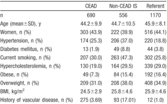
Hypercholester-Table 1. Clinical Characteristics
All France/Belgium Finland Italy
CEAD patients n 690 353 175 162 Age (mean⫾SD), y 44.2⫾9.9 43.8⫾9.0 45.1⫾10.0 44.3⫾10.8 Women, n (%) 303 (43.9) 168 (47.6) 64 (36.6) 71 (43.8) Carotid dissection, n (%) 446 (64.7) 240 (68.0) 96 (54.9) 110 (68.3) Cerebral ischemia, n (%) 541 (78.4) 287 (81.3) 128 (73.1) 126 (77.8) Cerebral infarct, n (%) 454 (65.8) 228 (64.6) 108 (61.7) 118 (72.8) Non-CEAD IS patients n 556 254 168 134 Age, y 44.7⫾10.5 45.1⫾9.4 46.1⫾12.0 42.3⫾10.2 Women, n (%) 222 (39.9) 103 (40.5) 62 (36.9) 57 (42.5) TOAST subtype, n (%) Large-artery atherosclerosis 75 (13.5) 27 (10.6) 20 (11.9) 28 (20.9) Cardioembolic 205 (36.9) 115 (45.3) 55 (32.7) 35 (26.1) Small-vessel disease 41 (7.4) 13 (5.1) 13 (7.8) 15 (11.2)
Other determined cause 9 (1.6) 3 (1.2) 1 (0.6) 5 (3.7)
Undetermined cause 226 (40.6) 96 (37.8) 79 (47.0) 51 (38.1)
Referents
n 1170 692 269 209
Age, y 45.9⫾8.1 45.8⫾7.1 45.9⫾10.5 46.1⫾7.9
Women, n (%) 516 (44.1) 330 (47.8) 104 (38.7) 82 (39.1)
CEAD indicates cervical artery dissection; IS, ischemic stroke; and TOAST, Trial of Org 10172 in Acute Stroke Treatment.23
Table 2. Prevalence of Vascular Risk Factors
CEAD Non-CEAD IS Referent
n 690 556 1170 Age (mean⫾SD), y 44.2⫾9.9 44.7⫾10.5 45.9⫾8.1 Women, n (%) 303 (43.9) 222 (39.9) 516 (44.1) Hypertension, n (%) 174 (25.3) 206 (37.0) 220 (18.8) Diabetes mellitus, n (%) 13 (1.9) 49 (8.8) 44 (3.8) Current smoking, n (%) 207 (30.0) 263 (47.3) 302 (25.8) Hypercholesterolemia, n (%) 130 (19.0) 164 (29.5) 339 (29.0) Obese, n (%) 49 (7.3) 84 (15.4) 192 (16.4) Overweight, n (%) 209 (31.0) 208 (38.0) 408 (34.9) BMI, kg/m2 24.5⫾2.9 25.8⫾4.6 25.9⫾4.6
History of vascular disease, n (%) 275 (3.69) 93 (17.01) 12 (1.0) CEAD indicates cervical artery dissection; IS, ischemic stroke; and BMI, body mass index.
olemia, obesity, and overweightness were significantly less
frequent in CEAD patients, with a graded inverse effect of
obesity and overweightness. Increasing BMI was associated
with a reduced risk of CEAD, and this effect was linear
(P
⫽0.89 for test of nonlinearity). These results were similar
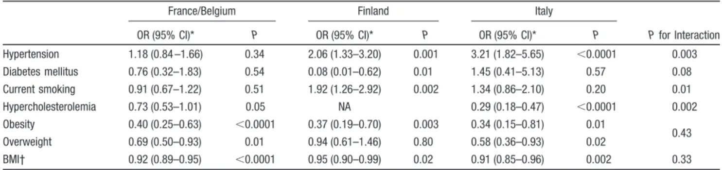
across countries (Table 4), although the magnitude of
asso-ciations with hypertension and hypercholesterolemia differed
significantly between countries.
When all risk factors were included in the same model,
there was still a significant positive association of
hyperten-sion with CEAD and an inverse association of
hypercholes-terolemia and obesity/overweightness with CEAD (Table 3).
In a stepwise logistic regression with age, gender, and
country of inclusion forced in, hypertension,
hypercholester-olemia, and obesity/overweightness were the variables
re-tained in the final model (Table I in the online-only Data
Supplement). In a secondary analysis, we observed an inverse
association with CEAD for both total cholesterol (odds
ratio
⫽0.51; 95% confidence interval, 0.44 to 0.59; P⬍0.0001
per 1-mmol/L increase) and low-density lipoprotein
choles-terol levels (odds ratio 0.51; 95% confidence interval, 0.42 to
0.62; P
⬍0.0001 per 1-mmol/L increase).
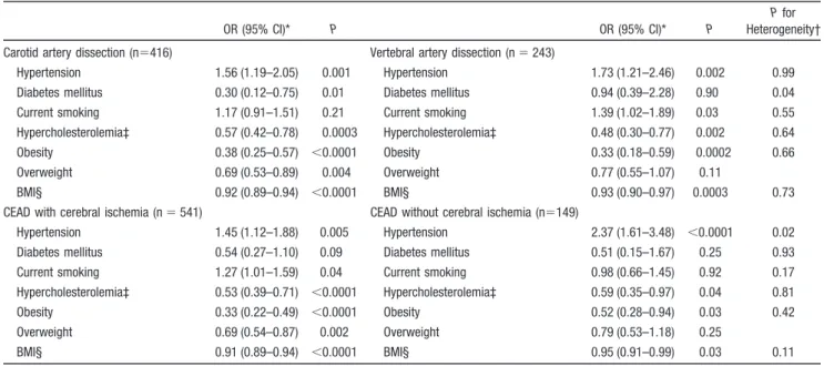
The associations of vascular risk factors with CEAD were
substantially unchanged when stratifying on dissection site
(carotid versus vertebral) and on the presence or absence of
cerebral ischemia (Table 5). Results were similar for patients
included retrospectively or prospectively and after exclusion
of participants
⬍35 years of age (Table II in the online-only
Data Supplement).
CEAD Patients Versus Non-CEAD IS Patients
Compared with non-CEAD IS patients, CEAD patients had a
significantly lower prevalence of hypertension, diabetes
mel-litus, current smoking, hypercholesterolemia, and obesity/
overweightness and a significantly lower BMI (Table 3).
Results were unchanged when all risk factors were included
in the same regression (Table 3). Associations were similar
across countries except for variability in effect size for
hypertension, current smoking, and hypercholesterolemia
(Table III in the online-only Data Supplement). Results were
also unchanged when the comparison was restricted to CEAD
patients with an IS (n
⫽454) versus non-CEAD IS patients
(Table IV in the online-only Data Supplement).
Table 3. Comparison of Vascular Risk Factor Prevalence Between Cervical Artery Dissection Patients, Referents, and Non–Cervical Artery Dissection Ischemic Stroke Patients
CEAD Patients vs Referents
Non-CEAD IS Patients vs Referents
CEAD Patients vs Non-CEAD IS Patients
OR (95% CI)* P P† OR (95% CI)* P P† OR (95% CI)* P P† Global P‡
Hypertension§ 1.67 (1.32–2.12) ⬍0.0001 0.0001 2.89 (2.27–3.68) ⬍0.0001 ⬍0.0001 0.58 (0.45–0.75) ⬍0.0001 0.004 ⬍0.0001 Diabetes mellitus 0.54 (0.29–1.02) 0.06 0.62 2.65 (1.72–4.08) ⬍0.0001 0.01 0.20 (0.11–0.38) ⬍0.0001 0.11 ⬍0.0001 Current smoking 1.22 (0.99–1.50) 0.07 0.58 2.53 (2.04–3.14) ⬍0.0001 ⬍0.0001 0.48 (0.38–0.61) ⬍0.0001 ⬍0.0001 ⬍0.0001 Hypercholesterolemia 0.55 (0.42–0.71) ⬍0.0001 ⬍0.0001 1.12 (0.86–1.46) 0.40 0.42 0.49 (0.36–0.67) ⬍0.0001 0.0006 ⬍0.0001 Obesity 0.37 (0.26–0.52) ⬍0.0001 ⬍0.0001 1.03 (0.76–1.40) 0.84 0.10 0.36 (0.24–0.53) ⬍0.0001 0.005 ⬍0.0001 Overweight 0.70 (0.57–0.88) 0.002 0.001 1.13 (0.89–1.43) 0.30 0.47 0.62 (0.48–0.81) 0.0003 0.04 BMI㛳 0.92 (0.90–0.95) ⬍0.0001 … 0.99 (0.97–1.02) 0.65 … 0.93 (0.90–0.95) ⬍0.0001 ⬍0.0001
CEAD indicates cervical artery dissection; IS, ischemic stroke; OR, odds ratio; CI, confidence interval; and BMI, body mass index. *Multinomial logistic regression adjusted for age, gender, and country of inclusion.
†Adjusted for all risk factors (except BMI), age, gender, and country of inclusion. ‡P for difference between the 3 groups.
§In total, 13.1% of CEAD patients, 22.7% of non-CEAD IS patients and 11.4% of referents were on antihypertensive treatment (P⫽0.02 for CEAD versus referents,
P⬍0.0001 for CEAD versus non-CEAD IS, and P⬍0.0001 for non-CEAD IS versus referents in multinomial logistic regression adjusted for country, age, and gender;
global P⬍0.0001). 㛳Per 1-kg/m2increase.
Table 4. Association of Vascular Risk Factors With Cervical Artery Dissection by Country of Inclusion
France/Belgium Finland Italy
P for Interaction
OR (95% CI)* P OR (95% CI)* P OR (95% CI)* P
Hypertension 1.18 (0.84 –1.66) 0.34 2.06 (1.33–3.20) 0.001 3.21 (1.82–5.65) ⬍0.0001 0.003 Diabetes mellitus 0.76 (0.32–1.83) 0.54 0.08 (0.01–0.62) 0.01 1.45 (0.41–5.13) 0.57 0.08 Current smoking 0.91 (0.67–1.22) 0.51 1.92 (1.26–2.92) 0.002 1.34 (0.86–2.10) 0.20 0.01 Hypercholesterolemia 0.73 (0.53–1.01) 0.05 NA 0.29 (0.18–0.47) ⬍0.0001 0.002 Obesity 0.40 (0.25–0.63) ⬍0.0001 0.37 (0.19–0.70) 0.003 0.34 (0.15–0.81) 0.01 0.43 Overweight 0.69 (0.50–0.93) 0.01 0.94 (0.61–1.46) 0.80 0.58 (0.36–0.93) 0.02 BMI† 0.92 (0.89–0.95) ⬍0.0001 0.95 (0.90–0.99) 0.02 0.91 (0.85–0.96) 0.002 0.33
OR indicates odds ratio; CI, confidence interval; BMI, body mass index.
*Multinomial logistic regression comparing cervical artery dissection (CEAD) patients with referents adjusted for age, gender, and country of inclusion (comparisons of CEAD versus non-CEAD ischemic stroke关IS兴 patients and of non-CEAD IS patients versus referents are shown in Tables III and V in the online-only Data Supplement).
Non-CEAD IS Patients Versus Referents
Hypertension, diabetes mellitus, and current smoking were
significantly more frequent in non-CEAD IS patients compared
with referents (Table 3). Similar results were observed when all
risk factors were included in the same model (Table 3). The
associations with hypertension, diabetes mellitus, and current
smoking were homogeneous across countries (Table V in the
online-only Data Supplement). Associations also did not differ
between patients with an IS of classic origin (large-artery
atherosclerosis, small-vessel disease, and cardioembolism) and
patients with an IS of another determined cause or undetermined
origin (Table VI in the online-only Data Supplement).
Discussion
Compared with country-, gender-, and age-matched referents,
CEAD patients were more frequently hypertensive and had a
lower prevalence of hypercholesterolemia, obesity, and
over-weightness. These associations were similar for internal carotid
and vertebral artery dissections and in CEAD patients with or
without cerebral ischemia. All vascular risk factors were less
frequent in CEAD patients compared with country-, gender-,
and age-matched patients with a non-CEAD IS. Young patients
with a non-CEAD IS were more often hypertensive, diabetic,
and current smokers compared with referents.
In the Context of the Current Literature
Although hypertension is usually considered one of the major
risk factors for aortic dissection,
25,26its association with CEAD
is controversial. Two studies described an increased prevalence
of hypertension in CEAD patients
15,16(restricted to CEAD
patients with cerebral ischemia in 1 study,
16which partly
overlaps with our Italian subsample), whereas 2 other studies
reported no association.
17,19In the present sample, we found a
significantly higher frequency of hypertension in CEAD patients
compared with referents, regardless of the presence or absence
of cerebral ischemia. Although a lower prevalence of
hypercho-lesterolemia has been described in CEAD patients compared
with non-CEAD IS patients,
10to the best of our knowledge, no
such association has been reported in comparison with referents.
The lower mean BMI and lower prevalence of obesity and
overweightness in CEAD patients compared with referents are
in agreement with recently published results from an
indepen-dent French group for 239 patients and 516 referents.
19The
lower prevalence of vascular risk factors in CEAD patients
compared with young non-CEAD IS patients is in line with
previous publications.
8 –10Although the association of vascular
risk factors with IS risk is well established in older individuals,
few data are available in young adults.
27We found that, as in
older individuals, hypertension, diabetes mellitus, and current
smoking were significantly more frequent in young non-CEAD
IS patients compared with referents regardless of IS subtype.
Underlying Mechanisms
As for dissection in other arteries, CEAD probably results from
multiple coexisting pathological processes, leading to a
weak-ening of or increased stress on the arterial wall.
26From earlier
observations that structural and functional arterial anomalies are
more frequent in CEAD patients than in referents,
14,28 –31it has
been postulated that CEAD patients could have a constitutional
weakness of the vessel wall, on top of which acute events such
as minor cervical trauma or infection could act as triggers.
1Elevated blood pressure could contribute to CEAD risk by
increasing carotid stiffness
32; alternatively, CEAD patients could
have a constitutionally elevated arterial stiffness,
31leading to an
Table 5. Association of Vascular Risk Factors With Different Subgroups of Cervical Artery Dissection
OR (95% CI)* P OR (95% CI)* P
P for
Heterogeneity† Carotid artery dissection (n⫽416) Vertebral artery dissection (n⫽ 243)
Hypertension 1.56 (1.19–2.05) 0.001 Hypertension 1.73 (1.21–2.46) 0.002 0.99
Diabetes mellitus 0.30 (0.12–0.75) 0.01 Diabetes mellitus 0.94 (0.39–2.28) 0.90 0.04
Current smoking 1.17 (0.91–1.51) 0.21 Current smoking 1.39 (1.02–1.89) 0.03 0.55
Hypercholesterolemia‡ 0.57 (0.42–0.78) 0.0003 Hypercholesterolemia‡ 0.48 (0.30–0.77) 0.002 0.64
Obesity 0.38 (0.25–0.57) ⬍0.0001 Obesity 0.33 (0.18–0.59) 0.0002 0.66
Overweight 0.69 (0.53–0.89) 0.004 Overweight 0.77 (0.55–1.07) 0.11
BMI§ 0.92 (0.89–0.94) ⬍0.0001 BMI§ 0.93 (0.90–0.97) 0.0003 0.73
CEAD with cerebral ischemia (n⫽ 541) CEAD without cerebral ischemia (n⫽149)
Hypertension 1.45 (1.12–1.88) 0.005 Hypertension 2.37 (1.61–3.48) ⬍0.0001 0.02
Diabetes mellitus 0.54 (0.27–1.10) 0.09 Diabetes mellitus 0.51 (0.15–1.67) 0.25 0.93
Current smoking 1.27 (1.01–1.59) 0.04 Current smoking 0.98 (0.66–1.45) 0.92 0.17
Hypercholesterolemia‡ 0.53 (0.39–0.71) ⬍0.0001 Hypercholesterolemia‡ 0.59 (0.35–0.97) 0.04 0.81
Obesity 0.33 (0.22–0.49) ⬍0.0001 Obesity 0.52 (0.28–0.94) 0.03 0.42
Overweight 0.69 (0.54–0.87) 0.002 Overweight 0.79 (0.53–1.18) 0.25
BMI§ 0.91 (0.89–0.94) ⬍0.0001 BMI§ 0.95 (0.91–0.99) 0.03 0.11
OR indicates odds ratio; CI, confidence interval; BMI, body mass index; and CEAD, cervical artery dissection.
*Multinomial logistic regression comparing subgroups of CEAD patients with all referents adjusted for age, gender, and country of inclusion. †Assessed with logistic regression restricted to CEAD patients with the CEAD characteristic as the outcome variable.
‡n⫽323 for carotid artery dissection, n⫽164 for vertebral artery dissection, n⫽413 for CEAD with cerebral ischemia, and n⫽102 for CEAD without cerebral ischemia.
increase in systolic pressure. The inverse association of CEAD
with hypercholesterolemia and BMI, as well as the young age of
occurrence of CEAD, are in contrast to aortic dissection, which
is most commonly associated with old age and atherosclerosis.
26Although we did not screen specifically for atherosclerotic
lesions, the vascular risk factor profile of CEAD patients
suggests that atherosclerosis is probably not a predisposing
condition to CEAD. Recently, an increase in wall material
stiffness with a heterogeneous echostructure was described in
CEAD patients.
31With aging and arteriosclerosis, the
echostruc-ture becomes more homogeneous, with an increase in collagen
and elastin cross-links, making it less prone to dissection.
31This
process could be accelerated in individuals with
hypercholester-olemia and elevated BMI, analogous to diabetes mellitus, which
is associated with an increased synthesis and reduced
degrada-tion of the extracellular matrix,
33an increased number of
covalent cross-links between proteins,
34and a reduced incidence
of abdominal aortic aneurysms.
35One could also speculate that
lean persons, with less adipose tissue protecting the arteries from
minor cervical traumas, might be more prone to developing
CEAD as a result of increased vulnerability to such traumas.
Low cholesterol and BMI could also be mere confounders
reflecting a common underlying genetic disorder or
susceptibil-ity factor. Patients with inherited connective tissue disorders
such as Marfan syndrome tend to be taller and have a lower BMI
than referents.
36Although Marfan syndrome itself seems to be
only marginally associated with CEAD, there is some evidence
that other connective tissue disorders, and possibly other genetic
susceptibility factors in connective tissue genes, may be
impor-tant predisposing conditions to CEAD.
37– 40Strengths and Limitations
The main strengths of this study are the large sample size and
the comparison to both referents and age-matched non-CEAD
IS patients. Despite being one of the main causes of IS in
young adults, CEAD is rare in the general population
(inci-dence, 2.6/100 000 per year)
41; thus, only an international
multicenter effort could achieve a sufficiently large sample
size for this analysis. The coherence of associations in
different patient subgroups and countries strengthens our
findings. CEAD and non-CEAD IS patients were recruited in
the same centers according to a unique protocol, thus
allow-ing optimal comparisons. We were limited by the
heteroge-neity in recruitment methods and risk factor evaluation for
referents across countries; on the other hand, this
heteroge-neity enabled us to test the robustness of our findings. For
Italy and Finland, the fact that referents were selected to be
free of vascular disease may have inflated the association of
CEAD and non-CEAD IS with vascular risk factors, but this
is unlikely to have affected our results substantially because
the prevalence of vascular disease is very low in this age
category in the general population. In the Finnish sample,
some of the associations, especially with BMI and obesity/
overweightness, may have been weakened by assortative
mating, but this effect should be marginal because only 6% of
Finnish referents were spouses of patients. The imbalance in
the proportion of participants
⬍35 years of age between
patients and referents in the French-Belgian and Italian
samples could have artificially inflated the inverse
associa-tions of hypercholesterolemia and obesity/overweightness
with CEAD. However, these associations were still
signifi-cant after the exclusion of all individuals
⬍35 years of age.
Another limitation is that weight and height were measured in
French-Belgian and Italian referents, whereas reported values
were used for the other groups; reported weight tends to be an
underestimation of the true measure, which could artificially
inflate the inverse association of BMI with CEAD. We also
cannot formally exclude nonrandom misclassification of
hy-pertension history if the number of individuals with less prior
access to care and therefore less opportunity for diagnosis of
hypertension differed between groups. Our study sample is
not perfectly representative of the general population.
Pa-tients in the CEAD and non-CEAD IS groups were recruited
through neurology departments, often in tertiary centers,
which are biased toward more complicated cases and rare
causes. Persons with CEAD causing only local signs or minor
strokes, which may be underdiagnosed, and CEAD patients
with very severe strokes requiring intensive care were less
likely to be included. Referents recruited through health
surveys and epidemiological studies generally have fewer
risk factors and less disease than persons who do not
participate.
42Finally, we did not correct for multiple
compar-isons, but given the strength of the associations and the
homogeneity of findings across countries and subgroups,
false-positive associations seem unlikely.
Implications
Our findings, if confirmed in independent data sets, could
improve the understanding of the mechanisms underlying
CEAD, a major cause of IS in young adults, in whom the
impact of stroke-related disability is particularly dramatic
from a personal and socioeconomic point of view.
Hyperten-sion was associated with CEAD, but the relationship seems
weaker than with IS resulting from other causes in young
adults of the same age. Further studies testing whether
hypertension is also associated with an increased risk of
CEAD recurrence could be important for preventive
pur-poses. These studies should include long-term follow-up of
consecutive CEAD patients and ascertainment of
hyperten-sion on the basis both of history before the dissection and on
blood pressure measurements at a distance of the vascular
event. The inverse association of CEAD with
hypercholes-terolemia could have implications in terms of secondary
stroke prevention, because statins are commonly prescribed
after an IS, including in CEAD patients in some instances. In
addition to validating these associations, future studies could
include a simultaneous assessment of the carotid wall
struc-ture and genetic susceptibility factors of hypertension,
obe-sity, and hypercholesterolemia to explore the underlying
mechanisms.
Conclusions
The vascular risk factor profile of CEAD patients differs from
referents and young adults with a non-CEAD IS.
Hyperten-sion was associated with an increased risk of CEAD, whereas
an inverse association with hypercholesterolemia, obesity,
and overweightness was observed.
Appendix
CADISP Investigators
Belgium: Department of Neurology, Erasmus University Hospital; Laboratory of Experimental Neurology, Université Libre de Brux-elles, Brussels (She´rine Abboud, Massimo Pandolfo); Leuven Uni-versity Hospital, Leuven (Vincent Thijs). Finland: Department of Neurology, Helsinki University Central Hospital, Helsinki (Tiina Metso, Antti Metso, Turgut Tatlisumak). France: Departments of Neurology, Lille University Hospital-EA2691, Lille (Marie Bode-nant, Ste´phanie Debette, Didier Leys, Paul Ossou); Sainte-Anne University Hospital (Fabien Louillet, Jean-Louis Mas, Emmanuel Touze´); Pitie´-Salpeˆtrie`re University Hospital, Paris (Sara Leder, Anne Le´ger, Sandrine Deltour, Sophie Crozier, Isabelle Me´resse, Yves Samson); Amiens University Hospital, Amiens (Sandrine Canaple, Olivier Godefroy, Chantal Lamy); Dijon University Hos-pital, Dijon (Yannick Be´jot, Maurice Giroud); Besançon University Hospital, Besançon (Pierre Decavel, Elizabeth Medeiros, Paola Montiel, Thierry Moulin, Fabrice Vuillier); Inserm U744, Pasteur Institute, Lille (Philippe Amouyel, Jean Dallongeville, Ste´phanie Debette). Germany: Departments of Neurology, Heidelberg Univer-sity Hospital, Heidelberg (Caspar Grond-Ginsbach, Manja Kloss, Christoph Lichy, Tina Wiest, Inge Werner, Marie-Luise Arnold); University Hospital of Ludwigshafen, Ludwigshafen (Michael Dos Santos, Armin Grau); University Hospital of Mu¨nchen, Munich (Martin Dichgans); Department of Dermatology, Heidelberg Univer-sity Hospital (Ingrid Hausser); Department of Rehabilitation, Schmieder-Klinik, Heidelberg (Tobias Brandt, Constanze Thomas-Feles, Ralph Weber). Italy: Department of Neurology, Brescia University Hospital, Brescia (Elisabetta Del Zotto, Alessia Giossi, Irene Volonghi, Alessandro Padovani, Alessandro Pezzini); Perugia University Hospital, Perugia (Valeria Caso); Milan University Hos-pital, Milan (Anna Bersano, Silvia Lanfranconi, Pierluigi Baron); University of Milano Bicocca, San Gerardo Hospital, Monza (Si-mone Beretta, Carlo Ferrarese); Milan Scientific Institute San Raf-faele University Hospital, Milan (Maria Sessa, Giacomo Giacolone); Department of Rehabilitation, Santa Lucia Hospital, Rome (Stefano Paolucci). Switzerland: Department of Neurology, Basel University Hospital, Basel (Stefan Engelter, Felix Fluri, Florian Hatz, Domin-ique Gisler, Margareth Amort, Philippe Lyrer). United Kingdom: Clinical Neuroscience, St. George’s University of London (Hugh Markus). Turkey: Department of Neurology, University Hospital of Istanbul (Ayse Altintas). Argentina: Department of Neurology, University Hospital Sanatorio Allende, Cordoba (Juan Jose Martin).
Groups Collaborating With the
CADISP Consortium
MONA-LISA-Lille (Philippe Amouyel, Jean Dallongeville); MONA-LISA-Strasbourg (Dominique Arveiler, Aline Wagner); Vo-barno Study, Clinica Medica, Brescia University Hospital, Brescia, Italy (Enrico Agabiti Rosei, Maria Lorenza Muiesan, Massimo Salvetti, Mara Giacche`, Maurizio Castellano).
Acknowledgments
We thank the staff and participants of all CADISP centers the MONA-LISA-Lille, MONA-LISA-Strasbourg, and Vobarno cohort for their important contributions. We are particularly grateful for the contribution of Marja Metso, RN, Department of Neurology, Hel-sinki University Central Hospital, HelHel-sinki, Finland; Laurence Bel-lengier, MS, Sabrina Schilling, MS, P. Christian Libersa, MD, PhD, and Dominique Deplanque, MD, PhD, Centre d’Investigation Clin-ique, University Hospital of Lille; Nathalie Fievet, PhD, Inserm U744, Pasteur Institute, Lille, France; Jean-Christophe Corvol, Sylvie Montel, and Christine Re´my, Centre d’Investigation Clinique, Pitie´-Salpeˆtrie`re University Hospital, Paris, France; Alessandra Pan-arotto and Luigi Mori, (Vobarno Study, Clinica Medica, Brescia University Hospital, Brescia, Italy); Ana Lopes Da Cruz, Laboratory of Experimental Neurology, ULB, Brussels, Belgium; and Annet Tiemessen, MS, Stroke Team, University Hospital Basel, Basel, Switzerland.
Sources of Funding
The CADISP study has received funding from the Contrat de Projet Etat-Region 2007, Centre National de Genotypage, Emil Aaltonen Foundation, Paavo Ilmari Ahvenainen Foundation, Helsinki Univer-sity Central Hospital Research Fund, Academy of Finland, Helsinki University Medical Foundation, Pa¨ivikki and Sakari Sohlberg Foun-dation, Aarne Koskelo FounFoun-dation, Maire Taponen FounFoun-dation, Aarne and Aili Turunen Foundation, Lilly Foundation, Alfred Kordelin Foundation, Finnish Medical Foundation, Projet Hospital-ier de Recherche Clinique Re´gional 2004, Fondation de France, Ge´nopoˆle de Lille, Adrinord, EA2691, Institut Pasteur de Lille, Inserm U744, Basel Stroke Funds, Ka¨the-Zingg-Schwichtenberg-Fonds of the Swiss Academy of Medical Sciences, and Swiss Heart Foundation. The Vobarno Study is supported in part by grants from the European Community Network of Excellence (InGenious HyperCare, 2006 to 2010); Italian University and Research Ministry, Regione Lombardia, and Fondazione della Comunita` Bresciana Onlus. The MONA-LISA Study was made possible by an unre-stricted grant from Pfizer and by a grant from the ANR (ANR-05-PNRA-018). Dr Thijs is supported by FWO Flanders.
Disclosures
Dr Metso received research grants from the Helsinki Central Hos-pital Research Fund, Finnish Medical Foundation, University of Helsinki, Aarne and Aili Turunen Foundation, Emil Aaltonen Foun-dation, Paaro Illmari Aimenainen FounFoun-dation, Pa¨ivikki and Sakari Sohlberg Foundation, Aarne Koskelo Foundation, Maire Taponen Foundation, Lilly Foundation, and Alfred Kordelin Foundation. Dr Metso also received research grants from the Finnish Medical Foundation and Alfred Kordelin Foundation. Dr Caso received payment for speakers’ bureau appointments from Boehringer Ingelheim-Sanofi. Dr Engelter received a research grant from the Swiss Academy of Medical Sciences and Swiss Heart Foundation.
References
1. Debette S, Leys D. Cervical-artery dissections: predisposing factors, diagnosis, and outcome. Lancet Neurol. 2009;8:668 – 678.
2. Rubinstein SM, Peerdeman SM, van Tulder MW, Riphagen I, Haldeman S. A systematic review of the risk factors for cervical artery dissection. Stroke. 2005;36:1575–1580.
3. Leys D, Bandu L, Henon H, Lucas C, Mounier-Vehier F, Rondepierre P, Godefroy O. Clinical outcome in 287 consecutive young adults (15 to 45 years) with ischemic stroke. Neurology. 2002;59:26 –33.
4. Gordon T, Castelli WP, Hjortland MC, Kannel WB, Dawber TR. Diabetes, blood lipids, and the role of obesity in coronary heart disease risk for women: the Framingham study. Ann Intern Med. 1977;87:393–397.
5. Gordon T, Kannel WB. Predisposition to atherosclerosis in the head, heart, and legs: the Framingham study. JAMA. 1972;221:661– 666. 6. Kannel WB, Wolf PA, Verter J, McNamara PM. Epidemiologic
assessment of the role of blood pressure in stroke: the Framingham study. JAMA. 1970;214:301–310.
7. Kannel WB. Role of blood pressure in cardiovascular disease: the Fra-mingham Study. Angiology. 1975;26:1–14.
8. Grau AJ, Brandt T, Buggle F, Orberk E, Mytilineos J, Werle E, Conradt, Krause M, Winter R, Hacke W. Association of cervical artery dissection with recent infection. Arch Neurol. 1999;56:851– 856.
9. Barbour PJ, Castaldo JE, Rae-Grant AD, Gee W, Reed JF III, Jenny D, Longennecker J. Internal carotid artery redundancy is significantly asso-ciated with dissection. Stroke. 1994;25:1201–1206.
10. Guillon B, Berthet K, Benslamia L, Bertrand M, Bousser MG, Tzourio C. Infection and the risk of spontaneous cervical artery dissection: a case-control study. Stroke. 2003;34:e79 – e81.
11. Grau AJ, Aulmann M, Lichy C, Meiser H, Buggle F, Brandt T, Grond-Ginsbach C. Increased cytokine release by leucocytes in survivors of stroke at young age. Eur J Clin Invest. 2001;31:999 –1006.
12. Smith WS, Johnston SC, Skalabrin EJ, Weaver M, Azari P, Albers GW, Gress DR. Spinal manipulative therapy is an independent risk factor for vertebral artery dissection. Neurology. 2003;60:1424 –1428.
13. Beletsky V, Nadareishvili Z, Lynch J, Shuaib A, Woolfenden A, Norris JW. Cervical arterial dissection: time for a therapeutic trial? Stroke. 2003;34:2856 –2860.
14. Lucas C, Lecroart JL, Gautier C, Leclerc X, Dauzat M, Leys D, Dek-lunder G. Impairment of endothelial function in patients with spontaneous cervical artery dissection: evidence for a general arterial wall disease. Cerebrovasc Dis. 2004;17:170 –174.
15. Longoni M, Grond-Ginsbach C, Grau AJ, Genius J, Debette S, Schwaninger M, Ferrarese C, Lichy C. The ICAM-1 E469K gene poly-morphism is a risk factor for spontaneous cervical artery dissection. Neurology. 2006;66:1273–1275.
16. Pezzini A, Caso V, Zanferrari C, Del Zotto E, Paciaroni M, Bertolino C, Grassi M, Agnelli G, Padovani A. Arterial hypertension as risk factor for spontaneous cervical artery dissection: a case-control study. J Neurol Neurosurg Psychiatry. 2006;77:95–97.
17. Konrad C, Muller GA, Langer C, Kuhlenbaumer G, Berger K, Nabavi DG, Dziewas R, Stogbauer F, Ringelstein EB, Junker R. Plasma homo-cysteine, MTHFR C677T, CBS 844ins68bp, and MTHFD1 G1958A polymorphisms in spontaneous cervical artery dissections. J Neurol. 2004;251:1242–1248.
18. Metso TM, Metso AJ, Salonen O, Haapaniemi E, Putaala J, Artto V, Helenius J, Kaste M, Tatlisumak T. Adult cervicocerebral artery dis-section: a single-center study of 301 Finnish patients. Eur J Neurol. 2009;16:656 – 661.
19. Arnold M, Pannier B, Chabriat H, Nedeltchev K, Stapf C, Buffon F, Crassard I, Thomas F, Guize L, Baumgartner RW, Bousser MG. Vascular risk factors and morphometric data in cervical artery dissection: a case-control study. J Neurol Neurosurg Psychiatry. 2009;80:232–234. 20. Debette S, Metso TM, Pezzini A, Engelter ST, Leys D, Lyrer P, Metso
AJ, Brandt T, Kloss M, Lichy C, Hausser I, Touze E, Markus HS, Abboud S, Caso V, Bersano A, Grau A, Altintas A, Amouyel P, Tatlisumak T, Dallongeville J, Grond-Ginsbach C. CADISP-Genetics: an international project searching for genetic risk factors of cervical artery dissections. Int J Stroke. 2009;4:224 –230.
21. Wagner A, Sadoun A, Dallongeville J, Ferrieres J, Amouyel P, Ruidavets JB, Arveiler D. High blood pressure prevalence and control in a middle-aged French population and their associated factors: the MONA LISA study. J Hypertens. 2011;29:43–50.
22. Muiesan ML, Salvetti M, Paini A, Monteduro C, Rosei CA, Aggiusti C, Belotti E, Bertacchini F, Galbassini G, Stassaldi D, Castellano M, Rosei EA. Pulse wave velocity and cardiovascular risk stratification in a general population: the Vobarno study. J Hypertens. 2010;28:1935–1943. 23. Adams HP Jr, Bendixen BH, Kappelle LJ, Biller J, Love BB, Gordon DL,
Marsh EE III. Classification of subtype of acute ischemic stroke. Defi-nitions for use in a multicenter clinical trial: TOAST: Trial of Org 10172 in Acute Stroke Treatment. Stroke. 1993;24:35– 41.
24. Ay H, Benner T, Arsava EM, Furie KL, Singhal AB, Jensen MB, Ayata C, Towfighi A, Smith EE, Chong JY, Koroshetz WJ, Sorensen AG. A computerized algorithm for etiologic classification of ischemic stroke: the Causative Classification of Stroke System. Stroke. 2007;38:2979 –2984.
25. Wilson SK, Hutchins GM. Aortic dissecting aneurysms: causative factors in 204 subjects. Arch Pathol Lab Med. 1982;106:175–180.
26. Golledge J, Eagle KA. Acute aortic dissection. Lancet. 2008;372:55– 66. 27. Chong JY, Sacco RL. Epidemiology of stroke in young adults: race/ethnic
differences. J Thromb Thrombolysis. 2005;20:77– 83.
28. de Bray JM, Marc G, Pautot V, Vielle B, Pasco A, Lhoste P, Dubas F. Fibromuscular dysplasia may herald symptomatic recurrence of cervical artery dissection. Cerebrovasc Dis. 2007;23:448 – 452.
29. Tzourio C, Cohen A, Lamisse N, Biousse V, Bousser MG. Aortic root dilatation in patients with spontaneous cervical artery dissection. Cir-culation. 1997;95:2351–2353.
30. Guillon B, Tzourio C, Biousse V, Adrai V, Bousser MG, Touboul PJ. Arterial wall properties in carotid artery dissection: an ultrasound study. Neurology. 2000;55:663– 666.
31. Calvet D, Boutouyrie P, Touze E, Laloux B, Mas JL, Laurent S. Increased stiffness of the carotid wall material in patients with spontaneous cervical artery dissection. Stroke. 2004;35:2078 –2082.
32. Benetos A, Laurent S, Hoeks AP, Boutouyrie PH, Safar ME. Arterial alterations with aging and high blood pressure: a noninvasive study of carotid and femoral arteries. Arterioscler Thromb. 1993;13:90 –97. 33. Norman PE, Davis TM, Le MT, Golledge J. Matrix biology of abdominal
aortic aneurysms in diabetes: mechanisms underlying the negative asso-ciation. Connect Tissue Res. 2007;48:125–131.
34. Aronson D. Cross-linking of glycated collagen in the pathogenesis of arterial and myocardial stiffening of aging and diabetes. J Hypertens. 2003;21:3–12.
35. Shantikumar S, Ajjan R, Porter KE, Scott DJ. Diabetes and the abdominal aortic aneurysm. Eur J Vasc Endovasc Surg. 2010;39:200 –207. 36. Erkula G, Jones KB, Sponseller PD, Dietz HC, Pyeritz RE. Growth and
maturation in Marfan syndrome. Am J Med Genet. 2002;109:100 –115. 37. Grond-Ginsbach C, Debette S. The association of connective tissue
dis-orders with cervical artery dissections. Curr Mol Med. 2009;9:210 –214. 38. Brandt T, Hausser I, Orberk E, Grau A, Hartschuh W, Anton-Lamprecht I, Hacke W. Ultrastructural connective tissue abnormalities in patients with spontaneous cervicocerebral artery dissections. Ann Neurol. 1998; 44:281–285.
39. Ulbricht D, Diederich NJ, Hermanns-Le T, Metz RJ, Macian F, Pierard GE. Cervical artery dissection: an atypical presentation with Ehlers-Danlos-like collagen pathology? Neurology. 2004;63:1708 –1710. 40. Uhlig P, Bruckner P, Dittrich R, Ringelstein EB, Kuhlenbaumer G,
Hansen U. Aberrations of dermal connective tissue in patients with cervical artery dissection (sCAD). J Neurol. 2008;255:340 –346. 41. Lee VH, Brown RD Jr, Mandrekar JN, Mokri B. Incidence and outcome
of cervical artery dissection: a population-based study. Neurology. 2006; 67:1809 –1812.
42. 3C Study Group. Vascular factors and risk of dementia: design of the Three-City Study and baseline characteristics of the study population. Neuroepidemiology. 2003;22:316 –325.