Nitric oxide modulates the angiogenic phenotype of
middle-T transformed endothelial cells
Lucia Morbidelli
a, Sandra Donnini
b, Marina Ziche
a,*
aInstitute of Pharmacological Sciences, Uni6ersity of Siena, Via E. Piccolomini
170,53100Siena, Italy
bDepartment of Pharmacology, Uni6ersity of Florence, Florence, Italy
Received 8 August 2000; accepted 18 January 2001
Abstract
The role of nitric oxide (NO) in the induction of angiogenesis was evaluated in a murine heart endothelioma cell line (H.end.FB) carrying the mT oncogene. Two clonal derivatives of H.end.FB, H80 and H73, exhibiting different NO synthase (NOS) activities were selected and used in the study. The relationship among NOS activity and tumor cell behaviour (growth, and angiogenic capacity) and the molecular control of gene expression were investigated. H.end.FB and H80 on one side and H73 on the other side exhibited the highest and lowest NOS activity, respectively. Cell growth was inversely correlated to the amount of NO produced by the cell lines. Conversely, in the avascular rabbit cornea assay, H.end.FB and H80 cells were strongly angiogenic, while H73 were poorly angiogenic, indicating that the ability of the cells to induce neovascularization was associated with the extent of NO produced. Consistently, systemic administration to rabbits of the NOS inhibitor Nw
-nitro-L-arginine methyl ester (L-NAME) significantly reduced the angiogenicity of H.end.FB cells. RT-PCR evidenced that H.end.FB expressed mRNA for TGF-b1 and all VEGF isoforms, VEGF165 being predominantly expressed. NOS inhibition reduced the basal expression of VEGF isoforms, while it markedly potentiated TGF-b1 expression. These results indicate that the endogenous production of NO in tumor cells can serve as an autocrine/paracrine signalling mechanism of progression, by controlling angiogenic factor/modulator expression. © 2001 Elsevier Science Ltd. All rights reserved.
Keywords:Angiogenic phenotype; Nitric oxide; Transformed endothelium
www.elsevier.com/locate/ijbcb
1. Introduction
Angiogenesis is a tightly controlled process that regulates the formation of new capillaries from
preexisting blood vessels [1]. Increasing knowl-edge on the process of angiogenesis has led to the recent hypotesis that the end of tumor dormancy may be explained by a sudden conversion of a tumor toward an angiogenic phenotype [2]. This change may occur by an increase in the expression of angiogenesis stimulators, a decrease in angio-genesis inhibitors or a combination of the two events [3].
* Corresponding author. Tel.: + 0577-221255; fax + 39-0577-281928.
E-mail address:[email protected] (M. Ziche).
1357-2725/01/$ - see front matter © 2001 Elsevier Science Ltd. All rights reserved. PII: S1357-2725(01)00018-8
Hyperemia and vasodilation are persistent components of in-vivo angiogenesis. Nitric oxide (NO) is responsible for the endothelium-depen-dent vasodilation, and we reported that it plays a central role in the angiogenic cascade by de-mostrating that vascular endothelial growth fac-tor (VEGF) effect is linked to the NO synthase (NOS) pathway and that NO within the intra-cellular signalling cascades selectively mediates the angiogenic switch of the endothelium [4 – 6]. Moreover, experimental and clinical evidence in-dicate that the NOS pathway contributes to tu-mor progression and angiogenesis [7,8], suggesting that promotion of angiogenesis is a key event in linking NOS upregulation and tu-mor growth. However, the molecular mecha-nisms involved in NO-dependent tumor angiogenesis have not been completely eluci-dated.
It is known that transgenic mice expressing the polyoma virus middle T (mT) oncogene de-velop endothelial tumors [9,10]. Furthermore, in-fection of newborn and adult mice with a retrovirus carrying the mT oncogene resulted in the rapid formation of cavernous hemangiomas [10]. Middle T-transformed endothelial cells iso-lated from murine hemangiomas exibited an in-creased NOS activity [11]. However, these cells retained functional properties of normal endothelial cells [11 – 14]. Interestingly, the ad-ministration of a NOS inhibitor to endothe-lioma-bearing mice significantly reduced both the volume and the growth of the tumor [11].
The aim of our study was to investigate the role of the NOS pathway in the mechanisms of tumor progression and its correlation with the acquisition of an angiogenic phenotype in the murine heart endothelioma cell line (H.end.FB) carrying the mT oncogene. To address this is-sue, subclones of H.end.FB cells exhibiting dif-ferent NOS activity were selected. The angiogenic activity was tested in vivo in the rab-bit cornea assay, and cell replication was moni-tored in vitro. The effect of NOS inhibition on the pattern of gene expression for angiogenic factors and on in-vivo angiogenesis was also evaluated.
2. Materials and methods
2.1. Cell cultures
Murine heart endothelioma cell lines (H.end.FB) and their subclones H80 and H73 obtained by dilution cloning, were kindly pro-vided by Prof. Federico Bussolino, University of Torino, Italy. H.end.FB cell line was obtained from heart cells taken from a mouse fetus DBA/ 2Xc57b1/6 at the 13th day of gestation and im-mortalised by infection with N-TKmT retroviral vector containing mT antigen of polyoma virus [9,14]. This cell line was characterised as en-dothelial on the basis of CD31, VE-cadherin and factor VIII expression, and uptake of low-density acetylated lipoproteins [9,11]. Cells were grown in DMEM supplemented with 10% FCS and were split 1:8 twice a week.
2.2. Cell proliferation assay
Cellular proliferation was quantified by the total cell number as previously described [6]. Cells (103/ml) suspended in 10% FCS medium were seeded in 48-well plates and allowed to adhere for 6 – 12 h. Then, the media were re-moved and replaced with 500 ml/well of 1% FCS in the presence or in the absence of 1 mM of the NOS inhibitor Nw-monomethyl-L-arginine (L-NMMA). Proliferation was evaluated after 24, 48 and 72 h. Cells were fixed with methanol overnight at 4°C, and stained with Diff Quik (Dade SpA, Milano, Italy). The number of cells was counted in seven random fields of each well at a magnification of 200 with the aid of a 21 mm2 ocular grid. Data are expressed as the number (means9S.E.M.) of cells counted per well.
2.3. Angiogenesis in 6i6o: rabbit cornea assay
The angiogenic activity was assayed in vivo using the rabbit cornea model [15]. Angiogenesis was studied in the cornea of albino rabbits since this is an avascular and transparent tissue where inflammatory reactions, and growing capillaries can be easily monitored and changes quantitated by stereomicroscopic examination.
Corneal assays were performed in New Zealand white rabbits (Charles River, Calco, Como, Italy) essentially as described previously [15] and in accordance with the guidelines of the European Economic Community for animal care and welfare (EEC Law No. 86/609)
Briefly, after being anaesthetised with sodium pentothal (30 mg/kg), a micro pocket (1.5 × 3 mm) was surgically produced using a pliable iris spatula 1.5 mm wide in the lower half of the cornea. Rabbits received in the cornea 2.5 × 10⁵ cells/5 ml. Subsequent daily observation of the implants was made with a slit lamp stereomicro-scope without anaesthesia. An angiogenic re-sponse was scored positive when budding of vessels from the limbal plexus occurred after 3 – 4 days, and capillaries progressed to reach the implanted pellet as previously reported. The number of positive implants performed was scored during each observation. The angiogene-sis score was calculated (vessel density × distance from the limbus) as described previously [5].
To evaluate the effect of NO synthase inhibi-tion on the response to the angiogenic effectors,
Nw-nitro-L-arginine methyl ester (L-NAME) was given in the drinking water ad libitum. L -NAME solutions (0.5 g/l) were prepared freshly every day. The water intake was approximately 200 ml/day in the treated animals and was not different from the control group. Animals re-ceived L-NAME 1 week before surgery and 10 days following corneal implant. The effect of L -NAME treatment was compared to the treat-ment with both D-NAME and vehicle alone. The efficacy of L-NAME administration was evaluated at the end of the treatment as acetyl-choline-induced relaxation in rabbit aortic rings preconstricted with noradrenaline as previously described [16].
2.4. Measurement of nitrites/nitrates
Nitrite/nitrate production was evaluated in growing cell cultures in Phenol Red-free medium additioned with 1% FCS. Nitrite/nitrate produc-tion was measured by adding 0.5 ml of cell cul-ture medium concentrated 5 × to 0.5 ml of Greiss reagent [11]. After 30 min incubation at
room temperature in the dark, absorbance was measured at 540 nm in a spectrophotometer (UV/Vis, Perkin Elmer, Norwalk, CT). A nitrite calibration curve was made in the culture medium liophilized and concentrated as the samples.
2.5. RT-PCR analysis of angiogenic factor
expression
Cells at 90% confluence were stimulated for 15 h with test substances in the presence of 1% FCS. Total RNA from cells was isolated by the standard guanidine thiocyanate – phenol – chloro-form extraction [4]. cDNA was synthesized as described previously [6]. Differential RT-PCR for VEGF and transforming growth factor b1 (TGF-b1) was carried out by using 5 ml of cDNA and specific primers for VEGF and TGF-b1 as described previously [17]. Calibration was performed by co-amplification of the same cDNA samples with primers for glyceraldehyde 3-phosphate dehydrogenase (GAPDH) as inter-nal standard, with sequences as described previ-ously [6]. After amplification, aliquots of each sample product (20 ml) were electrophoresed on a 3% agarose gel and stained with ethidium bro-mide. The size of the amplification products was 196 bp for GAPDH, 275 bp for VEGF121, 380 bp for VEGF145, 407 bp for VEGF165, 479 bp for VEGF189 and 336 bp TGF-b1.
Image processing and analysis of the intensity of the bands were performed as described [6]. The results were analysed as ratio between the optical densities of the bands related to the am-plification products of the target genes and GAPDH.
2.6. Statistical analysis
Results are expressed as means9S.E.M. for (n) experiments. Multiple comparisons were per-formed by one-way ANOVA and individual dif-ferences tested by Fisher’s test after the demonstration of significant inter-group differ-ences by ANOVA. A P value B0.05 was taken as significant.
3. Results
3.1. Characterisation of nitrate/nitrite production
in H.end.FB clones
H.end.FB cell line expressed both isoforms of NO synthase, the constitutive (cNOS) and the inducibile (iNOS) one [11]. Subclones H80 and H73 were selected based on their growth rate. To assess the content of NO produced by each indi-vidual clone, NO levels were measured in condi-tioned medium by H.end.FB cells and H80 and H73 subclones by Griess reaction. After 48 h of incubation in medium containing 1 mM L -arginine, the nitrite/nitrate concentration was 12.1 nmol/mg protein in the supernatant of H.end.FB, whereas the NO levels were 10.8 nmol/mg protein for H80, and 5.9 nmol/mg protein for H73. Under the same experimental conditions, the addition of L-NMMA to the cells significantly decreased ni-trite/nitrate production in H.end.FB and H80
cells, and only slightly reduced the nitrite/nitrate levels released by H73 clone (Fig. 1).
3.2. The angiogenic acti6ity of H.end.FB clones is
positi6ely associated with the amount of NO
released in 6itro
The capacity of the H.end.FB clones to stimu-late an angiogenic response has been assessed in vivo in the avascular cornea of albino rabbits. H.end.FB cells implanted in the rabbit cornea were able to induce a rapid and sustained angio-genic response starting from day 4 from the im-plant (Fig. 2A and B). After 10 days from the surgical implant, seven implants out of eight ex-hibited a pronounced vascularization.
H80 clone, characterized by high NO levels, was also strongly angiogenic. H80 clone induced angiogenesis in six implants out of eight per-formed, and the extent of neovascular growth appeared to be consistently vigorous, although slightly lower when compared to that produced by H.end.FB cells (Fig. 2A and C).
H73 clone, characterized by low NOS activity, was the least angiogenic compared to H.end.FB and H80 subclone. Only three corneal implants out of eight hinted at an angiogenic response after 1 week from the implant (Fig. 2A and D).
3.3. Blockade of the NOS pathway in the host
results in inhibition of neo6ascularization induced
by H.end.FB clones
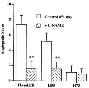
The role of NO production in the angiogenic response promoted by H.end.FB cells and sub-clones was evaluated by systemic administration of the NOS inhibitorL-NAME to the rabbits. The animals were treated 1 week before and 10 days after the corneal implant of H.end.FB cell lines. Treatment with L-NAME markedly reduced the angiogenic activity elicited by H.end.FB and H80 subclone. At day 8 from cell implant, the angio-genic score was reduced by the treatment by a 78% for H.end.FB cells and by a 69% for H80 clone (Fig. 3). Conversely, L-NAME treatment did not produce any significant modification in corneal implant containing H73 cells (Fig. 3). Fig. 1. Nitrite/nitrate accumulation in the supernatant of
H.end.FB cells and subclones. Constitutive nitrite/nitrate accu-mulation was measured in the cultured medium following 48 h incubation, by using the Griess reaction.L-NMMA (1 mM) was added to assess the specificity of the reaction. Data are means9S.E.M. of results obtained from three experiments performed in duplicate. *P50.05 versus basal condition.
Fig. 2. In-vivo angiogenesis by H.end.FB cell line and subclones. In-vivo angiogenesis was assessed in the avascular rabbit cornea model. (A) Angiogenesis by H.end.FB cells, H80 and H73 subclones. 2.5 × 10⁵ cells were surgically implanted in corneal
micropockets. The angiogenic response was assessed using a slit lamp stereomicroscope and capillary progression was monitored daily. Data (means9S.E.M.) are expressed as angiogenic score during time (days) (n=8). (B–D) Representative pictures of the angiogenic response induced by H.end.FB cell line (B), H80 (C) and H73 (D) clones at day 8. Original magnification 18 × .
Fig. 3. NO controls angiogenesis by H.end.FB cells. To evalu-ate the effect of NO synthase inhibition on the response to H.end.FB cells and their clonal derivatives, L-NAME was given in the drinking water ad libitum. Animals received L-NAME (0.5 g/l) 1 week before surgery and 10 days follow-ing corneal implant. Animal weight did not change durfollow-ing the treatment. Results are means (9S.E.M.) of the angiogenic score obtained at day 8 after implant of H.end.FB cell lines in animals treated (hatched column) or not (open column) with L-NAME. **P50.01 versus basal condition (n=3).
measured over time. Data are shown in Table 1. In vitro, the replication index of H73 clone was the highest compared to H80 clone and H.end.FB cells. To assess the potential role of NO in au-tocrinally affecting cell growth of endothelioma cells, clones were treated with 1 mM L-NMMA. No significant effect was observed in any of the clones compared to the controls, suggesting that endogenous NO was not directly involved in the control of cell growth (Table 1). Based on these results we conclude that the angiogenic activity of the high NOS expressing clones, H.end.FB cells and H80, was independent from their growth rate.
3.5. NOS upregulation modulates expression of
angiogenic factors in H.end.FB cell line
To assess the putative molecular mechanism associated with elevation of NO production and increased angiogenesis in endothelioma cells, gene expression for angiogenic factors was assessed. Two angiogenic factors were studied: the angio-genic stimulator VEGF [18] and the modulator TGF-b1 [19]. H.end.FB cells were thus investi-gated for the expression of VEGF and TGF-b1 mRNA by using differential RT-PCR. The role of NO was investigated by treating the cells with the NOS inhibitorL-NMMA. Cells were incubated in the presence or absence of 1 mM L-NMMA for 15 h. In unstimulated H.end.FB cells, we detected four VEGF isoforms: 121, 145, 165 and 189 aminoacids, with the VEGF165 isoform being predominantly expressed (Fig. 4A). Treatment with the NOS inhibitor L-NMMA of H.end.FB cell line decreased by a 20% VEGF165 mRNA expression, while it did not modify the other VEGF isoforms.
The expression of TGF-b1 was also investi-gated. As shown in Fig. 4B, H.end.FB cells ex-pressed low levels of TGF-b1 and the stimulation with L-NMMA significantly upregulated TGF-b1 mRNA gene expression.
Thus, NOS inhibition in H.end.FB cells elicited both angiogenic factor downregulation and angio-genic inhibitor upregulation, which might be re-sponsible for reduced in-vivo angiogenesis.
3.4. The pattern of cell growth of H.end.FB
clones is not associated with NOS acti6ity
To assess whether the pattern of cell replication could be responsible for the differences observed among the angiogenic profile induced by the cell lines, the growth rate of H.end.FB clones was Table 1
Cell replication of H.end.FB and subclonesa
Cell number (×10−3) 72 h 24 h 48 h 390.08 H.end.FB 1090.12 2090.15 990.09 3.290.04 +L-NMMA 1990.12 3.590.05 H80 1990.09 4290.23 +L-NMMA 3.490.06 1590.03 4090.15 2.790.09 2390.13 6090.24 H73 2090.12 390.05 6190.21 +L-NMMA
aThe total number of cells recovered after 24, 48 and 72 h
of growth in basal condition (1% FCS) is reported. Where indicated, cells were treated withL-NMMA (1 mM). Data are means9S.E.M. from four experiments run in duplicate.
Fig. 4. Differential RT-PCR for VEGF isoform (A), and TGF-b1 (B) expression in H.end.FB cells in the presence or in the absence of L-NMMA (1 mM). Representative RT-PCR amplification products for VEGF isoform (A) and TGF-b1 (B) (1 gel representative of 3 obtained with similar results). Cells were treated for 15 h as follows: C, none;L-NMMA, 1 mM L-NMMA. Numbers are the ratio between the optical densities related to the amplification products of the target gene (A, VEGF165, B, TGF-b1) and GAPDH9S.E.M.
lioma cells carrying the mT oncogene (H.end.FB) show an increase in basal NOS activity when compared to a wild-type cell line [11]. Based on reports showing a strong positive correlation be-tween the expression and activity of NOS and tumor angiogenesis and progression [7,8,21], the aim of the present study was to investigate (1) the angiogenic activity of tumor H.end.FB in relation to the levels of NO production and (2) the molec-ular mechanisms regulated by NO in the acquisi-tion of the angiogenic phenotype by H.end.FB cells. Our results indicate that murine heart en-dothelioma cell lines induce a strong and sus-tained neovascular response in vivo, which can be related to the extent of NO production. At the transcriptional level, the NOS pathway modulates the expression of the angiogenic factor VEGF, while its inhibition upregulates the expression of the TGF-b1 gene, suggesting that NO production might control angiogenesis in vivo by tuning an-giogenic factor expression in endothelioma cells.
Tumor cell proliferation and survival and their ability to induce angiogenesis are the key cellular processes that control tumor growth and spread-ing. We found that tumor H.end.FB cells are strongly angiogenic in the avascular rabbit cornea model. While the H80 subclone is able to induce angiogenesis, the H73 clone is poorly angiogenic when compared to H.end.FB cells. The growth rate of the endothelioma cell lines is inversely associated with their ability to elicit neovascular growth in vivo and to produce NO, suggesting that the growth rate of the two cell lines express-ing high NO levels is not involved in the angio-genic response. Furthermore, since the proliferation rate of any of the cell line in study is unaffected by NOS inhibition, the endogenous production of NO does not target the control of cell replication in tumor cells but targets other molecular functions relevant for angiogenesis.
The observation that H.end.FB cells and its H80 clone, expressing a high NOS activity, are strongly angiogenic, while the H73 clone, with a low NOS activity, is poorly angiogenic, implicates that the distinct angiogenic activity expressed in vivo by each clone could be positively associated to the extent of NO produced. The relevance of NO in the induction of the neovascular growth in
4. Discussion
Expression of the mT oncogene in either trans-genic [9] or chimeric [10] mice is associated with a significant subversion of normal blood vessel mor-phogenesis, which results in the rapid formation of multiple cavernous hemangiomas by recruit-ment of host cells [20]. Murine heart
endothe-vivo has been investigated through systemic NOS blockade. Inhibition of host NO production by systemic administration of L-NAME to the rabbits markedly reduces the angiogenic activity elicited by H.end.FB and H80 subclone. Angio-genesis in vivo requires a functioning NO path-way in the vasculature [5,7,16]. Thus, in our experimental model, we can speculate that there are two targets reached by systemic NO blockade: the host capillary endothelium, which does not enter the angiogenic switch when deprived of NO, and the tumor cell line, which changes its angio-genic phenotype losing NOS activity. While the first possibility is well documented and is likely to occur in our model, our data indicate that tumor cells can sense NOS inhibition as well.
The angiogenic phenotype of a tumor is the net result of the balance between positive and nega-tive regulators of angiogenesis [3]. Among the stimulators of angiogenesis, a pivotal role is played by VEGF [18], while TGF-b1 has been described with opposite functions in angiogenesis [19,22]. Since NO production is associated with the ability of the endotelioma cells to induce angiogenesis, the expression of angiogenic factors/ modulators has been investigated in H.end.FB cells, the cell line with the highest NOS activity and angiogenicity. We have measured the expres-sion pattern of the five isoforms of VEGF mRNA by semiquantitave RT-PCR. The cells express the four isoforms VEGF121, VEGF145, VEGF165 and VEGF189, but not VEGF206. We report the occurrence of a differential expression of VEGF165 mRNA transcripts in response to en-dogenous NO. The addition of the NOS inhibitor L-NMMA to the cell cultures downregulates VEGF165 expression, suggesting that the high NOS activity in transformed H.end.FB cell line might control the expression of this angiogenic factor. In addition, in H.end.FB cells, the expres-sion of TGF-b1 is strongly upregulated in re-sponse to NOS inhibition, indicating that NO constitutively produced by the cells downregulates TGF-b1 expression. At present, it is difficult to speculate which of the two factors affected by endogenous NO will be the predominant modula-tor of the angiogenic outcome in vivo: increased VEGF165 expression or modulation of TGF-b1
expression, or whether there are other additional mechanisms/mediators activated by NO.
In conclusion our results indicate that in en-dothelioma cells endogenous NO production modulates the angiogenic phenotype. A potential target of endogenous NO production appears to be the modulation of the balance among angio-genic factors at the transcription level in tumor cells. Additional experiments are needed to eluci-date whether the increased angiogenic activity linked to the NO production can be linked to a more aggressive behaviour of the endothelioma in vivo.
Acknowledgements
We thank Prof. Federico Bussolino for provid-ing us with the cells used in the study. This work was supported by funds from the Italian Ministry of University and Scientific and Technological Research (Cofinanziamento Programmi di Ricerca Scientifica di Rilevante Interesse Nazionale grant n. 9906217877), the National Re-search Council (Progetto Finalizzato Biotecnolo-gie) and the Italian Association for Cancer Research (AIRC) to M.Z. and Progetto Giovani Ricercatori, University of Siena to L.M.
References
[1] J. Folkman, Angiogenesis: Initiation and control, Ann. NY Acad. Sci. 401 (1982) 212 – 227.
[2] J. Folkman, Angiogenesis in cancer, vascular, rheumatoid and other disease, Nat. Med. 1 (1995) 27 – 31.
[3] E. Keshet, S.A. Ben-Sasson, Anticancer drug target: ap-proaching angiogenesis, J. Clin. Invest. 104 (1999) 1497 – 1501.
[4] M. Ziche, A. Parenti, F. Ledda, P. Dell’Era, H.J. Granger, C.A. Maggi, M. Presta, Nitric oxide promotes proliferation and plasminogen activator production by coronary venular endothelium through endogenous bFGF, Circ. Res. 80 (1997) 845 – 852.
[5] M. Ziche, L. Morbidelli, R. Choudhuri, H.-T. Zhang, S. Donnini, H.J. Granger, R. Bicknell, Nitric oxide-synthase lies downstream from vascular endothelial growth factor-induced but not basic fibroblast growth factor-factor-induced angiogenesis, J. Clin. Invest. 99 (1997) 2625 – 2634. [6] A. Parenti, L. Morbidelli, X.L. Cui, J.G. Douglas, J.
an upstream signal for vascular endothelial growth fac-tor-induced extracellular signal-regulated kinases1/2
acti-vation in postcapillary endothelium, J. Biol. Chem. 273 (1998) 4220 – 4226.
[7] O. Gallo, E. Masini, L. Morbidelli, A. Franchi, I. Fini-Storchi, W.A. Vergari, M. Ziche, Role of nitric oxide in angiogenesis and tumor progression in head and neck cancer, J. Natl. Cancer Inst. 90 (1998) 587 – 596. [8] L.C. Jadeski, K.O. Hum, C. Chakraborty, P.K. Lala,
Nitric oxide promotes murine tumor growth and metas-tasis by stimulating tumor cell migration, invasiveness and angiogenesis, Int. J. Cancer 86 (2000) 30 – 39. [9] V.L. Bautch, S. Toda, J.A. Hassell, D. Hanahan,
En-dothelial cell tumors develop in transgenic mice carrying polyoma virus middle T oncogene, Cell 51 (1987) 529 – 538.
[10] R.L. Williams, S.A. Courtneidge, E.F. Wagner, Embry-onic lethalities and endothelial tumors in chimeric mice expressing polyoma virus middle T oncogene, Cell 52 (1988) 121 – 131.
[11] D. Ghigo, M. Arese, R. Todde, A. Vecchi, F. Silvagno, C. Costamagna, Q.G. Dong, M. Alessio, R. Heller, R. Soldi, F. Trucco, G. Garbarino, G. Pescarmona, A. Mantovani, F. Bussolino, A. Bosia, Middle T antigen-transformed endothelial cells exhibit an increased activ-ity of nitric oxide synthase, J. Exp. Med. 181 (1995) 9 – 19.
[12] C. Garlanda, C. Parravicini, M. Sironi, M. De Rossi, R. Wainstok de Calmanovici, F. Carozzi, F. Bussolino, F. Colotta, A. Mantovani, A. Vecchi, Progressive growth in immunodeficient mice and host cell recruit-ment by mouse endothelial cells transformed by poly-oma middle-sized T antigen: implications for the pathogenesis of opportumnistic vascular tumors, Proc. Natl. Acad. Sci. USA 91 (1994) 7291 – 7295.
[13] E. Bocchietto, A. Guglielmetti, F. Silvagno, G. Tarabo-letti, G.P. Pescarmona, A. Mantovani, F. Bussolino, Proliferative and migratory responses of murine
mi-crovasculr endothelial cells to granulocyte colony stimu-lating factor, J. Cell Physiol. 155 (1993) 89 – 95. [14] F. Bussolino, M. De Rossi, A. Sica, F. Colotta, J.M.
Wang, E. Bocchietto, I.M. Padura, A. Bosia, E. De-jana, A. Mantovani, Murine endothelioma cell lines transformed by polyoma middle T oncogene as a target for and producer of cytokines, J. Immunol. 147 (1991) 2122 – 2129.
[15] M. Ziche, J. Jones, P.M. Gullino, Role of prostaglandin E1 and copper in angiogenesis, J. Natl. Cancer. Inst. 69 (1982) 475 – 482.
[16] M. Ziche, L. Morbidelli, E. Masini, S. Amerini, H.J. Granger, C. Maggi, P. Geppetti, F. Ledda, Nitric oxide mediates angiogenesis in vivo and endothelial cell growth and migration in vitro promoted by substance P, J. Clin. Invest. 94 (1994) 2036 – 2044.
[17] K.J. Brown, S.F. Maynes, A. Bezos, D.J. Maguire, M.D. Ford, C.R. Parish, A novel in vitro assay for human angiogenesis, Lab. Invest. 75 (1996) 539 – 555. [18] N. Ferrara, K. Houck, L. Jakeman, D.W. Leung,
Molecular and biological properties of the vascular en-dothelial growth factor family of proteins, Endocr. Rev. 13 (1992) 18 – 32.
[19] M.S. Pepper, J. -D. Vassalli, L. Orci, R. Montesano, Biphasic effect of transforming growth factor-b1 on in vitro angiogenesis, Exp. Cell Res. 204 (1993) 356 – 363. [20] R.L. Williams, W. Risau, H.-G. Zerwes, H. Dextler, A.
Aguzzi, E.F. Wagner, Endothelioma cells expressing the polyoma middle T oncogene induce hemangiomas by host cell recruitment, Cell 57 (1989) 1053 – 1063. [21] D.C. Jenkins, I.G. Charles, L.L. Thomsen, D.W. Moss,
L.S. Holmes, S.A. Baylis, P. Rhodes, K. Westmore, P.C. Emson, S. Moncada, Role of nitric oxide in tumor angiogenesis, Proc. Natl. Acad. Sci. USA 92 (1995) 4392 – 4396.
[22] L.F. Fajardo, S.D. Prionas, H.H. Kwan, J. Kowalski, A.C. Allison, Transforming growth factor b1 induces angiogenesis in vivo with a threshold pattern, Lab. In-vest. 74 (1996) 600 – 608.