Original research
Beneficial effects of oral pure caffeine on oxidative stress
Daniela Metro
a, Valeria Cernaro
b, Domenico Santoro
b,⇑
, Mattia Papa
a, Michele Buemi
b,
Salvatore Benvenga
c,d,e, Luigi Manasseri
aaDepartment of Biomedical and Dental Sciences and Morphofunctional Imaging, University of Messina, University Hospital Policlinico G. Martino, Padiglione G, Messina, Italy bDepartment of Clinical and Experimental Medicine, University of Messina, University Hospital Policlinico G. Martino Padiglione C, Via Consolare Valeria, 98100 Messina, Italy cDepartment of Clinical and Experimental Medicine, University of Messina, Italy
dMaster Program on Childhood, Adolescent and Women’s Endocrine Health, University of Messina, Italy
eInterdep Program of Molecular & Clinical Endocrinology and Women’s Endocrine Health, University Hospital Policlinico G. Martino, Padiglione H, Messina, Italy
a r t i c l e i n f o
Article history: Received 14 July 2017
Received in revised form 24 September 2017 Accepted 3 October 2017 Keyword: Oxidative stress Coffee Caffeine Lipid peroxidation Gluthathione Malondialdehyde
a b s t r a c t
Ingestion of coffee (which is a mixture of over 1000 hydrosoluble substances) is known to protect from type-2 diabetes mellitus and its complications, and other chronic disorders associated with increased oxidative damage in blood and tissues. This protection is generally attributed to polyphenols and mela-noidins. Very few studies were conducted on the amelioration of classic blood markers of oxidative stress induced after a few days of caffeine administration, but results vary.
To assess whether caffeine per se could account for antioxidant properties of coffee in the short-term, we tested the ability of pure caffeine ingestion (5 mg/kg body weight/day in two daily doses for seven consecutive days) to improve plasma levels of six biochemical indices in healthy male volunteers (n = 15). These indices were total antioxidant capacity (TAC), glutathione (GSH), oxidized glutathione (GSSG), GSH to GSSG ratio, lipid hydroperoxides (LOOH) and malondialdehyde (MDA).
We found that all indices changed significantly (P < .05 or < .01) in a favourable manner, ranging from !41% for GSSG to !70% for LHP levels, and +106% for GSH levels to +249% for the GSG/GSSG ratio. Changes of any given index were uniform across subjects, with no outliers.
We conclude that caffeine has unequivocal, consistent antioxidant properties.
! 2017 Published by Elsevier Inc. This is an open access article under the CC BY-NC-ND license (http:// creativecommons.org/licenses/by-nc-nd/4.0/).
Introduction
Oxidative stress is involved in ageing
[1–7]
and in various
dis-eases, including diabetes mellitus
[8–10]
, atherosclerosis
[11,12]
,
rheumatoid arthritis
[13–16]
, Alzheimer’s disease
[17–19]
,
Parkin-son’s disease
[20–22]
and cancer
[23–31]
. Coffee has an
antioxi-dant power three to five-fold greater than that red wine and tea
[32,33]
. Accordingly, coffee consumption is associated with a
decrease in incidence of the above disease, a beneficial effect that
is generally attributed to polyphenols and melanoidins
[34,35]
.
Concerning endocrine and metabolic disorders, coffee exerts a
protective effect on type-2 diabetes mellitus
[36]
, decreasing the
prevalence of newly detected hyperglycemia
[37]
. The antioxidants
contained in coffee also protect from lipid peroxidation
[38,39]
.
Studies in rats showed that green tea and coffee both inhibited
intestinal cholesterol absorption due to their content in
epigallo-catechin gallate and caffeine
[40]
. Coffee has recently aroused
interest also because supplementation studies have shown that
the consumption of coffee increased the concentration of plasma
total homocysteine (tHcy) in humans
[39,41]
. Elevated plasma
tHcy concentrations have been associated with increased lipid
per-oxidation
[42]
and it is also suggested to be an independent risk
factor for cardiovascular disease
[43]
.
Tools for obesity management including caffeine and green
tea have been proposed as strategies for weight loss and weight
maintenance. A green tea–caffeine mixture improves weight
maintenance, through thermogenesis, fat oxidation, and sparing
fat free mass.
[41]
. Coffee is a complex mixture of potential ‘‘
nutraceuticals.’’ Indeed, coffee contains about 1500 different
substances, approximately half of which are soluble
[36]
. In
order of abundance, typical values for the water-soluble
con-stituents are phenolic polymers (pulp) 8%, polysaccharides 6%,
chlorogenic acids 4%, minerals 3%, caffeine 1%, organic acids
0.5%, sugars 0.3%, lipids 0.2%, and aroma 0.1%. The
water-soluble constituents of coffee impair the intestinal absorption
of
L-thyroxine, most likely as a result of physical sequestration
of the hormone
[36]
.
https://doi.org/10.1016/j.jcte.2017.10.0012214-6237/! 2017 Published by Elsevier Inc.
This is an open access article under the CC BY-NC-ND license (http://creativecommons.org/licenses/by-nc-nd/4.0/).
⇑
Corresponding author at: Via F. Faranda n. 2, 98123 Messina, Italy. E-mail address:[email protected](D. Santoro).Contents lists available at
ScienceDirect
Journal of Clinical & Translational Endocrinology
The aim of the study is to assess in human volunteers whether
the short term administration of caffeine would be beneficial on
lipid peroxidation and a number of indices of oxidative stress.
Materials and methods
Study group
Male volunteers had to meet the following criteria in addition
to signing the consent form: being of age 18–25 years,
nonsmok-ers, nondrinknonsmok-ers, having normal body mass index (BMI), having a
diet that met the dietary reference values indicated by the Società
Italiana di Nutrizione Umana (Italian Society for Human Nutrition)
[46]
. Fifteen volunteers, regular coffee drinkers, were recruited.
The water solution of caffeine given to these volunteers was a
galenic formulation prepared by a local pharmacy. This caffeine
solution was administered orally, at room temperature, at the dose
of 5 mg/kg body weight/day in two daily doses (2.5 mg/kg in the
morning and 2.5 mg/kg after lunch) for seven days. The daily dose
was equivalent to five cups of coffee. We evaluated the
biochemi-cal oxidative markers specified below. Oxidative stress markers
were analyzed in plasma before and after the intake of caffeine.
Blood for the two time points (baseline and end of the study)
was drawn in the morning, with the baseline sample taken prior
to the first dose of caffeine and the final sample taken on the
morn-ing on day 8.
The markers of oxidative stress measured were (i) total
antiox-idant capacity (TAC); (ii) Glutathione (GSH); (iii) oxidized
glu-tathione (GSSG); (iv) GSH to GSSG ratio; (v) lipid hydroperoxides
(LOOH); (vi) malondialdehyde (MDA). As well known, decreased
oxidative stress is associated with an increase in TAC, GSH, GSH
to GSSG ratio, and a decrease in the remaining three indices.
Assays
Lipid peroxidation, was quantified by assessing the oxidative
state of the plasma through determination of the levels of lipid
hydroperoxides (LOOH, mmol/l) by means of spectrophotometric
technique analysis, and malondialdehyde (MDA) levels by
high-performance liquid chromatography (HPLC). For LOOH, we used
the Oxis Bioxytech
"LPO-560
TMAssay (Oxis International, Inc.,
Port-land, OR, USA). This assay is based on the oxidation of ferrous ions
(Fe
2+) to ferric ions (Fe
3+) by hydroperoxides under acidic
condi-tions. Ferric ions then bind with the indicator dye, xylenol orange,
and form a colored complex. The absorbance of the complex was
measured at 560 nm. For MDA measurement, 250
l
l serum was
added to 50
l
l NaOH 6 M and then incubated at 60
#C in water bath
for 30 min. Afterwards, proteins were precipitated with 125
l
l 35%
perchloric acid (v/v), with subsequent centrifugation and the
mix-ture was centrifuged at 2800 rpm for 10 min. Next, 250
l
l of the
supernatant were transferred into an Eppendorf tube and mixed
with 25
l
l DNPH, which had been prepared as 5 mM solution in
2 M hydrochloric acid. This mixture was incubated for 30 min at
room temperature in the dark and 50
l
l were analyzed by HPLC
[47]
.
The total antioxidant power (TAC,
l
mol/l) was determined by a
colorimetric technique, using a commercial kit (DIACRON
(Gros-seto, Italy).
The modulation of antioxidant defenses was determined by
analyzing plasma levels of reduced glutathione (GSH,
l
mol/ml),
oxidized glutathione (GSSH,
l
mol/ml) and GSH/GSSH ratio. GSH
and GSSH were measured by means of HPLC. This extraction
proce-dure requires that blood samples are collected in vacutainer tubes
containing K
3-EDTA. After collection, 100
l
l fresh blood were
mixed with 12
l
l phosphate buffer 10 mmol/l, pH 7.2 (for free
GSH), or 12
l
l phosphate buffer 10 mmol/l, pH 7.2, containing
10 mM N-ethylmaleimide (for oxidized GSH). One hundred
l
l of
this mixture were hemolyzed by adding 900
l
l distilled water
and immediately deproteinized by adding 200
l
l sulfosalicylic acid
(12% volume). The content of GSH was assessed in the acid-soluble
fraction
[48]
.
Statistics
For each group, the arithmetic mean of the values found and the
relative standard deviation (SD) were calculated. The significance
of differences between groups was evaluated by the analysis of
variance (ANOVA); P values < .05 were considered statistically
significant.
Results
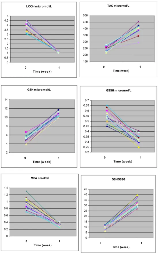
Data are illustrated individually in
Fig. 1
, and summarized in
Table 1
.
All indices changed in a favourable manner, ranging from !41%
for GSSG to !70% for LOOH levels, and +106% for GSH levels to
+249% for the GSG/GSSG ratio. We did not have any side effects,
except for a slight, non-statistically significant, increase in heart
rate.
Fig. 1
shows that changes of any given index were uniform
across subjects, with no outliers.
Discussion
As summarized in
Table 2
, the indices of oxidative stress we
have studied in the present paper are of relevance, including the
diabetes mellitus setting. Concerning the object of our study, viz.
caffeine, data from the literature show beneficial effects on TAC
and lipid peroxidation
[44,45,49,50]
with important additional
actions of DNA protection from on oxidative breakage by hydroxyl
radicals
[51]
and of decreased platelet aggregation
[52,53]
.
As recently reviewed
[54]
prior to us others
[39,43,55–57]
have
evaluated the short-term effects of drinking caffeine on the
oxida-tive stress. While in 4/5 such studies, the number of subjects is
lower than ours, only a few have evaluated all the six markers of
blood oxidative stress we did. Effects on DNA protection are
demonstrable as early as two hours after coffee ingestion
[43]
,
con-firming previous intervention studies that provided evidence for
long-term coffee consumption correlating with reduced DNA
back-ground damage in healthy volunteers. Continued coffee intake was
associated with further decrements in background DNA damage
within the 8 h intervention. Mean tail intensities (TIs%) decreased
from 0.33 TI% (baseline, 0 h) to 0.22 TI% (within 8-h coffee
con-sumption). The authors concluded that repeated coffee
consump-tion was associated with reduced background DNA strand
breakage, clearly measurable as early as 2 h after first intake
result-ing in a cumulative overall reduction by about one-third of the
baseline value
[43]
.
As reviewed elsewhere, the total antioxidant capacity of plasma
is the primary measure and marker to evaluate the status and
potential of oxidative stress in the body
[58]
. Lipid hydroperoxides
and MDA have been documented as a primary biomarker of free
radical mediated lipid damage and oxidative stress
[58]
. GSH, the
most abundant nonprotein thiol that defends against oxidative
stress, is considered as a biomarker of redox imbalance at cellular
level
[58]
. In contrast, GSSG is unable to perform antioxidant
func-tions. GSSG can be reduced back to GSH (and the GSH:GSSG ratio
maintained high) by glutathione reductase and associated
oxida-tion of NADPH to NAD+, unless such enzymatic activity is
over-whelmed by excessive amounts of reactive oxygen species (59).
The effect of coffee consumption on the modulation of plasma
antioxidant capacity was evaluated in 10 studies
[54]
. Eight studies
(seven chronic interventions and one acute trial) also investigated
the role of coffee in the modulation of blood GSH levels as a
sub-strate of GPx [glutathione peroxidase] and GST [glutathione
S-transferases] enzymes. Four out of seven chronic intervention
studies documented an increase in GSH levels
[42,58,28]
, while
two long-term studies
[12,22]
and one study performing both an
acute and a chronic intervention
[55]
did not show any significant
effect. Coffee ineffectiveness was attributed to the degradation and
Fig. 1. Individual data in the 15 volunteers for each of the six indices measured. Abbreviations are: GSH = glutathione; GSSG = oxidized glutathione; LOOH = lipid hydroperoxides; MDA = malondialdehyde; TAC = Total antioxidant capacity.
metabolic conversion of different coffee constituents in the body or
to the short duration of the intervention
[55]
.
The effect of coffee consumption on markers of lipid oxidation
has been investigated
[11,12,18,19,21–23,31,56]
. Five out of 12
studies investigated only the acute effect of coffee consumption
[16,19,21,23,56,61]
, five were chronic intervention studies
[12,18,19,21,22,61,62]
, while two studies investigated both acute
and chronic effects
[23,64]
. In these studies, isoprostanes (IsoPs)
and malondialdehyde (MDA) were the most frequently considered
markers of lipid damage. Besides 8-IsoPGF2 and MDA, further
markers of lipid damage and/or protection considered in the
pre-sent review were oxidized LDL, resistance to LDL oxidation, serum
LDL-conjugated dienes and hydroxyl fatty acids. The analysis of the
main findings revealed that most of the interventions failed to
demonstrate a significant decrease in markers of lipid damage with
exception of results found by Ochiai et al.
[57]
and Sirota et al.
[65]
.
The former reported a significantly reduced urinary 8-epiPGF2
fol-lowing consumption of a coffee beverage (providing 600 mg of
CGAs) when compared with placebo in healthy men. Results
showed that consumption of 200 mL Turkish roasted coffee during
a meal based on red-meat cutlets resulted in a significant
inhibi-tion of postprandial plasma MDA. No effect between treatments
and control/placebo were instead found by other authors
[16,23,61]
. The investigation by Leelarungrayub et al.
[56]
deserves
a special mention, because it reports a significant higher level of
MDA in men consuming caffeinated coffee, when compared to
decaffeinated coffee or control, followed by a submaximal exercise
test. Authors reported that, similarly to what observed in previous
investigations, results demonstrated an increased intramuscular
fat oxidation following consumption of caffeine-rich foods. For
what concerns the other markers of lipid damage, only Yukawa
et al.
[66]
found a modest reduction of LDL oxidation susceptibility
and a decrease of MDA levels following consumption of 3
coffees/-day for 1 week. No significant effect was instead found by Mursu
et al.
[39]
on serum LDL-conjugated dienes and plasma hydroxyl
fatty acids, or by Teekachunhatean et al.
[55]
on MDA levels and
by Hoelzl et al.
[67]
on both MDA and oxidized LDL.
In their article
[54]
, Martini et al. conclude that, despite the high
inter-study heterogeneity, data suggest that consumption of coffee
may increase glutathione levels and reduce the levels of DNA
dam-age. These effects are more evident in chronic interventions than in
acute studies.
In summary, we have demonstrated that 7-day administration
of pure caffeine induces unequivocally beneficial changes in a
number of oxidative-stress biochemical indices, the magnitude of
these changes being the greatest for the GSH to GSSG ratio.
Table 2
Summary of indices of oxidative stress and diabetes mellitus [ricontrollare referenze].
Index General Pertinence for diabetes
Lipid hydroperoxides (LOOH) Peroxidation of lipids produces highly reactive aldehydes, including MDA, acrolein, 4-hydroxynonenal, 4-oxononenal, and isolevuglandins[68]. It has been reported that peroxyl radicals can remove hydrogen from lipids, producing hydroperoxides that further propagate the free-radical pathway[69]
Increased lipid peroxidation occurs in both type 1 and type 2 diabetes mellitus[38]
LOOH increase particularly in patients with vascular complica-tions[70]. Lipid peroxidation in diabetes induces many sec-ondary chronic complications including atherosclerosis and neural disorders[71,72]
Malondialdehyde (MDA) MDA is a three carbon, low molecular weight aldehyde representing the main product of polyunsaturated fatty acid peroxidation. It is characterized by a high toxicity due to its ability to react with other molecules like DNA and protein[54– 58,28,59,30,60–75]
MDA is documented as a primary biomarker of free radical mediated lipid damage and oxidative stress[74]
Increased MDA level in plasma and many tissues was reported in diabetic patients[76,77]
Increased levels of MDA in diabetics suggests that peroxidative injury may be involved in the development of diabetic compli-cations
Glutathione (GSH) GSH is the most abundant nonprotein thiol that defends against oxidative stress[76]. GSH is an efficient antioxidant present in almost all living cells and is also considered as a biomarker of redox imbalance at cellular level[78,79]
Reduced levels of GSH are found in diabetes[79]. Decreased GSH level may be one of the factors in the oxidative DNA damage in type 2 diabetics
Oxidized glutathione (GSSG) GSSG is reduced back to GSH by the nicotinamide adenine dinucleotide phosphate (NADPH)-dependent catalysis of the flavoenzyme GSH reductase
GSSG levels in plasma from diabetic subjects were higher than those from controls
GSH to GSSG ratio This ratio is used to evaluate oxidative stress status in biological systems
Plasma GSH/GSSG showed a significant decrease in type 2 diabetes as compared to normal. Hyperlipidemia, inflammation, and altered antioxidant profiles are the usual complications in diabetes mellitus as a result of decreased GSH/GSSG ratio Total antioxidant capacity (TAC) TAC is the primary measure and marker to evaluate the status
and potential of oxidative stress in the body
TAC is significantly lower in diabetic subjects with poor glycaemic control than healthy subjects, while patients with good glycaemic control had plasma antioxidative values similar to controls[66]. Decrease in TAC of plasma is associated with increased complications of diabetes, which include cardiovas-cular disease, nerve damage, blindness, and nephropathy TAC is markedly reduced in sciatic nerve homogenates of diabetic animals[61]
Table 1
Changes in the indicated indices of oxidative stress observed in 15 healthy male volunteers after one-week administration of 5 mg/kg body weight/day in two daily doses.
Index Caffeine administration Statistics
(P) Before After Lipid hydroperoxides (LOOH), mmol/L 3.88 ± 1.85 1.16 ± 0.35 (!70%) <.05 Malondialdehyde (MDA), nmol/ml 0.9 ± 0.3 0.3 ± 0.1 (!67%) <.01 Oxidized glutathione (GSSG), mmol/L 0.56 ± 0.3 0.33 ± 0.4 (!41%) <.01 Glutathione (GSH), mmol/L 5.1 ± 1.5 10.5 ± 2.7 (+106%) <.01 GSH to GSSG ratio 9.11 ± 2.8 31.8 ± 3.4 (+249%) <.01 Total antioxidant
capacity (TAC), mmol/L
244.5 ± 40.3 398.2 ± 37.0 (+163%) <.05
*The beneficial outcome after caffeine administration is a decrease for the first three
indices and an increase for the last three indices.
The authors declare no conflict of interest.
This research did not receive any specific grant from funding
agencies in the public, commercial, or not-for-profit sectors.
References
[1]Arranz L, Fernández C, Rodríguez A, Ribera JM, De la Fuente M. The glutathione precursor N-acetylcysteine improves immune function in postmenopausal women. Free Radical Biol Med 2008;45:1252–62.
[2]Hashimoto K, Takasaki W, Yamoto T, Manabe S, Sato I, Tsuda S. Effect of glutathione (GSH) depletion on DNA damage and blood chemistry in aged and young rats. J Toxicol Sci 2008;33:421–9.
[3]Christon R, Haloui RB, Durand G. Dietary polyunsaturated fatty acids and aging modulate glutathione-related antioxidants in rat liver. J Nutr 1995;125:3062–70.
[4]Maher P. The effects of stress and aging on glutathione metabolism. Ageing Res Rev 2005;4:288–314.
[5]Rebrin I, Bayne AC, Mockett RJ, Orr WC, Sohal RS. Free aminothiols, glutathione redox state and protein mixed disulphides in aging Drosophila melanogaster. Biochem J 2004;382:131–6.
[6]Rebrin I, Sohal RS. Pro-oxidant shift in glutathione redox state during aging. Adv Drug Delivery Rev 2008;60:1545–52.
[7]Samiec PS, Drews-Botsch C, Flagg EW, Kurtz JC, Sternberg Jr P, Reed RL, et al. Glutathione in human plasma: decline in association with aging, age-related macular degeneration, and diabetes. Free Radical Biol Med 1998;24:699–704. [8]Cerielo A, Motz E, Cavarape A, Lizzio S, Russo A, Quatraro A, et al. Hyperglycemia counterbalances the antihypertensive effect of glutathione in diabetic patients: evidence linking hypertension and glycemia through the oxidative stress in diabetes mellitus. J Diabetes Complications 1997;11:250–5.
[9]Dincer Y, Akcay T, Alademir Z, Ilkova H. Effect of oxidative stress on glutathione pathway in red blood cells from patients with insulin-dependent diabetes mellitus. Metabolism 2002;51:1360–2.
[10]Yoshida K, Hirokawa J, Tagami S, Kawakami Y, Urata Y, Kondo T. Weakened cellular scavenging activity against oxidative stress in diabetes mellitus: regulation of glutathione synthesis and efflux. Diabetologia 1995;38:201–10. [11]Margutti P, Matarrese P, Conti F, Colasanti T, Delunardo F, Capozzi A, et al. Autoantibodies to the C-terminal subunit of RLIP76 induce oxidative stress and endothelial cell apoptosis in immune-mediated vascular diseases and atherosclerosis. Blood 2008;111:4559–70.
[12]Signorelli SS, Neri S, Di Pino L, Costa MP, Pennisi G, Digrandi D, et al. Oxidative stress and endothelial damage in patients with asymptomatic carotid atherosclerosis. Clin Exp Med 2001;1:9–12.
[13]Hassan MQ, Hadi RA, Al-Rawi ZS, Padron VA, Stohs SJ. The glutathione defense system in the pathogenesis of rheumatoid arthritis. J Appl Toxicol 2001;21:69–73.
[14]Pedersen-Lane JH, Zurier RB, Lawrence DA. Analysis of the thiol status of peripheral blood leukocytes in rheumatoid arthritis patients. J Leukoc Biol 2007;81:934–41.
[15]Seven A, Guzel S, Aslan M, Hamuryudan V. Lipid, protein, DNA oxidation and antioxidant status in rheumatoid arthritis. Clin Biochem 2008;41:538–43. [16] Karelson E, Mahlapuu R, Zilmer M, Soomets U, Bogdanovic N, Langel U.
Possible signaling by glutathione and its novel analogue through potent stimulation of frontocortical G proteins in normal aging and in Alzheimer’s disease. In: Diederich M, editor. Cell Signaling, Transcription, and Translation as Therapeutic Targets. New York Academy of Sciences; New York; 2002. 973: 537–40.
[17] Liu HL, Wang H, Shenvi S, Hagen TM, Liu RM. Glutathione metabolism during aging and in Alzheimer disease. In: De Grey ADN, editor. Strategies for Engineered Negligible Senescence: Why Genuine Control of Aging May Be Foreseeable. New York Academy of Sciences; 2004. 1019:346–9.
[18]Resende R, Moreira PI, Proenca T, Deshpande A, Busciglio J, Pereira C, et al. Brain oxidative stress in a triple-transgenic mouse model of Alzheimer disease. Free Radical Biol Med 2008;44:2051–7.
[19]Lang AE. The progression of Parkinson disease: a hypothesis. Neurology 2007;68:948–52.
[20]Spina MB, Cohen G. Dopamine turnover and glutathione oxidation: implications for Parkinson disease. Proc Natl Acad Sci USA 1989;86:1398–400. [21] Yamamoto N, Sawada H, Izumi Y, Kume T, Katsuki H, Shimohama S, et al. Proteasome inhibition induces glutathione synthesis and protects cells from oxidative stress: relevance to Parkinson disease. J Biol Chem; 282:4364–72. [22]Barranco SC, Perry RR, Durm ME, Quraishi M, Werner AL, Gregorcyk SG, et al.
Relationship between colorectal cancer glutathione levels and patient survival: early results. Dis Colon Rectum 2000;43:1133–40.
[23]Kigawa J, Minagawa Y, Kanamori Y, Itamochi H, Cheng X, Okada M, et al. Glutathione concentration may be a useful predictor of response to second-line chemotherapy in patients with ovarian cancer. Cancer 1998;82:697–702. [24]Kumar A, Sharma S, Pundir CS, Sharma A. Decreased plasma glutathione in
cancer of the uterine cervix. Cancer Lett 1995;94:107–11.
[25]Wong DY, Hsiao YL, Poon CK, Kwan PC, Chao SY, Chou ST, et al. Glutathione concentration in oral cancer tissues. Cancer Lett 1994;81:111–6.
[26]Yeh CC, Hou MF, Wu SH, Tsai SM, Lin SK, Hou LA, et al. A study of glutathione status in the blood and tissues of patients with breast cancer. Cell Biochem Funct 2006;24:555–9.
[27]Droge W. Free radicals in the physiological control of cell function. Physiol Rev 2002;82:47–95.
[28]Hayes JD, Pulford DJ. The glutathione S-Transferase supergene family: regulation of GST and the contribution of the isoenzymes to cancer chemoprotection and drug resistance. Crit Rev Biochem Mol Biol 1995;30:445–600.
[29]Valko M, Rhodes CJ, Moncol J, Izakovic M, Mazur M. Free radicals, metals and antioxidants in oxidative stress-induced cancer. Chem Biol Interact 2006;160:1–40.
[30]Hayes JD, McLellan LI. Glutathione and glutathione-dependent enzymes represent a co-ordinately regulated defence against oxidative stress. Free Radical Res 1999;31:273–300.
[31]Dalle-Donne I, Rossi R, Colombo R, Giustarini D, Milzani A. Biomarkers of oxidative damage in human disease. Clin Chem 2006;52:601–23.
[32]Richelle M, Tavazzi I, Offord E. Comparison of the antioxidant activity of commonly consumed polyphenolic beverages (coffee, cocoa, and tea) prepared per cup serving. J Agric Food Chem 2001;49(7):3438–42.
[33]Metro D, Muraca U, Manasseri L. Role of green tea in oxidative stress prevention. Clin Ter 2006;157(6):507–10.
[34]Borrelli RC, Visconti A, Mennella C, Anese M, Fogliano V. Chemical characterization and antioxidant properties of coffee melanoidins. J Agric Food Chem 2002;50(22):6527–33.
[35]Sanchez-Gonzales I, Jimenez-Escrig A, Saura-Calixto F. In vitro antioxidant activity of coffees brewed using different procedures (Italian, espresso and filter). Food Chem 2005;90:133–9.
[36]Benvenga S, Bartolone L, Pappalardo MA, Russo A, Lapa D, Giorgianni G, et al. Altered intestinal absorption of L-thyroxine caused by coffee. Thyroid 2008;18:293–301.
[37]Van Dam RM, Hu FB. Coffee consumption and risk of type 2 diabetes: a systematic review. JAMA 2005;294:97–104.
[38]Davì G, Falco A, Patrono C. Lipid peroxidation in diabetes mellitus. Antioxid Redox Signaling 2005;7:256–68.
[39]Mursu J, Voutilainen S, Nurmi T, Alfthan G, Virtanen JK, Rissanen TH, et al. The effects of coffee consumption on lipid peroxidation and plasma total homocysteine concentrations: a clinical trial. Free Radical Biol Med 2005;38 (4):527–34.
[40]Wang S, Noh SK, Koo SI. Epigallocatechin gallate and caffeine differentially inhibit the intestinal absorption of cholesterol and fat in ovariectomized rats. J Nutr 2006;136:2791–6.
[41]Westerterp-Plantenga MS. Green tea catechins, caffeine and body-weight regulation. Physiol Behav 2010;100:42–6.
[42]Tiwari BK, Pandey KB, Abidi AB, Rizvi SI. Markers of Oxidative Stress during Diabetes Mellitus. J Biomarkers 2013;2013:378790.
[43]Bakuradze T, Lang R, Hofmann T, Schipp D, Galan J, Eisenbrand G, et al. Coffee consumption rapidly reduces background DNA strand breaks in healthy humans: Results of a short-term repeated uptake intervention study. Mol Nutr Food Res 2016;60:682–6.
[44]Devasagayam TP, Kamat JP, Mohan H, Kesavan PC. Caffeine as an antioxidant: inhibition of lipid peroxidation induced by reactive oxygen species. Biochim Biophys Acta 1996;1282(1):63–70.
[45]Lee C. Antioxidant ability of caffeine and its metabolities based on the study of oxygen radical absorbing capacity and inhibition of LDL peroxidation. Clin Chim Acta May 2000;295(1–2):141–54.
[46] LARN – Livelli di assunzione di riferimento di nutrienti ed energia per la popolazione italiana. Revisione 2012 – SINU (Società Italiana di Nutrizione Umana).
[47]Mateos R, Lecumberri E, Ramos S, Goya L, Bravo L. Determination of malondialdehyde (MDA) by high-performance liquid chromatography in serum and liver as a biomarker for oxidative stress. Application to a rat model for hypercholesterolemia and evaluation of the effect of diets rich in phenolic antioxidants from fruits. J Chromatogr B Analyt Technol Biomed Life Sci 2005;827(1):76–82.
[48]Pastore A, Piemonte F, Locatelli M, Lo Russo A, Gaeta LM, Tozzi G, et al. Determination of blood total, reduced, and and oxidized glutathione in pediatric subjects. Clin Chem 2001;47(8):1467–9.
[49]Natella F, Nardini M, Giannetti I, Dattilo C, Scaccini C. Coffee drinking influences plasma antioxidant capacity in humans. J Agric Food Chem 2002;50(21):6211–6.
[50]Bydlowski SP, Yunker RL, Rymaszewski Z, Subbiah MT. Coffee extracts inhibit platelet aggregation in vivo and in vitro. Int J Vitam Nutr Res 1987;57 (2):217–23.
[51] Azam S, Hadi N, Khan NU, Hadi SM. Antioxidant and prooxidant properties of caffeine, theobromineand xanthine. Med Sci Monit 2003. 9(9):BR325–30. [52]Choi JW. Influence of caffeine on the responsiveness of human plateled to
agonists. Thromb Res 2003;110(4):209–12.
[53]Varani K, Portaluppi F, Gessi S, Merighi S, Ongini E, Belardinelli L, et al. Dose and time effects of caffeine intake on human plateled adenosine A(2A) receptors: functional and biochemical aspects. Circulation 2000;102 (3):285–9.
[54]Martini D, Del Bo’ C, Tassotti M, Riso P, Del Rio D, Brighenti F, et al. Coffee consumption and oxidative stress: A review of human intervention studies. Molecules 2016;21(8).
[55]Teekachunhatean S, Tosri N, Sangdee C, Wongpoomchai R, Ruangyuttikarn W, Puaninta C, et al. Antioxidant effects after coffee enema or oral coffeeconsumption in healthy Thai male volunteers. Hum. Exp. Toxicol 2012;31:643–51.
[56]Leelarungrayub D, Sallepan M, Charoenwattana S. Effects of acute caffeinated coffee consumption on energy utilization related to glucose and lipid oxidation from short submaximal Treadmill exercise in sedentary Men. Nutr Metab Insights 2011;4:65–72.
[57]Ochiai R, Sugiura Y, Otsuka K, Katsuragi Y, Hashiguchi T. Coffee bean polyphenols ameliorate postprandial endothelial dysfunction in healthy male adults. Int J Food Sci Nutr 2015;66:350–4.
[58]Droge W. Free radicals in the physiological control of cell function. Physiol 2002;82:47–95.
[59]Mišík M, Hoelzl C, Wagner KH, Cavin C, Moser B, Kundi M, et al. Impact of paper filtered coffee on oxidative DNA-damage: results of a clinical trial. Mutat Res 2010;692(1–2):42–8.
[60]Catanzaro O, Capponi JA, Michieli J, Labal E, Di Martino I, Sirois P. Bradykinin B1 antagonism inhibits oxidative stress and restores Na+K+ ATPase activity in diabetic rat peripheral nervous system. Peptides 2013;44:100–4.
[61]Korkmaz GG, Konukoglu D, Kurtulus EM, Irmak H, Bolayirli M, Uzun H. Total antioxidant status and markers of oxidative stress in subjects with normal orimpaired glucose regulation (IFG, IGT) in diabetic patients. Scand J Clin Lab Invest 2013;3(8):641–9.
[62]Rein D, Paglieroni TG, Pearson DA, Wun T, Schmitz HH, Gosselin R, et al. Cocoa and wine polyphenols modulate platelet activation and function. J Nutr 2000;130(8S Suppl):2120S–6S.
[63]Schiffrin EL. Antioxidants in hypertension and cardiovascular disease. Mol Interv 2010;10(6):354–62.
[64]Sirota R, Gorelik S, Harris R, Kohen R, Kanner J. Coffee polyphenols protect human plasma from postprandial carbonyl modifications. Mol Nutr Food Res 2013;57(5):916–9.
[65]Yukawa GS, Mune M, Otani H, Tone Y, Liang XM, Iwahashi H, et al. Effects of coffee consumption on oxidative susceptibility of low-density lipoproteins and serum lipid levels in humans. Biochemistry (Mosc) 2004;69(1):70–4. [66]Hoelzl C, Knasmüller S, Wagner KH, Elbling L, Huber W, Kager N, et al. Instant
coffee with high chlorogenic acid levels protects humans against oxidative damage of macromolecules. Mol Nutr Food Res 2010;54(12):1722–33. [67] Guo L, Chen Z, Amarnath V, Davies SS. Identification of novel bioactive
aldehyde-modified phosphatidylethanolamines formed by lipid peroxidation. Free Radical Biol Med 2012. 53(6)6:1226–38.
[68]Lobo V, Patil A, Phatak A, Chandra N. Free radicals, antioxidants and functional foods: impact on human health. Pharmacogn Rev 2010;4(8):118–26; Fowler MJ. Microvascular and macrovascular complications of diabetes. Clin Diabetes 2008;26(2):77–82.
[69]Baynes JW. Role of oxidative stress in development of complications in diabetes. Diabetes 1991;40(4):405–12.
[70] Ramesh B, Karuna R, Sreenivasa RS, Haritha K, Sai MD, Sasi BR, et al. Effect of Commiphora mukul gum resin on hepatic marker enzymes, lipid peroxidation and antioxidants status in pancreas and heart of streptozotocin induced diabetic rats. Asian Pac J Trop Biomed 2012;2(11):895–900.
[71]Del Rio D, Stewart AJ, Pellegrini N. A review of recent studies on malondialdehyde as toxic molecule and biological marker of oxidative stress. Nutr Metab Cardiovasc Dis 2005;15:316–28.
[72]Shodehinde SA, Oboh G. Antioxidant properties of aqueous extracts of unripe Musa paradisiaca on sodium nitroprusside induced lipid peroxidation in rat pancreas in vitro. Asian Pac J Trop Biomed 2013;3(6):449–57.
[73]Moussa SA. Oxidative stress in diabetes mellitus. Romanian J Biophys 2008;18:225–36.
[74]Bandeira Sde M, Guedes Gda S, da Fonseca LJ, Pires AS, Gelain DP, Moreira JC, et al. Characterization of blood oxidative stress in type 2 diabetes mellitus patients: increase in lipid peroxidation and SOD activity. Oxid Med Cell Longevity 2012:819310.
[75]Lu SC. Glutathione synthesis. Biochim Biophys Acta 2013;1830(5):3143–53. [76]Chakravarty S, Rizvi SI. Day and night GSH and MDA levels in healthy adults
and effects of different doses of melatonin on these parameters. Int J Cell Biol 2011;2011:404591.
[77]Rahigude A, Bhutada P, Kaulaskar S, Aswar M, Otari K. Participation of antioxidant and cholinergic system in protective effect of naringenin against type-2 diabetes-induced memory dysfunction in rats. Neuroscience 2012;226:62–72.
[78]Calabrese V, Cornelius C, Leso V, Trovato-Salinaro A, Ventimiglia B, Cavallaro M, et al. Oxidative stress, glutathione status, sirtuin and cellular stress response in type 2 diabetes. Biochim Biophys Acta 2012;1822(5):729–36. [79]Dinçer Y, Akçay T, Alademir Z, Ilkova H. Assessment of DNA base oxidation and
glutathione level in patients with type 2 diabetes. Mutat Res 2002;505(1– 2):75–81.