DOTTORATO DI RICERCA
Science for Conservation
Ciclo XXII
Settore/i scientifico disciplinari di afferenza: CHIM/12
TITOLO TESI
BIOFILMS ON EXPOSED MONUMENTAL
STONES: MECHANISM OF FORMATION AND
DEVELOPMENT OF NEW CONTROL METHODS
Presentata da: Oana-Adriana Cuzman
Coordinatore Dottorato
Relatore
Prof. Rocco Mazzeo
Prof. Piero Tiano
In memory of my professor, Ion I. Băra
ABSTRACT ...7
I. RESEARCH OBJECTIVES ...9
II. INTRODUCTION ...10
II.1. Biofilms formation and evolution ...10
II.2. Biofilms interaction with the substratum, biodeterioration processes ...13
II.3. Biomolecules and biofilm control ...16
III. MATERIALS AND METHODS ...19
III.1. Investigated fountains ...19
III.2. Biofilms development ...20
III.2.1. Natural biofilm observation. ...20
III.2.2. Laboratory experiments. ...22
III.3. Study of biofilm composition ...24
III.3.1. Isolation and identification of microorganisms by traditional and molecular methods. ...24
III.3.1.1. Traditional methods. ...26
III.3.1.2. DNA extraction. ...27
III.3.1.3. PCR amplification and sequencing...27
III.3.2. Microbial community analysis by ARISA fingerprint method. ...29
III.3.2.1. DNA extraction. ...29
III.3.2.2. PCR amplification and ARISA. ...29
III.4. Stone material and color measurements ...31
III.5. Antibiofouling agents, coating preparations and their characterization ...32
III.6. Laboratory experiments ...43
III.6.1. Growing conditions. ...43
III.6.2. Indoor experiments. ...43
III.6.2.1. Immersion method. ...43
III.6.2.2. Serial dilution method. ...45
III.6.3. Outdoor experiments – immersion method. ...48
III.7. In situ experiments ...50
III.7.1. Cleaning procedures, application of treatments and optical evaluation. ...50
III.7.2. Molecular evaluation of treatments efficacy. ...53
IV. RESULTS AND DISCUSSIONS ...55
IV.1. Biofilms on monumental stones ...55
IV.1.1. Biofilm development. ...55
IV.1.2. Biodiversity of microorganisms on monumental fountains. ...61
IV.1.3. Microbial community study by ARISA fingerprint method. ...66
IV.2. Antifouling agents as an alternative method for prevention the biofouling on artistic fountains...68
IV.2.1. Characterization of antibiofouling agents and coatings. ...68
IV.2.1.1. Color tests. ...68
IV.2.1.2. FT-IR and SEM-EDX analysis. ...69
IV.2.2. Efficacy of antibiofouling agents against biofilm development...76
IV.2.2.1. Indoor laboratory experiments. ...76
IV.2.2.1.1. Immersion method. ...76
IV.2.2.1.2. Serial dilution method. ...78
IV.2.2.1.3. Diffusion method I and II . ...81
IV.2.2.2. Outdoor experiments – immersion method. ...84
IV.2.2.3. In situ experiments. ...87
IV.2.2.3.1. Optical observations. ...87
IV.2.2.3.2. Molecular evaluation of treatments efficacy. ...93
V. CONCLUSIONS ...97
VI. ACKNOWLEDGEMENTS ...100
VII. BIBLIOGRAPHY ...102
ABAs AntiBiofouling Agents AHL Acylated Homoserine lactone
ARISA Automated rRNA Intergenic Spacer Analysis au absorbance units
BA Barrier filter emission bp base pairs
CBE Ceramium botryocarpum Extract CCD Charge Coupled Device
CI Cinnamaldehyde
CLSM Confocal Laser Scanning Microscopy CS Capsaicin
DAPI 4',6-diamidino-2-phenylindole, a fluorescent stain that binds strongly to DNA, used in fluorescence microscopy
ddNTP dideoxyribonucleotide triphosphate DM Dichroic Mirrror
DNA Deoxyribonucleic Acid EM Epiflourescence Microscopy EPS Extracellular Polymeric Substances
ESI-MS Electrospray Ionization-Mass Spectrometry
EtBr Ethidium Bromide, a fluorescent stain for nucleic acids EtOH Ethanol
FT-IR Fourier Transform Infrared Spectroscopy
ITS Internal Transcribed Spacer, used to know the genetic diversity among different strains of microorganisms by sequencing the ITS gene LSM Laser Scanning Microscopy
MeOH Methanol OD Optical Density OM Optical Microscopy
PCA Principal Component Analysis PCR Polymerase Chain Reaction pAPS Poly-Alkyl Pyridinium Salts QQ Quorum Quenching
QS Quorum Sensing rfu relative fluorescent units RH Relative Humidity
rRNA ribosomal Ribonucleic Acid S Silres BS OH 100
SEM Scanning Electron Microscopy
SEM-EDX Energy-Dispersive X-ray spectroscopy analysis SYTO a group of fluorescent nucleic acid stains TRITC Tetramethyl Rhodamine Iso-Thiocyanate UV Ultraviolet
ZA Zosteric Acid W pure Silres BS 290
ABSTRACT
The biodeterioration processes are wide spread in the natural environments and therefore all the monumental assets are susceptible to this kind of risk. Within the stone monumental artefacts artistic fountains can be considered the most vulnerable because of their particular exposure conditions which are extremely favorable to formation of phototrophic biofilms, giving rise to biodegradation processes related with physical-chemical and visual aspect alterations. Microbial diversity of five fountains (two from Spain and three from Italy) was investigated. It was observed an ample similarity between the biodiversity of monumental stones reported in literature and that one found in studied fountains. The achievement of microbial ecological data for this artifacts are very useful to identify the most diffuse microorganisms dwelling on these monumental assets, to investigate their possible risk for stone conservation and to understand and predict, with some approximation, their behavior when maintenance intervention of a fountain is required.
Mechanical procedures and toxic chemical products are usually employed to remove such phototrophic patinas. Alternative methods based on natural antifouling substances are recently experimented in the marine sector, due to their very low environmental impact and for the bio settlement prevention on partially immersed structures of ships. In the present work groups of antibiofouling agents (ABAs) were selected from literature for their ability to interfere, at molecular level, with the microbial communication system “quorum sensing”, inhibiting the initial phase of biofilm formation. The efficacy of some natural antibiofoulants agents (ABAs) with terrestrial (Capsaicine - CS, Cinnamaldehyde - CI) and marine origin (Zosteric Acid - ZA, poly-Alkyl Pyridinium Salts – pAPS and Ceramium botryocarpum extract - CBE), incorporated into two commercial coatings (Silres BS OH 100 - S and Wacker Silres BS 290 - W) commonly used in stone conservation procedures were evaluated. The mixing of ABAs with a coating was considered necessary because their water solubility and considering the aqueous conditions of their application. The formation of phototrophic biofilms in laboratory conditions (on Carrara marble specimens and Sierra Elvira stone) and on two monumental fountains (Tacca‟s Fountain 2 - Florence, Italy and Fountain from Patio de la Lindaraja - Alhambra Palace, Granada, Spain) has been investigated in the presence or absence of these natural antifouling agents. Recent investigation methods for the biofilm development and efficacy evaluation of selected ABAs were
used as useful tools in understanding the morphology and behavior of biofilms and treatments, such as various microscopical techniques (OM, EM, CLSM, SEM), molecular biology (ARISA fingerprint method, molecular identification of the isolated strains), image analysis, FTIR and SEM-EDX.
The natural antibiofouling agents, at tested concentrations, demonstrated a certain inhibitory effect. The silane-siloxane based silicone coating (W) mixing with ABAs was more suitable with respect to ethyl silicate coating (S) and proved efficacy against biofilm formation only when incompletely cured. The laboratory results indicated a positive action in inhibiting the patina formation, especially for poly-alkyl pyridinium salts, zosteric acid and cinnamaldehyde, while on site tests revealed a good effect for zosteric acid.
However this partial curing most probably favors its removal in water environment with the consequence of eliminate the ABAs products, and hence more suitable supporting product must be investigated for this kind of application.
I. RESEARCH OBJECTIVES
Special attention to monumental fountains is given only when some others alteration types appear and their state of conservation must be evaluated (Not R., 1995; Cardilli L., 1979; Pietrini A.M., 1991; Peraza Zurita Y., 2002a). Usually, detailed data related with the microbial composition are not considered necessary for further restoration processes, biological patina/biofilms being controlled in the same way, by mechanical removal or biocide treatments. Taking into account that the colonization mechanisms, in exposed environment, is an endless process that restarts after each removal, the study of microbiological components can contribute to understanding which are the most adapted genera/species dwelling on these kind of artefacts, which are the most dangerous as biodeteriogens, and consequently to develop new methods for control this unwanted biological growth. The most suitable approach should be to block its initial formation. In order to achieve this aim, groups of antibiofouling agents (ABAs) were selected from literature for their ability to interfere, at molecular level, with the microbial communication system “quorum sensing”, inhibiting the initial phase of biofilm formation. Such kind of innovative products were for the first time tested in the monumental heritage field.
The main objectives of this research activity are:
- to study the main microbial components (algae, cyanobacteria and fungi) of the phototrophic biofilms developed on monumental fountains by traditional and molecular methods and to compare the biodiversity of different fountains biotopes;
- to study the biological patina structure and the mechanism of its development on sound
stone material;
- to characterize the selected natural antibiofouling agents and to develop and verify their efficiency and behavior in laboratory and on site, as innovative natural antibiofouling agents for inhibition of patina formation.
II. INTRODUCTION
II.1. Biofilms formation and evolution
Biofilms occur on all solid surfaces in aquatic habitats. This complex biocenosis consist principally of water (70 to 95% of the fresh weight), extracellular polymeric substances (Flemming H.C., 1993; Wahl M., 1998) and microorganisms such as phototrophs (algae, cyanobacteria, diatoms) and heterotrophs (bacteria, fungi, protozoa, nematods), which are embedded in this hydrated matrix (Norton T.A., 1998). The proportion of each group is varying seasonally and it is influenced by different habitats (Underwood A. J., 1984; Anderson M. J., 1995; Roeselers G., 2007). The biofilm contains cells debris, airborne particles, bacteria and spores together with amounts of inorganic material adsorbed from the substratum (Warscheid Th., 2008). The biopolymers act also in sticking the cells to the substratum, and their adhesive properties contribute to the formation and cohesion of biofilms (Albertano P., 2003).
The extracellular polymeric substances (glyocalyx, sheath or envelope) protect the microorganisms composing a biofilm by balancing changes in: humidity, temperature, osmotic pressure and pH. The ability of bacteria and cyanobacteria to release EPS is related to their nutritional status. In cyanobacteria, particularly, limitation in light or nutrients can increase production of polysaccharides (Albertano P., 2003). Based on its ion-exchange capacity, the biofilms even resist to the penetration of biocides, detergents or antibiotics, hindering the possible control of microbial biodeterioration processes in the long-term (Warscheid Th., 2008).
It is found that the surfaces exposed to river water are covered with bacteria and microcolonies after only 3 days (Lawrence J. R., 2003). The photosynthetic microorganisms develop easily on the stone surfaces and, once established, allow the growth of more complex microbial consortia formed by heterotrophic microroganisms which can exercise stronger deteriorating activity (Tiano P., 1993; Tomaselli L., 2000a; Crispim C. A., 2003; Zurita Y. P., 2005). A study related with the early stages of biofilm succession in a lentic freshwater environment reports the abundance of the green algae in the first 1-4 days, followed by the one of diatoms (5-7 days) and cyanobacteria (10-15 days) (Sekar R., 2004). Some authors give an important role to diatoms in the first steps of colonization (Wetherbee R., 1998; Peraza Zurita Y., 2002a).
Organisms or cells may be attracted by a substratum in a variety of ways, especially if they are motile, whereas not motile microorganism (many cyanobacteria, and diatoms) presumably are deposited on a substratum by gravity or water movement. This initial contact may result in a transitory chemical attraction that is difficult to characterize. Cell control attachment and release are depending on the habitability of the microhabitat, non-motile microorganism, such as diatoms, adjust their position on substrata by a complex mode of cell motility called “gliding” (Edgar L. A., 1984). The adhesive molecules of diatoms, have been fully characterized and seems that the polysaccharides are the candidates for the binding domain of the initial adhesion (Wetherbee R., 1998). Many authors consider very relevant the role of EPS in early development stages of a biofilm also for blue-green algae (Scott C., 1996). It seems that the capsular EPS has an important role during the initial stages of biofilm formation, since it facilitate the attachment of cells to the substrate (Decho A. W., 2000, Barranguet C., 2005).
Many bacterial species, in a population density-dependent, coordinate their responses to environmental changes using complex cell-to-cell communication mechanism, by secretion and uptake of small diffusible molecules. These molecules, used as signals, are oligopeptides in Gram-positive bacteria, AHL derivatives (Acyl Homoserine Lactone) in Gram-negative bacteria (including cyanobacteria) and γ-butyrolactones in Streptomyces species. Recent studies in medical field revealed that fungi, like bacteria, use quorum regulation to affect population-level behaviors such as biofilm formation and pathogenesis, being most extensively studied in Candida
albicans (Hogan D. A., 2006).
Since biofilms typically contain high concentration of cells, cell-to-cell signals are believed to play an important role in its development . This phenomenon , known as Quorum Sensing (QS), influence the bacterial population coordinating important biological functions including motility, swarming, aggregation, plasmid conjugal transfer, virulence, sporulations, antibiotics biosynthesis, symbiosis, biofilm maintenance and differentiation (Gray K. M., 1997; Jiang X., 2006). In addition, recent findings have shown the ability to produce AHL autoinducers in bacteria isolated from prehistoric caves with wall paintings and in biofilms dominated by cyanobacteria (Laiz L., 1999).
The organization of fouling consists of three main levels: molecular fouling, micro-fouling and a macro-fouling (Barrios A. C., 2004). It is noted that the formation
of an organic molecular layer should be realized before the attachment of the microorganisms on a solid substrate (Yebra D. M., 2003). After that a reversible stage of adhesion of the primary colonisers starts, followed by their fixing and propagation. Finally a EPS matrix is formed and a three-dimensional structure is auto-organised (Fig. 1). The mature biofilm has a complex heterogeneity which confers stability and resistance to the biocoenosis (Nikolaev Y. A., 2007).
Fig. 1. Biofilm development : (1) - the establishment of the organic molecules (black spots); (2) – the
reversible adhesion of micro-organisms – primary colonizers (bacteria, green algae, coccoid cyanobacteria and diatoms); (3) – the transition to the irreversible adhesion, multiplication, the start of EPS (extracellular polymeric substances) production; (4) – the start of a three-dimensional development of the biofilm structure; (5) - adhesion of secondary colonizers (filamentous cyanobacteria, fungi) and continuous development of the biofilm; (6) – the mature biofilm with a expressed structure forms a specific micro-ecosystem; (7) - the homeostasis phase keeps in equilibrium the biofilm with a continuous growth of the structure and detachment of small parts which become free-living; some macro-organisms (i.e. invertebrates) can adhere.
The concept of biofilm architecture was introduced by Lawrence et al. in 1991 to define the unique structures formed by pure culture bacterial biofilms grown in continuous flow slide culture. The specific architecture of biofilm is a consequence of the interaction between the community members, mediated by the quorum sensing
communication and influenced by the physical-chemical and genotypic factors. Stevenson R. J. (1986) grouped periphyton community based on microbial components immigration and growth rate: the pioneers have a high initial abundance but decrease with time, the relative abundance of the late colonizers increases with time, and the intermediates have a relatively more stable abundance than the other groups. Biofilm evolution is also dependent by stochastic and mechanical processes, deterministic phenomena and temporal changes (Wimpenny J., 2000).
II.2. Biofilms interaction with the substratum, biodeterioration processes
The biological colonization of stone artifacts exposed under uncontrolled out-door conditions is always influenced by the favorable microclimate and the high biodiversity of the opportunistic airflora (Caneva G., 2007). Phototrophic biofilms can develop on monumental stone surface influencing their state of conservation both by aesthetical and physical-chemical point of view. The phototrophic microorganisms like phototrophic bacteria, algae, cyanobacteria and diatoms are the first colonizers of the stone surfaces followed, in favorable environmental conditions, by heterotrophic ones as associate consumers. This colored biocoenosis is stuck together by gel-like extracellular polysaccharide matrix (EPS) which confers resistance to dried periods and cleaning treatments.
The development of photosynthetic microorganisms depends on the combination of environmental location and climatic conditions, in addition to the chemical-physical properties of stones. The whole properties that contribute to biological colonization has been defined as “bioreceptivity” by Guillitte in 1995, who further defined different types, such as primary, secondary and tertiary bioreceptivity. The first pioneering microorganisms of a stone surface include ubiquitous photosynthetic microorganisms such as cyanobacteria, green algae, diatoms and lichens (Darlington A., 1981; Warscheid Th., 2000). A synergistic biodeteriorative effect on stone surfaces can be started by the concomitant growth of phototrophic and heterotrophic populations. In fact, bacteria and fungi metabolize the organic matter produced by phototrophs and release organic acidic compounds able to solubilise the minerals components of the substratum (Warscheid Th., 2000). The formation of biofilms intensifies microbial attack by weakening the mineral matrix through repeated
wetting and drying cycles and subsequent expansion and contraction (Warscheid Th., 1996).
The biofilm formation is related with the primary bioreceptivity. Some studies revealed that the primary receptivity is mainly controlled by chemical composition and physical characteristics of the substrates such as mineral composition, structure-texture, permeability, porosity, roughness, pH (Krumbein W. E., 1988; Albertano P., 2000; Miller A., 2006; Tiano P., 1995a). Naturally, also the intrinsic properties of the microorganisms appear to have an important role on the development of photosynthetic biofilms. The anionic nature of exopolymers can strongly adsorb cations and dissolve organic molecules from the materials, and can stabilize dust particles. The biofilm properties such as ion-exchangers and metabolic end-products like acids (and derived salts) can led to an increasing of water content of porous materials (Sand W., 1997). It was observed that calcium ions may be easily subtracted from rocks and precipitated on polysaccharide sheaths of cyanobacteria in the form of calcium carbonate (Albertano P., 2003, Crispim C. A., 2005). The metabolic activities of the organisms that are forming a biofilm are related with the production of extracellular polymeric substances, liberation of chelating compounds and organic/inorganic acids, the presence of coloured pigments and mechanic pressure exerted by shrinking/swelling phenomena. Therefore, the stony structure can be interested to pitting, ion transfer, leaching processes and dwindling (Tiano P., 1998). Through their growth, biofilms can change the chemical and mineralogical composition of the original rock. They can change the stability, permeability and colour of the stone as well the density (Kovacic L., 2000).
The monumental fountains, due to the constant contact with water, are particularly affected by the microbial colonization giving rise to biodegradation processes combined with visual aspect alterations. The different microenvironmental conditions with some areas constantly in contact with water or others only sporadically wet and some perpetually dry, offer ideal growth substrata for many microorganisms and organisms. This various habitats determine a different distribution of organisms on the fountain according to their specific requirements (Pietrini A.M., 2005). The most important factors determining biological growth are (Walsh J. H., 1968): primary energetic input (light), secondary energetic input (nutritive factors) and climate. In urban and industrial areas must be also considered the environmental pollution.
The biofilm directly participate in stones decay processes, causing both aesthetic and physical-chemical biodamages (Tomaselli L., 2000a), inducing alterations due to the fluid retention, to the aggressive action of both metabolic products and to atmospheric pollutants entrapped in the EPS, which can increase the chemical corrosion process.Besides the discoloration and staining processes (Fig. 2) produced by biogenic pigments (e.g. the green chlorophyll, brownish melanin, red carotenoids), the microflora leads to the change of materials characteristics with regard to their mechanical properties, superficial absorbency/hydrophobicity, diffusivity and thermal-hydric behavior (Warscheid Th., 2008). Within the complex biofilm not all inhabiting microorganisms that can affect the stone surface need to be in direct contact with the substratum (Gaylarde C. C., 1999).
Fig. 2. Biodeterioration aspects present on the monumental fountains: discoloration (a, b, c, d); staining
(b, arrow); encrustation (c, d).
A particular aspect of biodeterioration of stone monuments is linked to endolithic organisms colonizing the interior of rocks (Salvadori O., 2000). The presence of endolithic community, which can penetrate up to depths of several millimetres
(Saiz-b a
Jimenez C., 1999), with the diffusion of their excreted products into the intergranular matrix enhance by weathering reactions and decrease mechanical properties, such as cohesion between grains (Alakomi H. L., 2004.). The endolithic microhabitat gives
protection from intense solar radiation and desiccation, and also provides mineral nutrients (Walker J. J., 2005).
II.3. Biomolecules and biofilm control
The microorganisms growing on the artistic fountains can be suppressed by indirect (e.g. water chlorination) or direct methods such as mechanical action (e.g. abrasion) and/or biocides treatments. The application of toxic substances can be hazardous to the environment and to public health other than for the stone itself. The biocide application can be harmful for conservators and the environment (Price C. A., 1996) and little is known about the consequences of repeated applications (Fortune I. S., 2008). The EC regulations (BPD 98/8/EC n 20 June 2004) have had as consequence the elimination from the market of the most active (and toxic) compounds applied to this aim and new approaches are made in several sectors in order to overcome this problematic.
In nature some organisms may be heavily fouled on much of their surfaces, while others can be totally fouling-free. This has generated interest in identifying the secondary metabolites that might repel or inhibit fouling organisms. Recently the discovery of biomolecules involved in cell-to-cell signaling has opened new possibilities for the study of microbial communities. Autoinduction has been defined as a form of communication, in which cells monitor the population density via small autoinducer signal molecules and regulate the expression of various genes in a QS manner (Davies D.G., 1998). Formation of autoinducer molecules (AHLs) has been reported to be widely distributed among bacteria and cyanobacteria. The presence of QS molecules for all cultures isolated from the catacombs of Rome (N-acyl homoserine lactone type) have been evidenced using both chemical and function-based assays. It still remain uncertain if the presence of these signal molecules is due to the cyanobacteria themselves or to contaminating microorganisms, or both are responsible for this production (Albertano P., 2003).
The possibility to interfere with this cell density-dependent communication mechanism constitutes a novel and promising strategy to control biofilm formation. The
inhibition of QS is a new concept, known as a Quorum Quenching (QQ) (Romero M., 2008). An example of QQ is the production of acyl homoserine lactone (AHL) antagonists, for example halogenated furanones are produced by the red alga Delisea
pulchra to inhibit its surface colonization by AHL-based „quorum sensing‟
microorganisms (Givskov M., 1996). Other marine organisms, such as sponges (Reniera sarai, Aaptos suberitoides., Acanthella cavernosa, Crella incrustans), corals (Sinularia polydactyla, Leptogorgia virgulata), seeweeds (Ceramium botryocarpum,
Sargassum muticum, Polysiphonia lanosa) and marine fungus (Ampelomyces sp.) are
reported for their antifoulant properties, attributed to the presence of various antifouling compounds (aaptamine and analogs, 3-alkylpyridinium derivatives, diterpenoid hydrocarbons – pukalide and exopukalide, respectively, 3-chloro-2,5-dihydroxybenzyl-alcohol) (Reddy M. V. R., 1993; Sepčić K., 1997; Herlt A., 2004; Hellio C., 2001; Kwong T. F. N., 2006). Most recently, various higher plants (Zostera marina, Pisum
sativum, Cinnamonum sp., Capsicum anuum) and freshwater alga (Chlamidomonas reinhardtii) were also shown to secrete AHL signal mimic substances and to interfere
with the QS mechanism (Zimmerman R. C., 1995; Teplitski M., 2000; Teplitski M., 2004; Xu Q., 2005; Niu C., 2006). The production of diverse AHL mimic compounds by various organisms seems reasonable if the biological aim of these eukaryots is to disrupt AHL-mediated QS. Therefore, the possibility of interfering with QS using mimic signals constitutes a novelty and a promising strategy to control biofilm development.
Another set of biomolecules that seem to offer promising possibilities for the control of biofilms in terrestrial environment are the siderophores. These are low molecular weight, high affinity chelators of ferric iron, synthesized and secreted by many microorganisms in response to iron deprivation. The presence of siderophores was reported for non-pathogenic bacteria (Staphylococcus carnosus, S. Xylosus) under low-iron conditions in growth media. The siderophore preparations inhibit the growth of Gram-negative microorganisms from catacombs (Albertano P., 2003).
Alternative new approach based on natural antifouling substances, are recently experimented in the marine sector for the prevention of bio settlement on ships submerged structures. Natural sources or synthetic analogues must be found to ensure supplying at a reasonable cost (Yebra D. M., 2004). These natural antifouling agents are active substances found in marine animals, plants microorganisms, and in terrestrial plants, which prevent the settlement of microorganisms and the patina formation on the
surface of their structures, and they are believed to function as natural chemical defence against fouling. These chemical agents are in fact mimic bacterial signals that interfere with the cell-to-cell communication network (QS), which plays an important role in the biofilm formation. Therefore, these substances may be expected to be new environmentally friendly antifouling agents (de Nys R., 2002; Omae I., 2003). In the last period many studies related with the new environmental-friendly antifouling technologies were reported especially in the marine sector, but also in the medicine, food or water industries. All this studies are based on the antibiofouling strategies in the natural environment (Evans L.V., 1993; Yebra D. M., 2004; Fusetani N., 2004).
Since „quorum sensing‟ plays such a substantial role in biofilm formation, one strategy considered for preventing biofilm formation is to coat or embed surfaces with compounds capable of interfering with related signaling mechanisms. Recent studies in the marine biofouling sector consider the fluoropolymers and silicones the best materials that can be used as fouling-release coatings (Yebra D. M., 2004).
In order to be applied in the cultural heritage field these new substances must be, other than active as settlement inhibitors, not dangerous for the integrity of the stone surface, i.e. not change the colour and without reaction with the stone components.
III. MATERIALS AND METHODS
III.1. Investigated fountains
In this research five monumental fountains (Plate 1) were selected for studying the phototrophic biofilms composition. Two of them (Tacca‟s Fountain 2 -Florence and Fountain from Patio de la Lindaraja - Granada) were chosen for the in situ experiments in order to evaluate the efficacy of the innovative antibiofouling treatments in real cases. A literature survey related with biodeterioration of fountains studied by various authors is presented in Table 1.
The five investigated fountains are:
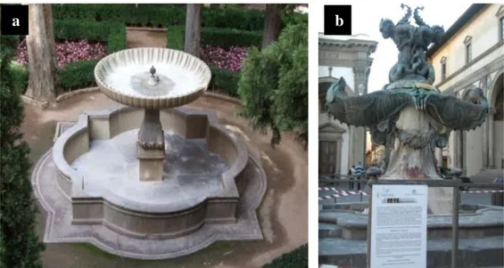
Tacca’s Fountains (1 and 2) - Florence, Italy. These two bronze baroque fountains with marine monsters were set up in 1620 by Pietro Tacca, one of the foremost fountain designer in Florence. They are located in a typical urban area in Santissima Annunziata Square. With an air of gothic exuberance, two fantastic creatures support each other back to back, while the basins of the fountain swell out like organic forms. In the centre of the two shells Tacca carved a water-filled run-off, which, being solid, is like an allegory of water. The pedestals and the border of the basins are made by Carrara marble.
Second Fountain from Villa la Pietra - Florence, Italy. The original villa was built in the fifteenth century by the Macinghi. In 1490 substantial renovations transformed the villa in the present state. During the period in which Florence was the capital of the Kingdom of Italy, the villa housed the Prussian Embassy. In 1904 Sir Arthur Acton, owner of the villa, started to build the garden located on the back of the building. This garden proposes the classical plan, based on three sloping terraces and on the topiary art shaped in a romantic manner, with various sculptures and two fountains. The so called Second Fountain is made by concrete.
Fountain from Patio de la Sultana, Generalife - Granada, Spain. This fountain is located in the Sultana Court at Generalife, Granada (Spain). The arcaded structure dates back to 1584. The area was originally the site of the now disappeared Palace Bath. Water from the irrigation canal, which at one time probably filled it while flowing to the adjacent courtyard, can still be seen pouring through a gap in the side wall. In the centre is a U-shaped pool of water, in the middle of which in the 19th century was placed a smaller pool, with a stone fountain, made on Macael marble and Sierra Elvira stone.
Fountain from Patio de la Lindaraja, Alhambra - Granada, Spain. It is located in the Patio de la Lindaraja, an open air garden with arcaded galleries on the ground floor. The effect is cloister-like, which is further enhanced by the design of the garden with its fountain in the middle. The base, ridge and pilaster of the fountain, in baroque style are made of stone from the Elvira Mountains. From around 1626 to 1995 the fountain had an adorned marble basin in Nasrid style, with epigraph inscriptions, which is currently kept in the Museum of the Alhambra.
Table 1. The investigated fountains reported by various authors
Fountain References
A. Trevi Fountain, Rome, Italy B. Pretoria Fountain, Palermo, Italy C. Fountain of Tritone , Rome, Italy D. Four Rivers Fountain, Rome, Italy E. Fountain of Bibatauin, Granada, Spain F. Lions Fountain, Alhambra Palace, Granada,
Spain
G. Fountain from Patio de la Lindaraja,
Alhambra Palace, Granada, Spain
H. El Bano de Comares and La sala de los
Abencerrajes, Alhambra Palace, Granada, Spain
I. Patio del Cuarto Dorado and Patio de los
Arrayanes, Alhambra Palace, Granada, Spain
J. Tacca‟s Fountains
K. Alcazares and Alhambra‟s Fountains
A. Pietrini (1991); Nugari et al. (1997) B. Not et al. (1995)
C. Barcellona Vero et al. (1979) D. Ricci et al. (1994)
E. Peraza Zurita et al. (2005) F. Sarro et al. (2005) G. Bolivar et al. (1997) H. Bolivar et al. (1998) I. Sanchez-Castillo et al. (1997) J. Tomaselli, et al. (2000b) K. Peraza Zurita (2002b, 2004)
III.2. Biofilms development
II.2.1. Natural biofilm observation
Many components of the biofilm contain different pigments, like chlorophylls, bacteriochlorophylls, carotenoids and phycobilins. The green algae contain chlorophyll a, b and carotenoids, the diatoms contain chlorophyll a, c, carotenoids and xanthophylls, and the cyanobacteria contains chlorophyll a and phycobilins (phycocyanins and phycoerytrins) (Neu T. R., 2003). Due to the specific autofluorescence of the pigments present into the different phototrophs they could be distinguished very easily by Epifluorescence Microscopy (EM) or Confocal Laser Scanning Microscopy (CLSM).
CLSM allows elimination of out-of-focus haze, horizontal and vertical optical sectioning, determination of 3D relationship of cells, and 3D computer reconstruction from optical thin section. Laser microscopy can be used to detect signals resulting from both reflection and autofluorescence in biofilm. Reflection provides information on the presence of colloidal and mineral materials or cell inclusions. Autofluorescence may be used to monitor specific population such as autotrophic algae and cyanobacteria. Most frequently, specific fluorescent molecules may be used directly to stain microorganisms (e.g. DAPI, SYTO series) or they may be conjugated to probes to identify specific biofilm targets such as the lectins used for examine the extracellular polymeric substances (Neu T. R., 1997; Lawrence J. R., 1998; Lawrence J. R., 2003, Wierzchos J., 2004). This technique was successfully applied for studying the aerophytic biofilms in cultural heritage (Hernández-Mariné M., 2002; Roldán M., 2004).
Epifluorescence microscopy, developed in the first half of this century and wide used with the beginning of 1990 for measuring the biomass of plankton (Sieracki M. E., 1990), was chosen to evaluate the covered area of the phototrophic patina, while the confocal laser microscopy allows the spatial semi-quantification of the micro-communities of the fully hydrated biofilms (Rodriguez S. J., 2007).
Aliquots of the biofilm samples collected from the investigated fountains (Table 3) were carefully observed by optical microscopy (Nikon Eclipse E600) in order to distinguish the main microbial components that are constituting phototrophic patinas and to observe their structure. Data related with the three-dimensional biofilm structure was collected using Zeiss LSM 510 META confocal laser scanning microscope. The autofluorescence of phototrophs and substratum was recorded in three channels using 488 nm, 543 nm and 633 nm excitation beams, with emission at 475-525 nm, 560-615 nm and 650 nm, respectively.
Cross-section of small solid parts of samples collected from Sultana Fountain were observed by epifluorescence microscopy (Nikon Eclipse E600) using UV-2A (excitation 330-380 nm, DM 400 nm, BA 420 nm) and TRITC (excitation 450-490 nm, DM 565 nm, BA 610 nm) filter cubes. A polystyrene resin (Mecaprex MA2, Presi, France) was used for samples embedding. The images were recorded using a Nikon DXM1200F digital CCD camera.
II.2.2. Laboratory experiments
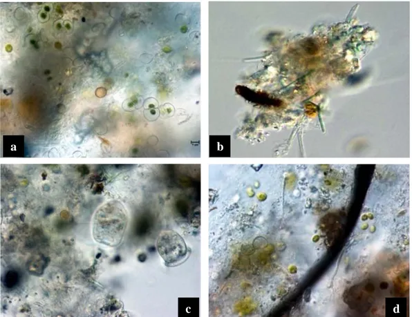
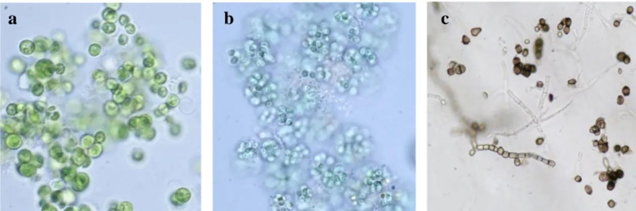
Carrara marble specimens completely (1x1x0.5 cm and 5x5x1 cm) or partially (10x5x1 cm) immersed in well water were used for these experiments. The biological inoculum (≈ 17x105 cells/ml) was composed mainly by Chlorella sp.,
Cosmarium sp., Navicula sp., Nitzchia sp., Leptolyngbya sp., Aphanocapsa sp., Gloeocapsa sp., Oscillatoria sp., protozoa (Fig. 3). The same mixed biofilm inoculum
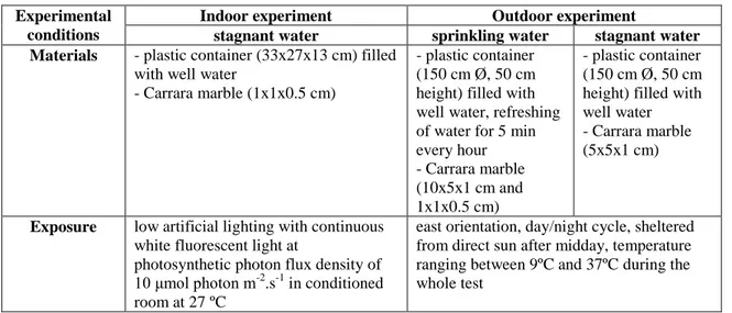
was used for the experiments carried out under indoor and outdoor conditions (Table 2). The outdoor experiment was started after one month after the water inoculation, and therefore the principal microbial components in the two simulation fountains was more stable and presented some differences: diatoms, filamentous algae and coccoid cyanobacteria were dominant in the simulation fountain with sprinkling water, while the palmeloid type algae and coccoid cyanobacteria were preponderant in the simulation fountain with stagnant water (Fig. 4).
Fig. 3. Biological start inoculum for the indoor and outdoor experiments with conjugatoficeae and
palemloid algal types, diatoms (a); filamentous cyanobacteria (b); protozoa (c) and coccoid cyanobacteria and fungal hypha(d).
a
c b
Fig. 4. Main microbial components after 1 month from the inoculum and before the immersion of stone
specimens in the two simulation fountains: with sprinkling water (a,b) and with stagnant water (c,d).
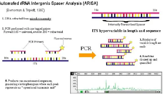
Table 2. The experimental conditions for indoor and outdoor experiments Experimental
conditions
Indoor experiment Outdoor experiment
stagnant water sprinkling water stagnant water
Materials - plastic container (33x27x13 cm) filled
with well water
- Carrara marble (1x1x0.5 cm)
- plastic container (150 cm Ø, 50 cm height) filled with well water, refreshing of water for 5 min every hour - Carrara marble (10x5x1 cm and 1x1x0.5 cm) - plastic container (150 cm Ø, 50 cm height) filled with well water - Carrara marble (5x5x1 cm)
Exposure low artificial lighting with continuous
white fluorescent light at
photosynthetic photon flux density of
10 μmol photon m-2
.s-1 in conditioned
room at 27 ºC
east orientation, day/night cycle, sheltered from direct sun after midday, temperature ranging between 9ºC and 37ºC during the whole test
The phototrophic biofilm development in the first stages was investigated on Carrara marble (1x1x0.5 cm)using the Leica TCS SP5, Leica TCS SP2, confocal laser scanning microscopes (CLSM) in fluorescence and reflection modes. Focusing the instrument at different depths it can be obtained optical slices without any mechanical
a
d c
interference with the patina structure and the colours chosen for each channel are all false colours.
As regard the indoor experiments, the images were recorded on collected samples with both microscopes. For the Leica TCS SP5 the following conditions were used: the reflective signal of the substratum was captured in the blue channel with the excitation at 488 nm and the emission at 490 to 500 nm; the auto fluorescence of photosynthetic organisms was observed in the green and red channel, using the excitation at 561 and 564 nm and emission between 565-615 nm and 665-790 nm respectively; extracellular polymeric substances (EPS) were labelled with the lectin Concanavalin-A conjugated with Alexa Fluor 488 (Invitrogen, Molecular Probes) and observed in the green channel (excitation 488 nm and emission at 493 to 517 nm). With the microscoope Leica TCS SP2 the following conditions were used: excitation at 488 nm and emission at 480 to 499 nm in the reflectance mode; excitation at 351-364 nm, 488 nm, 543 nm and emission at 400-508 nm, 500-561 nm, 555-700 nm respectively for capturing the natural fluorescence of the phototrophs.
The results of outdoor experiments were obtained by capturing images of phototrophic microorganisms autofluorescence, present in the collected samples, using Leica TCS SP5 (excitation beams at 488 nm, 543 nm, 458 nm and emission at 500-531 nm, 680-784 nm and 459-469 nm, respectively) in three channels (for phycoeritryn, chlorophyll a and substratum). The images from stacks were captured at 0.6 µm intervals, in RGB scale, filed in TIFF format.
The biofilm observation of microbiological development on 10x5x1 cmand 5x5x1 cmCarrara marble specimens was assessed through visual observations.
III.3. Study of biofilm composition
III.3.1. Isolation and identification of microorganisms by traditional and
molecular methods
A total of 35 samples were collected by scraping small areas of stone surface from different locations (Plate 2 and 3) of the five artistic fountains, using sterile scalpels and sterile vials. Table 3 contains the description of the collected samples. The samples coming from fountains of Florence, were immediately transported to the
laboratory for microbiological analysis, while the ones coming from Spain arrived as soon as possible, conserved in 2% formalin, to our laboratory by air express courier. Aliquots of the samples were processed for microscopical analysis, others were used for the inoculation in agarized cultural media for the growth and isolation of phototrophs and fungi, and others aliquots were used for the microbial community analysis by ARISA fingerprint method.
Table 3. Characteristics of the collected samples from five investigated monumental fountains
No. Fountain Encodes, typology and sampling position
1. Tacca’s
Fountain 1, Florence, Italy
1T – light green biofilm continuous wet because of the flowing water,
cylindrical marble pedestal, east orientation
2T – dark green biofilm sporadically wet due to the splashing of water, right
corner of the parallelepiped pedestal, east orientation
2. Tacca’s
Fountain 2, Florence, Italy
O1– brown biofilm below the water level, internal part of the basin, south
orientation
O2 – green-brown biofilm below the water level, internal part of the basin,
south orientation
O3 – brown biofilm above the water level, internal part of the basin, south
orientation
N1 – brown biofilm below the water level, sporadically wet due to the water
splashing, internal part of the basin, north orientation
N2 – green-brown biofilm above the water level, sporadically wet due to the
water splashing, internal part of the basin, north orientation
N3 – green-brown biofilm above the water level, sporadically wet due to the
water splashing, internal part of the basin, north orientation
3. Second Fountain of Villa la Pietra, Florence, Italy
VP1 – grey powder above the water level, dry internal part of the basin,
north-east orientation
VP2 – black crust, dry internal part of the basin, north-east orientation
VP3 – brown-green biofilm below the water level, internal part of the basin,
north-east orientation
VP4 – brown-green biofilm, interface water/air, intermittent wetted area,
internal part of the basin, north-east orientation
VP5 and VP6 – light green and turbid deposition, bottom of basin
4. Fountain from Patio de la Sultana Generalife, Granada, Spain
S1a – green biofilm under the water level, internal part of the central fountain,
east orientation
S1b – green biofilm, low part of the pedestal trunk of the central fountain with
constant high wetness, south orientation
S1c – green biofilm, the same level with the water one, corner of the pedestal
basement, north orientation
S1d – green biofilm, low part of the pedestal trunk of the central fountain with
constant high wetness, east orientation
S1e – green biofilm, low part of the pedestal trunk of the central fountain with
constant high wetness, north orientation
S1f – green biofilm, low part of the pedestal trunk of the central fountain with
constant high wetness, west orientation
S2 – green spots on grey patina, sporadically wet by splashed water,
No. Fountain Encodes, typology and sampling position
4. S5N – grey patina, rarely wet, continuous unshaded „surtidore‟, north
orientation
S5W – brownish patina, rarely wet, continuous unshaded „surtidore‟, west
orientation
S6N – dark-green patina, rarely wet, continuous unshaded „surtidore‟, north
orientation
S6S – grey patina, rarely wet, continuous unshaded „surtidore‟, south
orientation
S9W – red brownish patina, continuously wet, continuous shaded „surtidore‟,
west orientation
S9N – dark-green patina, continuously wet, continuous shaded „surtidore‟,
north orientation
S9S – green patina, continuously wet, continuous shaded „surtidore‟, south
orientation
S9E – green patina, continuously wet, continuous shaded „surtidore‟, east
orientation 5. Fountain from Patio de la Lindaraja Alhambra, Granada, Spain
1L – brown powdering patina, external part of the basin, north-west orientation 2L – green patina, sporadically wet, internal part of zig-zag ornaments of
superior marble basin, south-east orientation
3L – green patina, sporadically wet, internal part of superior marble basin,
south orientation
4L – green patina, internal part of the central fountain, west orientation 5L – green biofilm, sporadically wet, internal horizontal part of pilaster
ornaments, west orientation
6L – green patina, continuously wet, internal part of foundation, west
orientation
III.3.1.1. Traditional methods
Aliquots of the collected samples (Tacca‟s Fountain 1, Second Fountain from Villa la Pietra and Fountain from Patio de la Sultana) were suspended in standard saline solution and sown in Petri dishes containing the respective solid medium for the isolation of: (i) cyanobacteria (BG-11), (ii) nitrogen fixing cyanobacteria (BG-110), (iii) algae (modified BG-11 (5 ml/l NaNO3 diluted 1:1 with sterile water)) and (iv) fungi (PDA Difco). BG-11 type media were described in Rippka R. et al (1979).
The Petri dishes containing cultural media for phototrophs were incubated under continuous low white fluorescent light at photosynthetic photon flux density of 10 μmol photon m-2
s-1 at 27 ºC, while the ones containing PDA for fungi were incubated in darkness, at 26 ºC. The developed cyanobacteria, algae and diatoms colonies were transferred into tubes containing the same specified liquid cultural media, while the fungal strains were isolated on the PDA agar medium. All the isolated strains were maintained on slants with their specific cultural medium in appropriate conditions.
The morphological characterization and morphotypes identification were carried out with optical microscopy (Nikon Eclipse E600) according to Geitler L. (1932), Komarek J. and Anagnostidis K. (1988, 1998, 2005), Barnett H. L. (1998) and Bourrelly P. (1966) for all collected samples. Representative isolated cyanobacterial strains were also characterized by phylogenetic analysis of the 16S rRNA gene.
III.3.1.2. DNA extraction
DNA of isolated cyanobacterial strains and of collected samples from two fountains (Second Fountain of Villa la Pietra, Florence, Italy and Sultana Fountain from Generalife, Granada, Spain ) were extracted. 30 ml of liquid cultures (BG-11, or BG-110 media) of the isolated cyanobacterial strains were incubated with agitation in a Gallenkamp Orbital Incubator (CO2 4%, 27 ºC, light/dark cycles 14/10 hours). The biomass was harvested by centrifugation and the pellet was washed with standard saline solution (0.1% w/v) for two times or even more in case of the mucilaginous cultures, and then the pellet was divided in aliquots and frozen at - 20 ºC. For DNA extraction, pellets were thawed and treated with PowerPlantTM DNA Isolation Kit, according to the manufacturer‟s protocol (Mo Bio Laboratories Inc.).
Total community DNA of samples collected from the fountains was extracted using PowerSoilTM DNA Isolation Kit, according to the manufacturer‟s protocol (Mo Bio Laboratories Inc.).
All the DNA extracts were laden into 0.5% agarose gel, previously stained with ethidium bromide (10 mg/ml). The quality of DNA extracts was examined under UV light after 15 minutes at 150V of DNA molecules migration.
III.3.1.3. PCR amplification and sequencing
To amplify the rRNA 16S gene plus the ITS region, the encoding gene fragments of cyanobacteria, template DNA, universal primer 16S27F
(5′-AGAGTTTGATCCTGGCTCAG-3′) and cyanospecific primer 23S30R (5′-
CTTCGCCTCTGTGTGCCTAGGT-3′) were used. (Edwards U., 1989; Taton A., 2003). The genomic DNA (1 µl) was amplified using Hot Start Master (Larova GmbH, Germany). This PCR was carried out with an activation and initial denaturation step of 2 min at 94 ºC, followed by 30 cycles of denaturation for 45 sec at 94 ºC, annealing of 1
min at 55 ºC, and elongation of 2 min at 72 ºC, followed by a final extension step of 7 min at 72 ºC. All amplification reactions were performed in a TGradient Thermal Cycler (Biometra, Goettingen, Germany). The amplicons were visually quantified by comparison with DNA Molecular Weight Marker VI (0.15 - 2.1 kpb) (Roche, Mannheim, Germany) on agarose gel electrophoresis. The amplified DNA fragments were purified using ExoSAP-IT Clean-Up kit following the manufacturer‟s instructions (USB Corporation, Affimetrix, Inc, USA) and then sent to BMR Genomics, Padua, Italy (www.bmr-genomics.it) for sequencing.
The gene sequences for rRNA 16S were obtained using the three primers 16S979F, 16S544R and 16S1092R (Hrouzek P., 2005). Single reads were aligned in a unique 16S rRNA gene sequence using the software suite PHRED, PHRAP CONSED developed by the University of Washington (Gordon D., 2004) and consensus sequences imported in ARB (Ludwig W., 2004), which was used for the subsequent analytical steps. Most similar sequences included in the analysis were retrieved from the most recent SILVA database. The aligner on the SILVA website was also used to produce the sequence alignment for the ARB software. The alignment was later visually investigated and corrected under ARB. Phylogenetic relationships of the sequences were calculated using Neighbor-joining algorithm (Saitou N., 1987) using sequences over 1300 base pairs.
Sequencing and phylogenetic analysis were used for molecular identification of 35 cyanobacterial strains isolated from the monumental fountains. DNA sequencing is the process of determining the nucleotide order of a given DNA fragment. It has been performed using the chain termination method developed by Frederick Sanger (1977). This method consist in annealing and extension of a short complementary primer to a single stranded template DNA by DNA polymerase. The reaction is split into 4 tubes (called A - adenine, C - cytozine, G – guanine or T - thymine) each containing a low concentration of the indicated dideoxy nucleotide, in addition to the normal deoxynucleotides. Dideoxynucleotides, once incorporated, block further chain extension and so each tube accumulates a mixture of chains of lengths determined by the template sequence. The reactions are performed in a single tube containing all four ddNTP's, each labeled with a different color dye. Since the four dyes fluoresce at different wavelengths, a laser then reads the gel to determine the identity of each band according to the wavelengths at which it fluoresces (Howe C. J., 1995). The results are then depicted in the form of a chromatogram, which is a diagram
of colored peaks that correspond to the nucleotide in that location in the sequence, specific for each organism (Fig. 5) Therefore, these data are used for molecular phylogenetic analysis expressed in a phylogenetic tree, comparing the obtained sequences with the ones from public and specific databases.
Fig. 5. Chromatogram of a DNA sequence (…GTGAGGCGCTGC…)
III.3.2. Microbial community analysis by ARISA fingerprint method
III.3.2.1. DNA extraction
Total community DNA of the samples collected from the fountains was extracted using PowerSoilTM DNA Isolation Kit (Mo Bio Laboratories Inc.), according to the manufacturer‟s protocol. The quality of DNA extracts was observed as described in III.3.1.2.
III.3.2.2. PCR amplification and ARISA
PCR amplification of rRNA 16S gene for ARISA was performed using two specific primers sets, for eubacterial and cyanobacterial domain, respectively (Table 4). The reaction mixture (50µl each) for the PCR amplification contained: 5µl 10x DyNAzyme EXT buffer (with 15 mM MgCl2), 5µl dNTPmix (2mM), 1µl of each primer (10pmol/µl), 2.5µl BSA (Bovine Serum Albumin, BioLabs, 10 mg/ml), 33.5µl distillated water. Before adding 1µl of Taq DNA polymerase (Amersham-Pharmacia) and 1µl of genomic DNA, aliquots of the mix were placed in PCR tubes and sterilized under UV light (312 nm) for 8 min. Reactions (50µl final volume) were initially denaturated for 5 min at 94ºC, followed by 35 cycles of 94ºC for 1 min, 55ºC for 1 min and 72ºC for 2 min, and then a final extension step at 72ºC for 10 min was performed. The PCR products (5 µl each) mixed with bromophenol blue were visualized after 45 min of electrophoresis on 1.2% agarose gel stained with ethidium bromide (10 mg/ml). DNA Molecular Weight Marker VI (0.15 - 2.1 kpb) (Roche, Mannheim, Germany) loaded in the same gel (3 µl and 6 µl) was used for the approximate quantization of amplified DNA.
Table 4. Primer sequences and target sites
Primera Sequence (5’→ 3’) Target site Group Reference
16S1515F AGT CGT AAC AAG GTA
GCC GTA CC
1492-1515 cyanobacteria Cardinale et al.,
2004 (modified)
23S30R-6FAM CTT CGC CTC TGT GTG
CCT AGG T
30-52 cyanobacteria Lepère et al., 2000
ITSF GTC GTA ACA AGG TAG
CCG TA
1494-1513 eubacteria Cardinale et al.,
2004
ITSReub-HEX GCC AAG GCA TCC ACC 23-37 eubacteria Cardinale et al.,
2004
a R (reverse) and F (forward) designations refer to the primer orientation in relation to the rRNA
b
E.coli numbering of 16S rRNA or 23S rRNA nucleotides
The amplified DNA (2 ng/ µl each) was then sent to BMR Genomics for fragment separation in a capillary chromatographer. A fluorescent dye-labeled size standard GeneScan™ ROX500 (35, 50, 75, 100, 139, 150, 160, 200, 250, 300, 340, 350, 400, 450, 490, 500 base pair fragments) was added and the analyze was carried out with a GeneScan 3100 ABI Automated Capillary DNA Sequencer (Applied Biosystems).
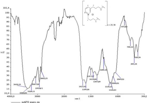
ARISA (Automated rRNA Intergenic Spacer Analysis) is a fingerprint molecular method for analyzing the community heterogeneity. This approach consist in extraction of total DNA from an environmental sample and then isolate homologous genes (usually rRNA genes) from the DNA mixture. Fig. 6 illustrates the ARISA principle. Each peak of an electropherogram represent an operational taxonomic unit, and therefore valuable information about the biodiversity of specific groups (e.g. using specific primers for eubacteria and cyanobacteria) present in the analyzed sample is obtained.
The computational analysis were performed with Peak Scanner TM (Applied Biosystems 1.0, 2006) and BioNumerics 2.5, (Applied Maths, Belgium) software. Only peaks higher than 50 relative fluorescence units and fragments between 100-650 base pairs were considered.
Principal component analysis (PCA) was performed on ARISA profiles obtained with BioNumerics software, in order to evaluate the similarities of microbial communities for fountains samples. The peaks intensities for each band were normalized in accordance with the total fluorescence of each sample ARISA profile. PCA was carried out using Unscrambler® Camo Software AS with the default parameters.
III.4. Stone material and color measurements
Tests were carried out with two different types of stones (Fig. 7) commonly used for creating artistic monuments in Italy and south of Spain:
- Carrara marble type P, a white metamorphic stone with a mean grain size <100 µm with an unimodal distribution. The water porosity was 0.57% (hydrostatic balance method- ISO 6783:1982). Specimens dimension: 1x1x0.5 cm or 5x5x1 cm.
- Sierra Elvira stone classified as pelsparite (Folk R. L., 1959; Folk R. L., 1962) or grainstone (Dunham R. J., 1962), a grey sedimentary stone with a pellet structure (pellet dimension 150-250 μm), presenting diffuse phenomena of dolomitization. The water porosity was 0.31%. Specimens dimension: 1x1x0.5 cm.
Fig. 7. Thin sections (crossed Nicols) for Carrara marble type P (a) and Sierra Elvira stone (b); scale bar
500µm.
To quantify the color change of the stones, before and after the applied treatments containing ABAs, the color parameters were calculated using CIELAB 1976 formula (Ohno Y., 2000). Reflectance values of the stone were measured with a Minolta Chroma Meters CR-200, a compact tristimulus color analyzer which measures the reflected colors of surfaces. The readings were taken three times and an average value was considered. Three main parameters (L*, a* and b*) were recorded, where L* is the lightness/darkness coordinate; a* the red/green coordinate, with + a* indicating red and - a* indicating green; b* the yellow/blue coordinate, with + b* indicating yellow and - b* indicating blue. ΔE is the total color difference calculated from CIELAB 1976 color difference formula: ΔE = [(ΔL*)2+(Δa*)2+(Δb*)2]1/2.
III.5. Antibiofouling agents, coatings preparations and their characterization
Five natural antibiofouling agents (ABAs) were selected as potential inhibitors of biofilm formation – three of them are present in marine environment (pAPS in a sponge, ZA in a marine plant and CBE in a red alga) and the others have a terrestrial origin (CI in cinnamon and CS in chilli pepper) (see Plate 4). Algophase (A) was used as a positive control even if this product has a biocide action and not an antibiofouling one.
(i) Poly-alkyl-pyridinium salts (pAPS) 1 (3-alkylpyridinium active compound) isolated from a marine sponge Reniera sarai (Sepčić K., 1997). In fact, the surface of this sponge is smooth and clean, veiled in a greasy secretion containing large amounts of pAPS that probably act as a protective coating against fouling (Turk T pers. obs.). Poly-APS are a mixture of two main polymers with molecular weights of 5520 and 18900 Da, corresponding to polymers composed of 29 and 99 3-octylpyridinium units, respectively (Fig. 8). Purified pAPS are soluble only in water. In aqueous solutions pAPS behave similarly to detergents that are classified as quaternary ammonium compounds. According to their critical micelle concentration and charge, pAPS resemble well-known cationic detergents like cetylpyridinium chloride and cetyltrimethylammonium bromide (Malovrh P., 1999). Therefore, these detergent-like properties of pAPS are capable of exercising a number of biological activities. The
surfactant activity towards artificial and natural membranes and acetylcholynesterase inhibitory activity might be involved in antifouling molecular mechanisms that prevent settlement of bacteria and metazoan onto the sponge surface (Sepčić K., 2006).
The aqueous extracts from this sponge possesses strong anticholinesterase and helmolytic activity as well as moderate antimicrobial and cytotoxic activities (Sepčić K., 1997). pAPS were tested for their potential anti-microfouling activity, for example their ability to prevent the formation of biofilm on submerged surfaces under laboratory conditions (Garaventa F., 2003). pAPS and some other natural 3-alkylpyridines were found to be very effective in preventing of microbial film formation (Sepčić K., 2006) and was also tested to prevent diatoms, algae and fungi settlement (Faimali M., 2003; Eleršek T., 2008). It was used as a water solution in the concentrations reported in Table 6.
Fig. 8. The FT-IR spectrum and the chemical formula of poly-alkylpyridinium salt
(ii) Zosteric acid (ZA) (p-sulphoxy-cinnamic acid) is a natural product present in the eelgrass Zostera marina. The eelgrass produces and continuously releases the water-soluble antifouling compound ZA. This antifoulant does not kill microorganisms but inhibits their adhesion trough its binding to special sites on cell-surfaces. The precise mode of action in producing the antifouling response remains undetermined; however, it should be noted that the extracellular polysaccharides
produced by many microorganisms are highly sulphated, and these sulphate esters play an important role in polymerisation (i.e. glue/gel formation) and therefore ZA could be operating at the atomic level by blocking sulphate-binding surface sensors, or by inhibiting the polymerization of extracellular glue (Zimmerman R., Patent 1995). Other authors have been reported also the preventive efficiency of ZA in microbial attachments and the antifouling effect has been attributed to the sulphate ester group (Todd J. S., 1993; Stanley M. S., 2002; Newby B. Z., 2006). ZA is very water soluble and generally, easily washes off the surfaces or leaches out of coatings (Elder S. T., 2007). ZA has shown to prevent biofouling from some marine and freshwater bacteria, algae and algal spores, barnacles, tubeworms and fungal spores adhesion (Todd J. S., 1993; Callow M. E., 1998; Stanley M. S., 2002; Newby B. Z., 2006).
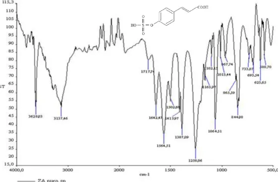
Zosteric acid was synthesised in our laboratory according to previously reported procedures with little modifications (Alexandratos S. D., 1999). The modifications were related with the incomplete purification of zosteric acid, performed only with diethyl ether and methanol. The synthesised chemical compound was characterised by ESI-MS2 (Fig. 9) and FT-IR (Fig. 10) and the resulting spectra were in agreement with the presence of zosteric acid. It was used as a water or methanol solution, in the concentrations reported in Table 6.
Spectrum 1B Plot - 9/23/2008 11:00 AM
1 B Scan 11 from c:\varianws\data\gianluca\acidozoostericosettembre2008.xms
200 300 400 500 m/z 0% 25% 50% 75% 100% 242.8 3.101e+8 243.9 3.189e+7 244.8 1.848e+7 388.8 4.914e+7 Spectrum 1B
0.185 min, Scans: 4-18, 150.0:550.0>(-), Ion: NA, RIC: 5.255e+8 BP: 242.8 (3.101e+8=100%), acidozoostericosettembre2008.xms
Fig. 9. ESI-MS spectrum of synthesized zosteric acid
2 GC-MS analysis was made by Prof. M. Bambagiotti of the Department of Pharmacology, University of
Fig. 10. FT-IR spectrum and chemical formula of zosteric acid
(iii) Extract of Ceramium botryocarpum (CBE)3 is a dichloromethane extract of a marine rhodophyta (Ceramium lanciferum var. monstruosum – this name is currently regarded as a synonym for C. botryocarpum). This extract and two other ones (ethanol and methanol) of this alga were successfully tested against marine bacteria, diatoms and microalgae (Ulva sp.) which are involved in the microfouling process (Bazes A., 2006). The effectiveness of this antifoulant was attributed to the releasing of allelopathically active compounds interfering with settlement and growth competitors. Allelopathy describes any direct or indirect effect of a living organism on another one through biochemical compounds released in the environment, including therefore all biochemicals interactions, both stimulatory and inhibitory (Gross E. M., 2003). The active compound is still unknown. CBE is soluble in water and presents a very high hygroscopicity. The FT-IR spectrum (Fig. 11) has similarities with one of pAPS, and therefore can be suggested its belonging to salts group, with characteristic signals at 1637 and near 3000 cm-1. Very recent studies report the presence of natural digeneaside in salt form in the extracts of red alga Ceramium botryocarpum (Claude A., 2009). It was used as a water or methanol solution in the concentrations reported in Table 6.
3
Fig. 11. FT-IR spectrum of Ceramium botryocarpum extract
(iv) Capsaicin (CS) (8-methyl-N-vanillil-6-nonenamide) is the natural extract from chilli pepper responsible for the „hotness‟ of the pepper, being concentrate mostly in the placental tissue of the pepper. Two very pungent synthetic capsanoid compounds are capsaicin and dihydrocapsaicin. It is an incredibly powerful and stable alkaloid seemingly unaffected by heat or cold (Watts J. L., US Patent, 1995). It has limited water solubility (≈60 mg/L). The best co-solvent for obtaining higher concentration of capsaicin in water was ethanol (Turgut C., 2004). It is registered by the US Environmental Protection Agency (EPA) in 1999, as a bird, animal and insect repellent and it was classified as a biochemical pesticide. Capsaicin has previously been assayed for antimicrobial activity (Cichewicz R. H., 1996; Molina-Torres J., 1998) and the efficacy against freshwater bacteria is also reported (Xu Q., 2005). Studies related with the toxicology and metabolism of capsaicin (Surh Y. J., 1995) indicated that capsaicin was toxic only when it was oxidized into an epoxy intermediate. The capsaicin employed in this study was purchased from Aldrich Chemical Co., and its composition was 65% capsaicin and 35% dihydrocapsaicin. Chemical formula and FT-IR spectrum are reported in Fig. 12. It was used as a water or ethanol solution in the concentrations reported in Table 6.