I CONTENTS Sommario II Summary IV List of abbreviations VI 1. Introduction 1
1.1 Mitochondria and mitochondrial DNA 1
1.1.1 Nucleus-mitochondria cross-talk 5
1.1.2 Mitochondrial role in aging 14
1.2 Frailty as aging phenotype 20
1.3 Aging epigenetics: DNA methylation 26
1.3.1 DNA methylation basic mechanisms 27
1.3.2 DNA methylation functions 31
1.3.3 DNA methylation role in aging and age-related diseases 34
1.4 Mitochondria as modulators of aging epigenetics 39
1.5 Plan of thesis 42
2. Global DNA methylation in old subjects is correlated with frailty. 43 3. The variability of mitochondrial DNA modulates global DNA
methylation levels 74
4. SIRT3 gene expression: A link between mitochondrial DNA
inherited variants and oxidative stress response. 111
5. Mitochondrial DNA variability modulates mRNA and
intra-mitochondrial protein levels of HSP60 and HSP75: experimental
evidence from cybrid lines. 148
6. Conclusive remarks 155
II
Sommario
La qualità e la velocità dell'invecchiamento umano dipendono dalla complessa interazione tra fattori genetici, epigenetici e ambientali. In questo contesto, numerose evidenze hanno dimostrato come un ruolo centrale sia giocato dai mitocondri e dalla variabilità del DNA mitocondriale (mtDNA). Essi hanno, infatti, un ruolo cruciale nel mantenimento dell'omeostasi energetica e redox, nella regolazione dei processi epigenetici e nell’ influenzare i meccanismi di risposta allo stress, elementi chiave per raggiungere la longevità.
Il presente lavoro fornisce una panoramica delle suddette assunzioni riportando, in quattro sezioni i risultati di studi condotti in vivo e in vitro.
Nelle prime due sezioni sono riportate una serie di evidenze sperimentali che dimostrano la correlazione tra il declino fisiologico tipico dell’età avanzato, le varianti ereditarie del DNA mitocondriale ed i livelli di metilazione globale del DNA. I dati ottenuti hanno dimostrato come i cambiamenti dei livelli di metilazione del DNA, la modificazione epigenetica più ampiamente caratterizzata, sono associati al declino funzionale di individui e correlano alla cambiamenti fisiologici che si verificano nell’uomo nel corso della vita. È emerso inoltre che il rimodellamento nello stato di metilazione del globale DNA durante l'invecchiamento è influenzato dalle varianti ereditarie del DNA mitocondriale, presumibilmente attraverso la regolazione differenziale della funzionalità della OXPHOS. Tali risultati hanno dunque dimostrato come processi epigenetici sono modulati da segnali bidirezionali tra genoma mitocondriale e nucleare, strettamente regolati dalla variabilità dell’mtDNA.
Le ultime due sezioni presentano i dati ottenuti da uno studio volto ad esplorare il ruolo della variabilità ereditaria del DNA mitocondriale nella regolazione dei profili di espressione di due classi di geni nucleari coinvolti nella risposta cellulare allo stress:
III sirtuine e Heat Shock Proteins (HSP). Tale studio è stato ispirato da dati di letteratura riportanti come l'efficienza della risposta cellulare allo stress, elemento chiave per il raggiungimento della longevità, è fortemente influenzata da segnali bi-direzionali tra il DNA mitocondriale e nucleo. In tali lavori, ci si è avvalsi della tecnologia dei cibridi per dimostrare che tali pathway di comunicazione, in condizioni di stress ossidativo e termico, sono stati in grado di influenzare, rispettivamente, l'espressione dei geni codificanti per le Sirtuine e le HSP, le prime sensori dell'omeostasi energetica e redox, le seconde regolatori dei processi di folding e migrazione proteica nonché dell’apoptosi. Dall’analisi dei profili di espressione delle sette sirtuine è emerso che il gene SIRT3 ha mostrato downregolazione in condizioni di stress ossidativo correlata alla sequenza del DNA mitocondriale alla compromissione della funzionalità mitocondriale stessa. Per quanto riguarda lo studio di espressione dei geni codificati per le HSP, è emerso come sia HSP60, sia HSP75 sono diversamente modulati in condizioni di shock stress da calore in base alla variabilità del mtDNA. Tali risultati suggeriscono che la correlazione tra la variabilità del DNA mitocondriale e livelli di espressione di geni nucleari coinvolti nella risposta allo stress può essere considerato come un fenomeno generale.
IV
Summary
The rate and quality of human aging depend on a complex interplay among genetic, epigenetic and environmental factors. In this scenario mitochondria and mitochondrial DNA (mtDNA) variability are emerging as major players. This is basically due to their crucial role in maintaining energetic and redox homeostasis, in modulating the intracellular epigenetic program and in influencing cell stress response mechanisms, which are key elements to achieve longevity.
The present work provides an overview of the above assumptions, reporting, in four sections, a series of in vivo and in vitro investigations. In the first two sections experimental evidences about a correlation among the age-specific functional decline, the mitochondrial DNA inherited variants and the global DNA methylation levels are reported. The data we show demonstrate that the remodeling of DNA methylation levels, that represent the best characterized epigenetic modification, is associated to the functional decline of aged individuals and correlates to their physiological changes occurring over the lifetime. We also demonstrate that the global DNA methylation remodeling during aging is influenced by mitochondrial DNA inherited variants, probably via the different regulation of OXPHOS machinery. So that, we prove as epigenetics processes are modulated in response to mtDNA-specific cross signaling between mitochondrial and nuclear genome.
The last two sections present data obtained in the studies on the role of mtDNA inherited variability in modulating the expression profiles of two classes of stress responder nuclear genes: Sirtuins and HSPs. These sections were inspired by evidences reporting that the efficiency of cellular stress response, a key element for attending longevity, is highly regulated at both nuclear and mitochondrial level through activating bi-directional signaling pathways between mitochondrial and nuclear DNA. In these
V works we availed of cybrid technology to demonstrate that the above pathways, in oxidative and heat shock stress conditions, were able to influence the expression of Sirtuin and HSP genes, respectively; the former are regulators of energetic and redox homeostasis, the latter are regulators of protein folding and migration as well as apoptosis in both stressed and non-stressed cells. As for sirtuin genes, we found that only SIRT3 gene was down-expressed depending on the mtDNA sequence in oxidative stress condition and that this down-expression was correlated to the impairment of mitochondrial function. As for HSPs, either HSP60 or HSP75 were differently modulated according to mtDNA variability, in heat shock stress conditions. Thus, the consistency of these results suggest that the correlation between mtDNA variability and expression levels of stress-responder nuclear genes is a general phenomenon.
VI
List of abbreviations
5-MCDG 5-methylcytosine DNA glycosylase
8-OH-dG 8-Hydroxyl-2'-deoxyguanosine
AceCoA Acetylcoenzyme A
AceCS2 Acetylcoenzyme A synthase 2
AD Alzheimer's disease
ADL Activities of daily living
AKAP12 A kinase (PRKA) anchor protein 12
ANT Adenine nucleotide translocaton
AP-1 Activator Protein-1
AP-2 Activator Protein-2
APOE Apolipoprotein E
ATP Adenosine triphosphate
ATP(6-8) ATP synthetase F0 subunit (6-8)
BER Base excision repair
BMD Bone mineral density
BMI Body mass index
BRCA1 Breast Cancer 1
C/EBPβ CCAAT/enhancer binding protein beta
CAMKIV calcium/calmodulin-dependent protein kinase IV
CAT Catalase
cGMP Cyclic guanosine monophosphate
CH3 Methyl group
VII
ClpP ClpP caseinolytic peptidase, ATP-dependent, proteolytic subunit homolog
c-Myc/Myn myelocytomatosis viral oncogene
CO(I-III) Cytochrome c Oxidase subunit (I-III)
CoQ Coenzyme Q
CR Caloric restriction
CREB cAMP response element-binding
CRP C-reactive protein
CVD Cardiovascular disease
Cytb Cytochrome b
DAPK Death-associated protein kinase
DHEA-S Adrenal androgen dehydroepiandrosterone-sulfate
D-Loop Displacement-loop
dmC Deoxymethylcytosine
DMEM Dulbecco’s modified eagle medium
DNMT1 Maintenance DNA methyltransferase
DNMT3(a-b) De novo methyltransferase (a-b)
DNMTs DNA Methyltransferases
DRD2 Dopamine receptor D2
E2F E2F transcription factor
eNOS Endothelial nitric oxide synthase
EORS Epigenetic oxidative redox shift
ERCs Extrachromosomal ribosomal DNA circles
ETC Electron transport chain
VIII
FMR1 Fragile X mental retardation 1
FOXO Forkhead box-containing protein type O subfamily
GAD1 Glutamate decarboxylase 1
GDH Glutamate dehydrogenase
GDS Geriatric depression scale
GH Growth hormone
GPx Glutathione peroxidase
GSH Glutathione
GSTP1 Glutathione S-transferase P1
H Heavy strand
H2O2 Hydrogen peroxide
HDAC1 Histone deacetylase 1
HDL High-density lipoprotein
HO Heme oxygenase
HOXB5 omeobox protein Hox-B5
HSP10 Heat shock protein 10
HSP60 Heat shock protein 60
HSPs Heat Shock Proteins
HVR(I-II) Hypervariable region (I-II)
ICDH2 Isocitrate dehydrogenase 2
ICR Imprinted control region
IGF-1 Insulin-like growth factor 1
IGF-2 Insulin-like growth factor 2
IL-6 Interleukin-6
IX
JNK c-Jun N-terminal kinase
Kaiso transcriptional regulator Kaiso
L Light strand
LHON Leber's hereditary optic neuropathy
MAPK Mitogen-activated protein kinase
MAT Methionine adenosiltransferase
MBD Methyl binding domain
MBD(1-3) Methylcytosine binding protein (1-3)
MBD2b Methyl-CpG-binding domain protein 2
MBD4 Methyl-CpG-binding domain protein 4
MBPs Methylcytosine binding proteins
MeCP2 methyl CpG binding protein 2
MEF2 myocyte enhancer factor-2
MEFs Mouse embryonic fibroblasts
MLH1 MutL protein homolog 1
MLSP Maximum lifespan
MMSE Mini mental state examination
MPO Myeloperoxidase
MPT Mitochondrial permeability transition
mtDNA Mitochondrial DNA
mtTFA Mitochondrial transcription factor A
mtTFB Mitochondrial Transcription Factor B
MZ Monozygotic
NADH Nicotinamide adenine dinucleotide
X
ND(1-6; 4L) NADH dehydrogenase subunit (1-6; 4L)
NFAT nuclear factor of activated T-cells
NF-KB Nuclear factor-kappa B
NO Nitric oxide
NRF1-2 Nuclear Respiratory Factor (1-2)
O2 Molecular oxygen
O2- Superoxide anion
OH H-strand replication Origin
OH. Hydroxyl radical
OL L-strand replication Origin
OMM Outer mitochondrial membrane
OXPHOS Oxidative Phosphorylation
p53 tumor protein p53
PDAC Protein deacetylase
PGC-1(α-β) Peroxisome proliferator-activated receptor co-activator 1 (α-β)
PGC-1α PPARγ coactivator 1α
PH H-strand transcription Origin
Pi Inorganic phosphate
PKC Protein kinase C
PL L-strand transcription Origin
PLAU Urokinase type plasminogen activator
PolgA mtDNA polymerase subunit A
PPARγ Peroxisome proliferator activated receptor γ
PPIEL Peptidiylprolyl isomerase E-like
coactivator-XI related 1
RASSF1 Ras association domain-containing protein 1
Rb Retinoblastoma associated protein
RELN Reelin
RNAi RNA interference
RNS Reactive nitrogen species
ROS Reactive oxygen species
rRNA Ribosomal RNA
Rtgs Retrograde proteins
S100A4 S100 calcium binding protein A4
SAM S-adenosyl-L-methionine
Sir2 Silent informator regulator 2
SIRT1-7 Sirtuin 1-7
SOD Superoxide dismutase
Sp-1 Sp1 transcription factor
SRHS Self-reported health status
STK11 Serine/threonine kinase 11
Tfam Mitochondrial transcription factor A
THO Tyrosine hydroxylase
THO Tyrosine hydroxylase
TNF-α Tumor necrosis factor ?
TRD Transcriptional repressory domain
tRNA Transfer RNA
tRNAAsp Aspartic acid transfer RNA
XII
UPR Unfolded Protein Response
USF Upstream stimulatory factor
VADC Voltage dependent anion conductance
VHL Von Hippel-Lindau tumor suppressor
1
1. Introduction
Over the past two decades, a growing interest in the research of the biological basis of human longevity has emerged, in order to clarify the biological and the environmental factors affecting the quality and the rate of human aging. In this scenario, many studies have focused on the involvement of mitochondrial function (and dysfunction) in aging. In fact, evidences are accumulating that these organelles are able to modulate numerous intracellular signaling pathways which appear to be critically important for the maintenance of cellular homeostasis.
In the frame of the research on aging and longevity, an emerging field that promises exciting findings is represented by the epigenetic changes affecting DNA during the lifetime. Also in this field of research, a complex interplay between mitochondrial function and epigenetic modifications in aging has been observed, although only a few aspects have been so far elucidated.
1.1 Mitochondria and mitochondrial DNA
Mitochondria are intracellular organelles located in the cytoplasm of eukaryotic cells. As it regards the structure, mitochondria are bounded by a double membrane. The outermost mitochondrial membrane (OMM) is smooth while the inner mitochondrial membrane (IMM) has many folds called cristae, that enhance the "productivity" of cellular respiration by increasing the available surface area.
Mitochondria are emerging to be involved in several cellular processes, including ion homeostasis, cell proliferation and differentiation, but their primary function is the generation of energy in form of ATP, via the electron transport chain (ETC) and the oxidative phosphorylation (OXPHOS) system located within the inner membrane
2 (IMM) of the organelle. The OXPHOS machinery is composed by five multiprotein complexes each composed by subunits encoded by both nuclear and mitochondrial genome, plus two molecules, ubiquionone (Coenzyme Q) and cytochrome c, that acts as diffusible electron carriers (Fig. 1).
Fig. 1: Electron Transport Chain (ETC). The reducing equivalents in NADH or
FADH2 enter in the electron transport chain through the complex I (NADH dehydrogenase) and Complex II (Succinate dehydrogenase) respectively. While electron are then transferred from NADH to coenzyme Q (CoQ) and to Complex III (Ubiquinine-cytochrome c reduttase), and the (Ubiquinine-cytochrome c to Complex IV (Cytochrome c oxidase), protons are translocated from matrix to the intermembrane space. The electrochemical grandient established across the IMM represents the driving force of ATP synthesis catalized by the Complex V (ATP syntase).
Every cell contains a variable number of mitochondria, and each mitochondrion harbors 2-10 copies of their own genome, the mitochondrial DNA (mtDNA) (Anderson et al. 1981).
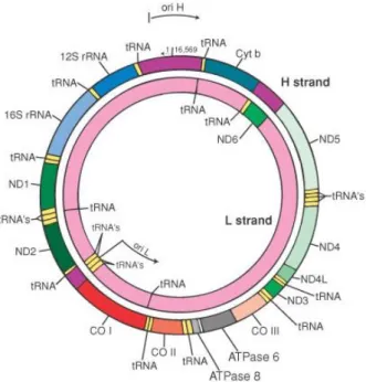
The human mitochondrial DNA is a 16569 bp closed-circular double-stranded molecule, containing 37 genes encoding for 2 ribosomal RNAs (rRNA 12S and 16S), 22 transfer RNAs and 13 subunits of the OXPHOS system (Fig. 2).
Fig. 2: Human mitochondrial DNA.
strand (H) and a cytosine
of these genes encode for the translational machinery of the mtDNA, including 22 tRNAs and 2 rRNAs. The remaining 13 genes encode for subunits of OXPHOS system, including seven subunits of
(Cytb), three subunits of Complex IV (COI 8).
The structure of mtDNA is very compact; in fact, mitochondrial genes have no introns, intergenic sequences are
termination codons are generated post
The sole non coding region, the Displacement containing two hypervariable
the origin of replication of H
(PL) transcription and regulatory elements for both mitochondrial replication and transcription.
MtDNA is inherited maternally, so that the paternal lineage does not contribute mtDNA to the offspring, and it is not subject to significant recombination (Elson et
Human mitochondrial DNA. mtDNA has two strands, a guanine
ine-rich light strand (L) carrying 28 and 9 genes
of these genes encode for the translational machinery of the mtDNA, including 22 tRNAs and 2 rRNAs. The remaining 13 genes encode for subunits of OXPHOS system, including seven subunits of Complex I (ND1-6 and ND4L), one subunit of Complex III (Cytb), three subunits of Complex IV (COI-III) and two subunits of Complex V (ATP6
The structure of mtDNA is very compact; in fact, mitochondrial genes have no introns, intergenic sequences are absent or limited to a few bases, some genes overlap and termination codons are generated post-transcriptionally from polycistronic transcripts. The sole non coding region, the Displacement-loop (D-Loop), is a region of 1121 bp containing two hypervariable regions, HVRI (nt 16024-16383) and HVRII (nt 57 the origin of replication of H-strand (OH), the promoter region for H (PH) and L
(PL) transcription and regulatory elements for both mitochondrial replication and
ed maternally, so that the paternal lineage does not contribute mtDNA to the offspring, and it is not subject to significant recombination (Elson et
3
guanine-rich heavy rich light strand (L) carrying 28 and 9 genes, respectively. 24 of these genes encode for the translational machinery of the mtDNA, including 22 tRNAs and 2 rRNAs. The remaining 13 genes encode for subunits of OXPHOS system, one subunit of Complex III III) and two subunits of Complex V
(ATP6-The structure of mtDNA is very compact; in fact, mitochondrial genes have no introns, absent or limited to a few bases, some genes overlap and transcriptionally from polycistronic transcripts. Loop), is a region of 1121 bp 16383) and HVRII (nt 57-372), strand (OH), the promoter region for H (PH) and L-strand (PL) transcription and regulatory elements for both mitochondrial replication and
ed maternally, so that the paternal lineage does not contribute mtDNA to the offspring, and it is not subject to significant recombination (Elson et al. 2001).
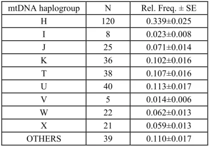
4 Therefore, in the course of evolution, inherited mutations have accumulated sequentially along mtDNA independent lineages. This inherited mtDNA variability has been extensively studied in human population genetics, representing an extraordinarily informative tool for the knowledge of human population history (Torroni and Wallace, 1994; Torroni et al. 1994). Indeed, groups of ancestral-associated polymorphisms (haplogroups) and cluster of these groups (haplogroup clusters) have provided additional insights about the origin and relationships of populations and the process of human colonization of continents, and have been used to define branches of the human phylogenetic tree for mtDNA (Torroni et al. 1996). The African haplogroup cluster L is the most ancient of all clusters, from which, approximately 65,000 years ago, have diverged two lineages (M and N) originating Asiatic and European haplogroups (Rose et al. 2002). As to European population, Torroni and coworkers (1996) found that about the 95% of the mtDNAs fall within nine different mtDNA haplogroup; of these, H is the most common once, followed by J, T, U, I, X, K, W and V.
The mtDNA inherited variants are likely to be non-neutral. In particular, several data have demonstrated that the different mtDNA lineages are qualitatively different from each other. In this field it is important to mention the study of Torroni and coworkers (1997) demonstrating that the penetrance of two primary mutations (11778 and 14484), which cause the Leber’s Hereditary Optic Neuropathy (LHON), is increased if they occurr on mtDNA molecules belonging to J haplogroup. In addition, a significant association has been found between common mtDNA polymorphisms and age-related pathologies, such as Parkinson and Alzheimer diseases (Ghezzi et al. 2005; Khusnutdinova et al. 2008; Maruszak et al. 2009; Takasaki, 2009).
Moreover, several studies have associated specific inherited variants of mtDNA with human longevity. For example, haplogroup J is over-represented in northern Italians,
5 Irish and Finnish centenarians (De Benedictis et al. 1999; Ross et al. 2001; Niemi et al. 2003). By contrast, the J haplogroup was underrepresented in southern Italians and in Chinese Uygur long-living people, supporting the idea that the effect of mtDNA inherited variants on longevity is population- and sex-specific, probably according to individual-specific nuclear genetic backgrounds and stochastic events (Dato et al. 2004; Ren et al. 2008).
Besides to this inter-individual variability, also an intra-individual mtDNA variability exists. Due to the multiple copy nature of mtDNA within a cell, somatic mutations in mtDNA molecules may coexist with the wild-type mtDNA, in a condition termed “heteroplasmy”.
1.1.1 Nucleus-mitochondria cross-talk
Although mitochondria contain their own genome, the vast majority of the mitochondrial proteins, including the entire complement of proteins involved in mtDNA replication and transcription, structural and transport proteins of mitochondrial membranes, the mitochondrial peptide involved in mitochondrial metabolism and TCA cycle, as well as most of the peptide subunits of the respiratory complexes (the protein-coding capacity of mtDNA is limited to 13 respiratory subunits) are encoded by nuclear DNA. Moreover, mitochondria are seat of multiple metabolic pathways, including β-oxidation of fatty acids and tricarboxylic acid and urea cycles, control intracellular Ca2+
metabolism and signaling, regulate thermogenesis and settle the cell fate by integrating numerous death signals. This implies that a sticky coordinated expression of two genomes occur to ensure the biosynthesis and the functional activity of mitochondria, in both physiological and pathological conditions (Garesse and Vallejo, 2001). Thus, a
6 complex interplay of bidirectional signaling linking nucleus and mitochondria emerges also by the crucial role played by mitochondria in cell physiology.
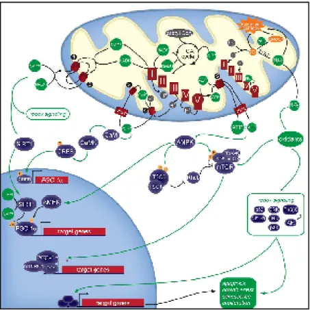
Signals from nucleus to mitochondrion are essential for maintaining an adequate mitochondrial structure and function. In fact, several nuclear-encoded transcription factors and co-activators have been identified as modulators of the mitochondrial replication and transcription. Two distinct classes of regulatory proteins regulate nucleus-mitochondria interactions at the transcriptional level. The first class comprises transcription factors that bind the promoter regions of both nuclear and mitochondrial genes. In particular, it is well known that the mitochondrial Transcription Factor A (Tfam) and the mitochondrial Transcription Factor B (mtTFB), isoforms TFB1M and TFB2M, work in conjunction with the mitochondrial RNA polymerase to confer promoter specificity and to enhance the rate of transcription initiation of mtDNA genes (Bonawitz et al. 2006; Scarpulla, 2006). Additionally, the second group of transcription factors, including the Nuclear Respiratory Factors NRF-1 and NRF-2 and the PGC-1 family coactivators (PGC-1alpha, PGC-1beta, and PRC), acts predominantly on nuclear genes whose products are required for respiratory chain expression and biological function (Fig. 3).
These findings indicated that nuclear signals are able to modulate the expression of both nuclear and mitochondrial genes in a coordinate way. However, mitochondrial activity also depends on a flow of information from mitochondria to nucleus. In fact, mitochondria are able to signal to the nucleus in response to metabolic disorders or damages that occur in mitochondria.
7 Fig. 3: Nuclear respiratory factors (NRF-1 and NRF-2) in the expression of nuclear genes governing mitochondrial respiratory function. NRFs act on the majority of
nuclear genes encoding for the subunits of the five OXPHOS complexes and on many other genes whose products direct the expression and assembly of the respiratory apparatus.
This signaling pathway, widely known as retrograde response (also referred to as mitochondrial stress signaling), is broadly defined as cellular response to changes in the functional state of mitochondria and results in wide-ranging changes in nuclear gene expression.
Most of the available data on the retrograde response mechanisms and function have been obtained by analyzing the consequences of mitochondrial dysfunctions in the budding yeast, Saccharomyces cerevisiae (Parikh et al. 1987). In particular, the decline of mitochondrial membrane potential, typical of yeast cells lacking of mtDNA (Rho0 petite), upregulates the Rtg pathway that, in turn, increases the expression level of several genes involved in biogenesis and function of mitochondria, including those of the Tricarboxylic Acid Cycle (TCA), the mitochondrial protein import and the
8 OXPHOS apparatus (Sekito et al. 2000; Traven et al. 2001). Moreover, it has been demonstrated that retrograde signaling is an important determinant of the yeast life span extension: the age-related accumulation of Extrachromosomal Ribosomal DNA Circles (ERCs) and their deleterious effects are mitigated by the retrograde response, with consequent increase of the lifespan (Jazwinski et al. 2005).
The mitochondrial retrograde response has been also documented in mammalian cells. Altered nuclear gene expression in response to mitochondrial dysfunctions has been extensively observed in different Rho0 cell lines (Marusich et al. 1997; Wang and Morais, 1997; Biswas et al. 1999).
Literature data have suggested that several forms of mitochondrial stress are able to activate different mitochondrial stress signaling (Fig. 4).
9 For example, the accumulation of mutant or unfolded protein in the mitochondrial matrix induces the activation of a mitochondrial Unfolded Protein Response (UPR), that results in the activation of mitochondrial chaperone and protease genes, including HSP60, HSP10 and ClpP, by the transcription factors CHOP and C/EBPβ thus reestablishing the normal cellular function (Zhao et al. 2002; Ryan and Hoogenraad, 2007). Experimental data suggest that the mitochondrial UPR is a two-stage regulatory process. First, the sensing of unfolded proteins in mitochondria leads to retrograde signaling to the nucleus and subsequent activation of CHOP gene. Second, CHOP, in conjunction to C/EBPβ, binds to target promoters and activates the transcription of mitochondrial-responsive genes.
Moreover, the mitochondrial release of molecules and/or metabolites, such as Ca2+, nitric oxide (NO), ROS and NAD+/NADH are able to activate several mitochondrial signaling pathways in response to a number of other types of mitochondrial stress such us alteration in energy production or structural damage.
Mitochondrial structural anomalies or disruption of OXPHOS machinery generally induce a drop in mitochondrial membrane potential (∆Ψm), that disrupts mitochondrial Ca2+ homeostasis and produce a rise in cytosolic free Ca2+. The latter event, in turn, activates calcineurin, several Ca2+-dependent kinases (PKC, JNK, MAPK, CAMKIV) and a wide spectrum of transcription factors, including NFAT, MEF2 and NF-KB, to induce transcriptional up-regulation and thus produce an appropriate cellular stress response (Butow and Avadhani, 2004; Mellström et al. 2008; Finley and Haigis, 2009). The mitochondrial loss of redox homeostasis and the resulting oxidative stress can also trigger signaling pathways. In this context, nitric oxide (NO) plays multiple effects. NO (or NO donors) have been found to increase the concentration of mitochondrial proteins and mtDNA in some cells in culture (Nisoli, 2003). NO stimulation of mitochondrial
10 biogenesis was apparently due to the cGMP upregulation of several transcription factors, including PGC-1 alpha (peroxisome proliferator-activated receptor gamma coactivator-1 alpha), NRF-1 (nuclear respiratory factor-1) and mtTFA (mitochondrial transcription factor A) (Nisoli et al. 2003). The role of NO in mitochondrial biogenesis has been also supported by several studies on model organisms. In particular, endothelial NO synthase (eNOS) knockout mice present lower mitochondrial levels in many tissues indicating that NO from eNOS physiologically regulates mitochondrial density in tissues (Nisoli et al. 2003). Moreover, calorie-restricted mice have increased eNOS expression and increased mitochondrial density and oxidative phosphorylation, and this increase was prevented in eNOS knockout mice (Nisoli et al. 2005). At moderate levels NO can also increase O2- and H2O2 production by inhibiting
mitochondrial respiration, while at higher levels it inhibits H2O2 production by
scavenging the precursor superoxide, resulting in peroxynitrite production (Brown et al. 2007). In this context it has been also reported that NO exerts a protective role against ROS and Reactive Nitrogen Species (RNS) damage by increasing the expression and the activity of the antioxidant protein Heme Oxygenase (HO) in several cell lines (Motterlini et al. 2002). It is important to note that the HO enzyme belongs to the family of Heat Shock Proteins (HSPs), which exerts a protective role in a wide variety of unfavorable conditions, including stress response (Calabrese et al. 2000).
Moreover, several transcription factors (AP-1, Sp-1, NF-KB) are redox sensitive, and the oxidation of conserved residues of these proteins by oxidants is able to induce changes in their transcriptional activity (Finley and Haigis, 2009).
Besides, based on the magnitude of the oxidative stress, pro-survival or pro-death pathways can be triggered directly by mitochondria (Finkel and Holbrook, 2000). In fact, when the stress response in not enough to counteract the intracellular impairment
11 principally due to a severe state of oxidative stress, mitochondria may directly influence cell viability and trigger both apoptotic and necrotic cell death (Gogvadze and Orrenius, 2006). In particular, a combination of increased mitochondrial Ca2+, intracellular
oxidative stress, ATP depletion, high inorganic phosphate (Pi) and mitochondrial
depolarization act synergistically to induce irreversibly the Mitochondrial Permeability Transition (MPT), a large conductance channel formed through a conformational change of several constituent proteins of mitochondrial membrane, including the Adenine Nucleotide Translocaton (ANT) in the IMM, the voltage dependent anion conductance (VADC) in the OMM and cyclophilin D in the mitochondrial matrix (Crompton, 2000). This process on the one hand could causes mitochondrial swelling, cytocrome c release, caspase activation and apoptotic cell death or, on the other hand, induces the collapse of the mitochondrial potential, ATP consumption and depletion and energetic collapse followed by necrotic cell death (Caroppi et al. 2009; Kitsis and Molkentin, 2010).
Additionally, the NAD+/NADH ratio, strictly connected to mitochondrial metabolism, can change the redox status and plays a critical role in various cellular functions, including regulation of calcium homeostasis and gene expression as well as in cell death by regulating numerous stress responders NAD+/NADH-dependent enzymes, such as dehydrogenases, poly(ADP-ribose) polymerases, Sir2 family proteins (sirtuins), mono(ADP-ribosyl)transferases, and ADP-ribosyl cyclases (Ying, 2006).
On the basis of the above consideration, the signaling network implemented by mitochondria brought out the idea that they can be considered as receiver/integrator organelles that play a pivotal role in cellular stress response and in determining the cell fate (Goldenthal and Garcia, 2004).
12 The mitochondria-nucleus signaling has been strictly correlated with mitochondrial and nuclear DNA variability in several age-related complex phenotypes (Gaweda-Walerych et al. 2008; Maruszak et al. 2009).
Several data have demonstrated that epistatic interactions with nuclear genetic background are a significant component of the mitochondrial genetics of aging. This epistasis may explain why some mtDNA mutations have very different phenotypic effects in different individuals, possibly obscuring the mtDNA effects in human aging and disease. A few examples of this epistatic interaction in humans are reported. In particular, De Benedictis and coworkers (2000), by analyzing the distribution of the mtDNA inherited variants by Tyrosine Hydroxylase (THO) genotypes in sample groups of increasing ages, observed a non-random association between the mtDNA and nuclear DNA variability in centenarians, and an over-representation of the U haplogroup in centenarians carrying a THO genotype unfavorable to longevity. Moreover, the penetrance of the maternally inherited deafness, associated with the A1555G mutation in the mitochondrial 12S ribosomal RNA (rRNA) gene, and of the Leber Hereditary Optic Neuropathy (LHON), a disease caused by missense mutations in the mitochondrial DNA (mtDNA), requires additional environmental or genetic changes for phenotypic expression so that the mitochondrial mutation appears to depend on additive effects of several nuclear genes (Bykhovskaya et al. 2000; Shankar et al. 2008). In the last case, linkage analysis in a large family harboring a homoplasmic G11778A mtDNA mutation on a haplogroup J background identified a novel LHON susceptibility locus on chromosome Xq25-27.2 (Shankar et al. 2008). These findings support the hypothesis that some human aging traits and diseases imply particular interactions between mtDNA and nuclear DNA.
13 Besides to the population data, in vitro models have been developed to understand how the inherited mtDNA variation can modulate cellular functionality. The best known of these models is represented by cytoplasmic hybrids also known as cybrids. Cybrid cell lines, first described by King and Attardi (1989), are engineered cells that share the same nuclear genome but have different mitochondrial genome. The preparation of cybrids starts from the creation of mtDNA-null cells (Rho0 cells) obtained by completely depleting cells of their own mitochondria though a long-term exposure to low concentration of Ethidium Bromide (EtBr). This compound inhibits mtDNA replication and transcription, without inducing any detectable effect on nuclear DNA division (King and Attardi, 1996). Rho0 cells are then repopulated with exogenous mitochondria derived from enucleated cells (often platelets) harboring particular type of mtDNA molecules. In this way it is possible to obtain different strain of cybrids with the same nuclear genome, that comes from the parental Rho0 cell and mtDNA of different sequence. By using this methodological approach it is possible clarify the influences that mtDNA variability has on the important cellular processes involving the mitochondrion-nucleus cross-talk. In particular, in cybrids with different mitochondrial genomes, the cell viability, the intracellular calcium dynamics, the mtDNA copy number, the mitochondrial reactive oxygen species (ROS) production, and the expression levels of several nuclear-encoded genes, including some cytokines, HSP60 and HSP75, have been demonstrated to be dependent by the interaction between nuclear and mitochondrial variability (Vives-Bauza 2006; Bellizzi et al. 2006, 2009; Kazuno et al. 2008; Suissa et al. 2009; Smits et al. 2010).
14 1.1.2 Mitochondrial role in aging
The role of mitochondria in the aging process has been a topic of intense interest for many years; in fact age-related changes in mitochondrial content, structure and function, as well as in mitochondrial DNA have been extensively documented.
A series of studies have demonstrated a decline of the mitochondrial respiration efficiency with age in human and primate and that this decline has been attributed either to a progressive down regulation of genes encoding for mitochondrial proteins such as several subunits of cytochrome-c oxidase, NADH dehydrogenase and ATP synthase, either to the decline of mitochondrial biogenesis with aging (Fernandez-Silva et al. 1991; Calleja et al. 1993; Barrientos et al. 1997; Welle et al. 2000; Short et al. 2005; Reznick et al. 2007).
It has been also well documented that the decline in OXPHOS activity correlates with a wide spectrum of mtDNA mutations, including point mutations, large scale deletions and duplications which progressively accumulate in post-mitotic tissues during human aging (Lee et al. 1994; Michikawa et al. 1999; Bua et al. 2006). For instance, an accumulation of mtDNA deletions has been found in skeletal muscle fibers deficient in electron transport activity in rodents and humans (Bua et al. 2006; Herbst et al. 2007). Studies on the substantia nigra neurons have shown high levels of somatic mtDNA deletions in both elderly control subjects and patients with Parkinson (Bender et al. 2006; Kraytsberg et al. 2006; Reeve et al. 2008). Moreover, an age-related decline in mtDNA content in skeletal muscle from mice and humans has been related to decreases in both mitochondrial ATP production rate and oxidative phosphorylation coupling (Short et al. 2005; Li et al. 2010).
Among the variety of mtDNA alteration, the most prevalent age-associated point mutations of mtDNA are A3243G and A8344G transition, while the most common
15 mtDNA deletion in aging human tissues is the 4977 bp deletion (Majamaa-Voltti et al. 2006; Pavicic and Richard 2009).
The relation between mtDNA mutation and aging phenotypes has been provided by several investigations using mtDNA mutator mice, that are knock-in mutant mice expressing a proofreading-deficient version of the mitochondrial DNA polymerase γ gene, that is the nuclear-encoded catalytic subunit of mtDNA polymerase (PolgA). Authors demonstrated that the accumulation of point mutations in mtDNA leads to the synthesis of respiratory chain subunits with amino acid substitutions that impair complex mitochondrial stability and cause progressive respiratory chain deficiency which, in turn, leads to premature aging (Trifunovic et al. 2004; Edgar et al. 2009). The large increase in somatic mtDNA mutations and the deficit in mitochondrial respiratory function have been extensively attributed to the progressive and irreversible accumulation of oxidative damage by Reactive Oxygen Species (ROS), a critical aspect of the aging process, as emerged in the mitochondrial theory of aging (Harman, 1972; 1973; Wei et al. 2001).
ROS are by-product of normal cellular metabolism; the mitochondrial respiratory chain is the major source of several ROS, including superoxide anions (O2-), hydrogen
peroxide (H2O2) and hydroxyl radicals (OH.), generated as by-products of cellular
energy. Moreover, ROS are produced by cytochrome P450 and peroxisomes metabolism, during the immune-inflammatory response, in the detoxification of xenobiotics and in response to several environmental agents, including γ-ray, ultraviolet light irradiation and non-genotoxic carcinogens (Franceschi et al. 2000; Inoue et al. 2003; Valko et al. 2006).
Mammalian cells possess a multi-level ROS defense network of enzymes, including superoxide dismutase (SOD), glutathione peroxidase (GPx) and catalase (CAT) that
16 cooperate to convert ROS into more stable molecules, such as water and O2. Besides
antioxidant enzymes, also non-enzymatic antioxidants, for instance ascorbic acid (Vitamin C), α-tocopherol (Vitamin E), glutathione (GSH) and carotenoids function as direct scavengers of ROS (Valko et al. 2007).
A growing body of evidences have shown that ROS play a dual role within cells (Valko et al. 2006). At low/moderate concentrations ROS have beneficial effects regulating the cell cycle, the processes of cellular defense against infectious agents and a number of cellular signaling pathways involved in oxidative stress response, in immune response and in apoptosis (Wojcik et al. 2010). In contrast, an excessive ROS production and/or a decline in the capacity of intracellular antioxidant defense result in the establishment of a state of oxidative stress that damages various cellular constituents, including proteins, lipids, and DNA. Generally, the ROS-induced protein modifications may change the structure and the catalytic activity of key enzymes whereas the lipid and DNA oxidation may alter the fluidity of membranes and the transcriptional processes respectively. Mitochondrial DNA is especially susceptible to attack by ROS, for several reasons: i) its close proximity to the electron transport chain, the major site of ROS production; ii) the size and the compactness of the genome; iii) the lack of protective histones; iv) the absence of adequate mitochondrial DNA repair systems. At the DNA level, ROS react with both purine and pyrimidine DNA bases, as well as the deoxyrybose backbone. The most studied mtDNA base lesion induced by oxidative stress in aging is the formation of 8-Hydroxyl-2’-deoxyguanosine (8-OH-dG) and an inverse correlation between the steady-state concentration of 8-OH-dG in mitochondrial DNA and the maximum lifespan (MLSP) of a wide range of mammalian species has been found (Barja and Herrero, 2000; Hamilton et al. 2001; Stevnsner et al. 2002).
17 As previously reported, mtDNA mutations accumulate progressively during lifetime and influence directly the cellular oxidative phosphorylation activity, thus leading to an enhanced ROS production. In turn, increased ROS production results in an increased rate of mtDNA damage and mutagenesis, thus causing a “vicious cycle” which ultimately culminates in cell death (Fig. 5).
Fig. 5: “Vicious Cycle” of mtDNA damage. ROS produced from the OXPHOS activity
damage mtDNA, thus altering mtDNA transcription and ETC activity, resulting in even higher levels of ROS production.
The link between cellular ROS and lifespan has been suggested by in vitro and in vivo studies in which the oxidant scavenging systems were enhanced. Indeed, Serra and coworkers (2003) found that the superoxide dismutase overexpression in human fibroblasts with low antioxidant capacity induce a decrease of the intracellular peroxide content, a slowdown of the telomere shortening rate, and an elongation of the life span of these cells, confirming also the causal role of oxidative stress for age-related telomere shortening. Similarly, Schriner and coworkers (2005), by using transgenic mice over-expressing human catalase observed a significant increase in mice lifespan,
18 associated to a lower susceptibility for mtDNA to ROS damage. Conversely, knockdown of SOD by using RNAi was demonstrated to induce cellular senescence through p53 (Blander et al. 2003). These results have provided strong support of the “Mitochondrial theory of aging”, reinforcing the role that oxidative stress has in mammals lifespan determination (Harman, 1972; 1973).
The oxidative stress hypothesis to explain senescence proposes that reducing the production of reactive oxygen species within the mitochondria concomitantly decreases their deleterious effects on survival. It has been shown that the magnitude of the proton gradient across the inner mitochondrial membrane is directly correlated with superoxide production by the electron transport chain (Korshunov et al. 1997); therefore, a possible method to altering free radical production may be manipulating the proton gradient across the mitochondrial inner membrane. Accordingly, in the last few years several evidences have demonstrated the emerging role of uncoupling proteins (UCPs) in this field of search. UCPs belong to a family of anion transporters located in the inner mitochondrial membrane and are involved in the uncoupling of respiration from energy production generating a leakage of protons into the mitochondrial matrix with heat production. This activity decreases the mitochondrial membrane potential and inhibits the generation of ROS. The role of UCP in modulating the mitochondrial ROS production has been suggested by a number of data confirming the “uncoupling-to-survive” hypothesis, proposed by Martin Brand (2000). As results, new items have emerged shedding light on a possible mechanisms implicated in the buffering of ROS and consequently in the process of aging (Feng et al. 2001; Holzenberger et al. 2003; Fridell et al. 2005; Andrews and Horvath, 2009). Interestingly, recent observations have suggested that the mitochondrial uncoupling process has similar effects of the caloric restriction, the sole known non-genetic enhancer of lifespan (Caldeira da Silva et al.
19 2008). Indeed, also the limitation in dietary calories uptake extends lifespan in several model organisms stimulating the respiratory rate, that, in turn, decreases the coupling between oxygen consumption and oxidative phosphorylation, supporting the central role for mitochondrial metabolism in the aging process (Xiao et al. 2004).
Aging is often associated with a sedentary lifestyle and it is known that if there are no demands for the extra energy that can be produced by aerobic oxidative phosphorylation, cells may down regulate the ETC components and survive adequately on glycolysis. Also the increased in sugar consumption may enforce the reliance on glycolysis (Johnson et al. 2009). As consequence, an epigenetic oxidative redox shift (EORS) has been recently proposed by Brewer (2010) to ensure ample supplies of NAD+ for glucose oxidation and maintain redox balance with impaired mitochondrial NADH oxidoreductase by upregulating other oxidoreductases. The activity of these other oxidoreductases has the 100% efficiency in generating oxyradicals (DeGrey, 2005). Thus, to avoid this catastrophic cycle, lactate dehydrogenase is upregulated at the expense of lactic acid acidosis. Overall, the oxidative redox shift is able to change the activity of numerous redox-sensitive transcription factors, including NF-KB, SP1, HOXB5 and USF and enzymes, including oxidoreductases and lactate dehydrogenase as previously reported. Also enzyme involved in the control of the epigenetic mark are reprogrammed. These includes histone acetylases, deacetylases (with their substrate requirement for NAD+) and methyltransferases. Together, these mediators impose the metabolic shift away from use of mitochondrial energy toward reliance on glycolysis. Thus, the EORS in aging results in a spiral of inability to respond to energy demands or stress which leads to stress-induced initiation of cellular death pathways and organ failure (Brewer, 2010).
20
1.2 Frailty as aging phenotype
As people age, they progressively accumulate impairment in multiple physiological systems, and become more prone to adverse health outcome. Thus, by analyzing the heterogeneity of health status amongst elderly people, a different degree of successful aging and pathological aging phenotypes can be observed, depending on the different capability that everyone has in counteracting extrinsic and intrinsic factors and restoring the physiological balance. Moreover, as a reflection of multisystem deficits, a wide range of semi-pathological (or intermediate) phenotypes, largely recognized as frailty, is also observed in population.
Frailty belongs to the family of geriatric syndromes, and therefore it can be considered as the resultant of a multidimensional interplay of genetic, biological, psycosocial and environment factors (Rockwood and Mitnitski, 2007). In fact, it has been defined as “a
state of increased vulnerability to stressors that results from decreased physiological
reserves and multi-system dysregulation, limited capacity to maintain homeostasis and
to respond to internal and external stresses. Frailty is an aggregate expression of risk
resulting from age- or disease-associated physiological accumulation of subtreshold
decrements affecting multiple physiologic systems resulting in adverse outcomes”
(Fried et al. 2004).
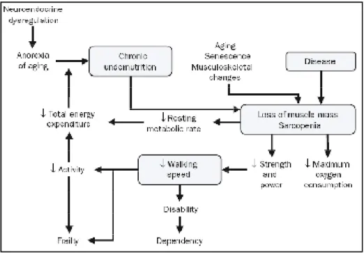
Current data has identified a wide range of geriatric phenotypes that are common in frailty syndrome, including weight loss and sarcopenia, muscle weakness with low grip strength, low activity level, exhaustion, low body mass index, blood pressure instability and balance and gait abnormalities (Fried el al. 2004; Bergman et al. 2007; Topinková 2008; Davis et al. 2010). Although this syndrome is generally due to the reduced levels of physical activity, it is also influenced by multisystem impairments including
21 immune/inflammatory, multiple hormonal and neuromuscular dysregulations, as well as metabolic and vascular alterations and oxidative stress that commonly occur with aging (Fig. 6) (Ferrucci et al. 1999; Leng et al. 2004; Schalk et al. 2004; Walston et al. 2006; Barzilay et al. 2007; Landi et al. 2008; Blaum et al. 2009; Hubbard et al. 2009; Desai et al. 2010; Evans et al. 2010; Hyde et al. 2010; Lustosa et al. 2010; Maggio et al. 2010).
Fig. 6: Frailty Cycle. This cycle combines elements such age-related musculoskeletal
changes, neuroendocrine dysregulations, diseases and nutrition into a patho-physiologic pathway (Singh et al. 2008).
Inflammation is one of the most recognized factors influencing frailty (Ershler and Keller, 2000). Hallmarks of inflammation, such as C-reactive protein (CRP), inflammatory cytokines and leukocytosis have been widely associated with aging and chronic age-related disease, including cardiovascular diseases (CVD), Alzheimer’s disease and diabetes (Lombardi et al. 1999; Alexandraki et al. 2006; Tuomisto et al. 2006). It has been also observed that the increase of the inflammatory markers could at
22 least partially explain the sarcopenia, the unintentional weight loss and the reduction of physical activity, that represents the most physiological characteristics of frailty (Pel-Littel et al. 2009). In particular, high serum levels of IL-6, TNF-α, and CRP are associated with mobility limitation, disability and mortality in older persons (Ferrucci et al. 1999; Penninx et al. 2004; Gallucci et al. 2007). What is more, stress-responsive genes in monocyte-mediated inflammatory pathway results significantly upregulated in the frailty syndrome (Qu et al. 2008).
Furthermore, several data associate the dysregulation of lipoprotein metabolism to inflammation and frailty of elderly individuals. Recently, elevated systemic levels of myeloperoxidase (MPO), a pro-oxidant enzyme that catalyzes the initiation of lipid peroxidation and affects nitric oxide levels, have been associated with unfavourable clinical outcomes in frail people (Giovannini et al. 2010). In addition, Landi and coworkers (2008) have demonstrated that high levels of high-density lipoprotein (HDL) cholesterol, a powerful anti-inflammatory agent, are associated with better survival, reporting that it can be considered as a reliable marker of frailty and poor prognosis among the oldest elderly. Moreover an interaction between the inflammatory system and the coagulation pathways has been suggested occurring in aging. In fact, procoagulant markers, including D-dimer, fibrinogen and factor VIII, increase in population with age and the incidence of venous thrombosis and pulmonary emboli increase sharply in geriatric population, and represent a major cause of morbidity and mortality in the elderly (Walston et al. 2002; Folsom et al. 2007; Tita-Nwa et al. 2010). This is noteworthy, because it could be explain: i) why comorbidities, like heart failure, myocardial infarction, peripheral vascular diseases, and hypertension increase the risk of frailty (Klein et al. 2005); ii) why the presence of a chronic inflammatory status links frailty to the development of several age-related diseases, such as atherosclerosis,
23 cardiovascular diseases, predementia syndromes, and diabetes (Libby, 2002; Panza et al. 2006; Fulop et al. 2006).
Like the inflammatory and the cardiovascular system, endocrine and neuroendocrine systems seem to influence both aging and frailty. In fact, the age-related decline of the adrenal androgen dehydroepiandrosterone-sulfate (DHEA-S), growth hormone (GH) and insulin-like growth factor (IGF-1) serum levels as well as the resulting reduction of their intracellular signaling efficiency has been broadly associated with the progressive loss of muscle mass, bone mineral density (BMD) and body mass index (BMI) (Perrini et al. 2010). Furthermore, signs of neurological dysfunction, often associated with reduced physical activity, falls and depression, are frequently observed in older people free of any form of neurological disease (Perrin et al. 1997; Panza et al. 2005).
Several genetic variants have been associated with the development of frailty. A significant association between frailty syndrome and polymorphisms of genes involved in vitamin B12 transport and metabolism, APOE gene, inflammation-related genes and mitochondrial DNA control region has been observed (Kulminski et al. 2008; Moore et al. 2010; Marioni et al. 2010; Matteini et al. 2010).
Recent investigations on the understanding the telomere biology have given rise the hypothesis that telomere shortening, that commonly occurs with aging, may form the biological basis for frailty. Although Cawthon and coworkers (2003) have demonstrated that telomere shortening in human beings contributes to mortality in many age-related diseases, no association between telomere length and i) frailty index (a parameter summarizing the physical, psychological, and functional deficits), ii) overall survival, iii) death from several cause (osteoporosis or fractures, infectious diseases, cancer, or cardiac and cerebrovascular diseases), has been observed (Woo et al. 2008; Sanders et al. 2009; Njajou et al. 2009). Thus, these findings suggest that although telomere length
24 could be considered a biomarker of cellular senescence, this relationship may not be extrapolated to the functional level represented by the frailty phenotype.
Lastly, lifestyle, including nutritional deficiencies or excesses, contributes to frailty (Khaw et al. 2008). Low serum levels of vitamins A, D, E, B(6), B(12), 25-hydroxyvitamin D and other micronutrients such as carotenoids, folate and zinc are associated with frailty amongst older adults (Semba et al. 2006; Wilhelm-Leen et al. 2010). The high incidence of type 2 diabetes in older people is usually due to excess of nutrients (Morley, 2000). In addition, a sedentary lifestyle and malnutrition inexorably reduces the mobility of aged people (Lee and Tanaka, 1997).
It is noteworthy that these physiological pathways are modulated in a complex manner with aging in response to genetic predispositions, diseases, reactive oxygen species production and mitochondrial dysfunctions. Indeed, as well as in aging, mitochondria influences the functional decline in later life, thus contributing to frailty.
As previously mentioned, sarcopenia is a prime characteristic of the frailty phenotype. This progressive atrophy in skeletal muscle is complex and has not been yet clearly defined, although recent evidences have indicated that in aging it occurs mainly through enhanced activation of apoptosis (Whitman et al. 2005; Dupont-Versteegden, 2005). Given the central role of mitochondria in oxidative stress and in regulating apoptosis, their integrity and functionality give a fundamental contribution to sarcopenia. In particular, Wanagat and coworkers, (2001) reported that reduction in the activity of Complex I and IV of the ETC, due progressive accumulation of mtDNA mutations, correlates with sarcopenia. More recently, Herbst and coworkers (2007) found that a progressive accumulation of mtDNA mutations occurs in muscle fibers with aging, inducing their dysfunction and breakage. Lastly, Moore and coworkers (2010)
25 demonstrated that some mtDNA polymorphisms in the DNA control region significantly correlate with frailty.
Also mitochondrial biogenesis decreases with aging, and the expression of the mitochondrial encoded ETC subunits decreases as well (Zahn et al. 2006; Reznick et al. 2007); these processes result in an overall loss of mitochondrial function determining a progressive loss of muscle mass (Dirks et al. 2006).
Besides the role played by mtDNA, mitochondrial ROS can results in more direct deleterious effects for cells; for example, an immediate consequence of oxidative damage is the oxidation of cardiolipine, a lipide of the inner mitochondrial membrane, that can directly promote the release of apoptogenic factors from mitochondria (Petrosillo et al. 2003; Gonzalvez and Gottlieb, 2007). Reactive oxygen species are also involved in the direct activation of inflammation via NFKB pathways (Kunsch and Medford, 1999).
Over the years, different multidimensional methodological approaches have been developed for establishing the presence of frailty and for regrouping subjects with homogenous phenotype. In general, these methods incorporates items of physical, cognitive and psycosocial signs, as well as diseases and disabilities, to classify the subject analyzed in a well defined phenotypic class of frailty. However, the wide heterogeneity in the quality of aging population that occur in different population as consequence of genetic variations and sociocultural differences, has revealed the need to develop population-specific models for monitoring aging (Jeune et al. 2006).
In recent years, many operational definitions of frailty have been proposed. Some of them consider frailty as a continuum accumulation of self-reported deficits, reflecting the proportion of potential disabilities present in a person (Mitnitski et al. 2002; Rockwood 2005, 2006); others, consider frailty as a distinct clinical syndrome non
26 synonymous of co-morbidity or disability and are based on direct measurement of functional parameters as grip strength, walking speed, unintentional weight loss and low physical activity (Fried et al. 2001). In this context, a novel methodology in frailty definition has been developed by Montesanto and coworkers (2010). In particular, well established geriatric parameters, including Mini Mental State Examination (MMSE), Hand Grip strength and geriatric Depression Scale (GDS), Activities of Daily Living (ADL) and Self-reported health status (SRHS) have been used as parameters in a hierarchical cluster analysis (CA), in order to define different aging phenotypes. The diagnostic and predictive soundness of this classification were confirmed by a detailed survival analysis showed higher survival chance for subjects characterized by lower frailty.
1.3 Aging epigenetics: DNA methylation
In the frame of the research on aging and longevity, an emerging field that promises exciting revelations about the determinants of cell senescence and organism aging is represented by the “Aging epigenetics”.
Epigenetics refers to the study of mitotically and, in some cases meiotically, hereditable changes of a phenotype that are unrelated to alterations in the DNA sequence. In vertebrates these changes are crucial for all the biological processes, as they regulate the expression of genetic information.
During the early stages of the life, the epigenetics status undergoes several changes to ensure an appropriate process of cell development and differentiation. However, non-random mechanisms such as environmental stimuli or stochastic errors in maintaining fixed the epigenetics patterns are able to induce changes in epigenetics profiles at both early and later in the life. Indeed, epigenetics anomalies have been found to be cause of
27 congenital disorders and multifactorial pediatric syndromes and adult-onset diseases as well (Schumacher and Petronis 2006; Vidal et al. 2007). Although epigenetics role in development, differentiation and pathological states has been extensively investigated, little is yet known about the relationship between epigenetics and aging.
The best known epigenetic modifications refer to DNA methylation and histone modifications, including methylation, acetylation, ubiquitylation and phosporylation. Both these processes are interdependent and cooperating in chromatin remodeling thus leading a dynamic regulation of gene expression in higher eukaryotic cells.
1.3.1 DNA methylation basic mechanisms
DNA methylation is a covalent biochemical modification that consists of the addition of a methyl group to the aromatic ring of a single DNA base. The methyl group protrudes into the major groove of the DNA double helix, preventing the binding of transcription factors that otherwise bind locally DNA sequences (Bell and Felsenfeld, 2000; Hark et al. 2000) and facilitating the binding of methyl-binding proteins (Jorgensen and Bird, 2002).
In eukaryotes, methylation occurs at 5-carbon position of deoxycytosine thus forming deoxymethylcytosine (dmC). In particular, in humans, this process accounts for 3-6% of the total cytosine (Callian and Feinberg, 2006).
In mammalian genome DNA methylation takes place predominantly when cytosine base is located 5’ to a guanosine, a so-called CpG dinucleotide (CpG) (Clark et al. 1995).
CpG dinucleotides are vastly under-represented as compared with what would be expected, probably because they act as a hotspot mutation. In fact, a depletion of about
28 20% of the expected frequency of CpG dinucleotides results from the process of deamination of cytosines in uracils that, in turn, are replaced by thymine after DNA replication, or from the direct hydrolytic deamination of 5-methylcitosine to thymine (Singal and Ginder, 1999) (Fig. 7).
Fig. 7: Molecular processes contributing to the under-representation of cytosine in the vertebrate genome. Both methylated and non methylated cytosines can be converted
to thymine by deamination processes (Liu et al. 2003)
Within mammalian genome about 70% of CpG dinucleotides are methylated. This process regards prevalently CpGs located into CpG-poor regions, localized in intergenic and intron region of genome. Furthermore, CpG methylated are localized in repetitive sequences, most of which derived from transposable elements, thus hindering the event of amplification and new insertion in the genome (Callian and Feinberg, 2006). On the other hand, most unmethylated CpG pairs are found in CpG-rich regions, termed CpG
islands, sequences of about 1 kb in length and having a CG content greater than 55%,
with an observed/expected CpG ratio of 0.65 (Jones, 1999; Takai and Jones, 2002). In the humans about 30,000 CpG islands were estimated. They are not distributed throughout the genome, but appear most often associated to the promoter regions and to the first exons of almost 60% of genes, including most housekeeping genes and half of all tissue-specific genes (Ioshikhes and Zhang, 2000).
29 In mammals, the process of DNA methylation takes places after DNA replication and is mediated by a family of DNA Methyltransferases (DNMTs) that includes DNMT1,
DNMT3a, DNMT3b and DNMT3L (Klose et al. 2006; Cheng and Blumenthal, 2008).
These enzymes catalyze the transfer of the methyl group from S-adenosyl-L-methionine (SAM) to deoxycytosine, producing 5-methylcytosine and S-adenosylhomocysteine (Bestor et al. 1988).
In particular, DNA methylation patterns are established during development by the de
novo methyltransferases DNMT3a and DNMT3b and their regulator DNMT3L, which
have high affinity for previously unmethylated DNA (Okano et al. 1999). Conversely, these patterns are replicated in somatic cells during mitosis by a semiconservative
maintenance DNA methyltransferase, DNMT1 (Fig. 8) (Bestor et al. 1988).
Fig. 8: DNA methylation reactions. De novo methyltransferases inserts methyl group
(CH3) in CpG sites that were not previously methylated. Once DNA methylation pattern are set by de novo methyltransferases and demethylases, they are maintained by DNMT1 during DNA replication. In absence of DNMT1, cells undergo to a passive DNA methylation (D’Alessio and Szyf, 2006).
30 DNMT2 is an highly evolutionary conserved member of methyltransferases. In particular this enzyme in Drosophila (dDNMT2), mouse (mDNMT2) and human (hDNMT2) contains the conserved methyltransferase motifs, and maintains the methyltransferase activity (Hung et al. 1999; Okano et al. 1999). However, recent studies have revealed that the primary target of DNMT2 proteins are non-CpG sites (Liu et al. 2003; Kunert et al. 2003); In fact, in human, DNMT2 does not methylate DNA but instead methylates a small RNA. In fact, mass spectrometry showed that DNMT2 specifically methylates cytosine 38 in the anticodon loop of the aspartic acid transfer RNA (tRNAAsp) (Goll et al. 2006).
The activity of DNMTs is crucial in establishing and maintaining methylation patterns; homozygous loss of DNMT1, DNMT3a or DNMT3b in mice results lethal (Okano et al. 1999; Li et al. 1992).
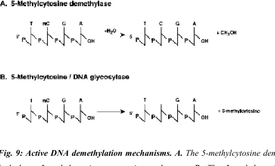
DNA methylation patterns are also regulated by DNA demethylation proteins, that operate by at least two different mechanisms:
i) Passive DNA demethylation
During DNA replication, symmetrically methylated strands of DNA in the parental chromosome segregate to daughter chromatids, thus containing hemimethylated DNA. Normally, the symmetric methylation is restored by the maintenance methyltransferase activity of DNMT1. However it is possible that regulatory nucleoprotein complexes are recruited in the sites of DNA methylation, preventing the access of DNMT1 to the methylation sites and thus leading to the progressive demethylation of DNA (Fig. 8) (Matsuo et al. 1998; Hsieh, 1999);