UNIVERSITÀ DEGLI STUDI DELLA TUSCIA DI VITERBO DIPARTIMENTO DI SCIENZE ECOLOGICHE E BIOLOGICHE
Corso di Dottorato di Ricerca in
Genetica e Biologia Cellulare - XXVII Ciclo
DOWN – rEgulation of the lamin a/C in neuronal
cells correlates with immature phenotype
(BIO/11)
Tesi di dottorato di:
Dr. Marta Nardella
Coordinatore del corso Tutore
The best way to know God is to love many things
Vincent Van Gogh
To my parents,
my sisters
and my grandparents
To my husband
and
my son
INDEX
ABSTRACT
I
1 INTRODUCTION
1
1.1 ARCHITECTURE OF THE NUCLEUS AND IMPLICATION
IN DISEASE
1
Nucleus and nuclear lamina
1
Nuclear lamins
2
Functions of Lamins in Nuclear and cellular architecture
5
Lamins during development and differentiation processes
8
Lamins and diseases
8
Lamins and cancer
10
1.2 NEUROBLASTOMA
11
Neuroblastoma as a sympathetic nervous system-derived tumor
12
1.3 CANCER STEM CELLS AND NB-TUMOR INITIATING
CELLS
16
The cancer stem cell theory
16
Isolation and characterization of neuroblastoma tumor initiating cells
18
1.4 microRNAS
19
miRNa biogenesis
20
miRNA may function as oncogenes or tumor suppressor genes in NB
22
2 AIM
24
3 MATERIALS AND METHODS
25
Human NB biopsy RNA extraction and real-‐time RT-‐PCR
25
Cell line maintenance and sphere formation
25
Primary cell coltures of Granule Cells
26
Generation of Lentiviral Infection of Granule Cells
26
Glutamate toxicity and neuronal survival
26
Colony-forming assay
27
Tumor sphere CD133 immunofluorescence
27
Immunoistochemistry
27
Western blot analysis
28
N-Myc FACS analysis
28
Side population
29
In vivo experiments
29
Histopathological analysis
29
Total RNA preparation
30
PCR Array
30
miRNA Assays
30
Hsa-miR-101 inhibitor and mimic
31
Real-‐time RT-‐PCR analysis
31
Statistical analysis
32
4 RESULTS
33
Abundance of LMNA in NB tumors is inversely correlated with that of
MYCN gene
33
LMNA gene knock-down induces a stem-like phenotype in SH-SY5Y
cells
40
LMNA-KD
sphere-derived
adherent
cells
maintain
stemness
The LMNA-KD sphere-derived adherent cell line is able to initiate tumors
in vivo
44
LMNA-KD
sphere-derived
adherent
cells
maintain
stemness
characteristics and acquire a more aggressive phenotype.
46
Lamin A/C increased during GCs maturation process both in vivo and in
vitro
48
Lamin A/C silencing prevents the complete maturation of GCs
50
5 DISCUSSION
52
6 BIBLIOGRAFY
55
ABSTRACT
Le lamine nucleari sono proteine che appartengono alla classe dei filamenti intermedi di tipo V. Esse formano una struttura reticolare al di sotto della membrana nucleare interna e svolgono diverse funzioni, alcune delle quali non sono state ancora del tutto chiarite. Le lamine di tipo A sono espresse nei tessuti differenziati e sono coinvolte nei processi differenziativi che avvengono durante lo sviluppo embrionale. La loro espressione risulta ridotta o assente in molti tumori umani, tuttavia il loro ruolo nella tumorigenesi non è stato ancora caratterizzato. La perdita della Lamina A/C è di particolare interesse, poiché la progressione del tumore è spesso associata ad una regressione dello stato differenziativo delle cellule tumorali.
Nel nostro laboratorio è stato precedentemente dimostrato che la Lamina A/C è necessaria per il differenziamento delle cellule di neuroblastoma e che la sua perdita è associata ad un aumento dell’aggressività tumorale. Il neuroblastoma (NB) è un tumore altamente aggressivo del sistema nervoso autonomo ed è il più comune tumore solido extracranico in età pediatrica.
Nonostante l’identificazione di marker molecolari - come l’amplificazione del fattore di trascrizione MYCN - si sia rivelata molto utile dal punto di vista della stratificazione dei pazienti e della realizzazione di protocolli terapeutici, sono necessari nuovi target molecolari per rendere maggiormente chiara l’eterogeneità e la progressione del tumore.
Sulla base dei dati precedentemente ottenuti e considerando che la Lamina A/C si esprime esclusivamente nei tessuti differenziati, l’obiettivo del lavoro è consistito nell’individuare il ruolo della Lamina A/C nei processi di maturazione delle cellule di origine neuronale. In particolare, è stato osservato che l’assenza della Lamina A/C predispone le cellule a un fenotipo staminale, favorendo così lo sviluppo di cellule capaci di iniziare il tumore con le caratteristiche di auto-rinnovamento.
Inoltre, prendendo in considerazione il ben noto marcatore del NB, MYCN - normalmente amplificato nei tumori poco differenziati che mostrano caratteristiche immature - abbiamo esaminato la possibile esistenza di una relazione inversa tra LMNA e MYCN nel NB.
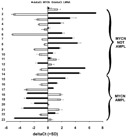
In questo studio abbiamo osservato una relazione inversa tra i livelli di espressione di LMNA e MYCN in 23 biopsie di NB. Utilizzando due modelli cellulari di NB che esprimono o LMNA o MYCN abbiamo dimostrato come questa correlazione inversa sia da attribuire ad un’alterazione del profilo di espressione di microRNAs che controllano la proliferazione e il differenziamento cellulare. Inoltre, abbiamo dimostrato che il silenziamento della Lamina A/C nelle cellule di NB SH-SY5Y comporta lo sviluppo di una popolazione di cellule inizianti il tumore (TIC), ovvero una
piccola popolazione di cellule responsabili della crescita del tumore, delle metastasi e delle recidive, nota come popolazione di cellule staminali tumorali. Tali cellule hanno molte caratteristiche specifiche delle cellule staminali - come la capacità di formare sfere e di espellere coloranti - ed esprimono marcatori di cellule staminali: POU5f, che codifica Oct4, Nanog, SOX2, nonché PROM-1, che codifica per CD133, e ABCG2. Inoltre, questa popolazione cellulare – diversamente da quella di origine – presenta un elevato potenziale tumorigenico in quanto le cellule sono in grado di generare un tumore quando inoculate in topi nudi. La caratterizzazione di NB TIC comporta un passo cruciale per il miglioramento delle terapie antitumorali.
Inoltre, come osservato nelle biopsie di NB, lo sviluppo di una popolazione di TIC in linee di NB correla all’incremento dei livelli di espressione del gene MYCN, come osservato in queste cellule. Infatti, sovraesprimendo MYCN, le cellule di controllo SH-SY5Y acquisiscono un fenotipo con caratteristiche staminali; al contrario, regolando negativamente i livelli di espressione di MYCN nelle cellule SH-SY5Y silenziate per Lamina siamo stati in grado di ripristinare il fenotipo più maturo.
Coerentemente con i nostri dati nei modelli di NB, il silenziamento della Lamina A/C nelle colture primarie di cervelletto di ratti neonati blocca la morte neuronale glutammato-mediata, che rappresenta un segnale della completa maturazione dei granuli cerebellari. Tale risultato rafforza l'ipotesi che l’espressione della Lamina A/C sia una caratteristica esclusiva delle cellule che iniziano il processo di maturazione e di quelle terminalmente differenziate.
Questo lavoro di tesi fornisce un modello di sviluppo di una popolazione di TIC nel neuroblastoma. Strategie mirate a colpire la popolazione di cellule staminali all’interno del tumore rappresentano una sfida per la riduzione della massa tumorale e/o dell'incidenza di recidive. Questi studi potrebbero consentire lo sviluppo di protocolli terapeutici personalizzati sulla base del profilo molecolare del singolo tumore.
Nuclear lamins are type V intermediate-filament proteins that form a meshwork-like scaffold underlying the inner nuclear membrane providing mechanical support to the nucleus. It is now clear that lamins exert many different functions within the cell, most of them still largely unknown. Among lamins, the A-type ones (Lamin A/C) are expressed in differentiated tissues and are involved in differentiation processes during the embryonal development. Their expression is reduced or absent in several human malignancies, even though their role in the tumorigenesis has not been characterized yet. It is widely accepted that tumor progression is often associated with regression from a more differentiated to a less differentiated state, therefore loss of Lamin A/C expression is of particular interest.
We previously demonstrated that Lamin A/C is necessary for the acquisition of a differentiated phenotype in neuroblastoma cells and its loss is associated to an increased cell aggressiveness.
Neuroblastoma (NB) is a highly aggressive embryonic tumor originating from the autonomic nervous system and represents the most common extracranial solid cancer in childhood. Despite the identification of molecular markers, such as MYCN amplification, turned out to be greatly useful in terms of patients’ stratification and therapeutic protocols implementation, new molecular targets are required to clarify the heterogeneity and progression of this tumor.
Thus, based on our previous results and considering that Lamin A/C expression uniquely in differentiated tissues, we intended to examine a possible role of Lamin A/C in the maturation of cells of neuronal origin. In particular, we reasoned that NB differentiation impairment in the absence of Lamin A/C was mainly due to the acquisition of a stem-like phenotype and a concurrent development of tumor-initiating cells with self-renewal features. In addition, considering the well-known hallmark of NB MYCN, normally amplified in poorly differentiated tumors displaying immature characteristics, we intended to investigate whether an inverse relationship between LMNA and MYCN gene could exist in NB.
In this study, we observed an inverse relationship between LMNA and MYCN expression levels in 23 biopsies of NB. Using two NB cellular models, expressing LMNA or MYCN alternatively, we demonstrated that this inverse correlation is due to changes of members of miRNA signatures controlling cell proliferation and differentiation. Moreover, we provide evidence that the down regulation of Lamin A/C expression in the SH-SY5Y NB cells allows the development of a tumor-initiating cells (TIC) population, a rare tumorigenic cell population known to be responsible for sustaining tumor growth, metastases and relapse, and usually named cancer stem cell population. Stem cell characteristics, such as sphere-forming, dye exclusion ability and an increased expression of the stem cell regulatory network (POU5f, which encodes Oct4, NANOG and SOX2) as well as of PROM-1, encoding for CD133, and ABCG2 were detected in our TIC population.
Furthermore, TIC-derived tumors obtained after TIC cells implantation in nude mice showed a more malignant phenotype with respect to the parental cell line. The characterization of NB TICs may be a crucial step for the improvement of antitumoral therapies.
In agreement with the aforementioned NB biopsies, the development of a TIC population in NB correlates with the increased expression of MYCN gene observed in these cells. Indeed, by up-regulating MYCN, control cells acquired a stem-like phenotype; while down-up-regulating MYCN in the LMNA silenced cells, we were able to restore a more mature phenotype. Consistently with our data in the NB models, we also showed how Lamin A/C knock down in primary cells explanted from the cerebellum of neonatal rats prevents glutamate-mediated neuronal death which is a signal of the complete maturation of granule cells. This evidence strongly supports the idea that Lamin A/C expression is an exclusive characteristic of the cells that begin the maturation process and terminally differentiate.
This thesis work provides new mechanistic insight into the development of a stem-like NB TIC population. Targeting of the TICs in a tumor represents a challenge for the reduction of the tumor mass and/or the incidence of tumor recurrences. Therefore, our data may provide opportunities to develop new personalized therapeutic strategies based on the molecular profile of the tumor.
1 INTRODUCTION
1.1
ARCHITECTURE
OF
THE
NUCLEUS
AND
IMPLICATIONS IN DISEASE
Nucleus and nuclear lamina
The nucleus is the defining feature of eukaryotic cells and is separated from the cytoplasm by the nuclear envelope (NE). The NE is composed of three distinct elements, i.e., the nuclear membrane, nuclear pore complexes, and the nuclear lamina. The nuclear membrane is a double-unit nuclear membrane, in which the outer nuclear membrane (ONM) is continuous with and shares biochemical and functional properties with the endoplasmic reticulum (ER). In contrast, the inner nuclear membrane (INM) is distinct from both the ONM and ER and is defined by a subset of integral membrane proteins, termed nuclear envelope transmembrane proteins (NETs) that are anchored to the INM during interphase. The ONM and INM are separated by a luminal space of 100 nm in width (perinuclear space). The nuclear membrane is punctuated by nuclear pore complexes (NPCs), which regulate the passage of macromolecules between the nucleus and the cytoplasm. At NPCs the ONM and INM converge at the so-called pore membrane, which again is defined by its own subset of integral membrane proteins. Underneath the INM is the nuclear lamina (NL). The NL is a polymeric protein meshwork of 10 nm filaments. It is a complex network of type V intermediate filaments (IF) proteins, the nuclear lamins, and inner nuclear membrane–associated proteins. The NL provides the mechanical and structural support for the nuclear membrane and anchoring sites for chromosomes and nuclear pore (Gerace and Burke, 1998).
Schematic representation of nucleus structures.
http://www.slideworld.org/slideshow.aspx/Cytoskeleton-2865193
Nuclear lamins
The major components of the NL are type V intermediate filament (IF) proteins, the nuclear lamins. Like all proteins, they are synthesized in the cytoplasm and then transported into the nucleus, where they are assembled before being incorporated into the existing network of NL (Aebi et al., 1986).
Lamins are divided into A and B types based on sequence homologies. A-type lamins are 70 and 60 kilodaltons (Gerace and Blobel, 1980; Goldberg et al., 2008), are encoded by the LMNA gene located on chromosome 1q21.2-q21.3 (Wydner et al., 1996; Lin and Worman, 1997) and are generated by alternative splicing to produce different isoforms, lamins A, A∆10, C and C2, which are all found in different cell types (Foster and Bridger, 2005; Martin et al., 2009). Lamin C2 is restricted only to spermatocytes during rat spermatogenesis whereas, A∆10 is found only in a few carcinoma cell lines (Alsheimer and Benavente, 1996). A∆10 results from the deletion of exon 10 (Hutchison, 2002) and is localised at the nuclear envelope (Broers et al., 1999).
The LMNA gene contains 12 exons. Exon 1 codes for the N-terminal head domain and the first part of the central rod domain of this gene. Exon 2-6 code for the central α-helical rod domain. Exons 7-9 code for the C-terminal tail domain for lamin A and lamin C. The nuclear localisation
signal (NLS) is contained in exon 7. Exon 10 contains a splicing site to generate lamin A and lamin C. Exon11 and 12 are specific for lamin A and coding for the CAAX box of pre-lamin A (Lin and Worman, 1993).
There are three types of B-type lamins (67 kilodaltons) in mammals: lamin B1, lamin B2 and lamin B3. Lamins B1 and lamins B2 are regulated and expressed in both embryonic and somatic cells during development whereas lamins B3 is regulated and expressed only in spermatocytes (Gerace and Blobel, 1980; Hutchison, 2002). Lamin B1 is encoded by LMNB1 and lamin B2 and lamin B3 are encoded by LMNB2 genes on chromosomes 5q23.2-q31.3 and 19p13.3, respectively (Lin and Worman, 1995; Dechat et al., 2008). LMNB1 gene contains 11 exons. Exon 1 codes for the N-terminal head domain and the first part of the central rod domain of this gene. Exon 2-6 code for the central α-helical rod domain. Exons 7-11 code for the C-terminal tail domain of lamin B. The nuclear localisation signal (NLS) is contained in exon 7. Exon11 contains a specific sequence for the CAAX box lamin B for post-translational farnesylation (Lin and Worman, 1993).
Little is known about the regulation of lamins gene expression. Some information are available on the regulation of LMNA gene expression. LMNA promoter presents different regulatory motifs among which a retinoic acid-responsive element (Okumura et al., 2000), binding sites for various transcription factors such as c-Jun, c-Fos and Sp1/3 (Okumura et al., 2004) or transcriptional coactivator such as CREB-binding protein (Janaki and Parnaik, 2006). Within the first intron of LMNA gene there are also binding sites for two transcription factors, the hepatocyte nuclear factor-3β and the retinoic X receptor β (Arora et al., 2004).
The nuclear lamins have a well-defined conserved domain structure consisting of an amino (N)-terminal globular domain, a central α-helical rod comprising four coiled-coil domains separated by linker regions L1, L12 and L2, and a globular carboxy (C)-terminal tail domain. Like the components of other intermediate filaments, the lamin alpha-helical domain is used by two monomers to coil around each other, indeed forming a dimer structure called “coiled coil”. Their assembly starts with the formation of coiled coil dimers which is associated with head to tail overlap. Two of these dimer structures then join side by side, in an antiparallel arrangement, to form a tetramer called “protofilament”. Eight of these protofilaments are laterally combined and twisted to form the characteristic 10 nm intermediate filament structure. These filaments can be assembled or disassembled in a dynamic manner. Although A- and B-type lamins interact each other in vitro (Sasse et al., 1998), little is known about their composition and structure in the cells within the lamina.
Schematic structure of lamin proteins.
Main characteristics are four central rod domains (1A, B, 2A, B), flanked by a globular head and a globular tail domain. In the globular tail domain, a nuclear localization signal (NLS) can be identified, as well as a CaaX motif, which is absent in lamin C but present in lamin A and in B-type lamins. Broers et al., Physiol Re, 2006.
The globular COOH-terminal domain of lamins shows an Ig-like structure. Between the COOH-terminal part of the rod domain and the Ig-like domain, lamins contain a nuclear localization signal sequence, not present in other IF proteins (Frangioni et al., 1993). Lamins, with the exceptions of lamins C and C2, are expressed as a pre-form and end in a C-terminal CAAX cassette which undergoes a highly structured sequence of posttranslational modifications before mature lamins are incorporated into the nuclear lamina. The CAAX cassette consists of a cysteine, two aliphatic amino acids and any COOH-terminal amino acid (Davies et al., 2009). The maturation process starts with farnesylation at the terminal cysteine site and is followed by cleavage of the last three amino acid residues of the CAAX motif and methylation of the carboxy-terminal cysteine. While the maturation of B-type lamins is terminated at this step, resulting in permanent farnesylation and carboxymethylation, an additional 15 amino acids are removed from the carboxyl terminus of farnesylated/carboxymethylated prelamin A. The last cleavage probably takes place during or after incorporation of this molecule into the nuclear lamina.
Besides farnesylation and carboxymethylation, during mitosis lamins are also posttranslationally modified by phosphorylation (Ottaviano and Gerace, 1985), sumoylation (Zhang and Sarge, 2008), ADP-ribosylation (Adolph et al., 1987), and possibly by glycosylation (Ferraro et al., 1989). In fact, during interphase the lamins are localised in the nuclear envelope resulting in a rim at the nuclear periphery and become dephosphorylated and polymerised at the nuclear periphery during telophase (Burke and Gerace, 1986; Foisner and Gerace, 1993).
Post-translational processing of the carboxyl terminus of prelamin A.
Processing of prelamin A to mature lamin A involves several steps, including farnesylation at the terminal cysteine site, followed by cleavage of the last three COOH-terminal amino acid residues of the CAAX motif, in this case SIM, probably by Zmpste24; methylation of the cysteine that is COOH terminal after cleavage, followed by a second cleavage of the last 15 amino acids, including the newly added isoprene group. The last cleavage probably takes place during or after incorporation of this molecule into the nuclear lamina. Lamin C is not processed, whereas B-type lamins are farnesylated but not further processed. Broers et al., Physiol Re, 2006.
Functions of Lamins in Nuclear and cellular architecture
While the most obvious localization of lamins during interphase is at the nuclear periphery, a growing number of studies suggest that in interphase cells lamins can be found in nucleoplasmic areas as well. Three levels of lamin organization can be distinguished:
1) lamins associated with the nuclear membrane,
2) lamins organized into intranuclear tubules and aggregates,
3) lamins visible as dispersed (veil-like) structures in the nucleoplasm.
The roles of lamins are mediated by interactions with numerous lamin binding proteins both at the nuclear periphery and/or in the nucleoplasm. The lamins and associated NE proteins are scaffolds for proteins that regulate DNA synthesis, gene transcription, responses to DNA damage, cell cycle progression, cell differentiation, and cell migration (Broers et al., 2006; Verstraeten et al., 2007; Razafsky et al., 2014). There are many INM proteins binding to lamins either directly or indirectly. For example, INM proteins such as emerin, MAN1, LBR, LAP1, LAP2β, nesprin, otefin, and SUN1 have been demonstrated to bind lamins (Foisner, 2003). Lamins can also bind non
integral proteins including chromatin, histones (H2A/H2B; Höger et al., 1991), transcription factors such as E2F, RNA polymerase II transcription machinery (Mattout-Drubezki and Gruenbaum, 2003), pRb (Dorner et al., 2007), BAF (Barrier to Autointegration Factor; Furukawa, 1998), LAP2α, extracellular signal-regulated kinase (Erk), nuclear actin, and proteins of the nuclear pore complex such as nucleoporins (e.g. Nup153) (Vlcek et al., 2001; Mattout-Drubezki and Gruenbaum, 2003; Dechat et al., 2008; Olins et al., 2010). The actual in vivo interactions between lamins, DNA, chromatin and the INM remain unclear. However, interaction must be specific and reversible to allow nuclear growth and disassembly during mitosis. After mitosis and during interphase, the reorganization of nuclear lamina and its assembly at the nuclear periphery is elicited by the IMPs. Most of these proteins can bind directly to lamin A, lamin B or both (Wilson and Foisner, 2010).
Model of the location of nuclear lamins and their interaction with nearby localized proteins.
Lamins bind directly to lamina-associated proteins (LBR, LAP2, emerin, MAN1, nesprins-1 and -2), but also to BAF, Rb, SREBP1, histone proteins, and DNA, and thereby mediate association with a scale of interacting structural proteins, including SUN1, actin, and possibly tubulin and intermediate filament proteins. Broers et al., Physiol Re, 2006.
Lamins/NET complexes have at least three clearly defined or emerging functions in maintaining cellular architecture. Lamin/NET complexes are important for organizing peripheral chromatin. The interaction between the lamins and chromatin involve their non-α-helical C-terminal tail domain and the N- and C-terminal tail domains of core histones (Mattout et al., 2007; Goldberg et al., 1999). Several proteins have been also described as lamin-binding proteins, which directly link both A- and B-types lamins to DNA (Schirmer and Foisner, 2007). Among these proteins are a Lamin B Receptor (LBR; Ye and Worman, 1994), the LAP2α protein, that binds specifically to Lamin A/C, the LAP2β protein, that interacts exclusively with B-type lamins (Dechat et al., 2000; Furukawa and Kondo, 1998), and BAF, which has been reported to bind to dsDNA and to histones (Margalit et al., 2007).
The lamins also have important functions in positioning NPCs within the NE, and have a crucial role in organizing the cytoskeleton. Specifically, the connection between the cytoplasm and nucleoplasm might be mediated by the interaction between SUN proteins (SUN1 and SUN2) and nesprins (nesprin-1, nesprin-2, and nesprin-3) in the luminal space (Padmakumar et al., 2005; Ketema et al., 2007). In the nucleoplasm Sun proteins interact with Lamin A (Haque et al., 2006), while in the cytoplasm, nesprins are thought to bind to actin (Warren et al., 2005) and possibly microtubules (Wiche, 1998). This assembly is referred to as the LINC complex (LInker of Nucleoskeleton and Cytoskeleton) and establishes a physical connection between the nucleoskeleton and the cytoskeleton (Crisp et al., 2006).
A model for the LInker of Nucleoskeleton and Cytoskeleton (LINC) complex.
Nuclear components, including lamins, bind to the inner nuclear membrane SUN domain proteins, which in turn bind to the KASH domain of the actin-associated giant nesprins on the outer nuclear membrane. The LINC complex establishes a physical connection between the nucleoskeleton and the cytoskeleton. Meinke et al., Biochem Soc Trans., 2011.
Lamins during development and differentiation processes
Different expression between nuclear Atype lamins and B-type lamins support the idea of their alternative functions during development and differentiation. The absence of lamin A/C in undifferentiated cells is responsible for the deformation of their nuclei. For example, lamin A is present in late embryos but not expressed in blastocysts whereas lamin A/C is expressed in oocytes (Prather et al., 1989; Bridger et al., 1993). Foster et al., in 2007, have shown that A-type lamins and B-type lamins are present at the nuclear envelope in early porcine embryos and that lamin A is also found in large intranuclear aggregates in two-cell to eight-cell embryos but is lacking in later embryonic stages (Foster et al., 2007). The regulated expression of A- and B-type lamins is also evident during differentiation of stem cells in culture. In fact, during the in vitro differentiation processes, human ES cells appear to express Lamin A/C before a complete down-regulation of the pluripotency marker Oct-4, suggesting that Lamin A/C expression is an early indicator of ES cell differentiation (Constantinescu et al., 2006). Differential expression of A- and B-type lamins has also been shown during neurogenesis in the adult rat brain (Takamori et al., 2007).
The developmental regulation of Lamin A/C expression has led various laboratories to hypothesize that these proteins play a role in differentiation. This has been recognized in muscle and adipocyte differentiation processes (Favreau et al., 2004, Frock et al., 2006, Lloyd et al., 2002; Hubner et al., 2006).
Lamins and diseases
Many aspects of nuclear as well as cytoplasmic activity are affected by modifications of the NE and the nuclear lamina. Processes as fundamental as DNA replication, transcription, and cell survival are altered in response to disruption of nuclear lamina, overexpression of mutant or truncated lamins, and loss-of-function mutations of the LMNA gene. Given the diversity of functions affected by these alterations, it is not surprising that complex patterns of tissue-specific pathologies are associated with lamin defects in humans. At present, no mutations in the Lamin B genes have been linked with human diseases, thus presuming that mutations in B-type lamins could be embryonic lethal. Conversely, A-type lamins have been in the limelight since the discovery that LMNA mutations or defective post-translational processing of pre–lamin A causes the majority of human diseases termed laminopathies, which include systemic disorders and tissue-restricted diseases (Capell and Collins, 2006; Verstraeten et al., 2007).
Classification and clinical phenotype of laminopathies
To date, over 200 mutations from more than 1000 individuals have been identified in the LMNA gene, and a database on the nuclear envelopathies can be found at http://www.umd.be.
LAMINOPATHIES CLINICAL MANIFESTATIONS
Systemic
HGPS Premature aging, hair loss, loss of subcutaneous fat, premature
atherosclerosis, myocardial infarction, stroke
Atypical Werner's syndrome
Premature aging, cataracts, scleroderma-like skin changes, premature atherosclerosis, hair graying
Restrictive dermopathy
Intrauterine growth retardation, skin alterations, multiple joint contractures, skull defects
MAD Skull/face abnormalities, clavicular hypoplasia, joint
contractures/lipodystrophy, alopecia, insulin resistance
Tissue restricted
EDMD
Early contractures of the neck/elbows/Achilles tendons, muscle contractures, wasting of skeletal muscle, cardiomyopathy with conduction disturbance
DCM Ventricular dilatation, systolic dysfunction, arrhythmias, conduction
defects
Limb-girdle
muscular dystrophy 1B
Slowly progressive shoulder and pelvic muscle
weakness/wasting,contractures, cardiac defects
Charcot-Marie-Tooth neuropathy type 2B1
Axonal degeneration, lower-limb motor deficits, walking difficulty, secondary foot deformities, reduced/absent tendon reflexes starting in the second decade of life
Dunningan-type FPLD
Dramatic absence of adipose tissue in the limbs/trunk and accumulation in the neck/face, hypertriglyceridemia, increased susceptibility to atherosclerosis/diabetes
Different mutations in LMNA gene are associated with different diseases.
Most of the mutations resulting in the diseases affecting striated muscles are distributed throughout the gene. The majority of the mutations that result in Mandibuloacral disease, Dunnigan-type familial partial dystrophy, and Hutchinson Gilford Progeria syndrome cluster in the region of the LMNA gene encoding the C-terminal globular domain.
Lamins and cancer
Alterations in the nuclear lamina proteins are thought to be involved in malignant transformations and cancer processes because of their role as guardian of the genome, their role in regulating basic nuclear activities that are implicated in tumorigenesis, their interactions with cancer gene pathways and their role in chromosomal reorganization. Thus, lamin expression in cancer cells may serve as a biomarker for diagnosis, prognosis and tumor surveillance. The expression of the A-type lamins is often reduced or absent in various A-types of cancer and varies widely depending on the type of cancer, its aggressiveness, proliferative capacity and degree of differentiation. Changes in A-type lamins have been well documented in the current literature concerning various tumor types such as leukemia, lymphomas, some skin cancers, including basal cell and squamous cell carcinoma, adenocarcinoma of the stomach and colon, squamous and adenocarcinoma of the
1990; Broers et al., 1993; Moss et al., 1999; Venables et al., 2001; Oguchi et al., 2002; Coradeghini et al., 2006). In ovarian cancer cells, VEGF promotes the invasion, partially via the down-regulation of Ezrin and Lamin A/C caused by increased expression of miR-205 (Li et al., 2014). We have previously demonstrated that knockdown of Lamin A/C in human neuroblastoma cells inhibits retinoic acid-mediated differentiation and results in a more aggressive phenotype (Maresca et al., 2012).
In general, the expression of A-type lamins has been correlated with a non-proliferating, differentiated state of cells and tissues (Tilli et al. 2003). Given that tumor progression is often associated with regression from a more differentiated to a less differentiated state, loss of Lamins A/C expression may not be surprising. Even though the altered lamins expression are currently emerging as an additional event involved in malignant transformation and tumor progression, the role of A-type lamins in the tumorigenesis is not yet characterized and the molecular defects underlying the loss of A-type lamins in human cancer remain unknown.
Agrelo et al. (2005) has shown that CpG Island promoter hypermethylation of the LMNA gene is a significant predictor of poor outcome in some lymphomas (Agrelo et al., 2005). Cancer may also be considered an epigenetic disease and patterns of aberrant DNA methylation are now recognized to be a common hallmark of human tumors. Since A-type lamins present an important dual role as protector of chromatin from damage and as multifunctional regulators of gene transcription, the epigenetic silencing of LMNA gene in hematologic malignancies could aid understanding how lamin dysregulation could contribute to cellular transformation.
1.2 NEUROBLASTOMA
Neuroblastoma (NB) is an extra-cranial heterogeneous tumor of the sympathetic nervous system (Esiashvili et al., 2009). The first description of pediatric tumors under the term “neuroblastoma” was made by Dr. James Homer Wright of the Massachusetts General Hospital as early as 1910 (Modak et al., 2010). Today approximately 120 new cases are diagnosed in Italy each year, rendering this tumor the most common extra-cranial pediatric cancer. In 95% of cases, NB is diagnosed before the age of 5 years where it leads to 15% of all cancer-related fatalities in infancy and childhood (Mueller et al., 2009). Despite a century of extensive clinical and basic research efforts, the biology and clinical progression of the disease continues to present a multi-faceted clinical enigma in pediatric oncology. The most common site for primary NB tumors is the adrenal medulla; however, tumors can arise anywhere along the sympathetic branch of the autonomic
nervous system (Alam et al., 2009). At presentation, the disease can be limited to a single organ, locally or regionally invasive, or widely disseminated; more than 50% of cases are metastatic at presentation (Alam et al., 2009). The most common metastatic sites are lymph nodes, bone marrow, bone, and liver (Alam et al., 2009).
The clinical behavior of NB is highly variable, ranging from highly aggressive phenotypes to benign tumors with a high propensity for spontaneous regression (Brodeur, 2003). Intriguingly, NB is both disproportionally lethal despite very aggressive multimodal therapy and associated with a highest rate of spontaneous and complete regression in a subset of cases (Alam et al., 2009; Brodeur et al., 2003; Mueller et al., 2009).
Three broad risk categories (low, intermediate, and high risk) have been proposed on the basis of analysis of age at diagnosis, histology category, grade of tumor differentiation, DNA ploidy, and copy-number status at the MYCN oncogene locus at chromosome 11q. In particular the MYCN oncogene is target of high-level amplifications at chromosome band 2p24 observed in about 20% of NB and since such amplification has been found to profoundly affect the patients’ clinical outcome, it is routinely used as prognostic biomarker and for treatment stratification (Seeger et al. 1111-16). MYCN belongs to a family of oncogenes called Myc oncogene-family members, whose most studied member is the MYC oncogene and include also MYCL oncogene. They are transcription factors, which activate, together with their dimerization partner Max, gene expression of a number of genes in an E-box dependent manner. MYCN and MYC probably have very similar molecular functions (Bonnet et al., 1997). The Myc oncogene-family members are involved in cell growth through protein synthesis, transcriptional regulation of ribosomal RNA processing, cell adhesion and tumor invasion.
Neuroblastoma as a sympathetic nervous system-derived tumor
The human nervous system consists of the Central Nervous System (CNS), comprising the brain and the spinal cord, and the Peripheral Nervous System (PNS), which links the CNS with the body’s sense receptors, muscles, and glands. The PNS is divided in two components: the somatic or skeletal nervous system, which controls voluntary movement, and the autonomic nervous system, which regulates inner organ function via the sympathetic, parasympathetic or enteric ganglia.
The nervous system originates from the neural plate, an embryonic structure evolving from the ectodermal germ layer during the third week of gestation. During the process of neurulation, the neural plate invaginates ventrally and closes in order to form the neural tube, which will give rise to
the CNS. During this closure, neural crest cells originate at the interface between the closing neural tube and the dorsal ectoderm (LaBonne and Bronner-Fraser, 1999).
Nervous system development.
Invagination of the dorsal ectoderm, closure of the neural tube, neural crest formation at the interface of the closing neural folds and dorsal ectoderm, and migration of the pluripotent crest cells. Wislet-Gendebien et al., J Biomed Biotechnol., 2012.
The neural tube is initially composed of a single layer of cells. However, as development proceeds and extensive cell division occurs, the neural tube becomes multilayered, with precursor cells dividing in the medial portion of the neural tube adjacent to the central cavity, which will give rise to ventricles. The portion of the neural tube adjacent to the ventricles becomes the Ventricular Zone (VZ) and contains neural stem cells and dividing progenitors (Greene and Copp, 2009). These stem cell populations are capable of self-renewing through symmetric cell divisions or can differentiate through asymmetric cell divisions. The neural tube expands in the head of the embryo to form the brain and in the trunk to form the spinal cord. The early mammalian neural tube is a straight structure. However, even before the posterior portion of the tube has formed, the most anterior portion of the tube is undergoing drastic changes. In this region, the neural tube balloons into three primary vesicles: forebrain (prosencephalon), midbrain (mesencephalon), and hindbrain (rhombencephalon). By the time the posterior end of the neural tube closes, secondary bulges—the optic vesicles—have extended laterally from each side of the developing forebrain. The prosencephalon becomes subdivided into the anterior telencephalon and the more caudal diencephalon. The telencephalon will eventually form the cerebral hemispheres, and the
diencephalon will form the thalamic and hypothalamic brain regions that receive neural input from the retina. Indeed, the retina itself is a derivative of the diencephalon. The mesencephalon does not become subdivided, and its lumen eventually becomes the cerebral aqueduct. The rhombencephalon becomes subdivided into a posterior myelencephalon and a more anterior metencephalon. The myelencephalon eventually becomes the medulla oblongata, whose neurons generate the nerves that regulate respiratory, gastrointestinal, and cardiovascular movements. The metencephalon gives rise to the cerebellum, the part of the brain responsible for coordinating movements, posture, and balance (Gilbert, 2000).
Early human brain development.
The three primary brain vesicles are subdivided as development continues. On the right is a list of the adult derivatives formed by the walls and cavities of the brain. Gilbert, 2000.
The cerebellum is one of the best studied parts of the brain. The cerebellar cortex is composed of four main types of neurons: granule cells (GCs, the most numerous neurons of the entire central nervous system), Purkinje cells (PCs) and two types of inhibitory interneurons, the Golgi cells and the stellate/basket cells. The cerebellum develops over a long period, extending from the early embryonic period until the first postnatal years. The main cell types of the cerebellum arise at different times of development and at different locations. The PCs and the deep cerebellar nuclei arise from the ventricular zone of the metencephalic alar plate, whereas the GCs are added from the rostral part of the rhombic lip, known as the upper rhombic lip. The rhombic lip, is the dorsolateral part of the alar plate, and it forms a proliferative zone along the length of the hindbrain. Cells from its rostral part reach the superficial part of the cerebellum, and form the external germinal or granular layer (EGL) at the end of the embryonic period. Granule cells are formed in the EGL. The
granule cells form axons, the parallel fibres, and migrate along the processes of Bergmann glia cells (BGCs) to their deeper, definitive site, the internal granular layer (IGL). In the fetal period, the IGL is formed by further proliferation and migration of the external germinal cells. This layer, situated below the layer of Purkinje cells, is the definitive granular layer of the cerebellar cortex. A transient layer, the lamina dissecans, separates the IGL from the Purkinje cells. Ultimately, it is filled by migrating granule cells and disappears (Rakic and Sidman, 1970). During the inward migration of the postmitotic granule cells (16–25 weeks), the Purkinje cells enlarge and develop dendritic trees (Milosevic and Zecevic, 1998; Miyata et al., 1999). The EGL appears at the end of the embryonic period and persists for several months to 1–2 years after birth (Lemire et al., 1975).
Differentiation of cerebellar cortical neurons.
GCs (light red) arise (1) from granule cell precursor cells (GCPs) in the external granular layer (EGL), migrate in several steps (2–7) along the dendrites of Bergmann glia cells (BGCs) to the internal granular layer (IGL).The development of PCs (red) also involves several steps (a–e). Hatten et al., Curr Opin Neurobiol., 1997.
Neural crest cells delaminate from the neural tube and migrate extensively throughout the embryo to generate, differentiate, and populate numerous organs. During this migration, neural crest cells undergo differentiation and form Schwann or glial cells, melanocytes, and sympathoadrenal progenitor cells. From these sympathoadrenal progenitor cells is derived the sympathetic nervous system (SNS). SNS’s special feature relates to the organism’s “fight or flight” response. There are three types of cells present in the SNS: chromaffin cells (adrenal medullary cells), small intensely fluorescent cells, and sympathetic neurons (known as neuroblasts during embryogenesis). The migrating neural crest cells are influenced by the molecular guidance given by various external factors and ligands acting on the receptors present on the surface of the neural crest cells. The morphology, differentiation, and derivatives of the neural crest cells are also mediated by
local tissue interactions. Genetically regulated cell-autonomous factors or exposure to environmental factors result in disturbances of differentiation, which then give rise to uncontrolled cell cycle or ectopic tissue formation. Disturbances in neural crest cell regulation are involved in different serious diseases such as neuroblastoma (Takahashi et al., 2013; Etchevers et al., 2006).
1.3 CANCER STEM CELLS AND NB-TUMOR
INITIATING CELLS
The cancer stem cell theory
To explain the initiation and development of tumors two alternative models have been proposed: the stochastic model and the hierarchical model. The stochastic model posits that the tumors are composed of heterogeneous cells and all the cells have the potential to be a tumor-founding cell. In contrast, the hierarchical model proposes that malignancies are formed of a hierarchy of cells and are driven by rare subpopulation of cells referred to as cancer stem cells (CSC) or tumor initiating cells (TIC). Only this subset of cells in the tumor has the potential to form tumors (Vescovi et al., 2006).
Comparison between the stochastic model and the CSC model for tumor growth.
a) In the stochastic model, oncogenic events (represented by lightning bolts) in one or multiple clones give these cells a growth advantage. b) In the CSC model only a distinct subset of cells have the potential to proliferate extensively and form new tumors. Most of the cells lack this ability. Reya et al., Nature; 2001.
CSCs are defined as a subset of tumorigenic cells that are endowed with enormous proliferative, clonogenic, self-renewal, and multi-lineage differentiation potentials in vitro and tumor-initiating ability in immune-deficient animals such as NOD/SCID mice (Clarke et al., 2006; Tang et al., 2007; Visvader et al., 2008; Rosen et al., 2009).
CSCs, like their normal counterparts, possess two important and fundamental properties, which are self-renewal and differentiation. Self-renewal is the ability for CSCs to undergo symmetric division and replenish the cancer stem cell pool. During differentiation, CSCs may undergo asymmetric or symmetric division, therefore giving rise to the multi-lineage of differentiated cells that comprise the heterogeneous populations of cancer cells.
The experimental premise of the cancer stem cell theory is based on the ability to purify CSCs on their physical and/or functional characteristics, such as cell surface marker expression (CD133, CD34) or dye exclusion ability (Fábián et al., 2009; Patrawala et al., 2005). A second imperative requirement to study CSC biology is the development of a reliable assay to quantify CSC tumorgenicity and proliferation. Current standard in vivo assays employ immunocompromised rodents such as non-obese diabetic/severe combined immunodeficiency (NOD/SCID) or NOD/SCID interleukin-2 receptor gamma chain null (Il2rg-/-) mice for xenografts of human cancer cells (Quintana et al., 2008; Dick et al., 1991).
Using the aforementioned techniques, the first cancer type upon which the CSC theory has been established is acute myeloid leukemia (AML) (Bonnet et al., 1997). Since 2003, putative CSCs have been reported for many human solid tumors including brain tumor, breast, colon, pancreatic and prostate cancers (Singh et al., 2004; Schatton et al., 2008; Al-Hajj et al., 2003; O'Brien et al., 2007; Dalerba et al., 2007; Li et al., 2007; Hermann et al., 2007).
The cancer stem cell theory predicts that unless the cancer stem cell pool is eradicated by a given set of therapeutic approaches, the cancer will recur. It is believed that CSCs are inherently more resistant to standard therapies. This feature can in part be attributed to their functional similarities with normal stem cells (Pallini et al., 2008). Conversely, therapeutic targeting of these CSCs will give promising results with reduction in tumor mass and/or incidence of tumor recurrence (Piccirillo et al., 2006).
Cancer stem cell theory.
Conventional therapies usually target cells with limited proliferative potential which results in shrinkage of tumor. As the putative cancer stem cells escape these therapies, ultimately the tumor is re-established. In contrast, therapies targeting the CSCs result in loss of tumors ability to regenerate and grow. Reya et al., Nature; 2001.
Isolation and characterization of neuroblastoma tumor initiating cells
Extensive metastases and frequent relapse in NB patients following aggressive treatments gave rise to early hypotheses that NB contains a set of cells that were inherently more resistant to therapeutic regiments and had the self-renewal, proliferation and differentiation properties of cancer stem cells. In 2007, Hansford et al isolated a highly tumorigenic population of sphere-forming cells from bone marrow aspirates of low and high-risk NB patients with metastatic disease (Hansford et al., 2007). This population, termed NB tumor-initiating cells (NB-TICs), were expanded under serum-free neurosphere growth conditions containing basic fibroblast growth factor, epidermal growth factor and B27, a multi-factor neuronal growth supplement. Similar to brain, breast and colon CSCs, NB-TICs proliferate as suspension spheres in vitro. Furthermore, NB-TICs express neuroblastoma markers NB84 and tyrosine hydroxylase as well as the neural crest marker nestin and have genetic alterations commonly seen in neuroblastoma, further establishing this population as neuroblastoma-lineage cells (Hansford et al., 2007). Introducing genetically engineered oncolytic virus is the next generation anticancer therapy. Mahller et al (2009) used nestin-targeted oncolytic
herpes simplex virus (HSV), which killed TICs in neuroblastoma and also prevented the formation of the tumors in athymic nude mice (Mahller et al., 2009).
The identity of TICs in NB in vivo is not yet fully recognized because of their cellular heterogeneity, which is a significant feature of neuroblastoma. Seventeen cell lines have been studied and among them the existence of three different cell types was observed, which has been named N-type (neuroblastic), I-type (intermediate type), and S-type (substrate-adherent) cells (Rettig et al., 1987). N-type cells have neuritic processes, scant cytoplasm, neurofilaments, granin, pseudoganglia, and expression of neurotransmitter enzymes. S-type cells have extensive cytoplasm, vimentin, and CD44 expression. I-type cells express features of both N-type cells and S-type cells. I-type cells are significantly more malignant than N- or S-type cells, with four- to five-fold greater plating efficiencies in soft agar and six-fold higher tumorigenicity in athymic mice (Walton et al., 2004).
These striking characteristics have led to the suggestion that I-type cells represent neural crest cancer stem cells and, therefore, embody the truly tumourigenic component of neuroblastoma (Ross et al., 1995).
The presence of highly malignant CSCs could significantly diminish patient prognosis and long term survival. Indeed, it has been demonstrated that tumours identified as high–risk or exhibiting progressive disease contain a higher fraction of I-type cells than low-risk tumors . Cell-cell interaction between the different phenotypic cell variants can influence tumor viability, tumourigenicity and even response to therapy (Ross et al., 2003).
1.4 microRNAs
MicroRNAs (miRNAs) are evolutionally conserved, small non-coding RNAs that are about 19-22 nucleotides (nt) long. miRNAs modulate gene expression by repressing their targets’ translation or inducing mRNA degradation through binding to the complementary sequence in target messenger RNA. miRNAs are annotated and catalogued in the public-accessible web-based database miRBase (www.mirbase.org), which was founded at the Sanger Institute in England and is now managed by the University of Manchester. So far, 1881 mature miRNAs have been reported in humans (miRbase, Kozomara and Griffiths-Jones, 2014). The miRNA nomenclature is managed by miRBase and has been slightly changed with upcoming releases of the database. In general, miRNA names start with a 3-4-letter prefix to designate the species (e.g. hsa- for homo sapiens miRNAs). They are further assigned by a three-letter prefix, such as miR- or let-, followed by a sequential number (e.g., miR-1). By definition, the mature miRNA is labeled “miR” (Griffiths-Jones et al.,
2006) while the precursor is labeled “mir”; however, this discrimination is not stringently used in the literature, and it has been recommended to use “mature” or “precursor” when a clear distinction is necessary. Identical miRNAs transcribed from different genes are given a numeric suffix, e.g. miR-1–1 and miR-1–2. Very similar miRNAs (paralogous miRNAs), often sharing the same seed sequence, are designated as a “miRNA family” (e.g. mir-29 family) and discriminated by numeric and letter suffixes (e.g. mir-29a, mir-29b, mir-29c; Ambros et al., 2003). In some cases, two mature miRNAs are processed from the same stem-loop precursor, one from each arm, and are accordingly designated by an additional suffix “-5p” (for that released from the 5’-arm) and “-3p” (for that released from the 3’-arm); e.g., miR-199a-5p and miR-199a-3p. The star-forms (miR*), previously used for minor forms, have been “retired” according to the latest nomenclature convention (Kozomara et al., 2011).
miRNA biogenesis
The biogenesis of miRNAs is a complex multi-step process that starts in the nucleus and ends in the cytoplasm of cells. Most miRNAs are transcribed as long monocistronic or polycistronic primary transcription units (primary miRNA or pri-miRNA) by RNA polymerase II. Typically, a pri-miRNA is characterized by a hairpin structure, containing a double-stranded (ds) RNA stem of ∼33 base pairs (bp), a terminal loop, and single-stranded (ss) RNA flanking regions. The stem-loop structure contains the miRNA in the 5’ or 3’ half of the stem. The pri-miRNA is cleaved in the nucleus by a protein complex (the “microprocessor complex”) consisting of several proteins including the RNase III enzyme, Drosha and its co-factor DGCR8. DGCR8 functions as a molecular anchor and defines the binding site for the microprocessor, while Drosha cleaves the RNA approximately 11 bp from the ss-dsRNA junction, producing the shorter, ∼ 65-70-nucleotide long hairpin pre-miRNA.
Following completion of this nuclear processing step, the pre-miRNA is exported from the nucleus to the cytoplasm by the Exportin-5. Here, the pre-miRNA is cleaved by another RNAse III enzyme called Dicer. Dicer cleaves ∼22 nt from the pre-existing end of the pre-miRNA, producing ∼22 nt double-stranded RNA molecules. One of the two strands (the guide strand or mature miRNA) is, selected upon thermodynamic properties, loaded on an Argonaute (Ago) protein, the main constituent of the RNA-Induced Silencing Complex (RISC). The other strand (passenger strand) is degraded. The mature miRNA sequence guides the RISC complex to recognize and target partial complementary mRNA sequences, primarily within the 3’-untranslated region (3’UTR) (Kim et al., 2009; Siomi et al., 2009;Guarnieri et al., 2008).
miRNA biogenesis pathways and function in gene regulation. Winter et al., Nature Cell Biology; 2009.
The mechanisms of miRNA-mediated post-transcriptional regulation on target mRNAs include blocking the translation and/or inducing the degradation of mRNAs by deadenylation and decapping processes (“mRNA-destabilization scenario”). No matter which mechanism predominates, the overall output is the reduction of amount of protein encoded by the target messenger (Bartel, 2009).
As miRNAs tend to target many different mRNAs, and each mRNA may contain several to hundreds of different miRNA binding sites, it is obvious that the miRNA-mRNA regulatory network is extremely complex. It has been estimated that 30-60 % of all human genes are regulated by miRNAs (Lewis et al., 2005; Friedman et al., 2009). miRNA regulation is involved in almost all physiological processes, including stem cell self-renewal, differentiation, proliferation, metabolism, survival and death pathways (Guarnieri et al., 2008; Huang et al., 2011). As a consequence of this broad function, miRNA biogenesis has to be tightly controlled. Good functionality of the nuclear membrane and in particular nuclear lamins could play a crucial role in biological functions such as
miRNA processing. Deregulated miRNA expression has been associated with a variety of diseases, including cancer. Changes in the composition of the nuclear membrane occur during the progression of several types of cancers that could be associated to miRNA deregulation (Park et al., 2009; Weber et al., 2010). Furthermore, miRNA transcription is regulated by several transcription factors, including oncogenes like MYCN.
miRNAs may function as oncogenes or tumor suppressor genes in NB
Chen and Stallings demonstrated that miRNAs play a role in NB pathobiology (Chen et Stallings, 2007). The aberrant expression of miRNAs in NB suggests that these miRNAs may function as either oncogenes or tumor suppressor genes. For example, Fontana et al demonstrated that the five miRNAs mapping within the miR-17--92 polycistronic cluster (miR-17-5p, -18a, -19a, -20a and -92) were expressed at high levels in NB cell lines (Fontana et al., 2008). High expression of miR-17-92 cluster promotes several aspects of oncogenic transformation, including inhibition of apoptosis and oncogene-induced senescence (Hong et al., 2010), by targeting p21 WAF1, pRB and E2F1 (Cloonan et al., 2008; Hong et al., 2010). Moreover, recently it was shown that miR-20a, miR-9 and miR-92a, which are critical for cell differentiation and are modulated by MYCN oncogene, are able to modulate the apoptotic program at the early stage of NB differentiation by regulating the expression of various apoptosis-related genes (Guglielmi et al., 2014).
On the other hand, many miRNAs that are under-expressed in NB might exert tumor suppressor functions. For example, miR-34a functions as a potential tumor suppressor by inducing apoptosis in NB cells. miR-34a directly targets the mRNA encoding E2F3 and significantly reduces the levels of E2F3 protein, a potent transcriptional inducer of cell-cycle progression (Christoffersen et al., 2010). Other important tumor suppressive miRNAs include mR-101, which targets the proto-oncogene MYCN and inhibits cell proliferation in MYCN-amplified NB (Buechner et al., 2011).
miRNA regulation of CSC
Increasing evidences have suggested that miRNAs might be involved in regulating CSC properties, including self-renewal and differentiation, tumorigenesis, metastatic potential and chemoresistance. First, miRNA signature specific for CSC populations has been reported in several cancers. In breast cancer, Yu et al. reported that let-7 as well as a number of other miRNAs including miR-16, miR-107, miR-128 and miR-20b were significantly reduced in breast CSCs (BCSC) enriched by consecutively passaging breast cancer cell line SKBR3 in mice treated with
chemotherapy (Yu et al., 2007). Clarke’s group used the cell surface CD44 and CD24 marker expression to enrich BCSC, and identified a set of 37 miRNAs to be differentially expressed in CD44+/CD24-/lo BCSC population, in which three clusters, miR-200c-141, miR-200b-200a-429, and miR-183-96-182 were significantly downregulated (Shimono et a., 2009). miRNA deregulation has also been reported in glioblastoma and other brain CSCs (Gal et al., 2008, Godlewski et al., 2009; Garzia et al., 2009). For example, by comparing miRNA expression in CD133+ glioblastoma stem cells with the CD133- population, Gal et al. showed that 451 as well as 486, miR-425, miR-16, miR-107 and miR-185 level were increased in the CD133- population (Gal et al., 2008). In hepatic CSCs identified by EpCAM+AFP+ profile, researchers also discovered a unique miRNA signature in which miR-181 family and several miR-17-92 cluster members were up-regulated in the CSC population (Ji et al., 2009).
Several studies have reported that miR-155 is involved in one most critical step in the metastatic cascade, epithelial-mesenchymal transition (EMT). EMT is a remarkable example of cellular plasticity that involves the dissolution of epithelial tight junctions, the intonation of adherens junctions, the remodeling of the cytoskeleton, and the loss of apical-basal polarity. In cells undergoing EMT, the loss of epithelial cell adhesion and cytoskeletal components is coordinated with a gain of mesenchymal components and the initiation of a migratory phenotype (Wang et al., 2003; Zavadil et al., 2007). Kong et al. reported that miR-155 plays an important role in TGF-beta-induced EMT, cell migration and invasion by targeting RhoA, disrupting tight junction formation (Kong et al., 2010).
2 AIM
Based on our previous data showing that Lamin A/C is necessary for the differentiation of neuroblastoma cells and its loss is associated to increased cell aggressiveness, and considering that Lamin A/C is expressed uniquely in differentiated tissues, I intended to identify a role of Lamin A/C in the maturation of cells of neuronal origin.
I hypothesize that the lack of Lamin A/C could predispose cells toward a stem-like phenotype, thus influencing the development of tumor-initiating cells (TICs) with self-renewal features in neuroblastoma. In a tumor-mass, TICs constitute a rare tumorigenic cell population responsible for sustaining tumor growth, metastases and relapse. Moreover, the amplification of the MYCN gene, encoding for a known transcription factor, is one of the most important molecular features in neuroblastoma, correlating with rapid disease progression and poor prognosis. Hence, considering a possible opposite role of the LMNA and the MYCN genes I intended to investigate whether an inverse relationship between LMNA and MYCN gene expression could exist in neuroblastoma. In particular, I hypothesized a possible reciprocal regulation between the two genes mediated by microRNAs.
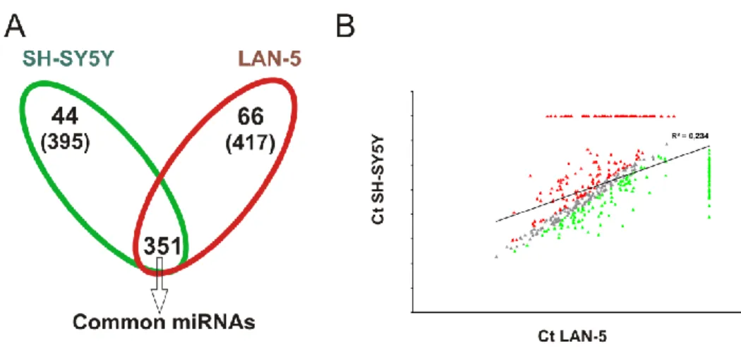
To address this hypothesis I used as experimental model the SH-SY5Y and LAN-5 human neuroblastoma cell lines expressing LMNA or MYCN alternatively. We also employed the SH-SY5Y model in which we have previously silenced LMNA gene.
3 MATERIALS AND METHODS
Human NB biopsy RNA extraction and real
‐time RT‐PCR
Total RNA was extracted from twenty-three frozen biopsies of human newly diagnosed NBs obtained from Department of Pediatrics and Infantile Neuropsychiatry of Sapienza University, Rome using TRIzol reagent (Life Technologies) according to the manufacturer’s instructions. RNA was reverse-transcribed, and real-time PCR performed as described above. Institutional written informed consent was obtained from the patient’s parents or legal guardians according to the local institutional guidelines.
Cell line maintenance and sphere formation
The SY5Y NB cell line was purchased from ATCC. In this study, we also used two SH-SY5Y-derived clones, LMNA-KD and control (CTR) cells that were grown as previously described (Maresca et al., 2012). The LAN-5 NB cell line (a gift from Dr. Doriana Fruci) was grown in RPMI-1640 medium (Gibco) supplemented with 10% FBS (Hyclone), 2 mML-glutamine and 1% penicillin/streptomycin in a fully humidified incubator containing 5% CO2 at 37°C. To obtain
spheres, cells were plated in a serum-free media in 60-mm, low-attachment culture dishes at a density of 9.5x10⁵ cells/cm2 and cultured in Neurobasal Medium (Gibco) supplemented with 20 ng/ml Epidermal Growth Factor (EGF, Sigma), 40 ng/ml basic Fibroblast Growth Factor (bFGF, Sigma), 1% neuronal supplement N2 (Gibco) and B27 (Gibco), and 2 µg/ml heparin. Three days after seeding, the cells formed floating neurosphere-like structures that grew rapidly until day 7. Before these structures became necrotic, we harvested and suspended them in Accutase enzymatic solution (Gibco) for 5 min at 37°C and then mechanically dissociated into a single-cell suspension. The cells were re-seeded in the same conditions as above, and the secondary spheres were allowed to form. As positive and negative controls, we used NB LAN-5 and SH-SY5Y cell lines, respectively. TICs were derived by harvesting the secondary spheres, treating them with Accutase enzymatic solution (Gibco) for 5 min at 37°C and then mechanically dissociating them into a single-cell suspension. These cells were seeded in adherent conditions in the presence of 1:1 mixture of Eagle's Minimum Essential Medium and F12 medium (Gibco) supplemented with 10% FBS (Hyclone), 2 mM L-glutamine, 0.5% non-essential amino acids, 0.5% sodium pyruvate and 1% penicillin and streptomycin.
Primary cell coltures of Granule Cells
Cultures enriched in granule neurons were obtained from dissociated cerebella of 8-day-old Wistar rats (CGC; Charles River, Calco, Italy) according to the procedure described by Levi et al. (1984). Cells were seeded (3 x 10⁶ cells/dish) on poly-L-lysine-coated 35-mm plastic dishes (NUNC, VWR International PBI s.r.l., Milano, Italy) in basal medium Eagle (BME; Life Technologies, Gaithersburg, MD, USA) supplemented with 10% heat-inactivated fetal calf serum, 2 mM glutamine and 25 mM KCl and 100 µg/ml gentamycin (Life Technologies, Gaithersburg, MD, USA). Ara-C (10 mM) was added to the culture medium 24 h after plating to prevent proliferation of nonneuronal cells.
Generation of Lentiviral Infection of Granule Cells
The cells were seeded into 35-mm plastic dishes previously coated with poly-L-lysine at a concentration 3,5 x 10⁵ cells/dish. After four hours, the cells were infected for 18 hours with the virus-containing supernatant from pLenti6/V5-GW/EmGFP-miR-LMNA or pLenti6/V5-GW/EmGFP-miRNeg (Maresca et al. 2012), and then fresh medium was added. The miRNA expression was monitored by checking the simultaneous coexpression of the EmGFP reporter gene by fluorescence microscopy.
Glutamate toxicity and neuronal survival
After 2, 5 and 8 days in culture, cells were washed once in Locke solution (in mM: 154 NaCI, 5.6 KCI, 3.6 NaHC03, 2.3 CaC12, 1.0 MgC12, 5.6 glucose, 10 HEPES, pH 7.4) and exposed at room temperature to a 100 µM glutamate pulse in Mg2+-free Locke solution for 30 min. Cells were subsequently washed in Mg2+-free Locke solution, replenished with their original medium and returned to the incubator. After 18 h, viable cells were assessed by counting the numbers of intact nuclei as described by Soto and Sonnenchein (1985), modified for counting cerebellar granule cells by the procedure of Volontè et al. (1994). This method has been shown to be reproducible and accurate and to correlate well with other methods of assessing cell survival-death (Stefanis et al. 1997; Stefanis et al. 1999). Cell counts were performed in triplicate and are reported as means ± SEM. The data are expressed as the percentage of intact nuclei in the control cultures at each time point.


![[3] M. Ventura e G. Garboden, A brief history of concentrated hydrogen peroxide uses, AIAA Paper 99-2739, 35th AIAA/ASME/SAE/ASEE Joint Propulsion Conference and Exhibit, Los Angeles, USA, June 1999](data:image/gif;base64,R0lGODlhAQABAIAAAP///wAAACH5BAEAAAAALAAAAAABAAEAAAICRAEAOw==)