LARGE MAMMAL REMAINS FROM THE EARLY PLEISTOCENE SITE OF PODERE SAN LORENZO (PERUGIA, CENTRAL ITALY)
MARCO CHERIN1*, BEATRICE AZZARÀ1, MARZIA BREDA2, ALBA BROBIA ANSOLEAGA1,3, COSTANTINO BUZI4, LUCA PANDOLFI5 & FAUSTO PAZZAGLIA6
1*Corresponding author. Dipartimento di Fisica e Geologia, Università degli Studi di Perugia, Via A. Pascoli, 06123 Perugia, Italy.
E-mail: [email protected], [email protected], [email protected]
2Dipartimento di Studi Umanistici, Università degli Studi di Ferrara, Via Paradiso 12, 44121 Ferrara, Italy. E-mail: [email protected] 3Department of Geography, Universitat Autònoma de Barcelona, Carrer de la Fortuna s/n, 08193 Bellaterra, Barcelona, Spain.
4Dipartimento di Biologia Ambientale, Sapienza Università di Roma, P.le A. Moro 5, 00185 Roma, Italy. E-mail: [email protected] 5Dipartimento di Scienze della Terra, Università degli Studi di Firenze, Via G. La Pira 4, 50121 Firenze, Italy. E-mail: [email protected] 6Int. Geo. Mod. srl, Via Fratelli Cairoli 24, 06125 Perugia, Italy. E-mail: [email protected]
To cite this article: Cherin M., Azzarà B., Breda M., Brobia Ansoleaga A., Buzi C., Pandolfi L. & Pazzaglia F. (2019) - Large mammal remains from the Early Pleistocene site of Podere San Lorenzo (Perugia, central Italy). Riv. It. Paleontol. Strat., 125(2): 489-515.
Abstract. Most of the research on fossil mammals from Umbria (central Italy) has been carried out in the
southwestern branch of the Tiber basin, due to its paleontological richness. This portion of the basin extends from Perugia to Terni and corresponds to a well-defined half-graben filled with fluvial-lacustrine deposits.
The paleontological sample presented here was discovered in a sand and gravel quarry at Podere San Lorenzo, East of the town of Deruta. The stratigraphic succession exposed in the quarry is no longer visible, but we describe here a new outcrop (Palazzone), which is not far from Podere San Lorenzo and shows comparable facies associations. The two successions were deposited in a fluvial environment characterized by an average reduction of the hydrodyna-mic energy from the bottom upwards. They are referred to the Early Pleistocene Santa Maria di Ciciliano Subsynthe-me (Madonna dei Bagni Lithofacies).
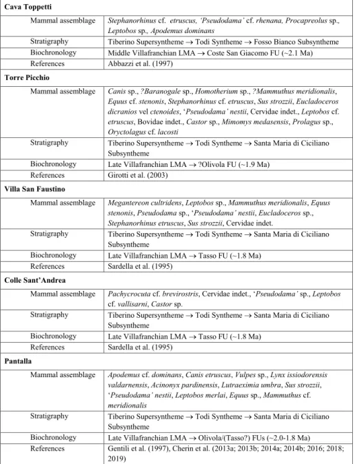
Large mammal remains are attributed to Mammuthus cf. meridionalis (Nesti, 1825), Stephanorhinus etruscus (Fal-coner, 1859), Equus stenonis Cocchi, 1867, Leptobos cf. etruscus (Falconer, 1868), ‘Pseudodama’ nestii (Azzaroli, 1947), and Sus strozzii Forsyth Major, 1881. Some hyena coprolites are also reported. The assemblage is typical of the early Late Villafranchian Land Mammal Age and can be referred to the Olivola/Tasso Faunal Units (about 2.0–1.8 Ma). This is in agreement with the alleged age of some other assemblages found in the southwestern branch of the Tiber basin (e.g., Torre Picchio, Villa San Faustino, Colle Sant’Andrea, Pantalla).
Received: January 30, 2019; accepted: May 03, 2019
Keywords: Early Pleistocene; Fossil mammals; Late Villafranchian; Tiber basin, Umbria.
I
ntroductIonDuring the Pliocene and Pleistocene, the pa-leogeography of central Italy was characterized by a set of NW-SE trending én echelon grabens whose genesis is due to the eastward migration of the ex-tensional deformation related to the opening of the Tyrrenian Sea. This caused the opening of basins in the western part of the Peninsula, where marine sedimentation occurred during the Pliocene and the beginning of the Pleistocene; at the same time,
con-tinental deposits started to fill the extensional basins located in the eastern part, close to the recently-formed Apennine Chain (Pascucci et al. 1999; Col-lettini et al. 2006). Consequently, Plio-Pleistocene marine sediments today crop out in the western part of Umbria, while continental deposits are mostly found in the central and eastern portions of the Re-gion (Ambrosetti et al. 1987).
The Tiber basin (Fig. 1) is the largest inter-montane continental basin along the Apennines. It is approximately 1800 km2 wide and extends from North to South describing an “upside-down Y” shape, splitting in a southeastern and a
southwest-ern branch south of Perugia (Basilici 1997). The first geological observations on the basin date back to the beginning of the 20th century, but for a long time it has been believed that the basin was represented by a single huge lake, called the “Tiberino Lake” (Lotti 1917). Conversely, research conducted in the last 40 decades (Conti & Girotti 1977; Ambrosetti et al. 1987; Basilici 1997; Pucci et al. 2014) as well as several geological mapping projects of the Umbria Region, demonstrate that the Tiber basin was a com-plex depositional system deriving from the coales-cence of smaller lacustrine, palustrine, and fluvial environments alternating in space and time.
Most of the fossils of Plio-Pleistocene mam-mals in Umbria have been recovered since the 19th century from continental deposits (gravel, sand, clay, lignite, calcareous tufa) that can be related to the Tiber basin (Cherin 2013). Even the famous French biologist Georges Cuvier, the putative “father” of vertebrate paleontology and comparative anatomy, cites some fossil proboscidean and rhinoceros re-mains from the Tiber basin in his book Recherches sur les ossemens fossiles de quadrupèdes (Paris, 4th edition, 1834).
The majority of recent scientific papers on
Umbrian fossil mammals concerns the southwestern branch of the Tiber basin, thanks to the outstanding richness of paleontological sites scattered in this area (Fig. 1, Tab. 1). This portion of the basin extends from the SE of Perugia to Terni and is delimited by Mount Peglia and the Narni-Amelia Ridge to the West and by the Martani Mountains to the East. It corresponds to a well-defined half-graben filled with alluvial-lacustrine deposits referred by Basilici (1997) to four lithostratigraphic units: (1) the Fosso Bianco Unit, mainly characterized by silty, clayey deposits, ascribable to a large deep lake system, chronologi-cally referred to the Gauss-Matuyama (i.e., Pliocene-Pleistocene) boundary (Abbazzi et al. 1997); (2) the Ponte Naja Unit, cropping out in the surroundings of the town of Todi and composed principally of gravel and sandy gravel bodies and clayey sandy silts formed on the distal part of an alluvial fan located along a lake margin around 2.15–2.10 Ma (Napo-leone et al. 2003); (3) the Santa Maria di Ciciliano Unit (Early Pleistocene), unconformably overlying the Fosso Bianco Unit and composed of silty clays and clayey silts deposited in alluvial plain environ-ments, alternating with sand lithosomes referred to meandering fluvial channels; (4) the Acquasparta Fig. 1 - a) Geographic location of the study area. The Tiber Basin is highlighted in light orange along the middle part of Umbria. Red points refer to the paleontological sites in Tab. 1: 1, Cava Toppetti; 2, Torre Picchio; 3, Villa San Faustino and Colle Sant’Andrea; 4, Pantalla. b) Geological map superimposed on a digital elevation model of the study area. PSL, Podere San Lorenzo; PZ, Palazzone.
Unit (Early Pleistocene), represented by continental carbonates (i.e., calcareous tufa), deposited within shallow-lake and wetland environments. The most recent Umbrian geological mapping projects at a scale of 1:10000 have adopted the lithostratigraphic units described by Basilici (1997) but have integrat-ed them into the UBSU (Unconformity Boundintegrat-ed Stratigraphic Units) system, widely used in continen-tal contexts (Salvador 1994). As a consequence, the Fosso Bianco, Santa Maria di Ciciliano, and Acquas-parta Units are currently classified as subsynthemes of the Todi Syntheme, while the Ponte Naja Unit is considered as a lithofacies of the Fosso Bianco Sub-syntheme (Regione Umbria 2013).
Apart from the Cava Toppetti assemblage (Tab. 1), virtually all the mammal remains found to
date in the southwestern branch of the Tiber ba-sin come from fluvial deposits of the Santa Maria di Ciciliano Subsyntheme. This work offers a further contribution to the knowledge of the paleontologi-cal, biochronologipaleontologi-cal, and paleoenvironmental fea-tures of this area of the basin thanks to the descrip-tion of a new mammal assemblage from the locality of Podere San Lorenzo (PSL) as well as of a new stratigraphic section (Palazzone; PZ) of the Santa Maria di Ciciliano Subsyntheme outcropping nearby (Fig. 1).
The Palazzone outcrop and notes on taphonomy.
In the area between Perugia and Todi (Fig. 1), the Tiber River course is currently set along the Tab. 1 - Selected Early Pleistocene
localities with fossil mammal remains in the southwestern branch of the Tiber basin (Umbria, central Italy). FU, Faunal Unit; LMA, Land Mammal Age. The approxi-mate ages of the FUs are from Rook & Martínez-Na-varro (2010) and Nomade et al. (2014).
Cava Toppetti
Mammal assemblage Stephanorhinus cf. etruscus, ‘Pseudodama’ cf. rhenana, Procapreolus sp., Leptobos sp., Apodemus dominans
Stratigraphy Tiberino Supersyntheme Todi Syntheme Fosso Bianco Subsyntheme Biochronology Middle Villafranchian LMA Coste San Giacomo FU (~2.1 Ma) References Abbazzi et al. (1997)
Torre Picchio
Mammal assemblage Canis sp., ?Baranogale sp., Homotherium sp., ?Mammuthus meridionalis, Equus cf. stenonis, Stephanorhinus cf. etruscus, Sus strozzii, Eucladoceros dicranios vel ctenoides, ‘Pseudodama’ nestii, Cervidae indet., Leptobos cf. etruscus, Bovidae indet., Castor sp., Mimomys medasensis, Prolagus sp., Oryctolagus cf. lacosti
Stratigraphy Tiberino Supersyntheme Todi Syntheme Santa Maria di Ciciliano Subsyntheme
Biochronology Late Villafranchian LMA ?Olivola FU (~1.9 Ma) References Girotti et al. (2003)
Villa San Faustino
Mammal assemblage Megantereon cultridens, Leptobos sp., Mammuthus meridionalis, Equus stenonis, Pseudodama sp., ‘Pseudodama’ nestii, Eucladoceros sp., Stephanorhinus etruscus, Sus strozzii, Cervidae indet.
Stratigraphy Tiberino Supersyntheme Todi Syntheme Santa Maria di Ciciliano Subsyntheme
Biochronology Late Villafranchian LMA Tasso FU (~1.8 Ma) References Sardella et al. (1995)
Colle Sant’Andrea
Mammal assemblage Pachycrocuta cf. brevirostris, Cervidae indet., ‘Pseudodama’ sp., Leptobos
cf. vallisarni, Castor sp.
Stratigraphy Tiberino Supersyntheme Todi Syntheme Santa Maria di Ciciliano Subsyntheme
Biochronology Late Villafranchian LMA Tasso FU (~1.8 Ma) References Sardella et al. (1995)
Pantalla
Mammal assemblage Apodemus cf. dominans, Canis etruscus, Vulpes sp., Lynx issiodorensis valdarnensis, Acinonyx pardinensis, Lutraeximia umbra, Sus strozzii,
‘Pseudodama’ nestii, Leptobos merlai, Equus sp., Mammuthus cf.
meridionalis
Stratigraphy Tiberino Supersyntheme Todi Syntheme Santa Maria di Ciciliano Subsyntheme
Biochronology Late Villafranchian LMA Olivola/(Tasso?) FUs (~2.0-1.8 Ma) References Gentili et al. (1997), Cherin et al. (2013a; 2013b; 2014a; 2014b; 2016; 2018;
and Pleistocene deposits. Tectonics mainly occurs through the action of normal faults with én echelon arrangement, which systematically drop the western part of the whole structure and confer a terraced morphology to the eastern side of the Tiber River Valley. There are also normal NE-dipping antithetic faults, which locally determine a horst and graben structure, typical of the deformations in an exten-sional tectonic regime (Cencetti 1990).
Much of the Deruta area (Fig. 1) is occupied by Pleistocene-Holocene alluvial deposits. These sediments crop out mainly in the northwestern part of the territory and form the present alluvial plain of the Tiber River. Conversely, on the hills SE of Deruta, Pleistocene deposits referred to the Santa Maria di Ciciliano Subsyntheme are mostly found (Fig. 1).
The paleontological collection here described was discovered in the 1990s in a sand and gravel quarry at PSL, East of Deruta (42°58’32.92” N - 12°26’20.87” E). Unfortunately, the quarry is cur-rently abandoned and completely covered by veg-etation, so it is not possible to describe the local stratigraphic succession, but only to make qualitative considerations based on excavation notes and pho-tographs taken during previous surveys. However, a new section (PZ) has been identified not far from PSL (Fig. 1). The two sites show facies associations that are almost completely comparable, as confirmed by Cencetti’s (1990) descriptions.
The PZ outcrop is divided into three areas, A-B-C (in ascending order), corresponding to some artificial walls excavated during slope consolidation operations (Fig. 2a). Area A develops in the West-East direction for about 30 m and for a height of about 3 m. The predominant lithology is represented by unsorted and clast-supported conglomerates or-ganized in tabular bodies (Fig. 2b), with poor sandy matrix. The pebbles are all rounded and heteromet-ric (centimeter to decimeter size), with a slight imbri-cation indicating a prevalent NE-SW paleocurrent in a high-energy hydrodynamic regime. The clasts are predominantly arenaceous, although carbonate and chert pebbles are also observed. These latter have a characteristic blackish color, an advanced state of alteration, and often a thin external oxidation red-dish patina. These black pebbles are mentioned by
lamination. Fossil plant remains are found in both lithologies, in the form of small branches and roots in the sands and larger fragments of branches in the conglomerates. In the eastern part of Area A, we discovered a fossil branch disposed obliquely (205/15 direction relative to the wall), with the low-er part lying in conglomlow-erates and the upplow-er part in sands (Fig. 2c). Probably the branch, carried by the water, was embedded obliquely into the gravel bottom of the river and was then covered by a sub-sequent sandy deposit. This demonstrates the high dynamism of the river environment, which, in a relatively short time (i.e., less than the time required for decomposition, fragmentation or removal of the branch by the current), has decreased hydrodynamic energy, as evidenced by the conglomerate-sand tran-sition. The fossil branch also confirms the prevalent NE-SW direction of the paleocurrent, as it is pos-sible to observe that on its left (NE) side, the sand granulometry is finer and more homogeneous than that on the right (SW) side, where larger clasts were “trapped” by the same branch. East of Area A, in Sub-area A’ (length 8 m, height 3 m), we detect a normal fault (dip notation 228/63) that puts in later-al contact the conglomerates of Area A with a mas-sive fine-grained sand (Fig. 2d). A fault with similar characteristics is reported and mapped by Cencetti (1990) very close to PZ.
The contact between Area A and Area B (length 50 m, height 1.8 m) is marked by an erosion-al surface that separates the conglomerates from the overlying deposits (Fig. 2e). The latter consist mainly of a tabular fine-grained sandy body, with-out channel geometry, rare cross laminations, and
Fig. 2 - Palazzone outcrop at Deruta (Perugia, Italy). a) Overall view of the outcrop, detailing the areas described in the text. b) Detail of Area A: conglomerate with sandstone lens in the middle. c) Remains of a fossil branch embedded obliquely between a conglomerate (below) and a sand lens (above). d) Normal fault in Sub-area A’ separating a conglomerate (left) and a fine sandstone (right). e) Stratigraphical contact be-tween conglomerates (Area A, below) and sandstone (Area B, above). f) Thin layer of whitish claystone with bottom and top reddish portions, within the massive sandstone of Area B. g) Altered fine sandstone and claystone in Area C.
grey-whitish laminated clay, delimited at the bot-tom and the top by reddish horizons. The three layers are related to short periods of stasis in river flow and/or periods of emersion, as suggested by the oxidation evidence in the reddish portions. The Sub-area B’, to the East of the main wall and per-pendicular to it, is crossed by the same normal fault identified in Sub-area A’. A thin cataclasite level consisting of red-blackish coated gravels, is visible along the fault plane.
Area C is about 50 m in length and 2.5 m in height. The outcrop is mainly composed of fine sands and silts with high clay content and poor presence of pebbles and it is crossed by numerous vertical fractures which are sub-parallel to the main fault seen in Sub-areas A’-B’. Bedding attitudes of these fractures are progressively changing from SW-NE to W-E, with a dip in direction from 180 to 230 degrees towards the fault.
The general sedimentological characteristics of the PZ and by extension, PSL sites can be refer-red to a fluvial depositional environment characte-rized by an average reduction of the hydrodynamic energy from the bottom upwards, as evidenced by the decreasing granulometry trend from Area A to C. Sand lenses within the conglomerate bodies can be related to periodic events of energy reduction and/or river bar formation. The stratigraphic di-scontinuity between Area A (predominantly con-glomerate) and B (predominantly sands) suggests a more or less prolonged interruption of the clastic input and a subsequent recovery of solid transport, although with lower hydrodynamic energy. The transition between Areas B and C is more gradual and it records a progressive reduction in granulo-metry, reaching fine sand and silt size at the top. Clear evidence of pedogenesis (root traces, CaCO3 nodules, reddish portions due to oxidation proces-ses) characterize the upper part of the sequence, testifying to more prolonged events of subaerial exposure. This may correspond to a transition to-wards an alluvial plain environment.
Although we did not participate in the collec-tion of the mammal fossils from PSL, we can rea-sonably state that they have been recovered from the lower part of the succession. Most likely, not all fossils come from the same level, as suggested
probably come from conglomerates. This is also supported by the advanced fragmentation and sur-face abrasion of the remains, related to transport in a high-energy regime. Other fossils, on the con-trary, show brownish-yellowish color, smaller av-erage dimensions and good state of preservation, suggesting that they have been preserved within finer grained deposits formed in lower energy con-text. In particular, they probably come from sand lenses within the conglomerate bodies. In fact (1) some finds still retain traces of sedimentary matrix, whose grain size and color are really similar to those of the sands observed in Area A at PZ; (2) these sands are probably deposited in fluvial bars (ideal contexts for the rapid burial and conservation of the remains; see plant macro-fossils described for Area A); (3) the tabular sandy complex observed in the higher part of the PZ section presents finer granulometry than that of the sediment associated with the fossil bones.
MaterIalsandMethods
Fossil preparation. Prior to the analytic study, fossils
de-scribed herein were prepared using the following products and pro-cedures:
For gluing and consolidation, we used solutions of Para-loidTM B-72 mixed with acetone following Davidson & Brown
(2012). In particular, we used Paraloid as “stock adhesive” (concen-tration 50%) to glue small to medium-sized fossil fragments (see below for large-sized specimens), as “dilute consolidant” (concen-tration ~5%) for the first consolidation sessions, and as “stock con-solidant” (concentration ~17%) for the final consolidation session.
For gluing large bone fragments (e.g., proboscidean long bo-nes), we used UHU® extra All-Purpose Adhesive (mixture of methyl
acetate and ethanol).
We chose to fill damaged areas with fine-grained plaster only when we found that these fractures jeopardized the stability of the specimens.
Paleontology. Identification of paleontological specimens
is based on anatomical feature descriptions and morphological and morphometric comparative analysis. All measurements are taken with digital calipers and are given with a precision of 0.1 mm. Mor-phological and morphometric data used for comparison are from the cited literature.
The biochronological framework is based on correlations of the Villafranchian Land Mammal Age (LMA) in its most recent definition (Rook & Martínez-Navarro 2010; Martínez-Navarro et al. 2015) and the late Cenozoic and Quaternary time scale (Cohen & Gibbard 2016).
Institutional abbreviations. IGF, Museo di Storia
Na-turale, Sezione di Geologia e Paleontologia, Università di Firenze; NHMB, Natural History Museum, Basel; SBAU, former Soprinten-denza per i Beni Archeologici dell’Umbria (now SoprintenSoprinten-denza Ar-cheologia, Belle Arti e Paesaggio dell’Umbria), Perugia.
s
ysteMatIc paleontologyClass MAMMALIA Linnaeus, 1958
Order Carnivora Bowdich, 1821
Family Hyaenidae Gray, 1821 Genus and species indet.
Fig. 3
Referred material: Three coprolites (SBAU 153505, SBAU
153506, SBAU 153529).
Description. The PSL collection includes
three coprolites, which show a rounded/ovoid shape and a relatively flat or slightly concave side, corresponding to the contact area with the adjacent fecal pellet. Bone splinters and cavities left by the decomposition of organic material are well visible in SBAU 153505 and SBAU 153529 (Fig. 3).
The maximum length (measured by placing the caliper between the flat/concave surface and the opposite vertex) and width (measured perpendicu-lar to the max length) of the three specimens are as follows: SBAU 153505 - 30.0 x 37.0 mm; SBAU 153506 - 50.8 x 52.2 mm; SBAU 153529 - 37.9 x 47.3 mm.
Discussion. The overall morphology of the
PSL coprolites resembles that of droppings of the recent African Crocuta crocuta (Brain 1981). Two bo-ne-cracking hyaenids are reported in the European Early Pleistocene: Pliocrocuta perrieri (late Pliocene– Early Pleistocene; about 4.2–2.0 Ma) (Turner et al. 2008; Vinuesa et al. 2014) and Pachycrocuta
breviro-stris (Early–Middle Pleistocene; about 2.0–0.8 Ma) (Turner et al. 2008; Madurell-Malapeira et al. 2010). Unfortunately, it is not possible to assign the PSL coprolites to one or the other species on the basis of morphology alone. However, the dimensions of the analyzed specimens fit those of Pa. brevirostris drop-pings from different European sites (Ceyssaguet, France; Trlica, Serbia; Untermassfeld, Germany) (Argant & Bonifay 2011).
Order Proboscidea Illiger, 1811
Family Elephantidae Gray, 1821 Genus Mammuthus Brookes, 1828 Mammuthus cf. meridionalis (Nesti, 1825)
Figs 4-5
Referred material: Molar fragments (SBAU 153509, SBAU
153521, SBAU 153540, SBAU 153549, SBAU 153549bis), tusk ment (SBAU 153543), distal condyle of femur (SBAU 153541), frag-ments of femur diaphysis (SBAU 153542, SBAU 153553, probably belonging to the same bone), left trapezoid (SBAU 153546), fragmen-ted proximal epiphysis of left tibia (SBAU 153547), fragmenfragmen-ted pro-ximal diaphysis of right femur (SBAU 153550), fragmented propro-ximal diaphysis of right humerus (SBAU 153548), undetermined fragments of limb bone diaphyses (SBAU 153551, SBAU 153552, SBAU 153554, SBAU 153554bis).
Description. The available proboscidean
material is severely damaged and incomplete, ma-king any taxonomic attribution extremely difficult. The preserved fragments of limb bones and the left trapezoid SBAU 153546 are large and massive (Fig. 4). Specimen SBAU 153543 is a small and damaged tusk fragment (Fig. 5). Schreger lines are clearly vi-sible thanks to the natural break of the fragment along the transverse plane; the width of Schreger angles is around 85 degrees. The most complete molar fragment is SBAU 153549bis, which preser-ves four almost complete lamellae. We measured the enamel thickness in various positions along Fig. 3 - Hyaenid coprolites from
Podere San Lorenzo (Italy). a) SBAU 153505; b) SBAU 153506; c) SBAU 153529. Scale bar: 3 cm.
the occlusal lamellar surface following Palombo & Ferretti (2005) and we obtained an average value of 3.8 mm. A similar average thickness (3.7 mm) was obtained for the small tooth fragment SBAU 153521.
Discussion. Three proboscideans occur in
Europe in the Early Pleistocene: Anancus arvernen-sis, Mammuthus meridionalis, and Palaeoloxodon antiquus. The gomphothere A. arvernensis is a common ele-ment of Pliocene mammal assemblages and became extinct in the Early Pleistocene at the beginning of the Late Villafranchian (Palombo & Ferretti 2005). However, the cheek tooth morphology of this spe-cies, characterized by lophs formed by pairs of thick conical cusps, does not fit that observed in the PSL specimens, which clearly show the typical lamellar morphology of the Elephantidae. Mammuthus
me-ridionalis occurs in Europe during the entire Early Pleistocene, whereas P. antiquus appears in the la-test Early Pleistocene (Galerian LMA) and survives until the Late Pleistocene (about 40 ka) (Palombo & Ferretti 2005; Stuart 2005). The two species are relatively similar in body size (Larramendi 2016). Although fragmented, we could take some measu-rements on two postcranial elements from PSL, the left trapezoid SBAU 153546 (width: 89 mm, depth: +112.5 mm, height: 77 mm) and the femur fragment SBAU 153550 (mid-shaft width: 182 mm, mid-shaft depth: 117 mm). The dimensions of both bones fall into the range of M. meridionalis (trapezoid - width: 72–99 mm, depth: 100–150 mm, height: 61–83 mm; femur - shaft width: 120–192 mm, mid-shaft depth: 90–133 mm) and P. antiquus (trapezoid - width: 71–100 mm, depth: 140–144 mm, height: 70–86 mm; femur - mid-shaft width: 116–242 mm, Fig. 4 - Mammuthus cf. meridionalis from Podere San Lorenzo (Italy). a) Proximal diaphysis of right femur (SBAU 153550) in anterior view. b) Proximal diaphysis of right humerus (SBAU 153548) in anterior view. c) Proximal epiphysis of left tibia (SBAU 153547) in anterior (c1) and dorsal (c2) views. d) Molar fragment (SBAU 153549) in occlusal view. e) Molar fragment (SBAU 153549bis) in occlusal view. f) Left trapezoid (SBAU 153546) in anterior (f1), dorsal (f2), and ventral (f3) views. Scale bar: 10 cm.
mid-shaft depth: 86–147 mm) (comparative data from Ferretti 1998 and Larramendi et al. 2017). Ho-wever, Schreger angles measured on the tusk frag-ment SBAU 153543 are characteristic of the genus Mammuthus (Palombo & Villa 2001). Similarly, the average enamel thickness of the PSL molar frag-ments agrees with the highest values recorded for M. meridionalis and is significantly higher than those of P. antiquus (Palombo & Ferretti 2005). For these reasons, considering the fragmentary nature of the available sample, it is here referred to Mammuthus cf. meridionalis.
Order Perissodactyla Owen, 1848
Family Rhinocerotidae Gray, 1821 Genus Stephanorhinus Kretzoi, 1942
Stephanorhinus sp.
Fig. 6a-c
Referred material: Fragment of upper tooth and fragment
of lower tooth (SBAU 153507), fragment of lower tooth (SBAU 153513), left m1 (SBAU 153514), left m2 (SBAU 153515).
Description. A few isolated and worn down
teeth are included in the studied material. Length of the lower molars SBAU 153514 and 153515 is
reported in Tab. 2. The preservation of the speci-mens and the absence of diagnostic morphological characters do not allow a specific taxonomic attri-bution.
Stephanorhinus etruscus (Falconer, 1868)
Fig. 6d-g
Referred material: Right M3 (SBAU 153510), left
mandibu-lar ramus with m1-m3 (SBAU 153511), left fragmentary maxilla with M2-M3 and right fragmentary maxilla with M1-M3 (SBAU 153516), left mandibular ramus with p4-m3 (SBAU 153517).
Description. The measurements are
summa-rized in Tabs 2 and 3.
The isolated M3 SBAU 153510 (Fig. 6d) is re-latively small (Tab. 3) and well preserved. It displays a mesial and a distal cingulum, a crochet, and weak accessory folds. The protocone is not constricted, the paracone fold is weak, and the parastyle is pro-minent.
The mandibular ramus SBAU 153511 (Fig. 6e) preserves the molars and the roots of the pre-molars. The surface of the bone is eroded and the ventral border of the ramus is gently convex. A me-sial cingulum is present on the molars, a vestibular cingulum is present on the anterior loph of m2 and m3, and a weak lingual cingulum is present below the base of the posterior lingual valley on m2. The posterior lingual valley on m2 and m3 is V-shaped, the anterior lingual valley on m3 is broad V-shaped. The vestibular groove on the molars stretched the neck and is shallow on m2 and m3.
The left and right maxillae SBAU 153516 (Fig. 6f) belong to the same old individual. The M1s and M2s are heavily worn, the internal folds and Fig. 5 - Mammuthus cf. meridionalis from Podere San Lorenzo (Italy).
Tusk fragment (SBAU 153543) photographed with side lighting to highlight the Schreger lines. Values of about 85 degrees can be measured for the outer Schreger angles. Scale bar: 3 cm.
Fig. 6 - (pag. 498) Rhinoceros remains from Podere San Lorenzo (Italy). a-c) Stephanorhinus sp.: (a) fragment of lower tooth embedded in sediment (SBAU 153513); (b) Left m1 (SBAU 153514) in occlusal view; (c) Left m2 (SBAU 153515) in oc-clusal view. d-g) Stephanorhinus etruscus: (d) Right M3 (SBAU 153510) in occlusal view; (e) Left mandibular ramus with m1-m3 (SBAU 153511) in labial (e1), lingual (e2), and oc-clusal (e3) views; f) Left fragmentary maxilla with M2-M3 in labial (f1), lingual (f2), and occlusal (f3) views and right fragmentary maxilla with M1-M3 in labial (f4), lingual (f5), and occlusal (f6) views (SBAU 153516); g) Left mandibular ramus with p4-m3 (SBAU 153517) in labial (g1), lingual (g2), and occlusal (g3) views. Scale bar: 5 cm.
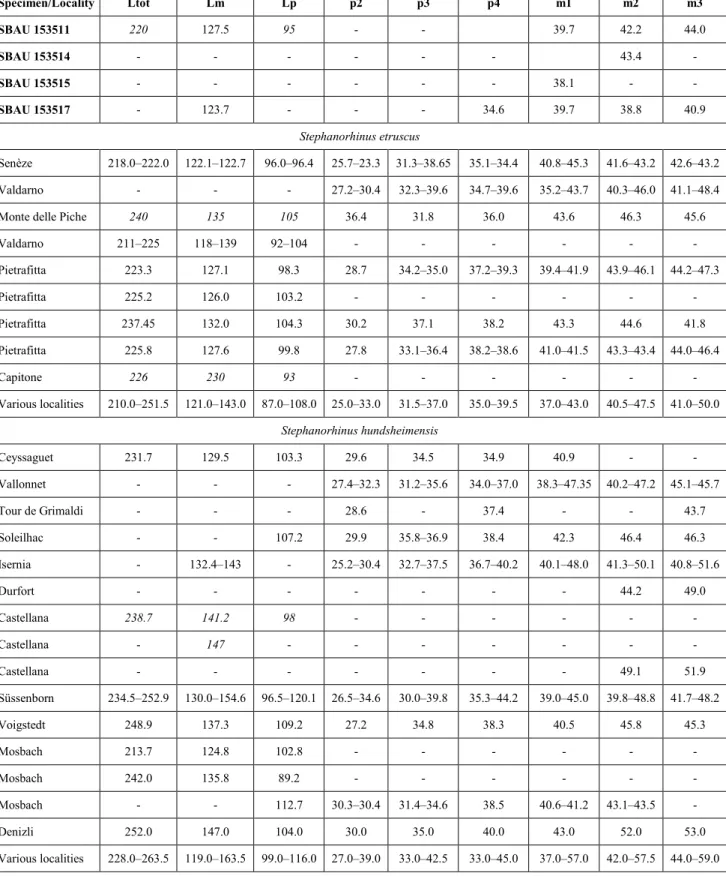
Specimen/Locality Ltot Lm Lp p2 p3 p4 m1 m2 m3 SBAU 153511 220 127.5 95 - - 39.7 42.2 44.0 SBAU 153514 - - - 43.4 - SBAU 153515 - - - 38.1 - - SBAU 153517 - 123.7 - - - 34.6 39.7 38.8 40.9 Stephanorhinus etruscus Senèze 218.0–222.0 122.1–122.7 96.0–96.4 25.7–23.3 31.3–38.65 35.1–34.4 40.8–45.3 41.6–43.2 42.6–43.2 Valdarno - - - 27.2–30.4 32.3–39.6 34.7–39.6 35.2–43.7 40.3–46.0 41.1–48.4
Monte delle Piche 240 135 105 36.4 31.8 36.0 43.6 46.3 45.6
Valdarno 211–225 118–139 92–104 - - - - Pietrafitta 223.3 127.1 98.3 28.7 34.2–35.0 37.2–39.3 39.4–41.9 43.9–46.1 44.2–47.3 Pietrafitta 225.2 126.0 103.2 - - - - Pietrafitta 237.45 132.0 104.3 30.2 37.1 38.2 43.3 44.6 41.8 Pietrafitta 225.8 127.6 99.8 27.8 33.1–36.4 38.2–38.6 41.0–41.5 43.3–43.4 44.0–46.4 Capitone 226 230 93 - - - - Various localities 210.0–251.5 121.0–143.0 87.0–108.0 25.0–33.0 31.5–37.0 35.0–39.5 37.0–43.0 40.5–47.5 41.0–50.0 Stephanorhinus hundsheimensis Ceyssaguet 231.7 129.5 103.3 29.6 34.5 34.9 40.9 - - Vallonnet - - - 27.4–32.3 31.2–35.6 34.0–37.0 38.3–47.35 40.2–47.2 45.1–45.7 Tour de Grimaldi - - - 28.6 - 37.4 - - 43.7 Soleilhac - - 107.2 29.9 35.8–36.9 38.4 42.3 46.4 46.3 Isernia - 132.4–143 - 25.2–30.4 32.7–37.5 36.7–40.2 40.1–48.0 41.3–50.1 40.8–51.6 Durfort - - - 44.2 49.0 Castellana 238.7 141.2 98 - - - - Castellana - 147 - - - - Castellana - - - 49.1 51.9 Süssenborn 234.5–252.9 130.0–154.6 96.5–120.1 26.5–34.6 30.0–39.8 35.3–44.2 39.0–45.0 39.8–48.8 41.7–48.2 Voigstedt 248.9 137.3 109.2 27.2 34.8 38.3 40.5 45.8 45.3 Mosbach 213.7 124.8 102.8 - - - - Mosbach 242.0 135.8 89.2 - - - - Mosbach - - 112.7 30.3–30.4 31.4–34.6 38.5 40.6–41.2 43.1–43.5 - Denizli 252.0 147.0 104.0 30.0 35.0 40.0 43.0 52.0 53.0 Various localities 228.0–263.5 119.0–163.5 99.0–116.0 27.0–39.0 33.0–42.5 33.0–45.0 37.0–57.0 42.0–57.5 44.0–59.0
Tab. 2 - Measurements (length in mm) of the rhinoceros lower teeth from Podere San Lorenzo (Italy), compared with the minimum, maxi-mum, and mean values of Stephanorhinus etruscus and S. hundsheimensis from different European sites. Ltot, length of the cheek tooth row; Lm, length of the molar row; Lp, length of the premolar row. Comparative data are from Pandolfi & Erten (2017). Estimated measurements are in italics.
the postfossette cannot be distinctly observed at this stage of wear and the ectoloph profile is not indicative of a specific taxonomic attribution. The left M1 has a rectangular shape, and cingula are not observable. The left M2 displays a weak inter-nal fold, probably a crista, a constricted protoco-ne, and a mesial cingulum. The left M3 is damaged on its distal side and the preserved protoloph has a weak mesial cingulum. The left M2 has a weak lingual cingulum at the base of the protocone and a weakly constricted protocone. The posterior side of the ectoloph is concave. The left M3 is relatively well preserved and less worn that the other molars. The tooth has mesial and distal cingula, a few pillars inside the median valley, and a small crochet. The protocone is slightly constricted and the paracone fold is weak.
The left mandibular ramus SBAU 153517 (Fig. 6g) is poorly preserved and lacks the first two premolars, while the m1 is damaged. The vestibu-lar groove on the teeth is stretched to the neck and shallow. The p4 displays a well-developed vestibular cingulum. The m2 and m3 show a vestibular cingu-lum on the anterior loph and a distal cingucingu-lum. The m2 has a lingual cingulum below the base of the an-terior lingual valley, whereas the m3 displays a weak mesial cingulum and a few pillars below the base of the lingual valleys. The posterior lingual valleys on m2 and m3 have a V-shaped morphology, as well as the anterior lingual valley on m2.
Discussion. Two species are commonly
re-ported in Europe during the Early Pleistocene: Stephanorhinus etruscus and S. hundsheimensis. Stepha-norhinus etruscus is one of the most recorded and wi-despread extinct European rhinoceros species and is documented since the latest Pliocene in Spain, Italy, France, and Romania (Pandolfi et al. 2017). The last appearance of S. etruscus is diachronic in the different Eurasian areas. Etruscan rhino po-pulations survived until the Jaramillo subchrone (around 1.1 Ma) in France, Romania, and Hunga-ry, and close to the Early-Middle Pleistocene tran-sition in Spain and Italy (Pandolfi et al. 2017 and references therein). Two size-morphs (a smaller late Early Pleistocene and a larger Middle Pleistocene) of S. hundsheimensis have been recognized by some authors (Fortelius et al. 1993; Lacombat 2006). Ne-vertheless, large-sized specimens of S. hundsheimensis have been also collected from late Early Pleistocene deposits of Central Europe suggesting a geographic pattern of distribution of the two sizes during the late Early Pleistocene as argued by Guérin (1980) for “Dicerorhinus etruscus brachycephalus”. The presen-ce of two evolutionary morphs of S. hundsheimensis is not confirmed by Pandolfi & Erten (2017) due to the presence of large-sized specimens (comparable to the early Middle Pleistocene ones) during the late Early Pleistocene and the re-attribution of several late Early Pleistocene findings to S. etruscus (Pandol-fi et al. 2017). The (Pandol-first appearance of S.
hundshei-Length of premolar row 100 135 - 96 119.5 - - - -
Length of molar row 126 145 - 128 159.5 - - 126 -
P2 length 29.0 35.0 32.0 28.5 37.5 34.5 - - - P2 width 32.0 42.5 38.1 33.0 44.0 38.6 - - - P3 length 35.0 41.0 38.3 35.0 46.0 40.9 - - - P3 width 42.0 54.0 48.7 45.0 57.5 50.0 - - - P4 length 37.0 42.5 39.9 39.0 48.5 43.1 - - - P4 width 45.0 63.0 54.7 50.0 59.0 54.7 - - - M1 length 41.0 50.5 48.0 44.0 57.5 49.9 - 43.9 - M1 width 48.0 60.5 55.6 51.0 63.0 57.4 - 59.1 - M2 length 45.5 57.0 49.9 47.0 63.0 53.9 - 49.2 45.4 M2 width 48.0 65.5 57.2 52.0 67.0 59.4 - 63.7 63.8 M3 length 47.0 59.0 53.3 50.5 66.0 55.7 46.7 - 54.7 M3 width 46.0 56.5 51.9 46.0 60.0 53.1 46.0 - 56.4 values of Stephanorhinus etruscus and S. hundsheimensis from different European sites. Comparative data are from Handa & Pandolfi (2016). Estimated measure-ments are in italics.
mensis in Europe is chronologically placed between 1.3 and 1.1 Ma; during that time span, the species is documented in Anatolia, Romania, Germany, Fran-ce, northern Italy, and northeastern Spain (Pandolfi & Erten 2017 and references therein).
The size of the rhinoceros specimens inclu-ded in this study falls within the dimensional range of S. etruscus and is generally smaller than S. hun-dsheimensis or close to its minimal values (Tabs 2 and 3). Diagnostic morphological characters cannot be observed on heavily worn rhinoceros teeth; among the studied material, only the M3s display a few characters (e.g., weak paracone fold; not constricted protocone; absence of crista) that confidently can be associated to S. etruscus. The length of the molar series falls within the dimensional range of S. etru-scus.
Concerning the lower teeth, deep and sharp vestibular grooves are commonly recorded in S. hundsheimensis (Lacombat 2006), whereas in S. etru-scus the vestibular grooves are normally open and shallow. The presence of vestibular cingula is a variable character in S. etruscus (Guérin 1980); ne-vertheless, in this species the cingula are generally absent or poorly developed. Vestibular and mesial cingula are usually present on m1s of S. hundshei-mensis and are more frequently absent in S. etruscus (Lacombat 2006). V-shaped lingual valleys are a common character observable in the lower teeth of S. etruscus. The dimensions of the lower teeth from PSL fit well with those of S. etruscus rather than with those of S. hundsheimensis.
Family Equidae Gray, 1821 Genus Equus Linnaeus, 1758 Equus stenonis Cocchi, 1867
Fig. 7
Referred material: Right P2 and fragmented left M3 (SBAU
153518), distal tibia (SBAU 153535).
Description. The measurements are
summa-rized in Tabs 4 and 5.
The right P2 (Fig. 7a) is moderately worn and has a sub-triangular occlusal outline. The root is bro-ken. In occlusal view, the anterostyle is pronounced and rounded; the parastyle is less pronounced, pointed and directed slightly distally; conversely, the
mesostyle is strong and squared, with a mesiodistal constriction in the middle; no clear metastyle is visi-ble. The prefossette is larger than the postfossette. The distolabial corner of the former is very close to the mesiolabial corner of the latter, but they are not in touch. The fossettes bear a total of six enamel plications (two plis protoloph, one pli protoconule, two plis prefossette, one pli postfossette). Both the protoloph and metaloph are crossed by a longitudi-nal groove. Mesially, the hypocolongitudi-nal groove is well developed and deep. The protocone is relatively small and sub-rounded in occlusal shape. A very small pli caballin is visible labially to the protocone.
The left M3 (Fig. 7b) lacks the lingual half of the crown and the entire root. The stage of wear is advanced. The occlusal surface slopes distally. The parastyle, mesostyle, and metastyle are well deve-loped and equal in size. The prefossette is slightly larger than the postfossette. There are six enamel plications (one pli protoconule, two plis prefossette, two plis postfossette, one pli hypostyle). The disto-lingual corner of the postfossette is elongated distal-ly, probably due to the fusion between the fossette and a hypoconal islet. The distal wall of the tooth shows a deep vertical groove. The molar length is 28.3 mm at the crown base.
The distal fragment of left tibia SBAU 153535 (Fig. 7c) is stout and massive. The bone surface is slightly abraded. Some marks, including two su-brounded holes, are visible on the anterior surface of the diaphysis (Fig. 7c) and can be interpreted as carnivore (hyena?) bite marks. In ventral view, the articular grooves are set at a marked angle to the sagittal plane of the shaft and the medial is dee-per than the lateral. The synovial fossa is confined to the intermediate ridge. The medial malleolus is thick, short, and shaped as a rounded knob. It is more developed than the lateral malleolus.
Discussion. The genus Equus is recorded in
several Early Pleistocene European faunal assem-blages with various species and subspecies, whose taxonomic and phylogenetic status and chronologi-cal distribution are still debated (Palombo et al. 2017 and references therein). The most common horses occurring in Europe during the Middle-Late Villa-franchian LMA (Gelasian-earliest Calabrian stages of the Early Pleistocene), belong to the “Equus ste-nonis group”. According to the comprehensive ta-xonomic review by Alberdi et al. (1998), stenonoid
horses possibly originated from the Early-Middle Villafranchian large-sized E. livenzovensis and then diverged into two groups, the first including relati-vely smaller forms (E. stenonis, E. senezensis, and E. altidens, in stratigraphical order), the second inclu-ding larger forms (E. major and E. suessenbornensis). Members of the first group are sometimes referred to a distinct genus or subgenus, namely Allohippus (e.g., Eisenmann 2004 and Amirkhanov et al. 2016, respectively).
The overall morphology of the equid teeth found at PSL is consistent with that reported for E. stenonis from various European sites. In
parti-cular, the short protocone and the relatively low number of enamel plications are considered typi-cal of E. stenonis (Azzaroli 1996). The number of plications counted in the PSL sample (6 in the P2 SBAU 153518 and 6 in the M3 SBAU 153518) is comparable to those for E. stenonis from other lo-calities, such as Saint-Vallier (8 and 6, respectively; Eisenmann 1980), La Puebla de Valverde (8/9 and 6/7; Eisenmann 1980), Líbakos (5 and 5; Steensma 1988), Dafneró (6 and 6; Koufos & Kostopoulos 1993), Gerakaroú (6 and 3; Koufos 1992), Vólax (5 and 6; Koufos & Vlachou 1997). Conversely, E. major and E. suessenbornensis are characterized by a ventral (c1), anterior (c2), and posterior (c3) views. Carnivore (hyena?) bite marks are well visible on the anterior surface of the tibia. Scale bar: 3 cm.
much more complicated enamel pattern on the up-per cheek teeth (Azzaroli 1990; Alberdi et al. 1998). Morphometrically, the P2 from PSL is similar in size to that of E. stenonis from different sites (especially the early Late Villafranchian Italian localities of Up-per Valdarno and Olivola) and is larger than that of E. senezensis and E. altidens (Tab. 4). The same goes for the distal tibia SBAU 153535, which falls in the upper range of variation of E. stenonis and is signifi-cantly larger than that of small-sized stenonoid hor-ses like E. senezensis, E. altidens, and E. wuesti (Tab. 5). In the light of morphological and morpho-metric data, the PSL equid sample is referred to E. stenonis.
Order Cetartiodactyla Montgelard, Catzeflis &
Douzery, 1997 Family Bovidae Gray, 1821 Genus Leptobos Rütimeyer, 1877–1878 Leptobos cf. etruscus (Falconer, 1868)
Fig. 8a-e
Referred material: Left p3 and m3 (SBAU 153519), left M3
(SBAU 153520), right astragalus (SBAU 153526), right proximal me-tacarpal (SBAU 153545).
Description. The measurements are
summa-rized in Tabs 6 and 7.
The left p3 and m3 SBAU 153519, probably belonging to the same individual, are well preserved but both lack the roots. The p3 is elongated, labio-lingually compressed, and sub-triangular in occlusal view. Mesially, the parastylid and paraconid are low and directed lingually and the groove between them is wide and shallow. The metaconid is well develo-ped and pronounced with a distolingual direction and is crossed by a vertical groove on the lingual side. It is not possible to distinguish the entostylid and entoconid because they form a single distolin-gual triangular cuspid, separated from the metaco-nid by a very deep fold. The hypocometaco-nid is bulging labially and is preceded mesially by a weak fold. The labial wall of the tooth is smooth and convex and interrupted by a vertical distal groove mesial to the hypoconid. Conversely, the lingual wall is crossed by vertical grooves of variable depth: that separating the parastylid and paraconid and that crossing the metaconid are shallow, whereas those distal to the paraconid and metaconid are very deep. The latter grooves taper towards the root and close before rea-ching the collar. The distal wall of the p3 is slightly concave. The left m3 is moderately worn and the tip of the metaconid is broken. In occlusal view, the parastylid is particularly sharp and directed labial-ly; the metastylid disappears about half centimeter above the collar, thus being not visible occlusally; the entostylid is pronounced and rounded, with a Specimen/
Taxon Locality (Reference)
Tooth length Protocone length Tooth width n min–max mean n min–max mean n min–max mean
SBAU 153518 Podere San Lorenzo (Italy) (a) 1 - 42.5 1 - 7.6 1 - 27.0 Equus stenonis
stenonis
Upper Valdarno
(Italy) (a) 5 35.7–44.4 40.4 5 6.8–8.5 7.6 5 24.5–29.0 27.0 Equus stenonis
stenonis Olivola (Italy) (a) 3 42.5–46.0 44.3 4 6.9–9.0 7.5 4 26.6–28.5 27.2 Equus stenonis Sésklo (Greece) (b) 4 42.0–47.0 43.9 2 7.6–7.7 7.7 4 25.5–29.0 27.5 Equus stenonis Sarikol Tepe (Turkey) (c) 2 43.5–43.8 43.7 2 7.3–8.1 7.7 2 28.5–28.8 28.7 Equus stenonis
guthi Chilhac (France) (d) 25 37.7–46.3 40.4 25 6.0–8.2 7.3 25 24.0–30.0 26.3 Equus senezensis
stehlini Upper Valdarno (Italy) (a) 5 34.1–40.5 36.8 6 6.9–8.6 7.4 6 23.2–24.9 23.8 Equus senezensis
senezensis Senéze (France) (e) 11 35.2–43.0 38.8 9 6.5–8.0 7.1 11 23.5–28.0 26.3 Equus senezensis
cf. stehlini Coste San Giacomo (Italy) (f) 1 - 32.0 1 - 8.8 1 - 23.6 “Equus stenonis
mygdoniensis”
Mygdonia Basin
(Greece) (g) 11 36.3–40.5 37.9 11 6.0–7.1 6.6 11 22.0–27.5 25.3 Equus altidens
altidens Cueva Victoria (Spain) (h) 5 34.8–43.6 40.1 5 6.4–7.0 6.6 5 24.4–28.0 26.3 Equus altidens
granatensis Venta Micena (Spain) (e) 20 35.0–45.0 39.1 20 5.5–8.0 7.0 20 23.8–28.0 25.8
Tab. 4 - Measurements (mm) of the equid P2 SBAU 153518 from Podere San Lorenzo (Italy) and of the comparative ma-terial used in this study. Ref-erences for morphometric data: a) this work; b) Atha-nassiou (2001); c) Kostopou-los & Sen (1999); d) Boeuf (1986); e) Vera Eisenmann on-line database (http:// vera-eisenmann.com); f) Palombo et al. (2017); g) Koufos (1992); h) Alberdi & Piñero (2012). Equus stenonis mygdoniensis probably belongs to the group of small-sized stenonoid horses like E. sene-zensis and E. altidens (Forsten 1999; Palombo et al. 2017).
distal direction. The metaconid and entoconid are mesiodistally compressed and protruding labially, as much as the parastylid and entostylid, respectively. Lingually, the protoconid and hypoconid are also strong and mesiodistally compressed. The mesial ectostylid between the protoconid and hypoconid is high, drop-shaped, and not fused with the conids themselves. On the other hand, the distal ectostylid between the hypoconid and hypoconulid, is not hi-gher than one centimeter, relatively narrower, and sub-circular in occlusal shape. The two central cavi-ties show a labiolingual compression in the middle. Distally, the hypoconulid is relatively straight labially and markedly convex lingually and exhibits a well-developed distal stylid directed distolabially.
The left M3 SBAU 153520 also lacks the root and is broken in the mesiolingual part. In occlusal view, the labial styles are rounded and bulging. The parastyle is perpendicular to the mesiodistal axis; the mesostyle and metastyle are almost equally pro-truding and oriented labiomesially and labiodistally, respectively. The metastyle is delimited distally by a
marked vertical groove on the distal wall of the to-oth. The paracone is smaller, mesiodistally shorter, and more pointed than the metacone. The metaco-nule has a triangular lingual outline, while the pro-tocone is unfortunately damaged. Between them, a strong entostyle develops; it has a complex, clover-shaped occlusal outline. Also, the central cavities show complex outline in occlusal view. The distal one has a very deep bubaline fold in distolingual po-sition. In labial view, the styles are strong and the ribs of the paracone and metacone become flatter and wider towards the root.
The right astragalus SBAU 153526 is broken in the distolateral part and is moderately weathered. In anterior view, the proximal trochlea is asymme-tric, with the lateral trochlear ridge higher and more pointed than the medial one. The proximal trochlear ridges are parallel to the sagittal plane. The intertro-chlear fossa is narrow and V-shaped. The posterior surface is almost completely occupied by the wide articular facet for the calcaneum, which is sub-qua-drangular and convex. The distolateral margin of
Equus stenonis
stenonis Upper Valdarno (Italy) (a) 3 74.0–76.0 75.2 3 47.5–54.0 49.8
Equus stenonis
stenonis Olivola (Italy) (a) 4 80.0–83.5 81.4 4 51.0–54.5 52.4 Equus stenonis Sésklo (Greece) (b) 23 73.3–86.0 79.1 27 49.0–56.9 52.3
Equus stenonis
guthi Chilhac (France) (c) 5 62.0–78.0 73.2 5 44.0–52.0 48.0
Equus stenonis viretii
St. Vallier (France)
- old coll. (d) 22 75.0–85.0 80.5 24 47.0–55.0 52.0
Equus stenonis
viretii St. Vallier (France) - new coll. (d) 14 74.0–88.0 82.7 16 45.5–max 52.0
Equus senezensis
stehlini Upper Valdarno (Italy) (a) 2 65.0–72.5 68.8 2 49.0–43.0 46.0
Equus senezensis
senezensis Senéze (France) (e) 15 60.0–80.0 71.0 10 44.0–49.0 47.6
“Equus stenonis
mygdoniensis” Mygdonia Basin (Greece) (f) 18 63.0–72.0 66.1 19 42.4–46.0 44.3
Equus altidens
altidens Cueva Victoria (Spain) (g) 1 - 72.5 1 - 48.5
Equus altidens Quibas (Spain) (g) 2 64.1–74.8 69.6 2 44.2–45.2 44.7
Equus altidens granatensis
Venta Micena
(Spain) (e) 40 68.0–79.0 73.2 40 43.0–51.0 48.0
Equus wuesti Untermassfeld (Germany) (h) 9 75.5–87.5 78.7 9 50.1–54.8 52.0
in this study. References for morphometric data: a) this work; b) Athanassiou (2001); c) Boeuf (1986); d) Eisenmann (2004); e) Vera Eisenmann on-line database (http://vera-eisenmann. com); f) Koufos (1992); g) Alberdi & Piñero (2012); h) Musil (2001). Equus stenonis mygdoniensis probably belongs to the group of small-sized stenonoid horses like E. sene-zensis and E. altidens (Forsten 1999; Palombo et al. 2017).
this facet is marked by a groove perpendicular to the sagittal axis, which separates the facet from the distal trochlea.
The right metacarpal SBAU 153545 is seve-rely weathered and lacks the distal half. However, the proximal articulation can be described. In pro-ximal view, the propro-ximal diaphysis is D-shaped and hosts two large articular facets, which border a wide nutrient foramen located at about one third from the posterolateral corner. The medial articular facet is wider than the lateral one and its anteroposterior diameter is almost equal to the mediolateral diame-ter. In posterior view, there is a shallow unobstructed groove connecting the proximal nutrient foramen with another faintly visible, foramen on the poste-rior surface.
Discussion. The extinct bovid Leptobos is
one of the most characteristic elements of Europe-an VillafrEurope-anchiEurope-an faunal assemblages. Masini (1989) and Masini et al. (2013) recognize two different groups. The first includes L. stenometopon, L. merlai, and L. furtivus, the second L. etruscus and L. vallisarni. However, the validity of L. furtivus as a good species has been questioned by Cherin et al. (2019). Two other European species, namely L. bravardi and L. elatus, are represented by very scanty remains, which are not sufficient to confidently refer them to one or the other groups (Cherin et al. 2019). The two groups of species (L. stenometopon-L. merlai and L. etruscus-L. vallisarni, respectively) can be easily distin-guished on the basis of morphological characters of the cranium, teeth, and some postcranial elements (Masini 1989; Duvernois 1990; Masini et al. 2013; Cherin et al. 2019). The morphology of the bovid teeth from PSL agrees with the information repor-ted in the above literature for L. etruscus, i.e., p3 with sub-triangular occlusal outline, shallow groove sepa-rating parastylid and paraconid, entostylid indistin-guishable from the entoconid, protruding hypoco-nid preceded by a vertical groove on the labial wall, deep sub-triangular grooves on the lingual wall; m3 with pointed parastylid, very low and faint meta-stylid, and large entometa-stylid, constricted lingual lobes, third lobe with straight labial margin, convex lingual margin, and marked distal stylid, presence of two ec-tostylids, with the distal much smaller than the me-dial; M3 with marked constriction of lingual lobes, frequent end deep infoldings in the central cavities, complex occlusal outline of the entostyle, strong
la-bial styles, metastyle followed by a vertical groove on the distal wall of the tooth. Measurements of the PSL m3 SBAU 153519 and M3 SBAU 153520 also fall into the variability range of L. etruscus from va-rious European sites (Tab. 6). The same goes for the astragalus SBAU 153526 (Tab. 7). Unfortunately, the proximal metacarpal SBAU 153545 is too damaged and weathered to be measured. Considering the re-latively poor preservation, the PSL bovid material is here referred to Leptobos cf. etruscus.
Family Cervidae Goldfuss, 1820 Genus ‘Pseudodama’ Azzaroli, 1992 ‘Pseudodama’ nestii (Azzaroli, 1947)
Fig. 8f-j
Referred material: Right frontal bone with almost
comple-te antler (SBAU 153527), basis of a left shed antler (SBAU 153528), fragment of left mandible with p4 and m1 (SBAU 153522), left m2
Specimen/Taxon Alveolar length Alveolar max width
n min–max mean n min–max mean p3 SBAU 153519 1 - 18.2 1 - 10.1 L. etruscus (a) 9 17.5–19.5 17.9 9 10.0–12.5 10.9 L. merlai (b) 2 15.5–17.0 16.2 2 10.5–10.5 10.5 “L. furtivus” (b) 10 13.5–17.5 15.7 10 10.0–12.0 10.8 L. elatus (b) 6 15.0–17.5 16.2 7 10.0–11.5 11.1 m3 SBAU 153519 1 - 38.5 1 - 16.5 L. etruscus (a) 10 37.5–45.0 41.0 11 13.5–19.5 16.1 L. vallisarni (c) 2 32.7–36.2 35.1 2 15.5–15.8 15.7 L. cf. stenometopon (c) 2 36.0–39.0 37.2 2 17.0–18.0 17.5 L. merlai (b) 2 37.5–39.0 38.2 2 17.5–17.5 17.5 “L. furtivus” (a) 13 33.0–37.5 35.4 14 14.5–17.5 16.1 L. elatus (b) 8 35.5–39.5 37.9 7 16.5–18.5 17.5 M3 SBAU 153520 1 - 30.4 1 - 24.7 L. etruscus (a) 10 28.0–34.0 30.0 10 18.5–27.5 22.8 L. vallisarni (c) 3 24.6–29.6 26.5 3 23.2–27.0 25.1 L. cf. stenometopon (c) 1 - 27.0 1 - 23.0 L. merlai (b) 4 25.5–29.0 27.0 4 24.0–26.5 24.9 “L. furtivus” (b) 7 24.0–30.0 26.7 7 19.5–25.0 22.1
Tab. 6 - Measurements (mm) of the bovid teeth from Podere San Lorenzo (Italy) and of the comparative material used in this study. References for morphometric data: a) Duvernois & Guérin (1989); b) Duvernois (1990); c) Masini (1989). The validity of the species “Leptobos furtivus” is debated (see Cherin et al. 2019).
(SBAU 153523), four isolated tooth fragments (SBAU 153524), right astragalus (SBAU 153530).
Description. The measurements are
summa-rized in Tabs 8 and 9.
The right frontal bone with antler SBAU 153527 is quite fragmentary, preserving basically only the pedicle, which ends anteriorly and medial-ly well before, respectivemedial-ly, the orbital rim and the suture surface to the other frontal. A portion of the suture surface to the parietal and temporal bo-nes is preserved. On the internal side of the bone, the endocranial cast can be seen. The burr is quite well developed but incomplete, missing the lateral portion. The antler preserves the very base of the first tine, located a few centimeters above the burr (33 mm from the burr, on the anterior side), the very base of the second tine and a stump of beam roughly 5 cm long. In lateral view, the beam is rou-ghly straight, with its posterior (ventral) edge mildly convex between the insertion of the two tines. In rostral view, the basal portion of the beam is ali-gned with the pedicle up to the base of the first tine; between first and second tine the beam mildly bends laterally, then it bends back medially at the level of the insertion of the second tine. The overall rostral outline is thus somewhat sigmoid.
The left shed antler SBAU 153528 preserves the shedding surface, the rather abraded burr, most of the first tine, departing at 20 mm from the burr and at roughly 80 degree angle to the beam, and a further piece of the beam. The tine points forward and slightly laterally and is regularly bent upward.
The left mandible SBAU 153523 consists in a
a medium wear stage. In the p4, the anterior wing of the paraconid elongates mesially creating a small enamel island with the parastylid, visible in occlusal view; the lingual wall is open (the so called “unmo-larized” condition), because of the lack of fusion between paraconid and metaconid; the entoconid elongates at about 45 degrees to the axis of the to-oth. The m1 has a developed mesial cingulum (with accessory tubercles), well evident in labial view, a mesial bending of the base of the entoconid, visible in lingual view, and a strong ectoconid between the mesial and distal lobes.
The isolated left m2 SBAU 153523 is almost unworn and the roots are missing, probably becau-se they were not completely formed yet. Like the above described m1, this tooth has a developed me-sial cingulum (with a visible accessory tubercle on it) and a mesially bent base of the entoconid, but a smaller ectoconid.
Both of the above dental specimens show a mild roughness of the labial wall. The isolated tooth fragments SBAU 153524 do not allow any measure-ment or morphological observation but are compa-tible in size with a fallow deer.
The right astragalus SBAU 153530 is comple-te but badly abraded, so that particular morpholo-gies cannot be detected. Its porous appearance sug-gests that it belonged to a juvenile specimen.
Discussion. The so called Dama-like deer
are an important element of the late Pliocene to Middle Pleistocene fauna of Europe, with roughly the size of modern fallow deer but three to four Fig. 8 - Cetartiodactyla from Podere San Lorenzo (Italy). a-e)
Lepto-bos cf. etruscus: (a) Right proximal metacarpal (SBAU 153545) in anterior (a1), posterior (a2), and dorsal (a3) views, (b) Left p3 and (c) left m3 (SBAU 153519) in occlusal (b1, c1), labial (b2, c2), and lingual (b3, c3) views; (d) Left M3 (SBAU 153520) in occlusal view; (e) Right astragalus (SBAU 153526) in anterior (e1) and posterior (e2) views. f-j) ‘Pseu-dodama’ nestii: (f) Right frontal bone with antler (SBAU 153527) in lateral (f1), anterior (f2), and medial (f3) views; g) Left shed antler (SBAU 153528) in anterior (g1), medial (g2), and lateral (g3) views; h) Right astragalus (SBAU 153530) in anterior (h1) and posterior (h2) views; i) Left mandible with p4 and m1 (SBAU 153522) in occlusal (i1), labial (i2), and lingual (i3) views; (j) Left m2 (SBAU 153523) in occlusal (j1), labial (j2), and lingual (j3) views. k) Sus strozzii: Right calcaneum (SBAU 153531) in medial (k1), anterior (k2), and lateral (k3) views. Scale bar: 5 cm.
L. cf. vallisarni (a) 3 63.6–66.6 65.6 3 42.0–45.0 43.5
L. cf. stenometopon (a) 10 59.2–67.7 62.7 10 37.0–44.0 41.2
L. merlai (b) 17 59.5–70.0 63.9 15 36.5–43.0 39.2
“L. furtivus” (b) 9 55.5–62.0 60.3 9 34.5–39.5 37.1
L. elatus (b) 9 60.5–67.5 63.6 9 36.5–40.0 38.9
Tab. 7 - Measurements (mm) of the bovid astragalus SBAU 153526 from Podere San Lorenzo (Italy) and of the comparative material used in this study. References for morphometric data: (a) Masini (1989); (b) Duvernois (1990). The validity of the species “Leptobos furtivus” is debated (see Cherin et al. 2019).
pointed and un-palmated antlers. Different species replace each other chronologically, but their taxo-nomic allocation and interrelationships are far from being clarified. Azzaroli (1992) proposed unifying all the species into the genus Pseudodama, type spe-cies P. nestii from the Upper Valdarno. This author suggested the presence of two lineages, each with ancestor-descendant relationships: an “Italian”
line-age with (in chronological order) P. lyra, P. nesti, P. farnetensis, and a “French” lineage with P. pardinen-sis, P. philisi (= P. rhenana), P. perolensis. In both the lineages, there is an evolution in the position and orientation of the basal tine that, in the earliest re-presentatives, departs at a certain distance from the burr and makes an acute angle with the beam, to be-came closer to the burr and creating an open angle Montopoli, Lower Valdarno IGF 1406 12.5 7.8 15.1 9.8
Montopoli, Lower Valdarno IGF 1407 12.6 8.2 12.7 9.8 Montopoli, Lower Valdarno IGF 1409 13.3 8.9 15.3 10.9 Lower Valdarno IGF 2805v 13.3 10.6 15.0 11.6 Lower Valdarno IGF 2806v 12.9 8.3 15.1 10.2 Lower Valdarno IGF 2817v 12.6 8.5 14.7 10.7
‘Pseudodama’ nestii Olivola IGF 1385 13.4 9.3 - - Olivola IGF 1388 - - 15.7 10.9 Olivola IGF 1394 13.4 9.2 15.0 10.5 Olivola IGF 1395 13.0 8.2 14.6 10.5 Olivola IGF 1396 12.9 8.6 15.0 9.9 Olivola IGF 1397 13.4 9.6 13.4 9.9 Olivola IGF 1476 12.5 8.5 13.5 11.2 Olivola IGF 1477 12.0 10.0 - - Olivola IGF 1479 12.0 8.2 - - Olivola IGF 1523 - - 15.0 9.9 Olivola IGF 1527 - - 15.6 10.1 Olivola IGF 1526 13.9 8.5 15.7 11.3
Upper Valdarno IGF 227 11.7 7.7 - -
Upper Valdarno IGF 228 - - 14.0 9.9
Upper Valdarno IGF 229 11.3 7.0 13.2 10.3 Il Tasso, Upper Valdarno IGF 230 13.0 9.5 13.8 10.5 Upper Valdarno IGF 231 13.0 8.9 14.3 10.9 Il Tasso, Upper Valdarno IGF 233 13.0 8.8 14.9 9.5 San Giovanni, Upper Valdarno IGF 237 11.2 7.8 12.6 9.8 San Giovanni, Upper Valdarno IGF 238 14.0 9.8 - - Il Tasso, Upper Valdarno IGF 240 14.1 10.4 14.0 11.6 Figline, Upper Valdarno IGF 242 12.2 9.0 14.9 10.1 Figline, Upper Valdarno IGF 243 12.6 8.1 14.5 9.7 Figline, Upper Valdarno IGF 243 13.3 8.4 14.9 9.7 Le Ville, Upper Valdarno IGF 251 12.1 9.4 14.3 10.0
Upper Valdarno IGF 252 13.3 9.0 13.8 9.1
Upper Valdarno IGF 253 - - 14.2 9.9
Figline, Upper Valdarno IGF 254 12.5 8.4 13.9 9.8 Upper Valdarno IGF 1156 12.4 7.3 13.8 9.9 Upper Valdarno IGF 1168 12.7 8.5 13.8 10.1
Upper Valdarno IGF 1170 13.8 8.2 - -
Upper Valdarno IGF 1175 12.5 7.8 14.9 10.3 Upper Valdarno IGF 1179 12.1 7.6 14.1 9.8 Upper Valdarno IGF 1187 12.6 8.2 14.0 10.1
Upper Valdarno IGF 1228 - - 15.4 10.6
Upper Valdarno IGF 5v 11.4 7.7 13.3 9.4
Upper Valdarno IGF no num. 11.5 7.3 14.1 10.4 Upper Valdarno IGF no num. 12.6 8.2 14.7 10.7
Upper Valdarno IGF 8920v 12.0 8.2 - -
Upper Valdarno IGF 8921v 12.4 7.9 14.6 10.0 Upper Valdarno IGF 8922v 12.6 9.2 14.0 11.7 Upper Valdarno IGF 8924v 11.4 7.4 13.4 10.2
with the beam in later representatives of the group (Azzaroli 1992; Van der Made 1999). However, Az-zaroli (1992) did not explain what distinguishes the Italian and French lineages. Surprisingly, Azzaroli (1992) does not include in Pseudodama (nor discusses at all) the small deer from Le Vallonnet which had been recently described as ‘C.’ vallonnetensis but alre-ady suggested to be related to the fallow deer group (De Lumley et al. 1988).
Most later authors use Azzaroli’s generic name Pseudodama (e.g., Lister et al. 2010; Kahlke et al. 2011), whereas some include all these species in Dama, confirming a strict relationship with modern fallow deer (e.g., Pfeiffer 1999; Van der Made 1999; Van der Made et al. 2014), or keep the genus Pseu-dodama for Azzaroli’s Italian lineage and prefer the name Metacervoceros for the French lineage (e.g., Pa-lombo 2014), or even reallocate the species of the two lineages into the Asiatic genera Axis and Rusa respectively (Di Stefano & Petronio 2003). Con-versely, Croitor (different papers) splits variously the Pseudodama group into many different taxa fol-lowing patterns and groupings different from paper to paper and from any other author, his last review including four apparently unrelated genera (Dama, Metacervoceros, Praeelaphus, Cervus) (Croitor 2018). We choose to use here the name ‘Pseudodama’ for this morphologically conservative group of deer which, as demonstrated by Breda & Lister (2013)
and Breda (2015), shares many features with the genus Dama, suggesting a strict relationship. Since the main morphological characters that differentiate ‘Pseudodama’ from later Dama are primitive features, ‘Pseudodama’ is probably a paraphyletic stem-group, so, in the absence of a proper phylogenetic analysis, we follow Breda & Lister (2013) and Breda et al. (2015), in using the name ‘Pseudodama’ within inver-ted commas.
The fallow deer material from PSL is consi-stent in size and morphology with ‘P.’ nestii as de-tailed below.
The weak pedicle and antler of SBAU 153527 suggests a juvenile age. The relatively high position of the first tine, which apparently formed an acu-te angle to the beam, is consisacu-tent with the earliest species of the genus ‘Pseudodama’, ‘P.’ nestii and ‘P.’ lyra. The sigmoid outline of the antler in frontal view suggests referral to the former species, since the latter species has a lyrate rostral outline inste-ad. Shed antler SBAU153528 probably belongs to a more mature individual, as suggested by the stron-ger diameter of the beam and by the relatively lower insertion of the first tine.
The p4 from PSL conforms to ‘Pseudodama’ due to its obliquely oriented entoconid, the entoco-nid being perpendicular to the tooth row in the red deer, aligned to it in modern Dama (Lister 1996) and variable from oblique to aligned to the tooth row Tab. 9 - Measurements (mm) of
the cervid astragalus SBAU 153530 from Podere San Lorenzo (Italy) and of the comparative material used in this study.
Locality Specimen Distal width Max lateral length
Podere San Lorenzo SBAU 153530 23.0 39.5
‘Pseudodama’ lyra
Montopoli, Lower Valdarno IGF 2809v 27.3 46.1
Montopoli, Lower Valdarno IGF 2810v 27.1 43.7
‘Pseudodama’ nestii
Olivola IGF 1389 27.5 43.9
Olivola IGF 1846 25.9 43.5
Olivola IGF 1847 25.5 41.2
Il Tasso, Upper Valdarno IGF 393 25.1 40.3
Il Tasso, Upper Valdarno IGF 398 24.9 40.3
Il Tasso, Upper Valdarno IGF 399 25.5 40.7
Upper Valdarno IGF 19v 26.1 41.7
Upper Valdarno IGF 107v 24.7 42.4
Upper Valdarno IGF 20v 26.9 41.6
Upper Valdarno IGF 59v 23.9 38.9