RESEARCH PAPER
Validation of genetic modi
fiers for Duchenne
muscular dystrophy: a multicentre study assessing
SPP1 and LTBP4 variants
Janneke C van den Bergen,
1Monika Hiller,
2Stefan Böhringer,
3Linda Vijfhuizen,
4Hendrika B Ginjaar,
4Amina Chaouch,
5Kate Bushby,
5Volker Straub,
5Mariacristina Scoto,
6Sebahattin Cirak,
6,7Véronique Humbertclaude,
8Mireille Claustres,
8Chiara Scotton,
9Chiara Passarelli,
10Hanns Lochmüller,
5Francesco Muntoni,
6Sylvie Tuffery-Giraud,
8Alessandra Ferlini,
9Annemieke M Aartsma-Rus,
2Jan J G M Verschuuren,
1Peter AC
’t Hoen,
2Pietro Spitali
2▸ Additional material is published online only. To view please visit the journal online (http://dx.doi.org/10.1136/ jnnp-2014-308409). For numbered affiliations see end of article.
Correspondence to Dr Pietro Spitali, Department of Human Genetics, Leiden University Medical Center (LUMC), PO Box 9600, Leiden 2300 RC, The Netherlands; [email protected] JCvdB and MH contributed equally to this study. Received 24 April 2014 Revised 16 September 2014 Accepted 9 November 2014 Published Online First 4 December 2014
To cite: van den Bergen JC, Hiller M, Böhringer S, et al. J Neurol Neurosurg Psychiatry 2015;86:1060–1065.
ABSTRACT
Objective Duchenne muscular dystrophy (DMD) is characterised by progressive muscle weakness. It has recently been reported that single nucleotide
polymorphisms (SNPs) located in the SPP1 and LTBP4 loci can account for some of the inter-individual variability observed in the clinical disease course. The validation of genetic association in large independent cohorts is a key process for rare diseases in order to qualify prognostic biomarkers and stratify patients in clinical trials. Methods Duchenne patients fromfive European neuromuscular centres were included. Information about age at wheelchair dependence and steroid use was gathered. Melting curve analysis of PCR fragments or Sanger sequencing were used to genotype SNP rs28357094 in the SPP1 gene in 336 patients. The genotype of SNPs rs2303729, rs1131620, rs1051303 and rs10880 in the LTBP4 locus was determined in 265 patients by mass spectrometry. For both loci, a multivariate analysis was performed, using genotype/ haplotype, steroid use and cohort as covariates. Results We show that corticosteroid treatment and the IAAM haplotype of the LTBP4 gene are significantly associated with prolonged ambulation in patients with DMD. There was no significant association between the SNP rs28357094 in the SPP1 gene and the age of ambulation loss.
Conclusions This study underlines the importance of replicating genetic association studies for rare diseases in large independent cohorts to identify the most robust associations. We anticipate that genotyping of validated genetic associations will become important for the design and interpretation of clinical trials.
INTRODUCTION
Duchenne muscular dystrophy (DMD) is a progres-sive muscle disease caused by protein truncating mutations in the DMD gene leading to dystrophin absence.1 Differences in disease progression, the reasons for which are largely unknown, have been described in the past.2 3 It has been hypothesised that variation in the inflammatory response to muscle damage could be responsible for the differ-ences among patients. Evidence for this hypothesis
was recently described in two papers reporting a relationship between the single nucleotide poly-morphism (SNP) rs28357094 in theSPP1 gene and disease severity.4 5 The same SNP was later reported to influence muscle size in healthy females.6It has also been shown that osteopontin, the SPP1 gene product, has pro-inflammatory effects and worsens the muscle phenotype of mice lacking dystrophin.7
Recently an haplotype composed of four non-synonymous SNPs in the latent transforming growth factor-β (TGF-β) binding protein 4 (LTBP4) gene has also been associated with delayed disease progression in patients with DMD.8 The SNPs involved are rs2303729, rs1131620, rs1051303 and rs10880 coding for V194I, T787A, T820A and T1141M amino acids, respectively (SNPs mapped to Uniprot isoform Q8N2S1-1, also known as LTBP-4L). Flanigan and co-authors described the IAAM haplotype to be associated with prolonged ambulation in patients with DMD. It has been postulated that the LTBP4 protein car-rying IAAM amino acids is able to reduce TGF-β signalling via altered TGF-β binding or release; this would benefit patients’ muscle regeneration cap-acity and reduce muscle fibrosis. These results are in line with previousfindings in a mouse model for γ-sarcoglycanopathy where LTBP4 was reported as a modifier for muscular dystrophy.9
In the current study, we aimed to validate whether theSPP1 rs28357094 SNP and LTBP4 IAAM haplo-types modulate disease progression in a large multi-centre cohort composed of five independent subcohorts from four different countries. Two cohorts were from the UK (Newcastle and London), one from the Netherlands (Leiden), one from France (Montpellier) and one from Italy (Ferrara). The approach took advantage of the resources and harmonised standard operating procedures devel-oped in the FP7-funded BIO-NMD consortium.
METHODS Patients
Patients with a mutation in the DMD gene and absence of dystrophin in the skeletal muscle were Open Access
Scan to access more free content
included from neuromuscular databases in Leiden (the Netherlands), Ferrara (Italy), Montpellier (France), Newcastle and London (UK). The genetic tests used to diagnose the patients were performed by each laboratory independently, using standardised assays as per international guidelines.10 Patients were selected giving priority to non-ambulant patients, thus enabling a more complete description of the disease course and reduce censoring during the analysis. Patients’ selection was also based on comparable historical periods to reduce the vari-ation that could be introduced by different standards of care; the mean year of birth per cohort is reported intable 1. Age at follow-up and age at which ambulation was lost (when applic-able) were documented. Information about corticosteroid use was noted, as it is known that steroid treatment is able to slow down disease progression.11 12 We considered corticosteroid-treated patients, the ones who started treatment at least 2.5 years prior to wheelchair dependence and continued treat-ment for at least 1 year. These cut-off points were chosen to ensure timing and duration of steroid treatment which could indeed impact age of wheelchair dependence. The study was approved by the local Research Ethics Committee prior to its start. Written informed consent for anonymised use of patients’ data for scientific purposes was obtained.
SPP1 genotyping
DNA samples from Newcastle, Leiden, London and Ferrara were amplified by real-time PCR followed by melting curve
analysis using the LightScanner (Idaho Technology Inc, Salt Lake City, Utah, USA; see online supplementary figure S1A). PCR reactions were performed in afinal volume of 10 mL con-taining 20 ng of genomic DNA, 1X PCR Buffer (Roche, Woerden, the Netherlands), 1X LCGreen Plus (Idaho Technology Inc), 200mM dNTPs (Life Technologies, Bleiswijk, the Netherlands), 2 mM MgCl2(Roche), 0.3μM forward and
reverse primer (Eurogentec, Maastricht, the Netherlands; see online supplementary table S1), 0.4 μM low calibrators (Eurogentec; see online supplementary table S1) and 0.5 U FastStart PCR Taq DNA Polymerase (Roche). The thermal profile was as follows: 10 min at 95°C, then 40 cycles of 20 s at 95°C, 30 s at 60°C, 40 s at 72°C and afinal step of 5 min at 72° C. PCR products were denatured at 95°C for 1 min, rapidly cooled down to room temperature and melted from 50°C to 98°C at a melt ramp rate of 2°C/s. Thefluorescence was mea-sured in real-time with the LightScanner software (Idaho Technology Inc). Low calibrators were used as internal controls to improve genotyping by melting.13 Three samples with a known genotype for rs28357094 (GG, TT and GT), determined by Sanger sequencing, were used as internal quality controls. This procedure was developed and performed at the Leiden University Medical Center (LUMC) in Leiden for all samples from Newcastle, Leiden, London and Ferrara.
DNA samples from Montpellier were genotyped by Sanger sequencing in Montpellier (see online supplementary figure S1B). The sequence of the primers used for amplification is indi-cated in online supplementary table S1.
LTBP4 genotyping
To assay SNPs rs2303729, rs1131620, rs1051303 and rs10880, we used the Sequenom MassARRAY platform (Sequenom Inc, San Diego, California, USA) according to manufacturer’s proto-cols, except for the PCR cycling protocol, which was performed as a step-down protocol (see online supplementary methods). Multiplex genotyping assays were designed using Sequenom MassARRAY Assay Design Suite, V.1.0 (Sequenom Inc). After a step-down PCR, a primer extension reaction was performed to introduce mass differences between alleles. Primer sequences are indicated in online supplementary table S2. The product was spotted onto a target chip with 384 patches containing matrix. Mass differences were detected using the MassARRAY Compact System (Bruker, Wormer, the Netherlands) by matrix-assisted laser desorption\ionisation time-of-flight mass spectrometry (MALDI-TOF). Data were acquired in real time with the MassARRAY RT software (Sequenom Inc) and genotypes were assigned using MassARRAY Typer 4.0.22 software (Sequenom Inc). Quality control was performed by duplicating 38 samples within and across plates and by the incorporation of control DNA as positive control and water as negative control.
Statistical analysis
SNPs were tested for Hardy-Weinberg equilibrium using a goodness-of-fit test. Statistical analysis of the SPP1 SNP was per-formed employing a multivariate Cox regression analysis using steroid use, cohort and genotype as covariates. A second regres-sion analysis was performed including the same covariates as in the first model but also including an interaction term between corticosteroid treatment and genotype. Patients who were still ambulant at the time of analysis were censored. We used a dom-inant model for the minor G-allele as previously described.4 5 LTBP4 haplotypes were reconstructed using the haplo.stats package in R. Haplotypes with a frequency below 10% were grouped together to form a single category. For two individuals Table 1 Characteristics of the cohorts for the analysis of single
nucleotide polymorphisms (SNPs) inSPP1 and LTBP4 genes
Leiden Ferrara Montpellier Newcastle London SPP1
Patients 65 43 107 68 53
Wheelchair dependent 47 43 107 45 46
Mean birth year 1993 1989 1989 1995 1995 Steroid use
Yes 27 13 7 34 21
No 38 30 100 34 32
Mean age of ambulation loss in non-ambulant patients (years)
Overall 9.6 10.1 9.5 10.5 10.8 Steroid treated 10.5 11.5 9.8 12.1 11.6 Non-steroid treated 9.2 9.4 9.5 9.7 10.3 SNP frequencies TT 52.3 65.1 61.7 64.7 62.3 GT/GG 47.7 34.9 38.3 35.3 37.7 LTBP4 Patients 63 43 38 69 52 Wheelchair dependent 46 43 38 46 45
Mean birth year 1993 1989 1987 1995 1995 Steroid use
Yes 27 13 2 34 20
No 36 30 36 35 32
Mean age of ambulation loss in non-ambulant patients (years)
Overall 9.6 10.1 9.5 10.6 10.8 Steroid treated 10.5 11.5 8.6 12.1 11.7 Non-steroid treated 9.1 9.4 9.6 9.8 10.3 Haplotype frequencies VTTT 52.0 49.3 51.2 47.6 41.9 IAAM 30.6 28.8 29.8 37.4 47.0 Other 17.4 21.9 19.0 15.0 11.1
haplotype reconstruction was ambiguous and the most likely configuration was chosen for these individuals. Cox regression analysis was performed using steroid use, cohort and theLTBP4 haplotype as covariates. Patients who were still ambulant at the time of analysis were censored. Data were analysed using the additive as well as recessive model. In the additive model diplo-types with one copy of the IAAM haplotype were considered intermediate, while IAAM/IAAM diplotypes were considered the extreme. Haplotypes with a frequency below 10% were grouped together for the analysis with the additive model; this enabled us to work with three haplotypes groups: VTTT, IAAM and other (table 1). The analysis with the additive model was performed using VTTT as well as other as reference haplotypes. The analysis with the recessive model was performed dividing patients into two groups: IAAM/IAAM diplotypes and other/ other diplotypes. Age at wheelchair dependence was used as dependent variable in all analyses. This parameter was chosen as it is a robust measurement of disease severity, and data about loss of ambulation were available for all cohorts. Statistical ana-lysis was performed and p values below 0.05 were considered significant. Analyses were performed using IBM SPSS software and R V.2.15.3.
RESULTS SPP1
TheSPP1 SNP rs28357094 was genotyped in 336 patients with DMD (see online supplementary figures S1A,B). Two hundred and eighty-eight patients were wheelchair dependent (85.7%), 43 patients (12.8%) were censored, among whom 27 carried the TT genotype, and 16 carried the GT/GG genotype. Five cases (4 TT and 1 GT/GG) were dropped (1.5%) since they were censored before the earliest event. The steroid-treated group consisted of 102 patients (30.4%; table 1). Corticosteroid-treated patients lost ambulation later compared to non-treated patients (11.9 vs 9.6 years; p<0.001). There was no significant deviation from the Hardy Weinberg equilibrium for the studied SNP. There was no significant effect of the SPP1 genotype on the age of ambulation loss ( p=0.73;table 2 and figure 1A, B), and no significant interaction between genotype and corticosteroid treatment ( p=0.44). A significant effect of cohort on age at wheelchair dependence was observed. Patients from Newcastle and London were significantly associated with a delayed age at ambulation loss compared to patients belonging to the other cohorts (table 2).
LTBP4
We genotyped SNPs rs2303729, rs1131620, rs1051303 and rs10880 in 270 patients (for an example of rs10880 see online supplementary figure S2). The number of patients included in this part of the study is lower compared to theSPP1 part due to a different technical set-up. Four samples failed and were excluded; another was excluded since the genotyping performed in duplicate was discordant; the other 37 patients in whom geno-typing was performed in duplicate were all consistent. For two patients only three SNPs were genotyped and the fourth SNP was imputed based on the other three SNPs. Thefinal number of patients considered for the analysis was 265. Two hundred and eighteen patients were wheelchair dependent (82.3%) and 47 patients (17.7%) were censored. The steroid group consisted of 96 patients (36.2%). There was no significant deviation from the Hardy Weinberg equilibrium for the studied SNPs. As shown by the study of Flaniganet al,8there was a strong linkage
disequilib-rium within theLTBP4 locus, causing all four SNPs to be usually inherited together. The frequencies of the reconstructed
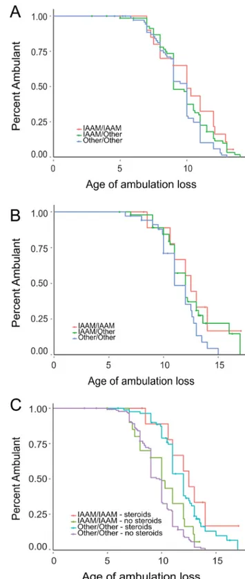
haplotypes are shown in online supplementary table S3 and were in concordance with those published.Table 1shows haplotype frequencies per cohort, highlighting how the IAAM haplotype was more frequent in the UK cohorts compared to the other cohorts. Corticosteroid treatment had a significant protective role ( p<0.001). The IAAM haplotype was associated with a sig-nificant delay in ambulation loss compared to the VTTT haplo-type ( p=0.046;table 2) and to the other haplotypes ( p=0.01; see online supplementary table S4) using the additive model. The additive effect of the IAAM haplotype is visible infigure 2A, B for corticosteroid naïve and also for corticosteroid-treated patients. The effect of the IAAM haplotype was confirmed using the recessive model ( p=0.01;table 2andfigure 2C). Differences in age of ambulation loss were also identified among cohorts; patients from Newcastle, London and Montpellier were signi fi-cantly associated with delayed ambulation loss compared to patients from Ferrara and Leiden (table 2).
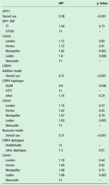
Table 2 Analysis of the effect of steroid use, cohort and genotype on age at wheelchair dependence
HR* p Value SPP1† Steroid use 0.38 <0.001 SPP1 SNP TT 1.04 0.73 GT/GG 1‡ – Cohort London 1.12 0.60 Ferrara 1.72 0.01 Montpellier 1.82 0.002 Leiden 1.8 0.006 Newcastle 1‡ – LTBP4† Additive model Steroid use 0.31 <0.001 LTBP4 haplotype IAAM 0.8 0.046 VTTT 1‡ – other 1.16 0.29 Cohort London 1.16 0.47 Ferrara 1.62 0.03 Montpellier 1.07 0.79 Leiden 1.83 0.005 Newcastle 1‡ – Recessive model Steroid use 0.31 <0.001 LTBP4 diplotypes IAAM/IAAM 1‡ – other diplotypes 1.3 0.01 Cohort London 1.18 0.44 Ferrara 1.69 0.02 Montpellier 1.08 0.75 Leiden 1.88 0.003 Newcastle 1‡ –
*HR above 1 point to an earlier loss of ambulation compared to the reference, while HRs below 1 point to a later loss of ambulation compared to the reference. †The reported values were obtained using a dominant model for the SPP1 SNP and additive and recessive models for theLTBP4 haplotype.
‡These conditions were used as reference, therefore their HRs are set at 1. SNP, single nucleotide polymorphism.
DISCUSSION
DMD is the most common muscular dystrophy and is charac-terised by progressive muscle degeneration. Disease course varies among patients with most of the patients losing ambula-tion between 6 and 12 years of age. The reason behind the dif-ferences in disease progression is so far not clear. However, genetic modifiers in the SPP1 and LTBP4 loci have been pro-posed to contribute to the observed differences. Pegoraro et al reported evidence connecting SNP rs28357094, lying upstream of theSPP1 gene promoter on chromosome 4, to age of wheel-chair dependence. The authors showed that the TT genotype was associated with delayed disease progression in a study of 262 patients derived from two independent cohorts (106 patients from Italy and 156 patients from the USA).5Later on
the same group published a second report in which patients par-ticipated in a 12- month longitudinal study where the 6 min
walk test and the North Start Ambulatory Assessment were used as functional outcome measure; the authors were able to show that patients with the TT genotype progress slower compared to the other group.4 We genotyped SNP rs28357094 in 336 Figure 1 Survival plots showing the effect of single nucleotide
polymorphism (SNP) rs28357094 (SPP1) for 336 patients with Duchenne muscular dystrophy (DMD). Survival plots for
corticosteroid-untreated (A) and corticosteroid-treated (B) patients with DMD. The two survival lines represent patients stratified according to theirSPP1 SNP genotype (blue=TT, green=GT/GG). Censored patients are indicated by a cross on the lines.
Figure 2 Survival plots showing the effect of the IAAM haplotype (LTBP4) in 265 patients with Duchenne muscular dystrophy. Survival plots showing the additive effect of the IAAM haplotype in steroid naïve (A) and corticosteroid-treated (B) patients. (C) Comparison of individuals homozygous for the IAAM haplotype with individuals carrying other diplotypes separated for corticosteroid-treated and corticosteroid-untreated patients.
patients from five independent European cohorts and were unable to replicate the published association between the SNP and age of ambulation loss. The association was also not signi fi-cant when the interaction between corticosteroid treatment and genotype was included in the model. The lack of a clear associ-ation between this SNP and disease progression is in accordance with a recent publication from Flanigan et al,8 who studied a US cohort composed of 254 non-ambulatory patients with DMD. In this study, the authors went further and presented evi-dence of a new genetic modifier composed of four SNPs located in theLTBP4 locus. In line with the scope of this paper to repli-cate known genetic associations in patients with DMD, we also genotyped these 4 SNPs in 265 patients with DMD. Interestingly, we identified a significant association between the LTBP4 haplotype and disease progression. As in the study by Flanigan et al, prolonged ambulation was associated with the IAAM haplotype, which is related to decreased TGF-β signal-ling. TGF-β is known to inhibit differentiation of satellite cells and increase fibrosis in muscle tissue.14–16 Several studies have shown that inhibition of TGF-β function decreases the presence of fibrosis and increases muscle strength in mdx mice.14–18 Reduced TGF-β signalling, as with the IAAM haplotype, could therefore slow-down disease progression in patients with DMD. As expected, in the SPP1 and LTBP4 analyses we observed a strong effect of steroid treatment in delaying disease progression ( p<0.001).11 12 We also observed a significant cohort effect
with patients from the UK and France performing better com-pared to the other cohorts. This may be due to differences in the corticosteroid treatment regimen, to differences in the stan-dards of care, given that the mean age of birth is different among the cohorts and that standards of care improve rapidly, or to differences in the definition of wheelchair dependence among the participating centres. In our study, the definition of ambulation loss was homogeneous within each centre, but it was not standardised among centres. It is therefore possible that part of the significant differences observed among the cohorts is due to different definitions of wheelchair dependence. This can be considered a limit of our study and a general limit in multicen-tre DMD studies where a significant number of patients is included to have sufficient statistical power. A limitation of our approach in replicating the published associations is constituted by the differences in percentage of corticosteroid-treated patients, which are lower compared to the published studies (54% in the study by Pegoraro et al, and 57% in the study by Flaniganet al) and which also greatly vary among the considered clinical centres (table 1). However, differences in the definition of steroid users may partly account for the differences in these percentages as our definition is more conservative compared to the one of Flaniganet al. Another limitation of our study is the use of age at loss of ambulation as the sole measure for disease severity. Although this is one of the most important disease mile-stones, it is not sensitive to small effects. Indeed disease modifiers with a small effect could therefore be overlooked. Future studies should ideally include data about more sensitive measures, such as differences in motor function measures.
In conclusion, in the present study we confirm the disease modifying effect of the IAAM haplotype of theLTBP4 gene, but could not replicate the previously described association between SNP rs28357094 in the SPP1 gene and disease severity in patients with DMD. Future trials in DMD should consider the LTBP4 haplotype before stratification, as it might confound the clinical trial results. This study underlines the possibilities and limitations of genetic association studies in rare diseases with inherently small cohort sizes.
Author affiliations
1
Department of Neurology, Leiden University Medical Center, Leiden, The Netherlands
2
Department of Human Genetics, Leiden University Medical Center, Leiden, The Netherlands
3
Department of Medical Statistics and Bioinformatics, Leiden University Medical Center, Leiden, The Netherlands
4
Department of Clinical Genetics, Leiden University Medical Center, Leiden, The Netherlands
5
Institute of Human Genetics, Newcastle University, International Centre for Life, Central Parkway, Newcastle upon Tyne, UK
6
Dubowitz Neuromuscular Centre, University College London, London, UK
7Children’s National Medical Center, Research Center for Genetic Medicine,
Washington DC, USA
8Laboratoire de Génétique de Maladies Rares, CHU Montpellier, Université
Montpellier 1, UFR Médecine, and Inserm U827, Montpellier, France
9Department of Medical Sciences, Section of Microbiology and Medical Genetics,
University of Ferrara, Ferrara, Italy
10Paediatric Hospital Bambino Gesù, Rome, Italy
Acknowledgements The authors would like to thank Helena ED Suchiman (Department of Molecular Epidemiology, Leiden University Medical Center, Leiden, the Netherlands) for her technical assistance with the Sequenom-based genotyping. Contributors MH, SB, LV, ST-G, AMA-R, PACtH and PS contributed to study concept and design. JCvdB, HBG, AC, KB, VS, MS, SC, VH, MC, CS, CP, HL, FM, ST-G, AF and JJGMV were involved in acquisition of the data. JCvdB, MH, AMA-R, PACtH and PS were involved in drafting of the manuscript. JCvdB, MH, SB, ST-G, AMA-R, PACtH and PS were involved in analysis and interpretation of the data. JCvdB, MH, LV, HBG, AC, KB, VS, MS, SC, VH, MC, CS, CP, HL, FM, ST-G, AF and JJGMV were involved in administrative, technical and material support. PS was involved in study supervision. All authors contributed to critical revision of the manuscript for important intellectual content.
Funding Thefinancial support used for salaries and consumables of the EU-FP7 BIO-NMD project (EC grant number 241665) to thefive participating centres is gratefully acknowledged. Thefinancial support used for consumables of the Association Française Contre les Myopathies (grant number 15092) to PACtH and PS is greatly acknowledged. Support is acknowledged from the European Union Seventh Framework Programme (FP7/2007-2013) under grant agreement No. 305444 (RD-Connect) and 305121 (Neuromics). The MRC Neuromuscular Biobank in London and Newcastle is also gratefully acknowledged.
Competing interests KB reports receiving trial funding from PTC Therapeutics and AVI BioPharma and has served on scientific advisory boards for AVI, GSK/Prosensa, Santhera, PTC Therapeutics, Acceleron, Biomarin and Summit. KB has consultancy agreements in place with Pfizer and Lilly. VS reports having served on scientific advisory boards for Acceleron Pharma, Genzyme, Prosensa, Santhera Pharmaceutical and NicOx. VS has received or receives funding for trials from GSK and Genzyme. All agreements are signed by Newcastle University. MS reports being involved in clinical trials for DMD for GSK and BHF. MS is employed and receives salary from UCL. FM reports having served on scientific advisory boards for Acceleron Pharma, Genzyme, AVI BioPharma, Debiopharma Group, GSK, Prosensa, Servier and Santhera Pharmaceutical. FM serves on the editorial board of Neuromuscular Disorders and Neuropediatrics; receives research support from the European Union, the MRC, the Wellcome Trust, the Association Française Contre les Myopathies (AFM), the Muscular Dystrophy Campaign, the GOSH Biomedical Research Centre and the Muscular Dystrophy Association USA, is receiving funding for trials from GSK and the British Heart Foundation, and has received funding for trials from AVI BioPharma, Trophos and PTC Therapeutics. AF reports being Principal Investigator of ongoing sponsored trials on DMD (Prosensa Therapeutics, NE and GSK, UK). AF is recipient of research funds from EU (BIO-NMD, Neuromics), Telethon Italy, Parent Project Italy and MIUR Italy. AF is a member of the Scientific Committee of ENMC. AMA-R reports being employed by LUMC and receiving salary from LUMC. LUMC has patents on exon skipping, some of which AMA-R is co-inventor on. On sublicensing some of these patents to Prosensa Therapeutics and GSK, AMA-R has received a share of royalty payments from LUMC. JJGMV reports being involved in clinical trials for Duchenne muscular dystrophy for GSK, Prosensa and Santhera. JJGMV is consultant for Prosensa on MRI studies, without receiving personal fees. All payments are made to LUMC. PACtH reports being employed by LUMC and receiving salary from LUMC. LUMC has patents on exon skipping, some of which PACtH is co-inventor on. On sublicensing some of these patents to Prosensa Therapeutics and GSK, PACtH has received a share of royalty payments from LUMC. Ethics approval Local Research Ethics Committee.
Provenance and peer review Not commissioned; externally peer reviewed. Open Access This is an Open Access article distributed in accordance with the Creative Commons Attribution Non Commercial (CC BY-NC 4.0) license, which permits others to distribute, remix, adapt, build upon this work non-commercially, and license their derivative works on different terms, provided the original work is
properly cited and the use is non-commercial. See: http://creativecommons.org/ licenses/by-nc/4.0/
REFERENCES
1 Mercuri E, Muntoni F. Muscular dystrophies. Lancet 2013;9869:845–60. 2 Humbertclaude V, Hamroun DF, Bezzou KF, et al. Motor and respiratory
heterogeneity in Duchenne patients: implication for clinical trials. Eur J Paediatr Neurol 2012;2:149–60.
3 Emery AE. The muscular dystrophies. Lancet 2002;9307:687–95.
4 Bello L, Piva L, Barp A, et al. Importance of SPP1 genotype as a covariate in clinical trials in Duchenne muscular dystrophy. Neurology 2012;2:159–62.
5 Pegoraro E, Hoffman EP, Piva L, et al. SPP1 genotype is a determinant of disease severity in Duchenne muscular dystrophy. Neurology 2011;3: 219–26.
6 Hoffman EP, Gordish-Dressman H, McLane VD, et al. Alterations in osteopontin modify muscle size in females in both humans and mice. Med Sci Sports Exerc 2013;6:1060–8.
7 Vetrone SA, Montecino-Rodriguez E, Kudryashova E, et al. Osteopontin promotes fibrosis in dystrophic mouse muscle by modulating immune cell subsets and intramuscular TGF-beta. J Clin Invest 2009;6:1583–94.
8 Flanigan KM, Ceco E, Lamar KM, et al. LTBP4 genotype predicts age of ambulatory loss in Duchenne muscular dystrophy. Ann Neurol 2012;4: 481–8.
9 Heydemann A, Ceco E, Lim JE, et al. Latent TGF-beta-binding protein 4 modifies muscular dystrophy in mice. J Clin Invest 2009;12:3703–12.
10 Bushby K, Finkel R, Birnkrant DJ, et al. Diagnosis and management of Duchenne muscular dystrophy, part 1: diagnosis, and pharmacological and psychosocial management. Lancet Neurol 2010;1:77–93.
11 Manzur AY, Kuntzer T, Pike M, et al. Glucocorticoid corticosteroids for Duchenne muscular dystrophy. Cochrane Database Syst Rev 2008;(1):CD003725.
12 Mendell JR, Moxley RT, Griggs RC, et al. Randomized, double-blind six-month trial of prednisone in Duchenne’s muscular dystrophy. N Engl J Med 1989;24:1592–7. 13 Gundry CN, Dobrowolski SF, Martin YR, et al. Base-pair neutral homozygotes can be discriminated by calibrated high-resolution melting of small amplicons. Nucleic Acids Res 2008;10:3401–8.
14 Allen RE, Boxhorn LK. Inhibition of skeletal muscle satellite cell differentiation by transforming growth factor-beta. J Cell Physiol 1987;3:567–72.
15 Bernasconi P, Torchiana E, Confalonieri P, et al. Expression of transforming growth factor-beta 1 in dystrophic patient muscles correlates withfibrosis. Pathogenetic role of afibrogenic cytokine. J Clin Invest 1995;2:1137–44.
16 Li Y, Foster W, Deasy BM, et al. Transforming growth factor-beta1 induces the differentiation of myogenic cells intofibrotic cells in injured skeletal muscle: a key event in musclefibrogenesis. Am J Pathol 2004;3:1007–19.
17 Bogdanovich S, Krag TO, Barton ER, et al. Functional improvement of dystrophic muscle by myostatin blockade. Nature 2002;6914:418–21.
18 Wagner KR, McPherron AC, Winik N, et al. Loss of myostatin attenuates severity of muscular dystrophy in mdx mice. Ann Neurol 2002;6:832–6.