Università degli Studi di Ferrara
DOTTORATO DI RICERCA IN
FARMACOLOGIA E ONCOLOGIA MOLECOLARE
CICLO XXIII
COORDINATORE Prof. Cuneo Antonio
LOCAL SUPPLEMENTATION OF FGF-2 AND
BDNF IN THE EPILEPTOGENIC HIPPOCAMPUS.
EFFECTS AND DELIVERY STRATEGIES.
Settore Scientifico Disciplinare BIO/14
Dottorando Tutore
Dott.ssa Bovolenta Roberta Prof. Simonato Michele
_______________________________ _________________________
I
TABLE OF CONTENTS
TABLE OF CONTENTS ... I TABLE OF ARTICLES...II RIASSUNTO ...1 ABSTRACT ...3GENERAL STRUCTURE OF THE THESIS ...5
1. Epilepsy...8
1.1 History and definition...8
1.2 Epidemiology. ... 9
1.3 Classification of epileptic seizures. ...10
1.4 Treatment. ... 11
1.5 Temporal Lobe Epilepsy. ... 13
1.6 Experimental models of TLE. ... 14
2. Epileptogenesis...19
2.1 Definition and peculiarities. ... 19
2.2 Genetic and epigenetic mechanisms of epileptogenesis. ...20
2.3 Acquired postinjury mechanisms. ...21
Neurodegeneration... 21
Neurogenesis (for details see ...23
Dendritic plasticity and changes in extracellular matrix. ... 24
3. Neurotrophic factors ...26 3.1 Family of neurotrophins... 26 p75NTR. ...29 BDNF...31 3.2 FGF-2. ... 33 3.3 Therapeutical application of NTFs...35
II
5. Influence of NTFs on mossy fibres sprouting ...56
6. References ...65
1. NTFs routs of administration. ...86
1.1 Generalities. ... 86
1.2 Viral vectors-based administration. ...87
1.3 Stem cell-based administration... 87
Mesoangioblasts (MABs). ...88
2. References ...149
OVERALL CONCLUSIONS AND FUTURE PERSPECTIVES...152
TABLE OF ARTICLES
Hippocampal FGF-2 and BDNF overexpression attenuates epileptogenesis-associated neuroinflammation and reduces spontaneous recurrent seizures……….. 39Localized overexpression of FGF-2 and BDNF in hippocampus reduces mossy fiber sprouting and spontaneous seizures up to four weeks after pilocarpine-induced status epilepticus………. 54
By-stander effect on brain tissue of mesoangioblasts producing neurotrophins………89
The biocopatibility of materials used in printed circuit board tecnologies with respect to primary neuronal and K562 cells………..151
1
RIASSUNTO
Tra le sindromi epilettiche, l’epilessia del lobo temporale (TLE) è sicuramente la forma più frequente nell’adulto. Si manifesta in seguito ad un danno cerebrale (infezione virale, ictus, trauma, neoplasie…), capace di innescare una cascata di eventi che culminano nella comparsa di crisi epilettiche spontanee, in molti casi difficilmente controllabili con la normale farmacoterapia. Il periodo che intercorre fra l’insulto iniziale e lo sviluppo di crisi spontanee viene definito “epilettogenesi”. Le alterazioni cellulari e tissutali che avvengono durante questa fase interessano principalmente la regione ippocampale e includono: neurodegenerazione, neurogenesi, neuroinfiammazione e gliosi reattiva, angiogenesi e riorganizzazione dei circuiti cerebrali.
Recentemente è stato dimostrato che la supplementazione di fattori neurotrofici (NTFs), quali FGF-2 (fibroblast growth factor-FGF-2) e BDNF (brain derived neurotrophic factor), produce effetti anti-epilettogenici riducendo la morte neuronale, favorendo una neurogenesi non aberrante e ristabilendo un corretto equilibrio tra circuiti inibitori ed eccitatori.
Nel principale lavoro riportato in questa tesi, abbiamo voluto osservare se tale trattamento è in grado di influire anche sui processi neuroinfiammatori. Per lo studio è stato utilizzato il modello sperimentale della pilocarpina, la cui somministrazione è in grado di indurre nell’animale uno stato epilettico (SE) e quindi una lesione epilettogena. Dopo tre giorni dallo SE, vettori virali erpetici esprimenti FGF-2 e BDNF sono stati inoculati nell’ippocampo.
Dopo 4, 11 e 25 giorni dal trattamento (DAI), gli animali sono stati sacrificati e i loro cervelli prelevati per analizzare l’espressione di tre marcatori dell’infiammazione: IL-1β, GFAP (marker di astrocitosi), Ox42 (marker di microgliosi). I risultati ottenuti dimostrano una riduzione particolarmente marcata di IL-1β, evidente già dopo 4 giorni dall’inoculazione del vettore virale, e un’attenuazione più ritardata, ma pur sempre significativa, degli altri due markers studiati.
Lo sprouting delle mossy fibers è un’altra caratteristica aberrante del tessuto ippocampale epilettico: gli assoni delle cellule dei granuli formano sinapsi eccitatorie con cellule normalmente non innervate, formando un circuito che potrebbe contribuire all’ipereccitabilità tissutale. I risultati ottenuti dimostrano che il trattamento con i fattori neurotrofici è in grado di ridurre lo sprouting aberrante delle fibre nervose, in un modo che correla con l’attenuazione del danno cellulare.
2 Studi comportamentali paralleli hanno inoltre evidenziato la capacità del trattamento di ridurre la frequenza e la gravità delle crisi spontanee che, in questo modello, insorgono dopo circa 21 giorni dallo stato epilettico.
Nonostante i promettenti risultati, l’applicabilità clinica dei fattori neurotrofici è limitata dalla scelta di una corretta via di somministrazione. Negli esperimenti riportati in questa tesi sono stati impiegati vettori virali erpetici, defettivi nella replicazione ed esprimenti i due NTFs. Tuttavia, la loro tossicità residua li rende inadeguati a essere applicati all’uomo. Cellule staminali modificate per esprimere i geni di interesse, tra cui i mesangioblasti (MABs), hanno dimostrato in vitro la capacità di promuovere la differenziazione, la sopravvivenza e la funzionalità neuronale. Non ultima, la capacità di localizzarsi nella sede cerebrale danneggiata, quando somministrati per via sistemica, rende queste cellule delle valide alternative a trattamenti più invasivi.
Sebbene siano richiesti ulteriori studi, i risultati raccolti in questa tesi rappresentano un importante contributo alla comprensione delle molteplici proprietà dei NTFs. Inoltre, la caratterizzazione di una via di somministrazione alternativa e maggiormente applicabile può avvicinare la terapia genica con fattori neurotrofici all’utilizzo clinico in numerose patologie neurodegenerative.
3
ABSTRACT
Among the epileptic syndromes, temporal lobe epilepsy (TLE) is the most common form in adults. It is the consequence of a brain damage (viral infection, stroke, trauma, cancer ...), capable of triggering a cascade of events culminating in the appearance of spontaneous seizures that are, in many cases, difficult to control with the usual drug therapy. The period that elapses between the initial insult and the development of spontaneous recurrent seizures (SRSs) is defined "epileptogenesis”. The cellular and tissue changes that occur during this phase mainly interest the hippocampal region and include: neurodegeneration, neurogenesis, neuroinflammation and reactive gliosis, angiogenesis, and reorganization of brain circuits.
Recently, it was shown that supplementation of neurotrophic factors (NTFs), such as FGF-2 (fibroblast growth factor-2) and BDNF (brain derived neurotrophic factor), has anti-epileptogenic effects by reducing neuronal death, favoring a correct neurogenesis and restoring a proper balance between excitatory and inhibitory circuits.
In the main study reported in this thesis, we examined if this treatment can also affect neuroinflammatory processes. We used the pilocarpine model, in which an episode of status epilepticus (SE) is followed by an epileptogenic lesion. After three days, herpes viral vectors expressing FGF-2 and BDNF were injected in the hippocampus.
Four, 11 and 25 days after treatment (DAI), animals were sacrificed and their brains removed to analyze the expression of three markers of inflammation: IL-1β, GFAP (a marker of astrocytosis), Ox42 (marker of microglia). The results show a very marked reduction of IL-1β expression, evident as early as 4 days after inoculation of the viral vector, and delayed, but significant, attenuation of the other two markers.
The sprouting of mossy fibers is another characteristic of the epileptic hippocampal tissue, in which the axons of granule cells form excitatory synapses with cells not usually innervated, forming a circuit that may favour hyperexcitability. The results show that treatment with neurotrophic factors reduce aberrant sprouting of nerve fibers, in a way that correlates with the attenuation of cellular damage.
4 Parallel behavioral studies have also highlighted the ability of the treatment to reduce the frequency and severity of SRSs that, in this model, begin to occur about 21 days after status epilepticus.
Despite the promising results, clinical applicability of neurotrophic factors is limited by the choice of an appropriate route of administration. In the experiments reported in this thesis, herpes viral vectors have been used. These vectors were replication defective and engineered to express the two NTFs. However, their residual toxicity makes them unsuitable for human application. Stem cells modified to express genes of interest, including mesangioblasts (MABs), have demonstrated, in vitro, the ability to promote differentiation, survival and neuronal function. Last but not least, the ability to localize in the damaged site when systemically administered makes these cells viable alternatives to more invasive treatments.
Although further investigations are required, the results collected in this thesis are an important contribution to the understanding of the multiple effects of NTFs. In addition, the characterization of an alternative and more applicable route of administration renders gene therapy with neurotrophic factor more applicable for the treatment of several neurodegenerative diseases.
5
GENERAL STRUCTURE OF THE THESIS
Several histological features characterize an epileptic brain tissue. They include loss of neuronal cells, neurogenesis and reorganization of hippocampal neuronal circuits, like sprouting of mossy fibers (MFS). Epileptic tissue also presents strong neuroinflammation with an important reactive astrogliosis and microgliosis and expression of several pro-inflammatory cytokines.
Recently, it has been demonstrated that the local over expression of neurotrophic factors (NTFs), such as FGF-2 (fibroblast growth factor-2) and BDNF (brain derived neurotrophic factor), has antiepileptogenic effects (Paradiso et al. 2009).
This thesis is organized in two chapters. Works described in the first chapter, investigate the possibility that these anti-epileptogenic effects of NTFs might involve anti-inflammatory mechanisms and affect sprouting of mossy fibers.
The second chapter is a brief excursus on the routes of administration suitable for driving a localized, safe and non-invasive supplementation of NTFs. In particular, it focuses on a new promising approach based on stem-cells gene therapy that employs mesoangioblasts.
Finally, an “addendum” collects two other works. The first is a study, conducted in collaboration with an Italian pharmaceutical company, on the evaluation of the central side effects of new generation phosphodiesterase 4 inhibitors. The second deals with the analysis of in vitro compatibility of materials used in lab on a chip (LOAC) production. Biocompatibility was analyzed on cells growing in suspension and on hippocampal primary cultures, the latter obtained following a protocol optimized in our laboratory.
6
Chapter I
7 The history of epilepsy can be summarized as 4.000 years of ignorance, superstition and stigma, followed by 100 years of knowledge, superstition and stigma.
(Kale R.)
Chapter I
8
1. Epilepsy
1.1 History and definition.
About epilepsy has been spoken and written for over 4.000 years. Characteristics of the illness such as the forced cry, the loss of consciousness, the fall, the twitching and the foaming at the mouth, have suggested, for several centuries, an association with possession of the spirits or prophetic experiences.
Symptoms are mentioned in the Bible in the Gospel of Mark, wherein Christ expels demon from a young person suffering from seizures: “Teacher, I brought you my son, who is possessed by a spirit that has robbed him of speech. Whenever it seizes him, it throws him to the ground. He foams at the mouth, gnashes his teeth, and becomes rigid. I asked your disciples to drive the spirit out, but they could not.” (Mark 9:14-29).
Through the centuries, many misconceptions about this condition have been conveyed based mainly on the particular era or in particular part of the world.
Disagreeing with the idea that epilepsy is a curse or a visionary power, in the 4th century B.C., Hippocrates, in his “On Sacred Disease”, stated “ …it appears to me to be nowise more divine nor more sacred than other diseases, but has a natural cause from the originates like other affections. … And this notion of its divinity is kept up by their inability to comprehend it and the simplicity of the mode by which it is cured, for men are freed from it by purifications and incantations.”
Although the Greek physician recognized that epilepsy was a brain disorder, false ideas continued to exist. In 1494, two Dominican friars, under papal authority, wrote a handbook on witch-hunting, the “Malleus Maleficarum”, in which they said that one of the ways of identifying a witch was by the presence of seizures. This book guided a wave of persecution and torture, which caused the deaths of more than 200.000 women.
And misinterpretation continued for many years: in the early 19th century, people who had critical epilepsy and people with psychiatric disorders were cared for in asylums, but the two groups were kept separated because seizures were thought to be contagious.
For the first, clinically correct, definition of epilepsy is necessary to wait for the 1870, when the neurologist John Hughlings Jackson defined a seizure as “an occasional, excessive and disorderly
Chapter I
9 discharge of nerve tissue on muscles”. He also recognized that seizures can alter consciousness, sensation, and behaviour.
Even if several headways have been made to define epilepsy, some prejudices remain still now. Recently, in nineties, some U.S. states had laws forbidding people with epilepsy to marry or become parents, and some states permitted sterilization.
Today is well known that epilepsy is not one condition, but a diverse family of disorders, having in common an abnormally increased predisposition to seizures. For the International League Against Epilepsy (ILAE) and the International Bureau for Epilepsy (IBE), to define the illness is necessary to satisfy three elements: “Epilepsy is a disorder of the brain characterized by (1) an enduring predisposition to generate epileptic seizures and by (2) neurobiological, cognitive, psychological and social consequences. The definition of epilepsy requires also the occurrence (3) of at least one epileptic seizure” defined, the latter, as “a transient occurrence of signs and symptoms due to abnormal, excessive or synchronous neuronal activity in brain” (Fisher et al., 2005).
Unfortunately, the stigma associated to epilepsy has still a great influence on the education and social life of patients and quite often leads to isolation, restrictions and/or overprotection. For these and other reasons (i.e. economic, legislative…) epilepsy remains a central point in the scientific community.
1.2 Epidemiology.
Epilepsy is one of the most common chronic neurological conditions. About 1% of the world’s population is affected by epilepsy and up to 5% may have a single seizure at some time in their lives. Incidence and prevalence studies are critical to provide measures of frequency and therefore the burden of disease, and allow for proper planning of services.
Prevalence is an estimate of the number of people with epilepsy in a given population at a specified time (point prevalence), or during a defined time interval (period prevalence). In most countries worldwide, the prevalence of active epilepsy ranges from 4 to 10/1000.
The incidence, instead, is the number of new cases per year. The incidence of epilepsy ranges from 40 to 70/100.000 in most developed countries and is nearly double in developing countries. These
Chapter I
10 discrepancies are due to the higher risk of experiencing a condition which can lead to permanent brain damage like meningitis, malaria, pre and perinatal complications and malnutrition (from Epilepsy in the WHO European Region: Fostering Epilepsy Care in Europe, 2011).
The age distribution of the incidence of epileptic seizures is bimodal, with two peaks of frequency in childhood (in 50-60% of patients, epilepsy begins before the age of 16) and seniority. In Western countries there is evidence of a decreasing incidence in children, with a simultaneous increase in elderly, related to improved life expectancy associated with an increased risk for causes of epilepsy common in old age (Hauser, 1992; Stephen and Brodie, 2002).
Differences in incidence rates in males and females are not statistically significant. There is no evidence of racial predilection, even if the incidence is significantly higher in the lower socioeconomic classes. (Sridharan, 2002).
1.3 Classification of epileptic seizures.
In 1981 ILAE divided seizures into three typologies, with subtypes of each: partial, with seizures involving only part of the brain; generalized, with seizures involving both hemispheres, and unclassifiable seizures.
This relatively easy classification appeared soon inadequate to respond to the vast heterogeneity of seizure types. For this reason a supplement to the previous classification was proposed in 1989 in which appears, for the first time, the new term of “epileptic syndrome”: “An epileptic syndrome is defined as a disorder characterized by a cluster of signs and symptoms occurring together”.
According to this system, epileptic syndromes are divided into four broad groups: localization-related that involves one or more distinct parts of the brain, generalized that involves both hemispheres at the same time, undetermined whether localized or generalized and, finally, special syndromes.
Within the localized and generalized groups, there are further subdivisions into idiopathic, symptomatic or cryptogenic. In idiopathic epilepsies, causes are unknown but genetic factors provoking, for example, channelopathies, are presumed to have a major causative role in the development of seizures. In symptomatic epilepsies there is an identifiable lesion in the brain that triggers seizures. The term “cryptogenic” has been recently substituted with “presumed
Chapter I
11 symptomatic epilepsy” in which some brain pathology causing epilepsy is presumed to exist, but has not been identified using current techniques (Engel, 2006).
1.4 Treatment.
The most common therapeutic approach of epilepsy is with antiepileptic drugs (AEDs). During the last 20 years there has been a dramatic increase in the therapeutic options available (Table 1).
Antiepileptic medicines Year of introduction
Phenobarbital 1912 Phenytoin 1939 Ethosuximide 1955 Primidone 1960 Carbamazepine 1965 Valproate 1970
Vigabatrin 1989 serious side effects, use restricted Oxcarbazepine 1990
Lamotrigin 1991
Gabapentin 1994
Felbamate 1994 serious side effects, use restricted
Topiramate 1995
Tiagabine 1996 marketed at a very low scale Levetiracetam 2000
Pregabaline 2005
Zonisamide 2007
Lacosamide (Vimpat) 2008 Eslicarbazepine acetate 2009
Table 1: principal antiepileptic drugs currently available.
The success rate of both the older and newer drugs is similar and leads to seizures freedom for up to 70% of patients, even if, the newer drugs, may have less side effects and lead to a better quality of life. Moreover, new promising compounds are actually in clinical trials, they include Brivaracetam (UCB; phase III), Carisbamide (Johnson and Johnson; phase III), Retigabine (Valeant Pharmaceutical and GSK; phase III) and Valrocemide (Tera/Acorda; phase III).
Chapter I
12 Unfortunately all these drugs are only anticonvulsivant: they act on symptoms reducing recurrent seizures, but their mechanisms of action do not influence the causes of epilepsy. In some cases, monotherapy is sufficient to contain seizures but, more often, a cocktail of different drugs is necessary. The choice among different AEDs available depends on causes, history, evolution of pathology and crisis and, especially for polytherapy, possible pharmacological interactions. Despite the accessibility to a wide range of molecules, between 30 and 40 % of patients with epilepsy continue to have seizures that are not adequately controlled by pharmacotherapy (Kwan and Brodie, 2000).
A solution, in these cases, might come from surgery, with the resection of epileptic area. To be applied, it requires not only a history of intractable epilepsy, but also a focal and circumscribed lesion and a very low risk to develop neurosurgical or cognitive damages. When applied, surgery reaches 90% of success. In a study of surgical versus pharmacological treatment in poorly controlled temporal lobe epilepsy, has been report that, after one year, 64% of the patients operated, but only 8% of those medically-treated, are seizures free (Wiebe et al.; 2001).
In patients refractory to drug therapy and/or in which surgery could not be applied or results ineffective, a valid alternative may be the Vagus Nerve Stimulation (VNS). It consists of regular, mild pulses of electrical energy to the brain via the vagus nerve. These pulses are supplied by a device placed under the skin on the chest wall, connected with a wire to the vagus nerve in the neck. Little is known about the mechanism of action, but a significant reduction in frequency, intensity and duration of seizures, especially when associated to normal pharmacological treatment, has been demonstrated.
Based on dietary approach, ketogenic diet is indicated as an additional treatment in children with drug-resistant epilepsy. It consists of a diet high in fat and low in carbohydrates and proteins. Under these conditions, the liver converts fat into fatty acids and ketone bodies that, passing into the brain, replace glucose as an energy source. Elevated levels of ketone bodies in the blood, a state known as ketosis, lead to a reduction in the frequency of epileptic seizures. Even if side effects are less important than those of several drugs, patients treated for a long time with ketogenic diet may experience kidney stones, high cholesterol levels in blood, dehydration, bone fractures, slowed growth or weight gain and hypovitaminosis that require a careful control by physician. Although the mechanisms underlying anti epileptic effects are unknown, ketogenic diet has reported peaks of 90% of reduction in seizures frequency (Neal et al.; 2008).
Chapter I
13 Over the years, great progresses have been made in the treatment of epilepsy. With continued cooperation among clinicians, geneticists and basic scientists, will be possible to achieve the goal of curing epilepsy, or preferably, preventing the process that lead to the acclaimed illness.
Among epileptic syndromes, temporal lobe epilepsy (TLE) is the most common serious neurological condition in adulthood, probably affecting at least 20% of all patients with epilepsy.
1.5 Temporal Lobe Epilepsy.
TLE was defined in 1985 by the ILAE as a condition characterized by recurrent seizures originating from the medial or lateral temporal lobe. Medial temporal lobe epilepsy (MTLE) arises in the hippocampus, parahippocampal gyrus and amygdale, lateral temporal lobe epilepsy (LTLE) arises in the neocortex on the outer surface of the temporal lobe of the brain.
Seizures may involve only one or both lobes, giving rise to simple (without loss of consciousness) partial, complex (associated with loss of consciousness) partial or secondarily generalized seizures. About 40% to 80% of people with TLE also perform repetitive, automatic movements (called automatisms), such as lip smacking and rubbing the hands together. As seizures usually involve areas of the limbic system which control emotions and memory, some individuals may have problems with memory, especially if seizures have occurred for more than 5 years. However these memory problems are almost never severe. Seizures occur after an initial insult like an infection, stroke or trauma, vascular malformation or prolonged febrile seizures; a genetic cause is less frequent. Between the initial insult and the onset of the crisis, a so called latent period characterized by the absence of seizures, occurs. During this period, changes in structure and physiology of the brain tissue (“epileptogenesis”) happen.
The most common lesional abnormality identified in patients with TLE is the Hippocampal Sclerosis (HS) (Babb and Brown; 1987). It is characterized by severe loss of the principal neurones associated with widening of the granule cell layer of the dentate gyrus, termed granule cell dispersion (GCD), which is observed in about 40-50% of surgical temporal lobe specimens (Houser et al., 1990 b;Lurton et al., 1998;El Bahh et al., 1999;Thom et al., 2002; Blümke et al. 2002). The other molecular, cellular and plastic alterations in hardened and sclerotic hippocampus will be described in detail further on.
Chapter I
14 In spite of the large diffusion, the study of epilepsy can not be performed on humans due to several reasons: ethical issues, unavailability of controls and high costs of human research. Besides, TLE is the most common form of drug-refractory epilepsy. Studies on human specimens are affected by several bias because of patients go toward surgery when pharmacotherapy fails. In these conditions is, thus, difficult to discriminate between the effects of the illness and those of a prolonged therapy. This is the reason for whom, to study epilepsy, animal models are of central importance.
1.6 Experimental models of TLE.
An animal model for a pathology could be isomorphic, if duplicates the disorder but not the underlying aetiology, or predictive, if it does not resemble the human disorder but allows prediction about it or its response. In the case of epilepsy, several models (i.e. electrical, chemical, genetics…) are available; they are resumed in table 2.
cell culture models single nerve cells
acutely dissociated from animal and human brain In v it ro m od el s in vitro isolated guinea pig brain GABA • Pentylenetetrazole • Bicuculline • Picrotoxin • Glutamic Acid Decarboxylase (GAD) • inhibitors
• Beta carbolines and convulsant • benzodiazepine Ro 5-3663 • GHB (gamma-hydroxy-butyrate) Excitatory Amino-Acid • Kainic acid • Quisqualic acid/alfa-amino-3-Hydroxy-5- • Methyl-4-Isoxasole Propionic acid • N-Methyl-D-Aspartic acid (NMDA) • Homocysteine, homocysteic acid In v iv o m od el s chemical models of epilepsy
Acetylcholine related substances
• Pilocarpine, and litium-pilocarpine • Organophosphorus compounds Other drugs • Strycnine • Aminophylline • Insulin induced hypoglycemia • Ay-9944 • THIP (4,5,6,7 tetrahydroxyisoxazolo • (4,5,c) pyridine 3-ol)
Chapter I
15
Table 2: in vitro, in vivo and genetic experimental models of epilepsy.
Inhalants
• Fluorothyl
Topical application
• Metals (cobalt, zinc, antimony, alumina • cream, iron
• Antibiotic (penicillins and cephalosporins)
• Tetanus toxin
Electrical models
• electroshock seizures
• local electrical stimulation
• electrical kindling
• self sustaining status epilepticus by Perforant Path stimulation
• self sustaining status epilepticus by Amygdala stimulation
• focal status epilepticus by perforant path stimulation in anesthetized rats
• continuous hippocampal stimulation
Lesion models of epilepsy
• cortical freeze lesion model
• antiproliferative agents (5-azacytidine, methyl-mercury, nitrosureas and carmustine)
• Methylazoxymethanol acetate (MAM) model
• In-Utero irradiation as a model of cortical dysplasia
• Hypoxia-induced seizures and hypoxic encephalopathy in neonatal period
• Lateral fluid percussion brain injury
• Chronic Partial Cortical Isolation model
• Head Trauma: haemorrhage-Iron Deposit
• Stroke
Others
• Complex febrile seizures –
• experimental model in immature
• rodents
• Infection induced seizures
• Rasmussen’s encephalitis model
Absence model • GAERS rats
• WAG/Rij rats Spike wave • Tottering • lethargic • ducky • stargazer • SWE • Mocha2j • coloboma Convulsion • Dilute lethal • jimpy • jittery • megencephaly • quaking • staggerer • torpid • veritint waddler • wabbler-lethal • weaver • writher Genetic models
Convulsions evoked by sensory stimuli • frings
Chapter I
16 For TLE, the pilocarpine model is a highly isomorphic model, described for the first time in 1983 by Turski and colleagues.
In this model, administration of pilocarpine in rats induces a status epilepticus (SE) characterized by tonic-clonic generalized seizures, followed by a latent period of seizures free behavior and by a chronic period, with the occurrence of spontaneous recurrent seizures (SRSs).
In detail: within 5 minutes from the injection, animals begin to be motionless, display oro-facial movements, salivation, eye blinking, twitching of vibrissae and yawning. Discontinuous seizures are observed at 30 minutes and they last up to 90-150 minutes, until limbic motor seizures with intense salivation, rearing, upper extremity clonus and falling, begin (Turski et al., 1983). Around 60% of the animals treated, successfully develop SE (Cavalheiro et al., 1991). All these behavioral changes are correlated to an high voltage, fast electroencephalographic (EEG) activity that appear to originate in the hippocampus and to propagate to the amygdala and neocortex (Turski et al., 1983). SE spontaneously remits within 12 hours after pilocarpine administration (Cavalheiro, 1995), then the animals enter post-ictal coma, lasting 1-2 days (Turski et al., 1983). The dose of alkaloid that reproduces the syndrome in rats is 300-400 mg/Kg. With these concentrations the mortality rate, at the time of the injection, has been reported to be around 30-40% (Turski et al., 1983; Cavalheiro et al., 1991; Liu et al., 1994), even if it can be reduced by stopping SE with anticonvulsant drugs such as diazepam. Our observations demonstrate that, after a three hours SE, animals experience some occasional, self-limiting generalized seizures of less than 1 min duration for 1-3 days (Mazzuferi et al., 2010).
The latent period starts after status epilepticus recover. Its duration varies in function of the dose of pilocarpine (Liu et al., 1994), lenght of SE (Lemos and Cavalheiro, 1995; Fujikawa, 1996, Biagini et al., 2006, Goffin et al., 2007), strain and age of the animal (Biagini et al., 2006; Goffin et al., 2007). Cavalheiro and collaborators (1991) defined a mean time interval of 14.8 days. During the latent phase, tissue rearrangements related to epileptogenesis occur (Dalby and Mody, 2001; Pitkänen and Sutula, 2002).
Chronic period follows epileptogenesis and, as previously mentioned, is characterized by the appearance of spontaneous recurrent seizures. Gravity of seizures can be established by a score system developed in 1972 by Racine and recently revised (Veliskova, 2006). It is reported in Table 3. As schematized, it uses numbers from 1 to 6 to define seizure classes: the firsts three are representative of partial seizures, from 4 to 6 of generalized ones. SRSs begin as partial seizures and become secondary generalized. The recurrence of seizures is almost regular throughout the lifetime
Chapter I
17 of the animal appearing in a clustered way, in a cycle peaking every 5-8 days or more (Goffin et al., 2007; Arida et al., 1999). Seizures frequency is higher during the diurnal period (Arida et al., 1999), with an incidence of 67% (Arida et al., 1999; Goffin et al., 2007). In 90% of the cases, EEG trace is characterized by a cerebral activity that starts from the hippocampus and spreads to the neocortex, usually lasts less than 60 seconds (Cavalheiro et al., 1991).
Classes Features 1 Staring and mouth clonus
2 Automatisms
3 Monolateral forelimb clonus
P
ar
tia
l
4 Bilateral forelimb clonus
5 Bilateral forelimb clonus with rearing and falling
6 Tonic-clonic seizures G en er ali ze d
Table 3: modify Racine’s scale. Scores from 1 to 3 are representative of partial seizures, the last three of generalized ones.
Pilocarpine exerts its effects by binding M1 muscarinic subtype receptors, resulting in alteration in Ca2+ and K+ currents (Segal, 1988). The high concentration of intracellular Ca2+ promotes the release of glutamate from presynaptic termini that, in turn, provokes the SE. Once activated, seizures are subsequently maintained by activation of NMDA receptors. Glutamate, acting on AMPA/KA receptors, allows the entrance of Na+ and Ca2+ into the cells and, as consequence, the Mg2+, which blockades the NMDA receptor, is removed. In this way it can be activated by glutamate, allowing the entrance of more Ca2+ into the postsynaptic cells and inducing excitotoxic effects and cell death.
Recent observations have shown that activation of cholinergic neurons may not be the only factor triggering pilocarpine SE. Marchi and collaborators (2007 a, b) suggested that pilocarpine induces an early focal damage to blood-brain barrier (BBB) in regions highly sensitized to cholinergic agonists, that may contribute to the development of seizures, facilitating the entrance into the brain of blood-borne factors (e.g. K+). Moreover, peripheral activation of the immune system has been hypothesized, since high levels of serum IL-1β have been found after injection of pilocarpine. High concentration of the pro-inflammatory citokine is known to cause sudden rapid changes in excitability of both inhibitory and excitatory neurons (Plata-Salamán and Ffrench-Mullen, 1992; Sawada et al., 1992; Yang et al., 2005).
Chapter I
18 As reviewed by Curiaet al. (2008), age, strain, gender, dose, association with other drugs to reduce mortality or collateral effects, are variables that have to be carefully controlled to obtain a reliable model of the pathology. The lack of a standardized protocol has led some investigators to criticize this model. However it continues to be used in many laboratories, including our, because very easy, rapid and shows a high homology with human disease, involving the same mechanisms and brain areas and showing the same pattern of responsiveness to AEDs seen in TLE patients.
Chapter I
19
2. Epileptogenesis
2.1 Definition and peculiarities.
The term epileptogenesis refers to the phenomenon in which various kinds of brain insults (e.g. traumatic brain injury, stroke, infection, febrile seizure…) trigger a cascade of events that eventually culminate in the occurrence of spontaneous seizures. The period required for this long-lasting transformation of the brain, may vary from weeks to years. Epileptogenesis can occur in various ways, generally divided into genetic and acquired mechanisms. Genetic influence is supposed to be strongest in idiopathic epilepsies, whereas mechanisms of circuitry reorganization, after a brain insult, are most extensive in the acquired symptomatic ones. It should be noted that these mechanisms are not separate realities but, in some cases, functional consequences of brain injury can depend on genetic background (Fig. 1) (Pitkänen and Lukasiuk, 2009).
Chapter I
20
2.2 Genetic and epigenetic mechanisms of epileptogenesis.
Over 13 genes associated with human epilepsy have been identified so far and at least 33 single gene mutations in mice have been linked to an epileptic phenotype.
Benign familial neonatal convulsion (BFNC), generalized epilepsy with febrile seizures plus (GEFS+) and autosomal dominant nocturnal frontal lobe epilepsy (ADNFLE) are three idiopathic diseases in which gene mutations codify for mutated voltage-gated or ligand-gated channels.
In particular, in BFNC, mutations were identified in genes for potassium (K) channels (Singh et al., 1998; Biervert et al., 1998) named KCNQ2 and KCNQ3. These mutations are responsible of a loss of function that leads to a decrease in the size of the potassium current. It has been suggested that, even a moderate reduction (20-25%) of function, may be associated with epilepsy (Schroeder et al., 1998).
GEFS+ is associated with a point mutation in the gene coding for the b-subunit of a voltage-gated sodium channel (SCN1B) and, as found more recently, in the a1-sodium channel subunit (SCNA1A) (Wallace et al., 1998; Escayg et al., 2000). In vitro studies suggest that the mutation results in defective inactivation of the sodium channel, which could lead to failure to limit the sustained repetitive firing of a depolarized neuron (McNamara, 1999).
ADNFLE begins clinically in childhood and patients have brief, nocturnal seizures with motor features. Mutations affect genes codifying for nicotinic cholinergic receptors (Steinlein et al., 1997; Phillips et al. 1998) and result in decreased Ca2+ flux through the receptor, which may lead to a reduction in the amount of GABA released from presynaptic terminals, and trigger a seizure by synaptic disinhibition (Kuryatov et al., 1997).
Pathologies described up to here, are characterized by complex inheritance patterns, in which several loci have been identified, but the underlying genetic defects remain to be determined.
PME (progressive myoclonus epilepsy) is a group of rare single-gene epilepsies characterized by myoclonus, generalized tonic-clonic seizures and progressive neurological dysfunction mainly in the form of dementia and ataxia (Berkovic et al., 1986). Among these, Unverricht-Lundborg Disease (ULD) and Lafora Disease (LD) are the most well characterized. Specific genetic mutations are associated with a deficit of two proteins, cystatinB and laforin respectively, that result in epilepsy (Acharya, 2002).
Chapter I
21 Some other changes in genes occur without directly affecting DNA sequence but by chemical modification of DNA or chromatin, such as DNA methylation and alterations in methylation or acetylation status of histones. These mechanisms are termed epigenetic. Seizure or SE-induced histone modifications have been reported for promoters of a number of genes, including those involved in neuronal plasticity such as c-fos, c-jun and CREB (Tsankova et al., 2004; Sng et al., 2005; Sng et al., 2006). Deacetylation of histones at the GluR2 promoter leads to its decreased expression, resulting in enhanced epileptogenesis (Sanchez et al., 2001). Involvement of epigenetic mechanisms have been sustained by the discovery that some AEDs can influence gene expression and, consequently, cellular metabolism (Pitkänen and Lukasiuk, 2009).
As reported by Pitkänen and Lukasiuk, apart from lead to cellular and/or circuitry alterations and brain pathologies associated with epilepsy, these mutations can significantly modify the severity, influence the development or maintain changes associated with postinjury epileptogenic process (Fig. 1).
2.3 Acquired postinjury mechanisms.
In addition to SE, various other types of brain insults such as traumatic brain injury (TBI), stroke and tumors can trigger the epileptogenic progression. Although cellular alterations may be different in different patients, certain features are common and include neurodegeneration, neurogenesis, gliosis, invasion of inflammatory cells and neuoroinflammation, axonal sprouting, dendritic plasticity, angiogenesis and changes in extracellular matrix. These alterations are accompanied by a variety of molecular changes that lead, in addition to epilepsy, to a variety of functional impairments such as developmental delay, memory, emotional and behavioural impairment and somatomotor decline (Pitkänen and Lukasiuk, 2009) (Fig. 1).
Neurodegeneration. This is probably the best described change occurring in epileptogenesis. Areas
with neuronal damage typically include hippocampus, but the damage also extends to extrahippocampal regions, such as the entorhinal and pyriform cortices or the amygdale. The extend of neuronal damage is directly influenced by the severity of seizures (Ben-Ari and Dudek, 2010). The normal hippocampus consists of subfields, from CA1 to CA4 and the dentate gyrus. The primary neurons of the Cornu Ammonis are the pyramidal cells while those of the dentate gyrus are granule cells. The axons of granule cells are called mossy fibers and these are normally not seen in the inner molecular (IML, supragranular) layer where the dendrites of granule cells are present. The
Chapter I
22 hippocampal circuitry consists of a trisynaptic excitatory pathway from the entorhinal cortex to the dentate granule cells, which project to the CA3 pyramidal neurons via mossy fibers, and from there to the CA1 region through Schaffer collaterals. There are local circuits in each region with excitatory and inhibitory interneurones (Fig. 2).
Figure 2: schematic representation of hippocampal circuitry. MFs: mossy fibers.
In an epileptic hippocampus, several degenerating cells are identified in CA1 and CA3 pyramidal cell layer and hilus, with milder damage in CA2 pyramidal layer and granule cells. This pattern is also reproduced in several animal model, including pilocarpine model: pronounced cell loss in these regions, accompanied by oedema, is seen 3 days after the injection of the alkaloid (Paradiso et al., 2009). Several lines of evidence suggest that neuronal death affects specific GABAergic interneuron and glutamatergic neurons. These result, respectively, into permanent reduction (but not complete failure) of inhibitory drive to principal neurons and formation of new excitatory glutamatergic circuits by many of the remaining neurons. Synergistically, these two events increase the propensity for further seizures (Ben-Ari and Dudek, 2010).
The reduced inhibition has been also explained by the “dormant basket cell (DBC) hypothesis”
(Sloviter, 1991). In contrast to the loss of GABAergic interneurons, the author found that certain inhibitory cells (basket cells) are more resistant to seizure-induced death. Normally, they receive
Chapter I
23 excitatory input by mossy cells and provide inhibition to granule cells. According to this hypothesis, seizures cause death of mossy cells, resulting in lack of the normal excitatory input to the basket cells which, although preserved, cannot provide feedback inhibition to granule cells and remain in a dormant state. Other authors have attempted to test the DBC hypothesis but, in general, the results were not supportive. However, the complexity of the hippocampal circuitry makes it difficult to fully test or reject this theory.
Whatever the cellular phenotype involved, cell death is a substantial histological feature of epileptic tissue and its alleviation reduced behavioural consequences after SE (Brandt et al., 2003; Paradiso et al., 2009). Nevertheless, it was not sufficient to prevent epileptogenesis and appearance of SRS, probably due to limits of the therapeutical approaches used. However, these results underlie that neuroprotective treatments have an important role in enhancement of recovery.
Neurogenesis (for details see Kuruba R et al., 2009). Neurogenesis is a process of generation of
new neurons in the central nervous system through division of neural stem cells (NSCs) and neuronal differentiation of newly born cells. Although most neurogenesis occurs during initial development, certain regions of the brain maintain neurogenesis throughout life. These include the subgranular zone (SGZ) of dentate gyrus (DG) (Fig. 2) and the subventricular zone lining the lateral ventricles. A great fraction of these new cells differentiate into granule cells of the DG and incorporate into the functional hippocampal circuitry through establishment of granule cell-specific afferent and efferent synaptic contacts. Physical exercise, exposure to an enriched environment or pathological stimuli positively enhance neurogenesis. Studies in animal models clearly reveal that, at early time points after an initial precipitating injury such as acute seizures or SE, increased hippocampal neurogenesis occurs. This is consistent with studies on tissues from pediatric patients in the early phase of TLE (Blümcke et al., 2001). It is believed that the release of mitogenic factors from dying neurons and reactive glia, or that augmented levels of Neuropeptide Y (NPY) found after acute seizures, probably increase the proliferation of NSCs. It is also likely that amplified neuronal activity during and after seizures can directly influence the production of new neurons
(Deisseroth al., 2004).
As indicated in the previous paragraph, increased DG neurogenesis, after acute seizures, is associated with anomalous migration of a fraction of newly born granule cells into the dentate hilus and/or the dentate molecular layer (Houser, 1990b; Parent et al., 1997; Parent et al., 2006; Scharfman et al., 2007a; Scharfman et al., 2007b). Displaced new neurons establish atypical
Chapter I
24 connectivity with excitatory terminals (e.g. mossy fibres) (Pierce et al., 2005) and exhibit spontaneous bursts of action potential (Scharfman et al., 2000), promoting aberrant circuitry development which contribute to the evolution of an initial-seizure-induced hippocampal injury into chronic epilepsy (see ahead). The precise reasons for anomalous migration of newly born granule cells are still being examined. A recent study supports the involvement of reelin, a migration guidance cue, supposed to promote appropriate migration of newly born neurons into the GCL. It is expressed by interneurons that are typically lost in TLE. Reelin deficiency, after acute seizures, is then thought to contribute to ectopic chain migration and aberrant integration of newborn cells into the dentate hilus (Gong et al., 2007). A contribute may derive also from an aberrant glial scaffold (probably due to gliosis), that may guide new neurons into the hilus and not toward the molecular layer, as happen in naïve animals (Shapiro et al., 2005).
Generally, production of new neurons occurs, both in animals and in humans, during the first few weeks after the seizures episode (Parent et al., 1997) and returns to baseline levels by about two months after the initial episode (Jessberger et al., 2007). Decreased hippocampal neurogenesis is likely to be linked, at least partially, to learning and memory deficit and depressive behaviour observed in TLE and to persistence of seizures.
Actually, decreased neurogenesis is a consequence of dramatic lowering in the neuronal differentiation of newly born cells rather than decreased production of new cells or substantial diminution in numbers of NSCs. (Kralic et al., 2005; Hattiangady et al., 2006). This is probably related to the presence of an unfavorable hippocampal microenvironment, typified by depletion of the concentration of several factors (e.g. FGF-2, IGF-1 and BDNF) that promote neurogenesis. In accordance with this hypothesis, implementation of these factors may ease neuronal differentiation of NSCs and other various impairments associated to chronic TLE. This is exactly the goal reached by our laboratory, by viral-vector mediated implementation of FGF-2 and BDNF (Paradiso et al., 2009).
Dendritic plasticity and changes in extracellular matrix. Postinjury tissue remodelling is also
characterized by the loss of dendritic spines, changes in their morphology and reduction of branches. These alterations could affect the availability of various receptor types as well as their stochiometry and, thus, compromise the information flow from afferent input. Another important change in dendritic plasticity is correlated with neurogenesis. In epileptic conditions, newly generated granule cells, present hilar basal dendrites (HBDs) that persist in the mature cells
Chapter I
25 integrating into synaptic circuits and, being furnished with spines, probably contributing to additional recurrent excitatory circuits (Spigelman et al., 1998; Ribak et al., 2000). Moreover, in epileptic rats, HBDs are significantly longer and form a dense plexus in the hilus as compared to control animals in which the majority of dendritic processes from new-born cells are orientated along the SGZ-GL border (Shapiro et al., 2005). Alterations in postinjury remodelling of neuronal circuits are accompanied by changes in the extracellular matrix (ECM). A large number of enzymes contribute to ECM degradation and rearrangement, in particular the tissue-type and the urokinase plasminogen activator (tPA and uPA), their inhibitors TIMP-1 and -2, metalloproteinases appear to be involved (Lukasiuk and Pitkänen 2004;Lukasiuk et al., 2006; Lukasiuk et al., 2003).
Up to here, only a little part of the changes that occur during epileptogenesis. Neuroinflammation and gliosis, angiogenesis and damage to the BBB, mossy fibres sprouting represent other important features. This modifications will be described in detail further on, in separate paragraphs, referring to the works conduct during these years.
Each cellular alteration that occurs during epileptogenesis, involves alterations in synthesis and release of numerous substances including neurotransmitters, chemical mediators and proteins. Among the latter, of particular interest are the neurotrophic factors (NTFs).
Chapter I
26
3. Neurotrophic factors
Involvement of NTFs in epilepsy have earned particular interest in scientific community because of their dual aspect: a pro- (“bad”) and/or an anti- (“good”) epileptic effect. In fact, some NTFs seem to favor epileptogenesis, other, due to their trophic effects, oppose these processes, still other can exert both positive and negative effects or are devoid of any consequence. Among them, two proteins have demonstrated an important role in the pathology of epilepsy: the neurotrophin BDNF (brain derived neurotrophic factor) and FGF-2 (fibroblast growth factor-2).
3.1 Family of neurotrophins.
Some proteins secreted in the brain play a crucial role in the control of neuronal numbers and of dendritic growth. The best studied group is a family of structurally related molecules termed neurotrophins. The first neurotrophin was identified by the Nobel-prize Rita Levi-Montalcini in 1966 and named “the” nerve growth factor (NGF). To date, five member of the family have been identified: BDNF, NGF and neurotrophins (NT) 3, 4/5 and 6, the latter found only in fishes (Götz et al., 1994). Initially produced as proneurotrophins (MW∼30 kDa), they are cleaved into the mature protein by prohormone convertases, such as furin (Barde, 1990; Ibáñez, 1998). Mature molecules are noncovalently-linked homodimers, with molecular weight of about 28 kDa. In common, they have very basic isoelectric points, a somewhat unusual property for secreted proteins, which may serve the purpose of limiting their range of action. With the exception of NT4/5, neurotrophin sequences are highly conserved in mammals.
Baesed on their three dimensional structure (Fig. 3), neurotrophins are classified as part of the Cysteine Knot Superfamily due to a distinct structure, formed by cystein residues, involved in a double loop formed by two disulphide bonds penetrated by a third disulphide bond, known as the cysteine knot (McDonald et al., 1991). The N- and C-termini are highly variable in both sequence and structure. In particular, the high variability in the N-terminus region is thought to be involved in receptor binding specificity (Kullander et al., 1997).
Chapter I
27
Figure 3: the neurotrophin molecule. Dashed lines represent the three disulfide bonds of the cysteine knot. The N terminus is disordered in the unbound structures and is shown by a dashed line. From Butte et al.,2001.
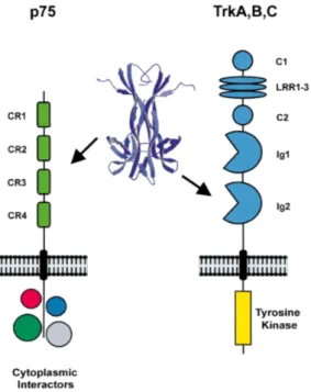
Neurotrophins bind to two different classes of transmembrane receptor proteins, the tropomyosin receptor kinase (Trk) and the neurotrophin receptor p75NTR (Fig. 4). A selective binding to these different receptors permits the transduction of very different signals. It is not excluded a direct interaction that allows fine tuning and cross talk.
Trk. The tropomyosin kinase receptors are transmembrane glycoproteins of ∼140 kDa. In mammals, three trk genes, codifying for the three different proteins TrkA, TrkB and TrkC, have been identified. A high, but not absolute, binding specificity has been seen among neurotrophins: NGF is the preferred ligand for TrkA, BDNF and NT4/5 for TrkB, NT3 for TrkC, even if the latter is also a ligand for TrkA and TrkB (Barbacid, 1994). As schematized in figure 4, five distinct motifs are recognized in the structure of these receptors (Schneider and Schweiger, 1991),but the most important for the interaction with ligands is an Ig-like domain of the extracellular portion, called Ig2 domain (Urfer et al., 1995; Urfer et al., 1998).
Chapter I
28
Figure 4: schematic representation of the p75 and Trk receptors. From Bibel and Barde, 2000.
Interaction with ligands induces dimerization of the receptor (Jing et al., 1992) that results in the phosphorylation of specific tyrosine residues, located in the juxtamembrane region and in the C terminus. This leads to an open conformation of the receptor and to the access of substrates to the kinase (Fig. 5).
Two complexes of adapter molecules bind to the tyrosine residue located in the juxtamembrane region: the Shc/Grb2/SOS and the FRS2/SHP-2/Grb2/SOS complex. The recent finding of Shc analogs (N-Shc, Sck; Nakamura et al., 1998) raises the question of recruitment of different Shcs that may be specific for TrkA, TrkB, or TrkC. Three main signalling cascades are activated by the Trk receptors and their substrates. The Ras/Raf/MEK/MAPK pathway induces the differentiation of neurons and neurite growth while the PKB/AKT pathway mediates the survival functions of the neurotrophins. Finally, the phosphorylated tyrosine in the C terminus recruits phospholipase C-γ (PLCγ) which, in turn, catalyses the cleavage of the substrate PIP2 to DAG and IP3, with DAG inducing activation of PKC and IP3 leading to release of Ca2+ from internal stores. The latter pathway seems to play an important role in neurotrophin-mediated neurotrophin release (Canossa et al., 1997) and in synaptic plasticity. It has also been reported that the PLC-γ track regulates the neuron-specific intermediate filament protein, peripherin (Loeb et al., 1994).
The first two pathways are interconnected with each other since activation of the MAPK pathway is mediated by FRS2, through the recruitment of the tyrosine phosphatase SHP-2/Grb2/SOS complex,
Chapter I
29 and also by phospho-Shc, via the Grb2-SOS complex. Other adapter molecules seem to form complexes with Grb2/SOS, for example rAPS, SH2-B (Qian et al., 1998) and SNT (Stephens et al., 1994), making possible to amplify and diversify receptor-mediated signals.
Figure 5: signalling through the Trk receptors; the main pathways. From Dawbarn and Allen, 2003.
Splice variants have been described for all three Trk receptors and include deletions in the extracellular domain and intracellular truncations or inserts. Some of these modifications were found to influence ligand specificity, as demonstrated by the fact that a TrkB splice variant, lacking exon9 in the extracellular domain, shows decreased interaction with NT4/5 and NT3 (Strohmaier, 1996), and a TrkA variant, with an increased specificity for NGF and a decreased specificity for NT3, has been described (Clary and Reichardt, 1994). Too little is known about the localization and the role of these variants, however they are often discussed as dominant negative modulators of Trk signalling (Eide et al., 1996; Ninkina et al., 1996).
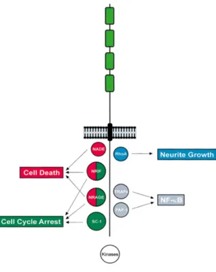
p75NTR. Codified by a gene on the human chromosome 17 (17q21-22) (Huebner et al., 1986), it is a transmembrane glycoprotein receptor of ∼75 kDa. It is characterized by four cysteine (CR1-CR4) repeats (Fig. 4) in the extracellular domain, and a palmitoylated conserved cysteine residue in the intracellular juxtamembrane region (Barker et al., 1994). Related to proteins of the tumor necrosis factor (TNFR) superfamily, binding to this receptor has been shown to affect cell survival (Barret and Bartlett, 1994), axonal outgrowth (Dechant and Barde, 2002) and to result in activation of NF-κB (Carter et al., 1996). It does not exhibit any intrinsic catalytic activity, so the signal is a
Chapter I
30 consequence of an association with, or dissociation from, cytoplasmic interactors. As represented in figure 6, they include NADE (p75NTR associated cell death executor), NRIF (neurotrophin receptor-interacting factor) and NRAGE (neurotrophin receptor-receptor-interacting MAGE homolog -MAGE is a protein family expressed in cancer tissues-) that mediate NGF-dependent apoptosis in sympathetic neuron precursor cells (Salehi et al., 2000). NRIF and NRAGE also lead to cell cycle arrest, as well as SC-1 (Schwann cell factor–1), whereas the GTPase RhoA modulates neurite growth.
Several variants of the p75NTR receptor have been found, such as soluble forms and truncated proteins lacking in the ability to bind neurotrophins (Dechant and Barde, 1997). The precise role is unknown, but their presence may be functionally relevant.
Neurotrophin receptor expression is highly regulated. In the central nervous system (CNS), few neurons express TrkA (mostly the basal forebrain cholinergic neurons) whereas TrkB is widely expressed, explaining the multitude of actions of BDNF. TrkC is typically expressed in the early phases of development (Tessarollo et al., 1993).
Also the expression profile of P75NTR is highly regulated, showing a down-regulation during postnatal development (Bothwell, 1995) and a rapid induction after injury, such as nerve lesion or seizures (Roux et al., 1999). This confirms the clear link between p75 receptor and cell death in pathological situations.
Chapter I
31
Figure 6: P75NTR interactors involved in transducing cell death. From Dawbarn and Allen; 2003.
Remarkably, Trk receptors and p75NTR are often co-expressed by the same cell and form a complex that can be immunoprecipitated after solubilization (Bibel et al., 1999). Both the intra- and the extracellular domains participate in this interaction, which is also dependent on the state of phosphorylation of the Trk receptors (Bibel et al., 1999). Receptor association leads to high-affinity neurotrophin binding, which is crucial in view of the limiting amounts of neurotrophins in vivo
(Hempstead et al., 1991; Benedetti et al., 1993; Davies et al., 1993;Barker and Shooter, 1994;Lee et al., 1994; Mahadeo et al., 1994; Horton et al., 1997). This association may also increase ligand discrimination by the Trk receptors, important in the case of TrkA and TrkB that bind more than one neurotrophin (Benedetti et al., 1993; Bibel et al., 1999). Finally, the proximity of p75NTR and Trk receptors in the membrane allows the signalling pathways, triggered by both receptors, to interact.
An interesting and neuron-specific aspect of neurotrophin is the retrograde transport of signals from axon terminals back to the cell body of neurons, with a process that seems to involve the internalization of the ligand-receptor complex (Bhattacharyya et al., 1997; Riccio et al., 1997)
probably via signalling endosomes (Grimes et al., 1996).
BDNF. Among neurotrophins, BDNF is the most abundantly expressed in the CNS. The gene for
Chapter I
32 UTR region and a 3’ exon which contains the coding sequence and the 3’UTR region. Alternative splicing gives rise to at least 20 different transcripts, all expressed in brain at different levels and in restricted neuronal population. Only the transcript containing exon IV has been found in heart and lung (Binder et al., 2001). Like many other growth factors and hormones, BDNF is generated from a precursor protein (pro-BDNF) cleaved by a protease in the mature form. In addition to the mature form, pro-BDNF is also biologically active.
BDNF mRNA has a widespread distribution in the central nervous system, including limbic forebrain, neocortex and, more than all, hippocampus (Lindvall et al., 1994). In this region it is expressed prevalently in granule cells, pyramidal cells and some hilar GABAergic neurons. BDNF protein immunoreactivity also appears preferentially in cell bodies and axons, compared to dendrites. Both physiologic, such as light and osmotic stimuli, physical exercise and the estrus cycle, and pathologic stimuli, enhance BDNF mRNA. Several evidences support a critical role of this neurotrophin in the pathogenesis of different diseases including epilepsy. After seizures, an increased BDNF synthesis and TrkB receptor activation is seen in granule cells of the hippocampus, both in humans (Mathern et al., 1997) and rodents (Binder et al., 2001). Many studies correlate these phenomena with proepileptogenic effects. Kokaia and collaborators (1995) reported a greater than twofold reduction in the rate of kindling development in BDNF heterozygous (+/-) mice, whereas kindling is completely abolish in absence of TrkB receptors (He et al., 2002). Moreover, application of NTFs, including BDNF, has been shown to potentiate synaptic transmission in vitro
(Lohof et al., 1993) and in vivo (Messaoudi et al., 1998); BDNF also enhances glutamatergic transmission (Lohof et al., 1993) and seems to reduce inhibitory synaptic transmission (Tanaka et al., 1997). The mechanism underlying synaptic potentiation remains unclear but may involve the facilitation of transmitter release or effects on ion channels and/or conductances. Also pro-BDNF, acting via p75NTR, could lead to seizure-induced apoptosis. As summarized by Binder et al. (2001), these alterations are responsible for a state of hyperexcitability that, in turn, could create a positive feedback loop in which increased activity results in upregulated levels of BDNF and of activation of TrkB.
However, other experimental evidence suggests an opposite, anti-epileptogenic, effect. For instance, BDNF is known to modulate the expression of neurotransmitters and neuropeptides, including neuropeptide Y. NPY is thought to inhibit seizure generation and is interesting to note that both kindling and kainate-induced seizures increase NPY immunoreactivity with a distribution that is strikingly similar to phospho-Trk immunoreactivity. This suggests that BDNF-induced Trk
Chapter I
33 activation could lead to NPY upregulation, which might subsequently limit excitability (Larmet et al., 1995). Contrary to the results published by Tanaka in 1997, a work of Palma and collaborators
(2005) demonstrates that BDNF amplifies GABA currents and prevents their run-down in Xenopus oocytes expressing GABAA receptors transplanted from surgically removed specimens of human epileptic brains. Moreover, mature BDNF favours survival or regeneration of hippocampal neurons damaged by SE (Simonato et al., 2006).
Several mechanisms have been proposed to explain the contrasting effects of BDNF. The same neurotrophin may induce different effects depending on splice variants, cellular site of action (dendritic localization is associated with the potentiation of active synaptic contacts and somatic distribution with cell survival and differentiation of neuronal precursors), local synthesis of the pro- and mature forms, and opposing effects resulting from the binding to TrkB or p75NTR (Simonato et al.,2006). Finally, different effects of neurotrophins may also depend on the duration of exposure, as demonstrated by the fact that chronic intrahippocampal infusion of BDNF inhibits the development of hippocampal kindling and reduces the duration of electrographic seizures (Larmet et al., 1995). Hence, acute BDNF could enhance synaptic transmission and neuronal excitability, whereas chronic treatment could promote survival and induce growth and morphological changes of synapses (Lu, 2004). Acute responses are mediated by cAMP activation (Li et al., 1999), whereas the reduced responsiveness, seen after long-term exposition, is probably due to TrkB receptors downregulation (Frank et al., 1996).
3.2 FGF-2.
Basic fibroblast growth factor, or fibroblast growth factor-2 (bFGF-2 or FGF-2), is a neurotrophic factor belonging to a family of 22 structurally related proteins (Itoh and Ornitz, 2004). It is a single-chain polypeptide composed of 146 amino acids, with a core region consisting of 120-130 aa, ordered into 12 antiparallel β-strands flanked by divergent amino and carboxyl termini. In common with other members of the family, FGF-2 owns a positively charged heparin-binding region that allows a high affinity binding to heparansulfate proteoglycans (HSPGs) sited in the extracellular matrix (ECM) and on the cell surface in the vicinity of the FGF receptors (FGFRs). The latter is required for high-affinity interactions with receptors, the former favours local storage of FGFs. Distinct isoforms of FGF-2 are described: the high molecular weight (HMW) forms, ranging from 20 to 24kDa, are predominantly localized in the nucleus, whereas the low molecular weight form (LMW, 18kDa) resides in the cytoplasm (Florkiewicz et al., 1991; Quarto et al., 1991; Sørensen et al., 2006).