UNIVERSITÀ DEGLI STUDI DI SASSARI
SCUOLA DI DOTTORATO IN
SCIENZE VETERINARIE
INDIRIZZO
:
Riproduzione, Produzione e benessere animale(
XXVIII CICLO)Study of DNA methylation dynamics
in a model of differential developmental competence
in the ovine species
Docente Guida Prof. Sergio Ledda
Direttore Tesi di dottorato di
Prof. Sergio Ledda Dott.ssa Laura Masala
Anno Accademico 2014-2015
La presente tesi è stata prodotta nell’ambito della Scuola di Dottorato in “Scienze Veterinarie” dell’Università degli Studi di Sassari, a.a. 2014/2015 – XXVIII ciclo, con il supporto di una borsa di studio finanziata con le risorse del P.O.R. SARDEGNA F.S.E. 2007-2013 - Obiettivo competitività regionale e occupazione, Asse IV Capitale umano, Linea di Attività l.3.1.
Ai miei genitori
Tear Gas Rag
Old Southern Rag, Blind Blake
Tear Gas Tear Gas Tear gas tore my throat Can’t say my mantra Tear Gas got my goat Tear Gas O Lord Tear Gas I can’t find my mind Bombing North Vietnam I’m stumbling around blind Tear Gas in Boulder Tear Gas in my heart Frightened on college hill Nixon’s poison Fart
Tear Gas here Tear Gas there Colorado and Saigon
They‘ll be droppin teargas Everytime I get a hardon
Allen Ginsberg May 10, 1972 Ft. Collins, Colorado
Table of contents
Chapter 1: Summary ... 7
Chapter 2: Introduction ... 8
2.1. Epigenetic mechanisms and nuclear DNA packaging: ... 8
2.2. Epigenetics modifications ... 9
2.2.1. Histone modifications ... 9
2.2.2. Non coding RNA ... 10
2.2.3. DNA methylation ... 11
2.3. Enzymes involved in DNA methylation remodelling ... 11
2.3.1. DNA-methyltransferases (DNMTs) enzymes ... 11
2.3.2. DNA demethylation dynamics ... 13
2.3.3. Others proteins and cofactors involved in DNA methylation remodelling ... 15
2.4. DNA methylation dynamics during development ... 18
2.4.1. DNA methylation reprogramming in the early embryo ... 18
2.4.2. DNA methylation reprogramming during germ-cell specification ... 20
2.5. Particular genome regions controlled by DNA methylation ... 21
2.5.1. X Chromosome inactivation ... 21
2.5.2. Genomic imprinting ... 22
2.5.3. Repetitive elements ... 23
2.6. Role of DNA methylation in disease ... 23
2.6.1. Epigenetic and assisted reproductive technologies (ART) ... 23
2.6.2. Disorders due to loss of imprinting ... 24
2.6.3. The role of DNA methylation in cancer ... 26
2.7. General introduction to in vitro embryo production technique ... 26
2.7.1. Oocyte collection ... 27
2.7.2. Oocyte maturation ... 28
2.7.3. Fertilization ... 29
2.7.4. Preimplantation Embryonic development ... 30
2.8. Developmental competence... 32
2.8.1. Oocyte quality ... 32
Chapter 3: Aim ... 35
Chapter 4: Materials and Methods ... 36
4.1 Sample preparation ... 36
4.1.1 Oocyte recovery and In Vitro Maturation ... 36
4.1.2. Semen processing and swim-up technique ... 37
4.1.3. In vitro fertilization and embryo development ... 37
4.1.4. Sheep and bovine tissue collection ... 38
4.1.5. Sperm collection ... 38
4.1.6. Blood collection ... 38
4.2. Gene expression analysis ... 39
4.2.1. Samples collection for gene expression analysis ... 39
4.2.2. RNA Isolation ... 39
4.2.3 Reverse transcription ... 39
4.2.4. Primers design and validation ... 40
4.2.5. Agarose gel electrophoresis ... 41
4.2.6. PCR products sequencing ... 42
4.2.7. Real time‐polymerase chain reaction ... 45
4.3. Immuno detection ... 45
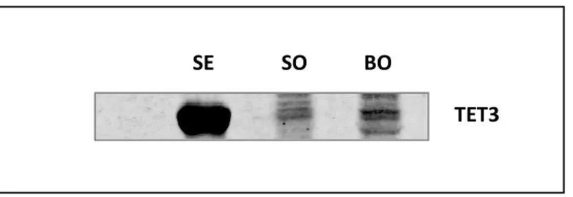
4.3.1. TET3 antibody validation ... 45
4.3.2. TET3 whole mount immunostaining ... 47
4.3.3. Methylation and hydroxymethylation whole mount immunostaining... 47
4.3.4. Confocal Analysis ... 48 4.4. Methylation analysis ... 49 4.4.1. DNA isolation ... 49 4.4.2. Bisulfite conversion ... 53 4.4.3. Pyrosequencing ... 53 4.5. Statistical analysis ... 55
4.5.1. Gene expression Statistical analysis ... 55
Chapter 5: Results ... 57
5.1. Gene expression analysis ... 57
5.2 Immunodetection ... 57
5.2.1 TET3 antibody specificity validation ... 57
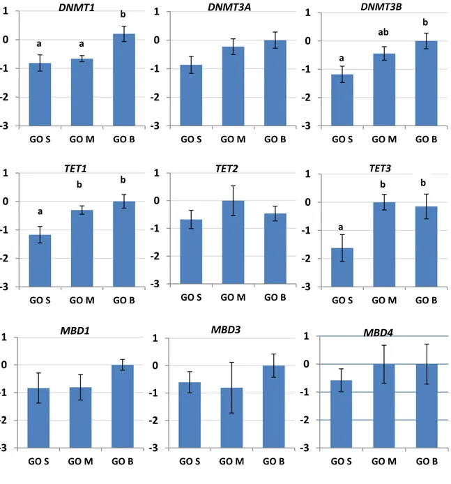
5.3. Methylation dynamics in growing oocytes ... 58
5.3.2. TET3 protein was not detected in GO ... 60
5.3.3. Global DNA Methylation analysis in GO ... 61
5.3.4. Global DNA Hydroxymethylation analysis in GO ... 62
5.4. Methylation dynamics during meiotic progression ... 63
5.4.1. Gene expression analysis in GV and IVM MII oocytes derived from P and A donors ... 63
5.4.2. TET3 protein was not detected at GV stage ... 65
5.4.3. Global DNA Methylation analysis during meiosis progression ... 65
5.4.4. Global DNA Hydroxymethylation analysis during meiosis progression ... 67
5.5. Methylation dynamics during early embryonic development ... 69
5.5.1. Gene expression analysis in IVC embryos at 2C and 4C stage ... 69
5.5.2. TET3 protein was detected only in embryos ... 71
5.5.3. Global DNA Methylation analysis during early embryonic development ... 73
5.5.4. DNA Hydroxymethylation analysis during early embryonic development ... 75
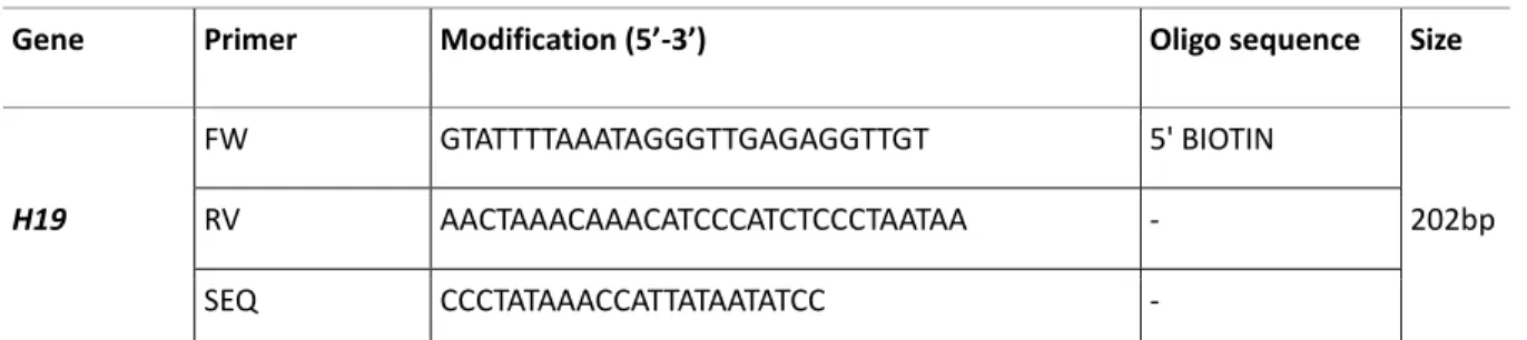
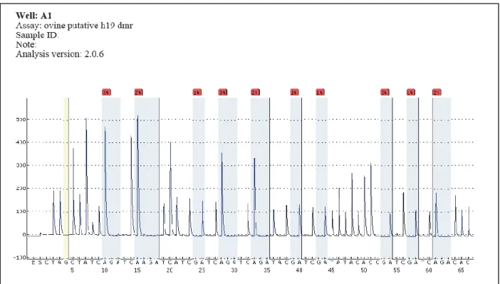
5.6. Pyrosequencing ... 78
5.6.1. H19 Assay validation with sperm and liver bisulfite converted DNA ... 78
5.6.2. H19 Assays validation with oocytes BC DNA from ... 80
5.6.3. Assessment of oocyte purity by H19 assay ... 82
Chapter 6: Discussion ... 83
Chapter 7: Conclusions ... 92
References ... 94
Chapter 1: Summary
Since 1956, when Conrad Waddington first used the term epigenetics, the understanding of
epigenetic phenomena is progressively growing year by year. In less than 80 years the
classical believe that everything was written in the DNA sequence has changed and now we
know that epigenetic modifications interact with the DNA sequence and together they
regulate the expression of genes in response to cell needs and environmental stimuli.
Different kinds of epigenetic modifications have been identified and part of the regulating
mechanisms are now clear, but at the same time several questions still need to be answered
in order to have a complete picture of the complex epigenetic mechanisms and regulations.
Sophisticated studies demonstrated that the epigenetic status undergoes dynamic changes
that are physiologic during embryo development (Messerschmidt et al. 2014), but they could
also be involved in the pathogenesis of certain diseases, such as cancer (Feinberg et al. 2002).
The most studied epigenetic modification is DNA methylation, that implicates the addition of
a methyl group (CH3) to the 5-carbon position of cytosine, frequently within a CpG
dinucleotide (Razin and Szyf 1984). Such modification is involved in the two waves of
epigenetic remodelling that take place during embryogenesis, one during early embryonic
development and the second one during primordial germ cell specification. DNA methylation
has been the focus of this thesis, as we studied the dynamics of this particular epigenetic
modification in relation to oocyte quality and during early embryo development in the ovine
Chapter 2: Introduction
2.1. Epigenetic mechanisms and nuclear DNA packaging
Epigenetics is the study of mechanisms involved in the regulation of gene activity that are not
determined by changes in DNA sequence. These mechanisms include the so called epigenetic
modifications that have an effect on DNA structure and how it is organized in the nucleus
rather than in the DNA sequence itself (Handy et al. 2011).
The nuclear DNA of eukaryotic cells is finely packaged in chromosomes. Each chromosome
consists of one continuous thread-like molecule of DNA coiled around specialized proteins
that bind to and fold the DNA. This organization of DNA and protein refers to a condensed
structure called chromatin that is assembled in a precise and ordered manner. The first level
of chromatin organization is the nucleosome, it is composed of an octameric core made up of
different kind of assembled histone proteins that tightly wrap the DNA (Mariño-Ramírez et al.
2005). Individual nucleosomes are then linked together by stretches of linker DNA and coiled
into chromatin fibers (Fig 2.1.), (Wu et al. 2007). Although this fine asset of DNA packaging, a
large amount of genes must be actively transcribed during life. Thus, chromatin could be
considered as a dynamic genome organization and, depending on cell demand, its
condensation state could be modulated in order to make the DNA accessible to the
transcription machinery in a time and tissue specific manner (Baker 2011). This general
introduction has been necessary to explain how chromatin is organized, in order to give a
chromatin structure. However, this thesis will focus on DNA methylation, a particular kind of
epigenetic modification that has been demonstrated to interact with the other epigenetic
mark to control chromatin structure modification and gene expression (Weissmann and Lyko
2003).
Figure 2.1: chromosome structure
2.2. Epigenetics modifications
Three different epigenetic modifications are involved in the complex epigenetic machinery:
Histone modification, Non coding RNAs, DNA methylation. Although the three modifications
are diverse, achieved by different chemical reactions and set up by distinct enzymes, studies
demonstrate that all of them interact with each other to regulate gene activity (Cedar and
Bergman 2009; Portela and Esteller 2010).
2.2.1. Histone modifications
As discussed above, nucleosomes are the first level of chromatin organization consisting of
acid tails that protrude out from the nucleosome. The function of terminal tails is to regulate
high–order chromatin by binding the DNA and also to regulate the interaction between
neighbour nucleosomes (Iwasaki et al. 2013). Terminal tails are subjected to
post-transcriptional modification at N-terminal ends, such as acetylation, methylation,
phosphorylation, ubiquitination, SUMOylation and ADP-ribosylation (Kouzarides 2007).
Histone modifications have important roles in transcriptional regulation and chromosome
condensation. The chromatin can be found in two main states: the actively transcribed
euchromatin and the transcriptionally inactive heterochromatin. The first state presents high
levels of acetylation and trimethylation of the tails, while the transcriptionally inactive state
is characterized by high levels of methylation and low acetylation (Li et al. 2007).
2.2.2. Non coding RNA
Non coding RNAs (ncRNAs) are RNA molecules are not translated into proteins, but have a
regulatory activity; in fact ncRNAs function is to regulate gene expression at both
transcriptional and post-transcriptional level. The ncRNAs involved in epigenetic processes
are the short ncRNAs [not longer than 30 nucleotides (nts)], such as microRNAs (miRNAs),
short interfering RNAs (siRNAs), and piwi-interacting RNAs (piRNAs), and the long ncRNAs
(longer than 200 nts) (Mattick and Makunin 2006). This particular class of RNAs plays a role in
targeting histone modifying complexes, DNA methylation and gene silencing. For instance, it
has been demonstrated that some ncRNAs interact with critical regions of CpG islands of
imprinted genes to promote the recruitment of methyltransferase 1 (Peschansky and
2.2.3. DNA methylation
DNA methylation is characterized by the covalent addition of a methyl group to the 5-carbon
position of cytosine, frequently within a CpG dinucleotide (Razin and Szyf 1984). DNA
methylation is usually associated with repression of gene transcription as this chemical
modification modulates the condensed state of chromatin by the interaction with histones
modifications (Cedar and Bergman 2009). CpGs dinucleotides are mostly distributed across
the whole genome and are usually methylated (Larsen et al. 1992). However, specific areas of
the genome are characterized by regions rich in CpG multiple repeats known as CpG islands;
they are often located in gene promoters and are unmethylated in somatic cells, with the
exceptions of imprinted genes and the inactive X chromosome in females (Deaton and Bird
2011).
2.3. Enzymes involved in DNA methylation remodelling
DNA methylation patterns undergo multiple rounds of dynamic changes during development
and are maintained stable in adult somatic cells. Establishment, erasure, re- establishment
and maintenance and of DNA methylation are coordinated by particular mechanisms that
regulate both DNA methylation and demethylation.
2.3.1. DNA-methyltransferases (DNMTs) enzymes
The addition of methyl groups is controlled at different levels in cells and is carried out by a
family of DNA-methyltransferase (DNMTs) enzymes. These enzymes are widely conserved
among eukaryotes (Kim et al. 2009). Three major types are responsible of the regulation of
DNA methylation: DNMT1, DNMT3A and DNMT3B. All of them have a catalytic domain that
(SAM), and the formation of 5-methylcytosine (5-mc). Two main mechanisms are involved in
DNA methylation: DNA methylation maintenance, performed by DNMT1, and de novo DNA
methylation, carried out by DNMT3A and DNMT3B. Mutant mice lacking either the
maintenance enzyme DNMT1 or both de novo methylases DNMT3A and DNMT3B exhibit
demethylation in the genome and die at the mid-gestation stage, indicating that methylation
is essential for embryogenesis (Okano et al. 1999)
2.3.1.1. DNMT1 and DNA methylation maintenance
DNMT1 was the first methyltransferase to be discovered (Bestor et al. 1988). It is responsible
of the maintenance of DNA methylation during cells division. In fact, DNA replication
implicates the loss of DNA methylation marks in the newly synthesized DNA strand. It has
been demonstrated that DNMT1 is the enzyme responsible of the maintenance of DNA
methylation, it has high affinity for hemi-methylated DNA (Yoder et al. 1997; Vilkaitis et al.
2005) and it interacts with the proliferating cell nuclear antigen (PCNA), a processivity factor
for DNA polymerase δ in eukaryotic cells, essential for replication. The targeted mutation of
the DNMT1 gene results in embryonic lethality (Li et al. 1992) suggesting that it has a major
role during embryo development.
2.3.1.2. DNMT3A and DNMT3B and de novo DNA methylation
Two enzymes are responsible of de novo DNA methylation: DNMT3A and DNMT3B (Okano et
al. 1998a); they possess the ability to methylate a previously unmodified cytosine residue.
DNMT3A transcript is provided as a maternal RNA by the oocyte and is important during
early preimplantation development and for the establishment of the differential DNA
(Kaneda et al. 2004; Kato et al. 2007). Conversely, DNMT3B is transcribed upon embryonic
genomic activation and is strongly expressed by the blastocyst stage, at which time its
presence is predominantly restricted to the epiblast lineage. As DNMT1, both DNMT3A and
DNMT3B are essential during embryo development. Deletion of DNMT3B causes embryonic
lethality, while DNMT3A knockouts are partially viable (Okano et al. 1999) Combined genetic
deletion results in earlier embryonic lethality, indicating at least partial functional
redundancy of the two enzymes (Okano et al. 1999).
2.3.2. DNA demethylation dynamics
In contrast to DNA methyltransferases enzymes, that are able to catalyse the addition of the
methyl group into the cytosine, a family of enzymes capable to directly remove the methyl
group does not exist or at list has not been yet identified. Two models of DNA demethylation
have been proposed, involving a passive or an active mechanism.
2.3.2.1. Passive replication-dependent demethylation
Passive DNA demethylation does not involve any active enzymatic process that alters the
5-mc itself. It refers to the loss of it during successive rounds of replication, in the absence of
functional DNA methylation maintenance, which results in a retention of the unmethylated
state of the newly synthesized DNA strand. Passive replication-dependent demethylation
occurs in maternal pronucleus (PN) during subsequent cleavage divisions (Mayer et al. 2000).
2.3.2.2. Ten-eleven translocation (TET) enzymes and active DNA demethylation
Active DNA demethylation involves an enzymatic process that modifies the methyl group.
called respectively TET1, TET2 and TET3, mediate the oxidation of 5-mc to 5
hydroxymethylcytosine (5-hmc) and further to 5-formylcytosine (5-fc) and 5-carboxycytosine
(5-cac) (Ito et al. 2011). All three TET enzymes have a CD (Cys-rich and DSBH regions) domain
that uses O2 to decarboxylate α-ketoglutaric acid generating a high-valet iron oxide that
converts 5-mc to 5-hmc (as well as 5-hmc to 5-fc and 5-fc to 5-cac). TET1 and TET2 are mainly
implicated in primordial germ cell (PGCs) reprogramming, but further studies are needed to
clarify the exact contribution of TET1 and TET2 in the PGC methylome reset. Recently,
(Yamaguchi et al. 2012) reported significant reduction of female germ-cell number and
fertility in TET1 deficient mice suggesting that TET1 is responsible of meiosis and meiotic
gene activation in female germ cells. Conversely, TET3 is responsible of global hydroxilation
of methyl groups in the male PN following fertilization. In 2011, Gu and colleagues reported
that TET3-deficient zygotes obtained from conditional KO mice were unable to undergo male
PN hydroxylation. Different ways of active DNA demethylation that involves the tree
intermediates that originate from TETs oxidation have been proposed. 5fc and 5cac can be
recognized and excised out of the DNA by the thymine DNA glycosylase enzyme (TDG) in a
process involving the base excision repair pathway (Maiti and Drohat 2011). It has been
demonstrated that depletion of TDG in mESCs leads to the accumulation of 5cac and 5fc
(Raiber et al. 2012; He et al. 2011). 5-hmc modification could aid passive DNA demethylation
even in the presence of DNMT1, which has low affinity for 5-hmc, so it is not able to maintain
the DNA mark trough replication (Valinluck and Sowers 2007). Recent studies suggest a
different role for DNA hydroxymethylation other than DNA 5-mc removal. Ruzov et al.,(2011)
development and suggest that 5-hmc may be an epigenetic modification with a specific
biological role during cell self-renewal and differentiation other than being only a transient
condition during active demethylation (Ruzov et al. 2011).
2.3.3. Others proteins and cofactors involved in DNA methylation remodelling
2.3.3.1. DNMT2
The role of DNMT2 protein in DNA methylation is controversial. DNMT2 protein was
originally assigned to the DNA methyltransferase family, due to the strong similarity with
DNMT1 and DNMT3B catalytic domain (Okano et al. 1999). Despite its high similarity with
others DNMTs, it has been shown that DNMT2 has only a weak DNA methylation activity
(Jeltsch et al. 2006). In fact, targeted deletion of the DNMT2 gene in embryonic stem cells
causes no detectable effect on global DNA methylation, suggesting that this enzyme has little
involvement in setting DNA methylation patterns (Okano et al. 1998b). However in 2006 it
has been shown that DNMT2 has an active role as RNA methyltransferase: it seems that
DNMT2 specifically methylates cytosine 38 of transfer RNAAsp (Goll et al. 2006). Methylation
of tRNAs has an impact on the folding and stability of the structure (Alexandrov et al. 2006).
DNMT2 has been shown to methyl other tRNAs and a protective function was hypothesized
(Schaefer et al. 2010). Researchers are currently examining whether DNMT2 acts on other
small RNAs.
2.3.3.2. DNMT3L
The DNA methyltransferase 3-like (DNMT3L) protein is a crucial cofactor for de novo
to the methyltransferases, which dimerises with the de novo enzyme DNMT3A (Jia et al.
2007). DNMT3A and DNMT3L, are largely involved in establishing maternal DNA methylation
imprints in mice and cow (Kaneda et al. 2004; Bourc'his et al. 2001). Conversely in human
DNMT3L mRNA expression was undetected until postfertilization (Huntriss et al. 2004), while
in rhesus monkey the transcript was barely detectable (Vassena et al. 2005); this suggests a
later role for DNMT3L in de novo methylation during early embryonic development and not
in the establishment of maternal imprints.
2.3.3.3. Methyl-CpG-binding proteins (MBDs)
Methyl-CpG-binding proteins (MBDs) are a family of proteins that consists in five members:
MeCP2, MBD1, MBD2, MBD3 and MBD4 (Berger and Bird, 2005). Each of these proteins, with
the exception of MBD3, are characterized by a binding domain which preferentially binds to
methylated CpG(s) (Hendrich and Bird 1998). MBD proteins possess also a CxxC domain that
helps the interaction with DNMTs and a transcription repression domain (TRD) that acts to
silence the gene transcriptional machinery. MBD interaction with methylated DNA is crucial
as it prevents binding of transcription factors and RNA polymerase and thereby represses
transcription (Hendrich and Tweedie 2003).
2.3.3.3.1. Methyl-CpG-binding domain protein 1 (MBD1)
MBD1 has high affinity for related to DNA methylation and chromatin modifiers such as the
Suv39h1–HP1 complex, to enhance DNA methylation-mediated transcriptional repression
(Fujita et al. 2003). The functional importance of MBD1 was demonstrated in human HeLa
cells, where MBD1 was shown to associate with the H3K9 methyltransferase SETDB1 (Sarraf
domain of MBD1 makes it a unique member of the MBD family due to its affinity to
unmethylated DNA. This binding allows the protein to trigger methylation of H3K9 and
results in transcriptional repression (Fujita et al. 2000).
2.3.3.3.2. Methyl-CpG-binding domain proteins 2 and 3 (MBD2 and MBD3)
MBD2 and MBD3 are components of the NuRD (Mi-2) histone deacetylase and nucleosome
remodelling complex (Tazi and Bird 1990; Zhang et al. 1999). MBD2 is required for
transcriptional silencing of methylated promoters and for proper nurturing behaviour in
mothers, while MBD3 is essential for successful embryogenesis. Both proteins were
identified as potential methyl-CpG binding proteins, although in mammals only MBD2
specifically binds to methylated DNA both in vitro and in vivo (Hendrich and Bird 1998).
Biochemical assays have shown that MBD3 cannot bind methylated DNA directly, but it is an
essential subunit of the Mi-2/NuRD chromatin remodelling complex together with MBD2
(Saito and Ishikawa 2002). The MBD3 protein is 70% identical to MBD2, but lacks the 152
amino-acid N-terminal extension that is implicated in methylated DNA binding (Hendrich and
Bird 1998; Hendrich et al. 1999). Both genes are activated in embryogenesis following
implantation and are widely expressed in adult mice (Hendrich et al. 2001; Hendrich and Bird
1998).
2.3.3.3.3. Methyl-CpG-binding domain protein 4 (MBD4)
MBD4, as others MBD proteins, seems to acts as a transcriptional repressor and is also
involved in DNA repair due to the presence of a glycosylase domain. The protein uses its
N-terminal MBD to bind methylated CpG and its C-N-terminal glycosylase (which is homologous to
spontaneous hydrolytic deamination of methylated cytosine which causes mCpG-TpG
transitions (Bird 1980), whereas non-methylated CpG mutates to UpG. MBD4 is able to excise
and repair both mutated nucleotides (Hendrich et al. 1999). Its repair function is suggested
by the two to three times higher number of mCpG-TpG transitions in MBD4-null mice,
showing that MBD4 acts to reduce mCpG-TpG mutations (Millar et al. 2002; Wong et al.
2002). Despite its well characterized DNA repair activity , its role as a transcriptional
repressor still needs to be confirmed. In fact, a study in zebrafish provided evidence for a role
of MBD4 in active demethylation (Rai et al. 2008).
2.4. DNA methylation dynamics during development
In mammals, DNA methylation patterns are relatively stable in somatic cells, but dynamic
changes occur during embryonic development and gametogenesis. DNA methylation
remodelling is essential to regulate mechanisms that are crucial for normal development and
the future health of the new born. (Saitou et al. 2012; Messerschmidt et al. 2014; Okae et al.
2014). During embryo development, different waves of DNA methylation reprogramming
occur, specifically after fertilization and during germ-cell specification, Fig. 2.2.
2.4.1. DNA methylation reprogramming in the early embryo
During early embryogenesis a massive erasure of DNA methylation occurs to maintain a
specific transcriptional state essential for pluripotency establishment (Paranjpe and Veenstra
2015). Soon after fertilization, the genome of the zygote undergoes massive demethylation.
The zygote is characterized by the presence of two PNs one originated from the sperm and
one form the egg. Both PNs undergo DNA demethylation (Mayer et al. 2000; Oswald et al.
proposed for the two PNs. The paternal genome, which is high methylated in the sperm, is
actively demethylated in the zygote. Recently, some authors strongly evidenced that the
active demethylation of the male PN is mediated by TET3 enzyme by hydroxylation of the
5-mc (Wossidlo et al. 2011; Guo et al. 2014) as previously mentioned (section 2.3.2.2).
Conversely, the maternal genome undergoes subsequent passive demethylation via DNA
replication during cleavage. It seems that the maternal genome is protected from Tet3
oxidation by an interaction between STELLA and H3K9me2, which is mostly present in female
PN. This interaction alters chromatin configuration and protects the DNA from TET3
hydroxylation (Nakamura et al. 2012).
Both active and passive demethylation continue through cell divisions, in fact genome-wide
methylation analysis demonstrated that the newly formed embryo reaches the lowest
methylation levels at the blastocyst stage (Messerschmidt et al. 2014).
Some sequences are resistant to the wave of global DNA demethylation : repetitive DNA
(Lane et al. 2003; Guibert et al. 2012), a subset of single-copy genes (Guibert et al. 2012;
Borgel et al. 2010) and imprinted loci (Nakamura et al. 2007). The mechanisms by which
these particular DNA sequences escape massive DNA methylation erasure is not known, but
it has been demonstrated that both maintenance and de novo methylation enzymes are
dispensable for the maintenance of imprinting (Hirasawa et al. 2008). Following blastocyst
implantation , the epiblast rapidly adopts a somatic epigenetic re-methylation of the
genome. This process is initiated via de novo methylation (Popp et al. 2010; Borgel et al.
germline-specific genes are tightly repressed by DNA methylation, to prevent ectopically gene
activation (Borgel et al. 2010; Maatouk et al. 2006; Seisenberger et al. 2012).
2.4.2. DNA methylation reprogramming during germ-cell specification
In post-implantation mammalian embryos, a population of pluripotent cells in the epiblast
gives rise to the PGCs precursors. The PGCs precursors then proliferate and migrate across
the embryo to reach the genital ridges. PGCs continue to proliferate until when they enter
into meiotic prophase in female gonads and mitotic arrest in male gonads (Saitou and Yamaji
2012). When PGC precursors initiate to differentiate in the epiblast, they undergo global DNA
demethylation, which is mostly concluded when the cells colonize the genital ridge. During
their migration, some sequences escape demethylation; these sequences include, but are not
limited to, the imprinted differentially methylated regions (DMRs) (Leitch et al. 2013). In fact,
depending on the sex of the organism, specific imprints are reset during the development of
germ cells. The methylation patterns in male germ cells are then fully established at birth and
are maintained throughout many cycles of mitotic divisions before the cells enter meiosis
(Davis et al. 2000; Henckel et al. 2009). On the contrary, in female germ cells re-methylation
is not complete at birth, but continues during the oocyte growth phase, while oocytes are
Figure 2.2: Dynamic changes of DNA methylation during embryonic development
After fertilization, the paternal genome (blue line) is rapidly demethylated by active mechanisms, whereas the maternal genome (red line) is passively demethylated. Differentially methylated regions (DMRs) associated with imprinted genes are protected from this erasure (dashed green line). De novo methylation occurs after implantation (black line), but primordial germ cells (PGCs) are not specified until the epiblast stage (shading at the top of the figure). This methylation must be reset in PGCs. The figure shows the methylation dynamics after the epiblast formation for germline cells only . After migration most sequences are demethylated in PGCs. But some sequences are subject to late demethylation and are not reprogrammed until after PGC sex determination. Following sex determination, the germ cells undergo de novo methylation, but the dynamics are sex-specific. Methylation is completed in the prospermatogonia before birth, whereas methylation in the oocytes is established during postnatal growth (Cowley and Oakey 2012).
2.5. Particular genome regions controlled by DNA methylation
2.5.1. X Chromosome inactivation
In mammals, females receive two copies of the X chromosome while males receive one copy.
To prevent female cells from having twice as many gene products from the X chromosomes
as males, one copy of the X chromosome in each female cell is inactivated (XCI). The
necessity of dosage compensation by XCI is likely to start at the onset of genome activation
(Huynh and Lee 2003). This process is regulated by a region on the X chromosome called the
expression seems to be upregulated in the future inactive X chromosome in order to trigger
the establishment of chromosome-wide silencing (Brockdorff 2002; Penny et al. 1996). DNA
methylation is involved in the transcriptional regulation of this gene. After Xist has been
upregulated and XCI triggered on one X chromosome in female cells, Xist repression of the
second allele of Xist (on the active X chromosome) is maintained by DNA methylation of its
promoter (Norris et al. 1994).
2.5.2. Genomic imprinting
Genomic imprinting is a unique regulated molecular process for controlling
parental-origin-specific gene expression, depending on the DNA methylation status of the differentially
methylated region (DMR) of each imprinted gene (Li et al. 1993). By this mechanism, genes
are expressed in a mono-allelic manner rather than from both chromosome homologues
(bi-allelic expression) as it happens for most genes. It means that if the DMR is methylated in the
allele derived from the sperm (paternal imprinting), the gene is maternally expressed and
vice versa. Imprinted genes are normally found in clusters and are controlled within DMRs,
which are CpG-rich sequences located within specific regions called imprinting control
regions (ICs). In the germ-line, parental-specific DNA methylation is erased during the
differentiation of primordial germ cells, is re-established during spermatogenesis or
oogenesis and subsequently maintained throughout development (Delaval and Feil 2004).
Imprinted genes have important roles in normal growth and foetal development (DeChiara et
2.5.3. Repetitive elements
A large portion of the genome is composed by repetitive elements, consisting of interspersed
repeats derived from transposable elements (Weisenberger et al. 2005) and tandem repeats
of simple (satellite DNA) or complex sequences. Repetitive elements comprise ∼45% of the
human genome. Those include 1 million alu sequences occupying ∼10% of the genome, and
LINE-1 elements that also represent a large genomic portion (Ehrlich 2002). These
interspersed repeated DNA sequences contain numerous CpG dinucleotides. Therefore, the
methylation status of these sequences represents the most global DNA methylation patterns
that have been found in the genome (Yang et al. 2004) and have an active role in a variety of
human diseases (Pogribny and Beland 2009). Loss of DNA methylation in these sequences
represents the global hypomethylation state that characterizes many types of cancers (Ross
et al. 2010).
2.6. Role of DNA methylation in disease
2.6.1. Epigenetic and assisted reproductive technologies (ART)
Assisted reproductive technologies (ARTs) refer to the methods aimed at overcoming
infertility problems (Pinborg et al. 2015). ART treatments involve several in vitro
manipulation steps in order to achieve pregnancy by artificial or partially artificial
procedures. Different procedures used for ARTs consist of: hormonal stimulation, used to
induce superovulation; in vitro oocytes maturation prior to fertilization; intracytoplasmic
sperm injection (CSI), in vitro culture of preimplantation embryos and cryopreservation of
either gametes or embryos (Iliadou et al. 2011; Wright et al. 2003), that based on the
widely accepted and implemented therapy for some forms of infertility. However, it has been
noticed that the use of ART is related with an increased risk of pregnancy disorders and birth
defects (Hansen et al. 2005). An association between ART and epigenetic defects has also
been proposed. This hypothesis is supported by experimental studies involving ART in mouse
and ruminants and by the increased incidence of imprinting disorders, such as
Beckwith-Wiedemann, Angleman and Prader-Willi syndromes, of individuals conceived with the use of
ART (Niemitz and Feinberg 2004). As discussed above, during embryonic development and
germ cells specification and maturation, a crucial reset and rearrangement of epigenetics
marks takes place; thereby, the manipulation of germ cells and/or embryos could influence
and disturb the process of methylation and⁄ or demethylation and lead to embryo defects.
Studies have shown that the culture media used for embryo culture have a role in the
deregulation of developmentally important genes (Khosla et al. 2001). A study published in
2000, comparing two different embryo culture media to determine the potential effect on
imprinted genes, showed that H19 expression and methylation were adversely affected by
culture media composition (Doherty et al. 2000). Although results from animal studies
support the association between ART and epigenetic alternations, it has also to be
mentioned that most children conceived through ARTs are developing normally (Lu et al.
2013). It has to be kept in mind that ART patients often have reproductive problems that may
be mainly responsible for the birth defects associated with ARTs
2.6.2. Disorders due to loss of imprinting
Imprinted genes are a particular class of genes expressed in a mono-allelic manner
imprinting control regions (ICRs). Imprinted genes have important roles in normal growth and
foetal development (DeChiara et al. 1991; Salas et al. 2004), A number of human diseases,
identified as imprinting disorders, have been found to be caused by incorrect establishment
of DNA methylation at DMR during embryo development(Cassidy and Schwartz 1998). The
first identified imprinting disorders are Prader-Willi (PWS) and Angelman (AS) syndromes.
Both disorders arise from an imprinting defect in the chromosome 15, but the abnormality is
on the paternally derived chromosome 15 for PWS and on the maternally derived 15 for AS.
The involved chromosomal region contains a differentially methylated region (SNRPN/SNURF
ICR), which regulates the expression of imprinted genes (Kalsner and Chamberlain 2015). The
loss of maternally expressed genes controlled by the SNRPN ICR results in AS that involves a
cognitive and neurologic impairment, while PWS results in behavioural and endocrine
disorders derived from the loss of those genes which are expressed from the paternal allele
(Cassidy and Schwartz 1998; Cassidy et al. 2000). Beckwith-Weidemann Syndrome (BWS) and
Silver-Russell Syndrome (SRS) are also due to imprinting defects. Both of them are caused by
changes in the level of methylation of at ICR which controls the expression of both H19 and
IGF2. BWS syndrome is caused by a hypermethylation of the H19 ICR that results in decrease
of H19 and increase of IGF2 expression, giving rise to a phenotype of overgrowth (Ko 2013).
By contrast, SRS is caused by a hypomethylation of the H19 ICR that results in decreased IGF2
and increased H19 expression, giving rise to a phenotype with growth problems (Searle and
2.6.3. The role of DNA methylation in cancer
As this thesis is focused on DNA methylation dynamics that physiologically occur during
embryo development, a brief overview of the pathological involvement of DNA methylation
in cancer will be described. During the last decade, numerous studies have shown that
defects in the epigenetics mechanisms could contribute to the neoplastic phenotype.
Regarding DNA methylation, tumours are usually characterized by low level of CpG
methylation in comparison with their normal-tissue counterparts, indicating that cancer cells
have a specific epigenome (Ehrlich 2009). Loss of methylation could be related to the faster
growth rate that is characteristic of cancer cells, and may be also related to reactivation of
transposable elements that are usually suppressed by DNA methylation (Ross et al. 2010).
Reactivation of transposable elements can promote their integration in to the genome,
leading o gene activity disruption. Nevertheless, some important regions that control the
expression of tumour suppressor genes have been found to be hypermethylated in cancer
cells. This results in the silencing of genes possibly involved in cell cycle control and DNA
repair (Lahtz and Pfeifer 2011; Kulis and Esteller 2010).
2.7. General introduction to in vitro embryo production technique
In vitro embryo production (IVP) is currently one of the most important biotechnologies in
ruminant breeding and husbandry. This technique allows to better control animal
reproduction, helping genetic improvement in livestock, but it is also an important research
tool for developmental biology studies in mammals (McEvoy et al. 2000b; Paramio and
oocyte in vitro maturation (IVM), in vitro fertilization (IVF) and in vitro culture of the resulting
embryos (IVC).
2.7.1. Oocyte collection
The oocyte is a high specialized type of cell that, together with the spermatozoon, allows the
generation of a new individual. Oocytes reside within the follicles, that constitute the
functional unit of the ovaries, since when they migrate into the germinal ridge during
embryonic development (Tingen et al. 2009). From foetal life until primordial follicle
activation, oocytes remain arrested at diplotene stage of meiosis into the primordial follicle
until luteinizing hormone surge, at puberty (Grondahl et al. 2013; Celik et al. 2015). In a
normal oestrous cycle, only one or maximum two follicles mature and ovulate the oocyte.
For the set-up of the IVF technique, large quantities of material are obtained by ovaries
collected at the abattoir. The transport of ovaries to the laboratory is a critical step, in fact it
has been demonstrated that low temperatures and/or long period of transportation slow
down or completely arrest metabolic activities (Naoi et al. 2007). The oocytes are then
collected by ovary aspiration or slicing, with an average collection of 5 to 10 oocytes from
each bovine ovary (Mermillod and Marchal 1999) and one to two oocytes from sheep or goat
ovaries (Cognie 1999; Paramio and Izquierdo 2014). Thereafter, the recovered oocytes are
screened under a stereomicroscope to be selected for IVM.
The oocytes are surrounded by several layers of somatic cells (granulosa cells). These cells
are very important for oocyte growth and maturation as they establish contacts with the
oocyte through gap junctions to transfer small metabolites and regulatory molecules into the
(COC) is one of the most important factors that influences a successful IVM (Merton et al.
2003; Dadashpour Davachi et al. 2014; Lonergan et al. 2003). Therefore, only COC with
complete and compact cumulus should be selected for IVF (Leibfried and First 1979; Stangl et
al. 1999).
2.7.2. Oocyte maturation
As mentioned above, since foetal life the oocytes are arrested in the ovaries at the diplotene
stage of meiotic prophase I. Several key factors, such as molecular and morphological events,
are needed to allow the oocyte to fully acquire developmental competence and maturation.
(Von Stetina and Orr-Weaver 2011). The developmental competence refers to oocyte ability
to mature, be fertilized and give rise to an embryo, up to the birth of a live offspring The
maturation events involved in oocyte growth and acquisition of developmental competence
include proliferation and differentiation of cytoplasmic organelles, synthesis and storage of
mRNA and proteins required to initial early embryogenesis, resumption and completion of
meiosis (Fair 2010; Eppig et al. 1994) and acquisition of epigenetic modifications (O'Doherty
et al. 2012; Hanna and Kelsey 2014). For example, maternal effect genes, that are
transcriptional regulators required only after fertilization, are transcribed and stored during
oocyte growth (Li et al. 2010; Fair 2010; Bebbere et al. 2014).
Oocytes collected from ovaries for IVP are blocked at the prophase stage of meiosis; as soon
as they are retrieved from the follicular inhibitory environment, meiotic resumption occurs
spontaneously and oocytes progress to metaphase II (MII) (Sirard 2001). In vitro maturation
is one of the most critical steps of IVP. Ruminant oocytes are usually in vitro matured at 39° C
medium supplemented with bicarbonate, minerals, carbon and energy sources (glucose,
glutamine) vitamins, amino acids , hormones and growth factors (foetal calf serum, serum of
female in oestrus, follicular fluid). The optimal maturation time is 22-24 hours (hrs). The first
visual sign that indicates oocyte maturation is the expansion of cumulus cells. As mentioned
above, before IVM granulosa cells in the COC are compacted in different layers around the
oocyte, and expand upon oocyte maturation (Van Soom et al. 2002). Usually, when
performing IVP, it is necessary to remove cumulus cells after IVM to allow the visualization of
cytoplasm vacuolization and granulation and of the first polar body extrusion, as they are
indicative of oocyte maturation (Van Soom et al. 2002).
2.7.3. Fertilization
In vivo, soon after the spermatozoa reach the female tract, the capacitation process takes
place. By the capacitation, the spermatozoa acquire fertilization ability by membrane
modifications (Ghersevich et al. 2015; Cognie et al. 2004) in order to penetrate the oocyte
membrane. Once a spermatozoa reach the oocyte, it takes contact with the oocyte’s coat
known as zona pellucida (ZP) and fuses with it through the acrosome reaction. The fusion of
the two membranes stimulates the oocyte to complete the second meiotic division, in order
to reduce the maternal genome to a haploid state by the extrusion of the second polar body
(Azzarello et al. 2012). At this stage the fused oocyte is denoted zygote and the maternal and
paternal PNs migrate toward each other and start to replicate their DNA. During in vitro
fertilization, oocytes and spermatozoa are incubated for 18-24 hrs at 39° C under a 5% CO2 in
a humidified atmosphere. The medium is commonly auditioned with capacitating agents
that ranges between 0.5 and 2x10⁶ spermatozoa per ml. Before spermatozoa and oocyte
incubation, the swim-up or sperm centrifugation in a discontinuous density gradient (Percoll)
techniques are used to collect the most motile sperms. In ruminants, such as cow or sheep, it
is not possible to identify the fertilized oocytes because the zygotes are not transparent due
to the high lipid content; therefore, after 24 hrs from IVF, all presumptive zygotes are
transferred to a new media that contains specific compounds needed for embryo
development.
2.7.4. Preimplantation Embryonic development
Preimplantation embryo development initiates with fertilization and concludes with
formation of the blastocyst (Rossant, J 2009). As mentioned above, after fertilization the two
PNs start to migrate toward each other and replicate their DNA. The zygote enters then the
first mitotic division and two identical embryonic cells are formed. The embryo continues
with cell divisions, this process is called cleavage. The fourth cleavage cycle results in a 16 cell
embryo, then the embryo starts to compact into a morula. At this stage the cells are called
blastomers and a series of inter- and -intra organization events occurs, such as polarization
and formation of particular junctions and cytoskeletal adherences between blastomers. Soon
after morula formation, a blastocelic cavity starts to develop, defining the transition from
morula to blastocyst stage. At the same time, the differentiation between the inner cell mass
(ICM) and traphectoderm (TE) occurs; the ICM will differentiate in the three primordial germ
layers that will later develop into the foetus, while the TE will be responsible for uterine wall
invasion and placenta formation. Just after fertilization, the transcriptome of the embryo
1996), and will later be specifically degraded and replaced by transcripts produced by the
embryo. This gradual transition, termed embryo genome activation (EGA), is a crucial step
during embryonic ontogeny and coincides with a developmental block observed within
mammalian embryo culture in vitro. The developmental stage at which EGA occurs in
mammalian species varies considerably: at the 1- to 2-cell stage in mice, at the 4- to 8-cell
stage in cows and humans, and at the 8- to 16-cell stage in sheep and rabbits (Schultz and
Heyner 1992). For in vitro embryo culture, after 24 hrs incubation in IVF media, presumptive
zygotes are checked and only cells that show cell division are transferred to a new culture
media. The remaining oocytes, not showing any cell division after 24 hrs from IVF, are
maintained in the incubator in IVF media for further 8 hrs (until 32 hrs) because a delayed
fertilization may occur. After 32 hrs, presumptive zygotes are checked again and late 2 cells
embryo are transferred to a new culture medium specific for IVC. Two different methods are
used for IVC. One provides the coculture of the embryos in a somatic cell support, while the
other one involves the use of semi-defined media. The development usually takes place at
39° C in droplets of culture medium overlaid with oil for 7 to 9 days. A high density of
embryos per volume seems beneficial to development (Ferry et al. 1994). When embryos are
cultured without cell support, the oxygen concentration in atmosphere should be reduced to
5% to avoid induction of oxidative stress on the embryos (Olson and Seidel, 2000; Voelkel
and Hu, 1992). The rate of cleavage is usually high (over 80% of the total oocytes in sheep
and bovine), when appropriate bull/ram and fertilization conditions are used. However, the
rate of development to blastocyst stage plateaus at around 40% of the total oocytes. It has
maturation conditions used) determines the rate of blastocyst production, whereas
developmental environment is determinant for embryo viability (Rizos et al. 2002).
2.8. Developmental competence
2.8.1. Oocyte quality
It is widely accepted that the earliest stages of life set the basis for the offspring future health
(Barker 1997). Well-developed embryos originate from high quality gametes. The
developmental competence of the oocyte is the final result of a complex biological process
that is hormonally regulated and driven by the follicular environment. Cumulus cells play a
pivotal role in this process, affecting the oocyte growth and maturation through a
biochemical/molecular exchange with the oocyte. This bidirectional communication has been
demonstrated and extensively studied (Moor et al. 1980; Gilchrist et al. 2006; Hussein et al.
2006) .Large scale gene expression analysis showed that this physiological interaction is
crucial for the uptake of molecules involved in the attainment of oocyte developmental
competence (Regassa et al. 2011). Recently, it has also been hypothesized that the
acquisition of epigenetic modifications could affect the gaining of developmental
competence during oocyte growth (Ptak et al. 2006; O'Doherty et al. 2012). During the final
phase of oocyte development, global transcriptional activity decreases, allowing the oocytes
to progress through meiosis (Brevini-Gandolfi et al. 1999) and bout 30% of the total mRNA is
degraded (Su et al. 2007; Bestor et al. 1988; Yuan et al. 2011) Either maintenance of
transcription or aberrant degradation during oocyte maturation could also be deleterious to
oocyte quality. Evidences indicate that transcriptional silencing process could be mediated by
system translates into specific molecular mechanism, whose deeper understanding is
fundamental for optimizing the safety and efficiency of in vitro reproductive technologies.
2.8.2. Sheep model of differential developmental competence
Several models have been proposed in order to understand the mechanisms underlying
developmental competence. We have developed a model consisting of ovine oocytes
deriving from adult versus prepubertal donors to study high versus low developmental
competence, respectively. The oocytes derived from adult animals are fully grown and
contain all the molecules necessary for maturation, fertilization and the first phases of
pre-implantation development. Conversely, oocytes deriving from prepubertal donors have not
completed their growth however, they can be fertilized, develop and, with a reduce
percentage, give rise to a live offspring, as observed in several species (Armstrong et al. 1992;
Armstrong et al. 1997; Ledda et al. 1999). This ability indicates that at least some prepubertal
oocytes contain a subset of molecules sufficient to support complete development, and may
be helpful to analyse the molecular pathways involved in oocyte developmental plasticity.
We have previously shown that prepubertal and adult ovine oocytes show differences in
mRNA abundance at germinal vesicle (GV) stage (Leoni et al. 2007), in accordance with
studies in cattle (Romar et al. 2011). These differences confirm the hypothesis that
prepubertal oocytes have not finished preparing the molecular machinery (RNAs and
proteins) involved in meiotic progression, fertilization and early development. We
hypothesized that oocytes from adult and prepubertal animals undergo the completion of
meiosis and the following stages of development employing differently the molecular
modifications could affect the gaining of developmental competence during oocyte growth,
we decided to analyse DNA methylation dynamics through this model of developmental
Chapter 3: Aim
The aims of this work were :
To contribute to the knowledge on DNA methylation dynamics during oocyte growth, meiotic progression and early embryo development in a large animal model
To evaluate DNA methylation dynamics in relation to oocyte quality in the ovine species, in order to understand whether the acquisition of epigenetic modifications affects oocyte developmental competence
Chapter 4: Materials and Methods
4.1 Sample preparation
4.1.1 Oocyte recovery and In Vitro Maturation
Oocytes were recovered from prepubertal (P) (30–40 days of age, body weight 6–10 kg) and
adult (A) ovine ovaries collected at a local slaughterhouse and transported to the laboratory
within 1 hour (hr) in Dulbecco’s phosphate buffered saline (PBS) with antibiotics. After
washing in fresh medium, ovaries were sliced using a micro‐blade and the follicle content
was released in TC‐199 medium (TCM-199) (with Earle’s salts and bicarbonate)
supplemented with 25 mmol Hydroxyethyl piperazine ethane sulfonic acid (HEPES), penicillin,
streptomycin, and 0.1% (w/v) polyvinyl alcohol. The COC showing several intact cumulus cell
layers and a compact cytoplasm were selected and matured in vitro (IVM) in TCM-199
supplemented with 10% heat treated oestrus sheep serum (OSS), 0.1 IU/ml
follicle-stimulating hormone (FSH) and 0.1 IU/ml luteinizing hormone (LH) (Pergonal, Serono Italy)
and 100 μM cysteamine Then, 30–35 COCs were cultured for 24 hours (hrs) in 5% CO2 in air
at 38.5° C in four‐well Petri dishes (Nunclon; Nalge Nunc, Roskilde, 100 Denmark) with 600 μl
maturation medium, layered with 300 μl mineral oil. After IVM, oocytes were vigorously
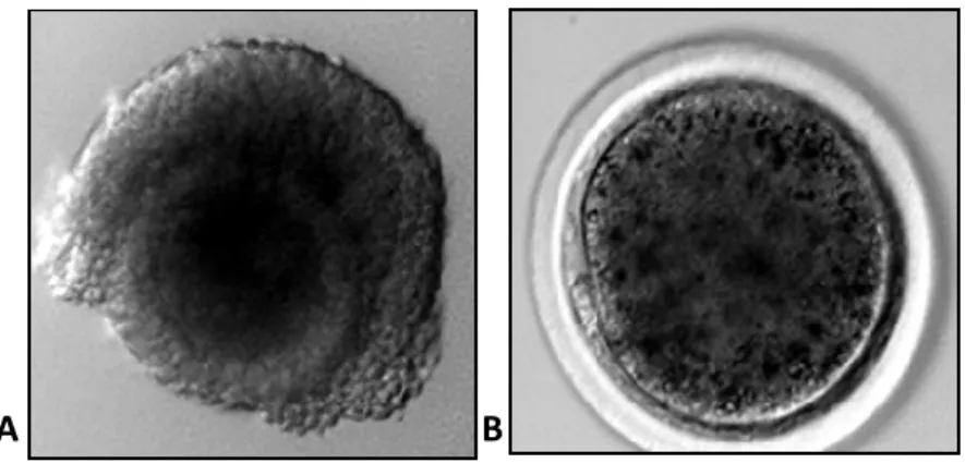
Figure 4.1.: Sheep oocytes: A) Germinal Vesicle (GV) and B) metaphase II (MII) stage
4.1.2. Semen processing and swim-up technique
Frozen–thawed spermatozoa (stored in liquid nitrogen in pellets of 250 µl), were used
following swim up procedure for all in vitro embryo production experiments. Two pellets per
in vitro fertilization (IVF) were thawed at 37° C for 30 seconds in a water bath. Afterwards an
aliquot of 200 µl of semen was deposited at the bottom of a glass vial under 1 ml of Synthetic
Oviductal Fluid (SOF) medium and incubated at 38.5° C for 15 min. After incubation 6 drops
of 100 µl of supernatant containing the most viable sperms were gently collected and placed
on a warm Petri dish. Only the drops containing the highest number of motile spermatozoa
and less debris were used for IVF.
4.1.3. In vitro fertilization and embryo development
Selected spermatozoa and IVM oocytes were incubated in fertilization medium (SOF medium
with 2% OSS, 1 μg/ml heparin and 1 μg/ml hypotaurine) for 22 hrs at 38.5° C in an
atmosphere of 5% CO2 and 5% O2 in N2 in four-well Petri dishes (Tervit et al. 1972).
Thereafter, presumptive zygotes were transferred and cultured for 8 days in four-well Petri
dishes containing SOF + essential and non-essential amino acids at oviductal concentrations
(Walker et al. 1996) +0.4% bovine serum albumin (BSA) under mineral oil, in a maximum
humidified atmosphere with 5% CO2, 5% O2 and 90% N2 at 38.5° C (Bogliolo et al. 2011). The
first cleavage was recorded between 24 and 26 hrs after fertilization.
4.1.4. Sheep and bovine tissue collection
Sheep liver, oesophagus and ovaries and bovine ovaries were collected at a local
slaughterhouse and transported to the laboratory within 1 hr in PBS. Subsequent tissues
were snap frozen in liquid nitrogen and stored at -80° C until required for further analysis.
4.1.5. Sperm collection
Ejaculates were collected from healthy adult (2-6 years old) Sarda rams (Genetic Centre of
AGRIS, Agenzia Regionale per la Ricerca in Agricoltura, Bonassai, Italy). The animals were of
proven fertility and trained to service an artificial vagina. After collection the semen was
immediately submitted to swim up technique following the protocol described in section
3.1.2. and immediately used for DNA extraction.
4.1.6. Blood collection
Blood was collected from 3 healthy Sarda sheep from the jugular vein. The wool on both
sides of the neck was removed and the area was carefully disinfected to avoid
contaminations. Blood samples (5 ml) were collected using Vacutainer®vials (BD, Becton
Dickinson and company) containing Ethylenediaminetetraacetic acid (EDTA) to prevent blood
4.2. Gene expression analysis
4.2.1. Samples collection for gene expression analysis
Gene expression analysis was performed on pools of growing oocytes (GO) at different
diameter (70/90 µm, 90/110 µm, 110/130 µm) derived from P animals, GV, IVM MII oocytes,
IVP (in vitro produced) embryos at two (2C) and four (4C) cell stage and blastocysts derived
from P and A donors were added to 30 μl RLT buffer *RNeasy Micro Kit (Qiagen, Hilden,
Germany)], snap frozen in liquid nitrogen, and stored at – 80° C until RNA isolation was
performed. As a control for IVM procedure, a pool of oocytes in each IVM run was cultured
until the blastocyst stage to assess the developmental competence of the fertilized eggs.
4.2.2. RNA Isolation
Total RNA was isolated from pools of oocytes, embryos and blastocyst with the RNeasy Micro
Kit (Qiagen, Hilden, Germany) following manufacturer’s instructions. Five pg of luciferase
mRNA (Promega) were added to each group prior to RNA extraction to account for RNA loss
during the isolation process. Importantly, during the procedure, RNA was treated with DNase
I (Qiagen, Hilden, Germany) to exclude any potential genomic DNA contamination. Isolated
RNA was eluted in 12 μL NUCLEASE‐free water (NF-H2O (Sigma-Aldrich)). Following
extraction, RNA was immediately transferred to the -80: C freezer were it was stored until
reverse transcription–polymerase chain reaction (RT‐PCR) was performed.
4.2.3 Reverse transcription
The reverse transcription allows the synthesis of DNA from an RNA template. This technique
template and a short primer complementary to the 3' end of the RNA to direct the synthesis
of the first strand cDNA, which can be used directly as a template for the Polymerase Chain
Reaction (PCR). The conversion of RNA into cDNA is fundamental because RNA is chemically
unstable and easily degraded by RNAses, that are ubiquitous in nature besides, PCR
amplification requires DNA as template. RT was performed in a final volume of 20 μL,
consisting of 50 mM Tris‐HCl (pH 8.3), 75 mM KCl, 3 mM MgCl2, 5 mM DTT, 1 mM dNTPs, 2.5
μM random hexamer primers, 0.05 μg oligo (dT), primers, 20 U RNase OUT and 100 U
SuperScript III RT (all purchased at Invitrogen Corporation, Carlsbad, CA). The reaction tubes
were incubated at 25° C for 10 min, then at 42° C for 1 hr and finally at 70° C for 15 min to
inactivate the reaction. One tube without RNA and one with RNA, but without reverse
transcriptase, were analysed as negative controls. To quantify the mRNA recovery rate, 5 pg
of luciferase mRNA (not subjected to RNA isolation) were subjected to cDNA synthesis as
well.
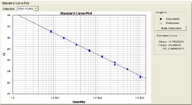
4.2.4. Primers design and validation
Primers were designed by Primer3, a free online tool to design and analyse primers for PCR
and real time PCR experiments (http://primer3.ut.ee/). All primers were designed in order to
amplify an intron-spanning region. To rule any potential genome DNA contamination. In fact,
intron-spanning primers allow to discriminate between genomic DNA and cDNA, since any
product amplified from genomic DNA would be larger than the one amplified from
intron-less cDNA. For each primer pair, the efficiency of the PCR reaction was determined by Real
Time PCR analysis. A standard curve with serial dilutions of a known amount of template, was
included the interval above and below the abundance of the targets. Only primers achieving
an efficiency of reaction between 90 and 110% (3.6 > slope > 3.1) and a coefficient of
determination r2 > 0.99 were used for the analysis (Fig 4.2).
Figure 4.2: Example of a Standard curve to asses Primers efficiency (3.6 > slope > 3.1 and a coefficient of determination r2 > 0.99).
4.2.5. Agarose gel electrophoresis
To further confirm the size of the amplicons, 3 μl of each PCR products were mixed with 5 μl
of Gel Loading Solution (Sigma Aldrich) before being subject to electrophoresis on a 2%
agarose (Sigma Aldrich) gel prepared using 0.5X TBE buffer. Such buffer from made using a
10X concentrated stock which consisted of 890 mM Tris-base, 20 mM EDTA (Sigma-Aldrich,),
890 mM Boric acid. Samples were run alongside TrackIt™ ΦX174RF DNA/Hae III Fragments.
Agarose gels stained with 20 μl syber safe (Invitrogen, Paisley, U.K) were imaged by Safe
4.2.6. PCR products sequencing
After gel checking PCR product were purification with MinElute PCR purification kit (Qiagen,
Hilden, Germany). 10 ng of purified PCR products and 3.2 pmol of each primer were put
together in a PCR tube and let dry at 65° C for no more than 5 min to allow water
evaporation to obtain a pellet. Subsequently the PCR products sequenced by singer
sequencing (Model 3130 xl Genetic Analyzer; Applied Biosystems, Foster City, CA, USA) by
BMR genomics s.r.l a company in DNA sequencing technology. After identities were
confirmed with BLAST (http://www.ncbi.nlm.nih.gov/BLAST/). Information on the primers
Table 4.1: Primers designed for real‐time PCR
Gene Primer Sequence (5’-3’) TA Size
DNMT1 FW CAGCTCTCGTACATCCACAG 60°C 158bps RV AATCTCGCGTAGTCTTGGTC DNMT3A FW GTGATGATTGATGCCAAAGA 60°C 165bps RV GGTCCTCACTTTGCTGAACT DNMT3B FW ATTGCAACAGGGTACTTGGT 60°C 122bps RV ATATTTGATGTTGCCCTCGT TET1 FW CAGTATGCTCCAGCTGCTTA 60°C 166bps RV TGCTCCCATTATTCATGTTG TET2 FW TACAAGAAACTCGCACCTGA 60°C 159bps RV CTGCATGTTCTGCAAGTCTC TET3 FW TGGAGCATGTACTTCAATGG 60°C 173bps RV GGTCACCTGGTTCTGATAGG MBD1 FW GAAGTGTCAGGTTGGACCTC 60°C 209bps RV GATTGAAAGCGATCCTCTGT MBD3 FW ACATCAGGAAGCAGGAAGAG 60°C 203bps RV GCTCTAGACGTGCTCCATTT MBD4 FW AACAGACGACAGCTCACAAA 60°C 204bps RV TCCTGGACGAGATTAAAAGG
Table 4.2: Sequence of each region amplified for gene expression analysis Gene Sequence DNMT1 CAGCTCTCGTACATCCACAGCAAGGTGCAGGTCATCTATAAGGCCCCCTCGGAGAACTGGGCC TTGGAGGGAGGCGTGGACCCCGAGGCCCTGATGTCGCAGGACGACGGGAAGACCTACTTCTA TCAGCTGTGGTACGACCAAGACTACGCGAGATT DNMT3A GTTGATTGATGCCAAAGAAGTGTCAGCTGCGCACAGGGCCCGCTACTTCTGGGGGAACCTTCC TGGTATGAACAGGCCATTGGCATCCACTGTGAATGATAAGCTGGAGCTGCAGGAGTGTCTGGA GCACGGCCGAATAGCCAAGTTCAGCAAAGTGAGGACC DNMT3B ATTGCAACAGGGTACTTGGTCCTCAAAGAACTGGGCATCAAAGTGGAGAAATACGTGGCCTCT GAAGTGTGGAAGAGTCCATTGCTGTTGGCACCGTTAAGCACGAGGGCAACATCAAATAT TET1 TTTTAGTATGCTCCAGCTGCTTATCAAAACCAGGTGGCGCTTGAACATATTGCCCGAGAATGTC GGCTTGGGAAGAAAGAAGGTCGTCCTTTCTCTGGGGTCACTGCTTGCCTAGACTTCtgTGCCCAt CCCCACAGGGACATTCACAACATGAATAATGGGAGCA TET2 TACAAGAAACTCGCACCTGATGCATACAATAATCAGATTGAATATGAACACAGAGCACCTGAGT GCCGTCTGGGTCTGAAGGAAGGCCGCCCATTCTCAGGGGTCACCGCATGTTTGGACTTCTGTG CGCATGCCCACAGAGACTTGCAGAACATGCAG TET3 TGGAGCATGTACTTCAATGGCTGCAAATATGCTCGGAGCAAGACTCCTCGCAAGTTCCGCCTCG CAGGGGACAACCCCAAAGAGGAAGAAGTGCTCCGGAAGAGTTTCCAGGACCTGGCCACCGAA GTTGCTCCCCTGTATAAGCGGCTGGCGCCCCAGGCCTATCAGAACCAGGTGACC MBD1 GAAGTGTCAGGTTGGACCTCAGAAGAGTGAAGTCAGGAAGGAGGCACCAAGGGATGATACCA AGGCTGACACTGACACAGTCCCAGCTTCACTTCCTGCCCCTGGGTGCTGTGAGAACTGTGGAAT CAGCTTTTCAGGAGATGGCACCCGAAGGCAGCGGCTCAAGACTTTGTGCAAGGACTGCCGAGC ACAGAGGATCGCTTTCAATC MBD3 ACATCAGGAAGCAGGAAGAGCTGGTGCAGCAAGTCCGCAAGCGGCTGGAGGAGGCGCTGAT GGCCGACATGCTGGCCCACGTGGAGGAGCTGGCCCGGGACGGTGAGGCACCGTTGGACAGG GCGGGCGCTGATGAGGAGGAAGATGAGGACGAGGAGGAGGAGGAGCCCGACCAGGACCCA GAAATGGAGCACGTCTAGAGC MBD4 AACAGACGACAGCTCACAAACAGAGAAAAACCCtTACGTCTGCGAAAGTATCTCAAGAAGACA CCGTCCCACGAACACAAATAGAAAAAAGGAAAACAAGCCTGTATTTTTCCAGCAAATACAACA AAGAAGCTCTTAGCCCCCCACGACGCAAGGCCTTTAAGAAGTGGACACCTCCGAGGTCACCTTT TAATCTCGTCCAGGA
4.2.7. Real time‐polymerase chain reaction
Relative quantification of transcripts was performed by Real‐Time polymerase chain reaction
(RT‐PCR) in a 7900HT Fast Real‐Time PCR System (Applied Biosystems). The PCR was
performed in a 15 μL reaction volume containing 7.5 μL 2× SYBR Green PCR Master Mix
(Applied Biosystems, Foster City, CA), 200 nM of each primer and cDNA equivalent to 0.25
oocytes or embryos. The PCR protocol consisted in two incubation steps (50° C for 5 min and
95° C for 2 min), followed by 40 cycles of amplification program 95° C for 15 s, 60° C for 30 s
and 72° C for 30 s, a melting curve programme 65–95° C, starting fluorescence acquisition at
65° C and taking measurements at 10‐s intervals until the temperature reached 95°C and
finally a cooling step to 4° C. Fluorescence data were acquired during the 72° C extension
steps. To minimise handling variation, all samples to be compared were run on the same
plate using a PCR master mix containing all reaction components apart from the sample. The
PCR products were analysed by generating a melting curve to check the specificity and
identity of the amplification product. The relative quantification of all transcripts was
performed after normalization normalised on the basis of Luciferase (Luc) mRNA levels.
4.3. Immuno detection
4.3.1. TET3 antibody validation
For the immunodetection of TET3 protein, an antibody specific for sheep is not commercially
available. Due to this inconvenient, a rabbit polyclonal antibody against Human, Bovine,