U
NIVERSITÀ DEGLIS
TUDI DIP
ISAS
CUOLA DIS
PECIALIZZAZIONEIN
M
ALATTIE DELL’A
PPARATOC
ARDIOVASCOLARETesi di Specializzazione
Management of patients with cardiovascular implantable
electronic devices-related infective endocarditis: towards a
tailored stepwise approach
R
ELATORIP
ROF.
M
ARIOM
ARZILLID
OTT.
SSAM
ARIAG
RAZIAB
ONGIORNIC
ANDIDATOD
OTT.
G
IOVANNIC
OLUCCIA2
A mia madre e a mio padre,
perché il loro esempio
rimarrà l’insegnamento più grande
3
S
UMMARYA
BSTRACTP
.
4
B
ACKGROUND P.
6
S
TUDYO
BJECTIVESP
.
18
M
ETHODSP
.
19
R
ESULTS P.
25
D
ISCUSSIONP
.
34
C
ONCLUSIONS P.
45
R
EFERENCES P.
47
4
A
BSTRACTBACKGROUND. With expanding indications for cardiac device
implantation, the prevalence of device-related infections is increasing. Patients with device infection and intracardiac vegetations represent a high-risk population with multiple comorbidities and significant mortality, regardless of the management strategy.
OBJECTIVES.The purpose of this study was to assess the feasibility, safety
and efficacy of a patient-tailored stepwise approach to the treatment of cardiac devices-related infective endocarditis (CDRIE), including antibiotic therapy and complete tranvenous device removal.
METHODS. Sixty-three consecutive patients with an established diagnosis
of CDRIE were prospectively enrolled in the study between January 2010 and August 2012. All patients underwent intra-cardiac echocardiogram before transvenous lead removal (TLR) to assess the presence, size and characteristics of lead-related vegetations. The treatment strategy consisted of a minimum period of at least two weeks of pre-operative specific antibiotic and anticoagulant therapy aimed to reduce the vegetation dimension and the infective burden of the intracardiac mass and surrounding tissues. Such treatment, in presence of stable hemodynamics, was continued until a TLR procedure was considered safe enough, without major risks of massive pulmonary embolism. After TLR, patients received prolonged antibiotic therapy and were followed for at least 6 months.
RESULTS. All patients had lead-related vegetations and 29% of them had
5 treatment before TLR, for a duration of 9,8 ± 5,4 weeks. TLR was attempted in all patients and was successful in the removal of all hardware in 97% of patients. Peri-procedural minor complications were observed in 10% of the patients; no major complication was observed. 30-days in-hospital mortality was 3%, due to overwhelming sepsis despite successful complete device removal. At a follow-up time of 19,4 ± 9,7 months, overall mortality was 10% (all deaths apart from in-hospital deaths were unrelated to CDRIE and its management). A composite end-point including peri-procedural complications and follow-up events was used to identify predictors of Outcome Events: the only independent predictor resulted the presence of a defibrillator lead.
CONCLUSIONS. Our experience suggests that treating patients with medical therapy (specific antibiotics and anticoagulants) also before TLR, even for prolonged periods, in a patient-tailored way, is a both feasible, safe and effective strategy to deal with CDRIE, possibly helping in reducing short-to-mid term mortality after device extraction, in patients with intracardiac lead-related vegetations.
6
B
ACKGROUNDInfection of cardiovascular implantable electronic devices (CIED), including permanent pacemakers (PM) and implantable cardioverter defibrillators (ICD), is a severe disease associated with high mortality [1]. Reported mortality rates of CIED–related endocarditis range from 31% to 66% if the infected device is not removed, and 18% or less with a combined approach consisting of complete device removal and antimicrobial therapy [2,3].
The rising number of patients with a CIED explains the increasing frequency of infective endocarditis (IE) in these patients. The reported incidence of CIED infection varies widely among studies [4]. A recent population-based study found an incidence of CIED infection of 1.9 per 1000 device-years and a higher probability of infection after ICD as compared with PM [5].
Both diagnosis and therapeutic strategy are particularly difficult in these patients.
A distinction should be made between local device infection (LDI) and cardiac device-related IE (CDRIE).
7 LDI is defined as an infection limited to the pocket of the CIED and is clinically suspected in the presence of local signs of inflammation at the generator pocket, including erythema, warmth, fluctuance, wound dehiscence, erosion, tenderness, or purulent drainage [6].
CDRIE is defined as an infection extending to the electrode leads, cardiac valve leaflets, or endocardial surface [7].
However, differentiating LDI and CDRIE is frequently difficult. In one study, culture of intravascular lead segments was positive in 72% of 50 patients with manifestations strictly limited to the implantation site [8]. However, the possibility of intra-operative contamination of the lead tip cannot be excluded in these patients
The main mechanism of CDRIE is contamination by local bacteriological flora at the time of device implantation. Then, the infection can spread along the electrode to the endocardium and the electrode tip. The consequence may be formation of vegetations, which can be found anywhere from the subclavian vein to the superior vena cava, on the electrode lead, on the tricuspid valve, but also on the mural endocardium of the right atrium and right ventricle.
Septic pulmonary embolism is a very frequent complication of CDRIE. Other possible mechanisms of CDRIE include haematogenous seeding
8 from a distant focus of infection. Several factors have been associated with CIED infections, including fever within 24 h before implantation, use of temporary pacing before implantation, and early reimplantation [7].
CDRIE is one of the most difficult forms of IE to diagnose. Clinical presentation is frequently misleading, with predominant respiratory or rheumatologic symptoms, as well as local signs of infection [9].
CDRIE must be suspected in the presence of unexplained fever in a patient with a CIED. Fever is frequently blunted, particularly in elderly patients. As in other forms of IE, echocardiography and blood cultures are the cornerstone of diagnosis [7].
Echocardiography plays a key role in CDRIE and is helpful for the diagnosis of both lead vegetation and tricuspid involvement, quantification of tricuspid regurgitation, sizing of vegetations, and follow-up after lead extraction.
Although trans-esophageal echocardiography (TEE) has superior sensitivity and specificity to trans-thoracic echocardiography (TTE), and is cost-effective, it is recommended to perform both in suspected CDRIE [7,10,11].
The generalization of multiplane technology for TEE has significantly improved exploration quality [12]. In addition, few other imaging
9 techniques are available, given that MRI is contraindicated in pacemaker carriers and CT has not been evaluated.
If TEE remains negative and there is still suspicion, it should be repeated within 1 week. A repeatedly negative study should virtually exclude the diagnosis.
However, both TTE and TEE may be falsely negative in CDRIE and a normal echographic examination does not rule out CDRIE [7].
In some experiences, intra-cardiac echocardiography (ICE) resulted to be useful for accurate intraluminal scanning of the right cardiac chambers and great venous vessels, increasing echocardiographic sensibility and specificity [13,14].
Regardless of the technique used, the echocardiographic finding considered to be a major criteria in the diagnosis of CDRIE is the documentation of a mobile, echodense mass attached to the valvular or to the mural endocardium/vessel wall or to the implanted leads [7].
However, it is usually very difficult to distinguish between a thrombus and an infected vegetation: recognizing that 5% of adherent masses were deemed thrombus in a retrospective survey [15], there will be some patients who are labeled as manifesting CDRIE, who may not have a lead infection. Masses that are detected in patients without positive blood
10 cultures or other suggestive features for infection are likely to represent thrombus and by themselves do not require lead removal or antibiotic treatment. In addition, the failure to visualize a mass adherent to a lead with TEE does not exclude lead infection.
Moreover, as a rule, if huge vegetations do not generally constitute a diagnostic difficulty, it is often not easy to discriminate between infectious lesions, thrombosis, fibrin deposits or pannus, in case of detection of microfilaments, sheath-like structures along the leads, strands and other uncommon structures whose nature is still unknown and significance is a matter of debate.
Additionally, echocardiography is important in assessing the maximum vegetation size prior to intervention, as many vegetations may embolize and break up into the lung circulation during transvenous lead removal (TLR).
Blood cultures are positive in 77% of cases of CDRIE [16]. Staphylococci are the most frequent pathogens, S. aureus being predominant in the acute forms of CIED infection [17].
Blood cultures can be positive with and without lead or valve vegetations or can be negative despite intracardiac vegetations.
11 The Duke criteria are difficult to apply in these patients because of lower sensitivity. Modifications of Duke criteria have been proposed, to include local signs of infection and pulmonary embolism as major criteria [9]. Finally, lung CT and lung scintigraphy are considered both useful to detect pulmonary septic embolism.
In the majority of patients, CDRIE must be treated by prolonged antibiotic therapy associated with device removal [2,7].
In the case of definite CDRIE, medical therapy alone, in fact, has been associated with high mortality and risk of recurrence. For this reason, CIED removal is recommended in all cases of proven CDRIE and should also be considered when CRDIE is only suspected, in the case of positive cultures obtained in different days (persistent bacteremia), when there is no clear source of the positive cultures in the heart, on the leads, or from other parts of the body (occult infection) [2,7].
CIED extraction can be performed percutaneously without need for surgical intervention in the majority of patients.
As newer technologies have emerged and the experience has grown, TLR has become the preferred method for removal of CIED hardware. However, these procedures involve significant risks, including cardiac tamponade, hemothorax, pulmonary embolism, lead migration, and
12 death, even in experienced hands. Thus, the performance of these procedures should be limited to centers with the appropriate facilities and training, which includes the presence and imminent availability of cardiothoracic surgery on site, to provide backup in the event of complications. In high-volume centers, TLR can be accomplished relatively safely with a high rate of success.
A primary surgical approach to lead removal in patients with CIED infection should be limited to patients who have significant retained hardware after attempts at percutaneous removal.
Another scenario in which a preference for surgical lead removal has been advocated is in patients with lead vegetations > 2 cm in diameter, because of concerns about the risk of pulmonary embolism with TLR, as a result of vegetation displacement during extraction, as it frequently occurs, particularly when vegetations are large [18].
Experience suggests, however, that percutaneous removal in patients with large vegetations can be done without precipitating a clinically apparent pulmonary embolism. These episodes, in fact, are frequently asymptomatic, and percutaneous extraction remains recommended even in cases of large vegetations, since overall risks are even higher with surgical extraction [2,7].
13 Some authors recommend surgery to be performed in patients with very large vegetations [16,19], when percutaneous extraction is technically impossible, or when severe tricuspid valve IE is associated.
When performed, surgery requires good exposure under extracorporeal circulation to allow complete removal of all foreign material. Excision of all infected contact lesions at the level of the tricuspid valve, right atrium, right ventricular free wall, and distal superior vena cava is essential.
However, mortality associated with surgical removal is high in these frequently elderly patients with associated co-morbidities [20].
With regard to vegetation size, the optimal approach is a matter of debate. Surgical extraction may be preferred for patients with larger vegetations; however, mortality rates up to 12.5% have been reported after surgical lead extraction [7].
It is thought that vegetations with a high probability of obstructing a main stem of the pulmonary artery (a vegetation >2 cm in diameter might be of borderline size) should be removed by open heart surgery.
However, several patients with vegetations >2 cm are described to have undergone successful TLR, especially patients with high perioperative risk and prior complex cardiac operations, often without major adverse events such as severe pulmonary embolism [21,22].
14 Moreover, a risk factor analysis [23] was conducted that examined clinical and echocardiographic variables that identified patients with CIED infections who were at increased risk of mortality: size and mobility of lead vegetations were not independently associated with mortality.
However, vegetation size, shape, and friability, presence of a patent foramen ovale, atrial or ventricular septal defect and other surgical indications and goals all need to be considered when making this decision. The largest vegetation size, in presence of which a TLR procedure can be safely performed, remains to be defined.
Decisions regarding percutaneous versus surgical removal of leads with vegetations larger than 2 cm in diameter should be individualized and based on patient’s clinical parameters and the extractor’s evaluation [22]. Antimicrobial therapy is adjunctive in patients with CIED infection, and complete device removal should not be delayed, regardless of timing of initiation of antimicrobial therapy. It should be individualized and the selection of the appropriate antimicrobial agent should be based on culture and susceptibility results if possible.
There are no clinical trial data to define the optimal duration of antimicrobial therapy for CIED infections, regardless of the extent of infection, or to determine when conversion to an oral agent is appropriate
15 once complete device removal has been achieved. Factors that influence medical decision making include the extent of device infection, the causative organism, the presence and duration of bloodstream infection, and associated complications such as valvular involvement, septic thrombophlebitis, or osteomyelitis.
Blood cultures should be obtained from all patients after device removal. When CIED infection is limited to the pocket site, 7 to 10 days of therapy after device removal is reasonable if the presentation is device erosion without inflammatory changes; otherwise, 10 to 14 days of antimicrobial treatment is recommended.
Therapy can be switched to an oral regimen once susceptibility results are known if there is an oral agent available that is active against the pathogen and the infected CIED has been removed.
At least 2 weeks of parenteral therapy is recommended after extraction of an infected device for patients with bloodstream infection.
Patients with sustained (>24 hours) positive blood cultures despite CIED removal and appropriate antimicrobial therapy should receive parenteral therapy for at least 4 weeks, even if TEE is negative for valvular vegetations.
16 It is intuitive that adequate debridement and control of infection at all sites, both at the generator site and metastatic, if present, sould be achieved before new device placement. The contralateral side is preferred for new device placement, if required.
It is Class I recommendation (Level of Evidence: C) a duration of antimicrobial therapy of at least 4 to 6 weeks for complicated infection (ie, endocarditis, septic thrombophlebitis, or osteomyelitis) or if bloodstream infection persists despite device removal and appropriate initial antimicrobial therapy [22].
It is imperative to make an assessment of the need for new device placement in each patient who needs to undergo the removal of an infected CIED.
One third to one half of patients in some series will not require new CIED placement. There are several factors, including reversal of the pathological processes that precipitated the need for CIED implantation, changing clinical circumstances, and lack of appropriate clinical indication initially, that obviate the need for new CIED placement and thus result in avoidance of new device infection.
On the other hand, removal of infected hardware should not be attempted until a careful assessment of a new implantation strategy has been
17 performed, particularly in patients with pacemakers for complete heart block and resynchronization therapy devices. When implantation of a new device is necessary, it should be performed on the contralateral side if possible to avoid relapsing device infection. If this is not possible, a transvenous lead can be tunneled to a device placed subcutaneously in the abdomen.
Implantation is usually postponed to allow for resolution of infection, but patients who are PM dependent represent a challenge, because they cannot be discharged home with a temporary pacemaker.
There have been no prospective trial data that examined timing of new device implantation and risk of relapsing infection; however, several investigators recommend waiting for blood cultures to be negative before a new device is placed [22].
18
S
TUDYObjectives
The treatment of patients with established diagnosis of CDRIE includes removal of all CIED hardware and a prolonged course of antimicrobial therapy.
Percutaneous extraction of infected devices presenting with large lead vegetations is associated with a variable risk of periprocedural pulmonary embolism and therefore, in cases of vegetation size of 2 cm in diameter or greater, an open surgical removal is usually to be considered [7, 22].
At our Institution, a multidisciplinary approach involving cardiac electrophysiologists experienced in TLR, infectious disease consultants and cardiac surgeons produced an original way of treating patients with CDRIE and large vegetations, that aims to start a medical treatment based on antibiotic and anticoagulant therapy before TLR, to continue it for a variable duration without keeping the patient hospitalized and to perform lead extraction percutaneously once vegetation size has decreased enough to avoid the risk of serious massive pulmonary embolism and once the bacterial burden of infected tissues has been significantly reduced. After
19 TLR, patients receive a course of antimicrobial treatment as recommended by the current guidelines.
In this study we had the objective of assessing the safety and efficacy of this patient-tailored stepwise approach.
Methods
We prospectively enrolled in the study all consecutive patients admitted at our Institution (mostly referred from other Italian Hospitals) from January 2010 to August 2012 with an established diagnosis of CDRIE, according to the modified Duke criteria [7,22].
At first admission, a complete characterization of patient’s clinical profile was obtained and all patients underwent TTE and ICE to confirm the diagnosis. ICE was performed to assess not only a better size quantification and characterization of the masses, but also to get some useful elements to guide the TLR procedure.
Intracardiac vegetation was defined as a discrete, echogenic, oscillating mass found on a valve, lead, or endocardial surface and confirmed in multiple views by echocardiography.
20 At least two sets of blood cultures were collected and an infectious disease consultant was contacted to set up an antibiotic treatment based on germ identification, when available.
TLR was performed only if vegetation size was < 2 cm in diameter and if the patient had already received a course of appropriate antibiotic treatment for at least two weeks before admission at our Institution, or fulfilled it during hospitalization.
The extraction procedure has been previously described [24,25]. After device removal, the leads were examined visually and by means of fluoroscopy in their intravascular segment; the proximal end was then clipped and a standard stylet was inserted into the lead. Lead extraction was attempted by means of gentle manual traction. If this proved unsuccessful, operators crossed to mechanical dilatation with a single-sheath (i.e. non-telescopic) technique. This involved inserting and advancing multi-sized dilators (Cook Intravascular Inc., Leechburg, PA, USA) through the venous entry-site, as first choice, or through the right internal jugular vein, if required [24,25]. A surgical debridement of the device pocket was performed, using electrocautery and blunt dissection. Procedural outcomes (i.e. success and complications) were defined according to previously published guidelines [2,26].
21 Patients with vegetation size of 2 cm or greater in diameter or who did not have already received an antibiotic treatment for at least two weeks, in presence of stable hemodynamics, were, instead, discharged; in fact, based upon the infectious disease specialist advice, it was usually selected a treatment that did not require patient’s hospitalization and could be orally administered or possibly given as an outpatient treatment.
The recommended duration of the medical therapy, in case of persistent stability of hemodynamics, was variable between at least 2 and 4 weeks and was decided based upon each patient’s specific characteristics; in case of huge vegetations, it was usually added an anticoagulant therapy in order to reduce mass dimensions.
Once completed the course of antibiotic and anticoagulant home treatment, a novel admission served to re-assess the status of the patient, their blood tests and cultures and vegetation size, by means of TTE and ICE.
Another cycle of 4-weeks antibiotic and anticoagulant home therapy would have been prescribed in case of vegetation size still > 2 cm, otherwise the patient underwent TLR, as above described.
In case of persistently large vegetations despite long periods of appropriate antibiotic and anticoagulant treatment, it was discussed the
22 indication to surgical removal, but, in our series of patients, either due to unacceptably high surgical risk (and lacking concomitant indications to surgery), or to patient refusal, a percutanous removal of the system was attempted also in several cases of very large vegetations.
The vegetation size considered for the study was the larger diameter of the mass that was observed at an ICE made at some of the pre-operatory echocardiographic assessments or at the ICE performed at the time of TLR. ICE was performed using a 9 F / 9 MHz ultra-ICE catheter-based ultrasound transducer (EP Technologies, Boston Scientific, San Jose, CA, USA). The Ultra ICE catheter was introduced percutaneously into a femoral vein through a short venous introducer (Cordis, Avanti+) and advanced under limited fluoroscopy.
Examination started with the ultra-ICE catheter positioned initially in the left or right subclavian vein, depending on the cardiac lead venous-entry side. Subsequently, the ultra-ICE catheter was withdrawn through the right atrium to the inferior vena cava in order to obtain the so-called axial imaging planes. Several slice sections on the axial plane were achieved by applying an ideal perpendicular angle of incidence of the ultrasound beam to the vessels and right atrium wall in the center of their cavity. Full interrogation included modified tomographic imaging planes at the vessel
23 (subclavian vein, innominate vein, superior vena cava), atrial, tricuspid, and ventricular plane.
ICE image quality was adequate to assess all structures involved in TLR [13,14]. In particular, all the following structures could be detected: cardiac leads; areas of fibrous adherence; vegetations: they can appear as variably shaped hyperechogenic structures, differently adherent to catheter or cardiac structures even if located at atypical intravascular locations (such as the venous tree); vein permeability: essential to detect total or partial obstruction and to check permeability of other veins (also in order to plan subsequent lead reimplantation).
Additionally, intraprocedural imaging with ICE was important for complication monitoring, complementary to fluoroscopy.
Following the extraction procedure, the patients received a prolonged course of antibiotic treatment, based on published guidelines [7,22]. Reimplantation was performed at the discretion of the treating physician. The strategies were different according to whether or not the patient was PM dependent. In the first case, if the patient was apyretic and the preoperative cultures were negative, an early reimplantation usually after 72 hours was performed; in the second, the new device was implanted in
24 the contralateral site after usually 1 week of post-extraction antibiotic therapy.
Patients were followed for at least 6 months from the date of TLR, by means of visits and/or phone calls.
Statistical analysis
Continuous variables were expressed as mean ± 1 standard deviation or median (range) when appropriate and categorical variables as percentages. Groups were compared for categorical data using Fisher’s exact test and for continuous variables using analysis of variance followed by Fisher’s protected least significant difference for multiple comparisons. All tests were 2-sided; a P<0.05 was considered to be significant. For non parametric variables, appropriate non-parametric tests (i.e. the Kruskall Wallis test and the Spearman correlation) were used. The predictors of peri-procedural adverse events were assessed at Logistic regression analysis. Only variables with a P<0.05 at univariate logistic analysis were entered in the multivariate model. Kaplan-Meier statistics followed by Log-Rank test was used to evaluate the predictors of adverse events. Statistical analyses were performed using JMP statistical software (SAS Institute Inc, version 4.0.0) and Stata software (Stata Statistical Software: Release 10, StataCorp. 2007, College Station, TX).
25 Results
A total of 63 patients were prospectively enrolled in the study. The patient demographics are summarized in Table 1.
Table 1
Number of patients 63
Sex: male 49 (78)
Age (years) 65 ± 16
Comorbidities
Congestive heart failure 32 (51)
Diabetes 18 (29)
Chronic kidney disease 17 (27)
Coronary artery disease 10 (16)
Previous cardiac surgery 7 (11)
Valvular prosthesis 4 (6)
Chronic obstructive pulmonary disease 7 (11) Type of device
Pacemaker 23 (36)
Implantable cardioverter defibrillator 19 (30)
Biventricular pacemaker 1 (2)
Biventricular defibrillator 20 (32)
Left ventricular ejection fraction 47 ± 13 Device implantation indication*
Atrio-ventricular block 14 (22)
Sinus node dysfunction 10 (16)
Primary prevention of sudden cardiac death 25 (40) Secondary prevention of sudden cardiac death 14 (22)
Resynchronization therapy 20 (32)
Number of leads/patient
Total 2 (1-4)
Targeted for extraction 2 (1-4)
Age of the oldest lead targeted for extraction (months) 96 ± 66 Months from most recent device-related procedure 26 ± 22
Data are expressed as “number (percentage)”; “mean ± standard deviation” or “median (range)”.
*The sum does not match with number of patients, due to multiple indication in some cases.
26 Data about the characteristics of CIED infection, its timing and medical treatment are summarized in Table 2.
Table 2
Infection history and characterization
Time from first device infection diagnosis to extraction (months) 11,3 ± 20,3
History of fever 59 (94)
Lead vegetation detected at echocardiograms 63 (100) Concomitant infection of device pocket 38 (60) Vegetation size at intracardiac echo (mm) 14,5 ± 9,4
Patients with vegetation size 20 mm or larger 18 (29)
Positive peripheral blood cultures 47 (75)
Atrial blood sampling during intracardiac echo 53 (84) Pre-extraction antibiotic treatment
Patients treated 58 (92)
Total duration of pre-extraction antibiotic therapy (weeks) 9,8 ± 5,4 Patients treated with oral antibiotics 42 (67)
Duration of oral therapy alone (weeks) 7,1 ± 5,5 Patients treated with oral plus parenteral antibiotics 51 (81)
Duration of combined (oral plus parenteral) treatment (weeks) 5,3 ± 4,1 Post-extraction antibiotic treatment (weeks) 3,8 ± 3,5
Data are expressed as “number (percentage)” or “mean ± standard deviation”
The majority of patients (60%) showed signs of device pocket infection and had positive blood cultures (75%). All patients had lead vegetations; 29% of patients had large vegetations, 20 mm in diameter or larger.
Infection of the device was usually present from several months (11,3 ± 20,3) and most patients (92%) had received a prolonged course (9,8 ± 5,4 weeks) of antibiotic treatment at the time of TLR. All patients received post-procedural appropriate antibiotic therapy, according to guidelines, for a duration of 3,8 ± 3,5 weeks.
27 The results of peripheral blood cultures, early taken during the diagnostic work-up, are summarized in Figure 1.
Figure 1
The majority of cases of CDRIE in our patient series, as shown, were due to Staphylococci (n=38, 61%), of which 10 cases to S. aureus (16%), 17 cases (27%) to S. epidermidis and 11 cases (18%) to Coagulase-negative Staphylococci other than S. epidermidis. A minority of cases were due to other Gram-positive species (4: S. viridans, P. acnes, C. jeikeium, Enterococcus spp), to Gram-negative germs (4: E. cloacae, P. aeruginosa, H. influenzae, Borrelia spp) and one patient resulted positive to C. albicans. Blood cultures tested negative in 16 cases (25%).
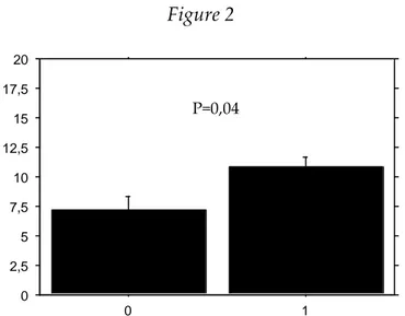
28 The identification of a microorganism in blood cultures resulted associated with a significantly longer period of antibiotic treatment before extraction, as shown in Figure 2. Figure 2 0 2,5 5 7,5 10 12,5 15 17,5 20 0 1
x-axis: 0=patients with negative blood cultures; 1=patients with positive blood cultures y-axis: duration of antibiotic treatment before extraction (weeks)
Moreover, the identification of a microorganism in blood cultures resulted associated with a significantly smaller dimension of the vegetation detected at ICE, as shown in Figure 3.
Figure 3 0 5 10 15 20 25 30 35 40 C e ll M e a n 0 1 Cell
x-axis: 0=patients with negative blood cultures; 1=patients with positive blood cultures y-axis: diameter of lead vegetation at ICE (mm)
P=0,04
29 TLR was attempted in all patients and was successful in removal of all hardware in the great majority of cases (n=61, 97%): the procedure was unsuccessful only in two cases. In one case the technique was not effective and the patient underwent surgical extraction of two pacing leads (148 months old), with concomitant implantation of two epicardial leads, without complications. In another case, a lead fragment was abandoned (the patient was carrier of a dual chamber ICD and had three leads, of which only two were completely extracted), and this did not bring complications. The patient underwent a subcutaneous ICD system implantation two weeks later and did not report adverse events in the follow-up.
Radiological success was not achieved also in another case, in which only the fractured atrial lead tip was abandoned in the auricle, without sequelae: in this case, however, the extraction procedure was considered clinically successful.
Minor peri-procedural complications occurred in 6 patients (10%) and are summarized in Table 3: two patients had symptomatic embolization of part of the vegetation into lung circulation, that was promptly resolved and passed without apparent consequences; two patients had hematomas at the pocket site, one of which requiring blood transfusion; in two cases
30 there was a minor pneumothorax that was resolved by means of thoracic drainage.
All patients entered the follow-up, but 61 (97%) were followed for at least six months (follow-up duration 19,4 ± 9,7 months), by means of visits and/or phone calls (2 patients were lost to follow-up). The events recorded in the follow-up are summarized in Table 3.
Table 3
Peri-procedural complications 6 (10)
Symptomatic pulmonary embolism 2 (3)
Pocket site hematoma 2 (3)
Need for blood transfusion 1 (2)
Minor pneumothorax 2 (3)
Events at follow-up 8 (13)
Death 6 (10)
Sepsis (in hospital death, < 1 month from extraction) 2 (3) Prolonged diarrhea (32 months after extraction) 1 (2) Dementia (23 months after extraction) 1 (2) End stage renal failure (7 months after extraction) 1 (2) Acute pancreatitis (19 months after extraction) 1 (2) Dislodged lead repositioning (1 month after reimplant) 1 (2) Decubitus of an abandoned epicardial lead 1 (2)
Data are expressed as “number (percentage)”
Only the two septic deaths occurred during hospitalization within one month from successful extraction, appear to be strictly related to the clinical problem of CDRIE.
Given the small number of events in the follow-up and the small amount of procedural complications observed, all these events were considered together in the statistics, to formulate a composite end-point, named
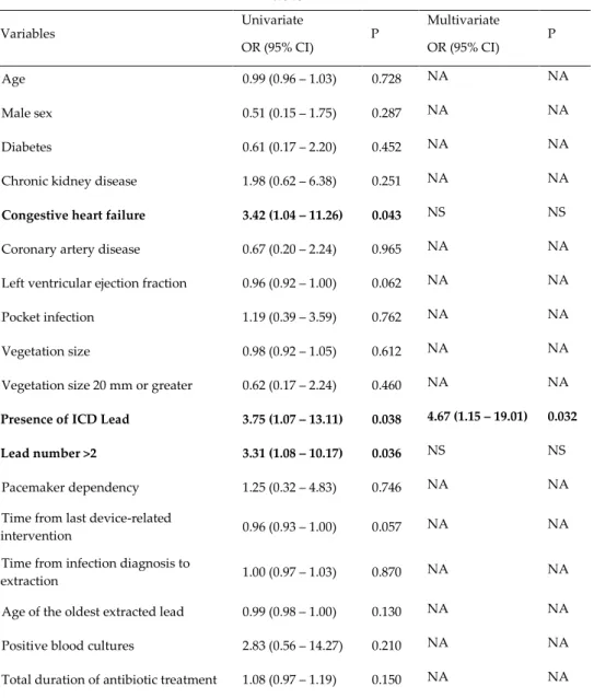
31 Outcome Events. Many clinical variables were tested as predictors of Outcome Events, as indicated in Table 4, that summarizes the results of the univariate and multivariate analysis performed.
Table 4 Variables Univariate OR (95% CI) P Multivariate OR (95% CI) P Age 0.99 (0.96 – 1.03) 0.728 NA NA Male sex 0.51 (0.15 – 1.75) 0.287 NA NA Diabetes 0.61 (0.17 – 2.20) 0.452 NA NA Chronic kidney disease 1.98 (0.62 – 6.38) 0.251 NA NA
Congestive heart failure 3.42 (1.04 – 11.26) 0.043 NS NS
Coronary artery disease 0.67 (0.20 – 2.24) 0.965 NA NA Left ventricular ejection fraction 0.96 (0.92 – 1.00) 0.062 NA NA Pocket infection 1.19 (0.39 – 3.59) 0.762 NA NA Vegetation size 0.98 (0.92 – 1.05) 0.612 NA NA Vegetation size 20 mm or greater 0.62 (0.17 – 2.24) 0.460 NA NA
Presence of ICD Lead 3.75 (1.07 – 13.11) 0.038 4.67 (1.15 – 19.01) 0.032
Lead number >2 3.31 (1.08 – 10.17) 0.036 NS NS
Pacemaker dependency 1.25 (0.32 – 4.83) 0.746 NA NA Time from last device-related
intervention 0.96 (0.93 – 1.00) 0.057 NA NA Time from infection diagnosis to
extraction 1.00 (0.97 – 1.03) 0.870 NA NA Age of the oldest extracted lead 0.99 (0.98 – 1.00) 0.130 NA NA Positive blood cultures 2.83 (0.56 – 14.27) 0.210 NA NA Total duration of antibiotic treatment 1.08 (0.97 – 1.19) 0.150 NA NA OR: Odds Ratio; CI: Confidence Interval; NA: not available; NS: not significant; ICD: implantable cardioverter defibrillator
At univariate analysis, three variables resulted significant predictors of Outcome Events: an history of congestive heart failure, the presence of an
32 ICD lead and having a number of leads > 2. At multivariate analysis, only the presence of an ICD lead resulted as an independent predictor of Outcome Events.
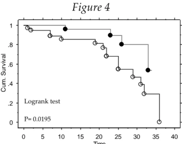
Kaplan-Meier statistics to evaluate the predictors of Outcome Events showed significant results in differentiating event-free survival curves, with the grouping variables of presence of an ICD lead (Figure 4) and number of leads > 2 (Figure 5).
Figure 4 0 .2 .4 .6 .8 1 Cu m . S u rv iv a l 0 5 10 15 20 25 30 35 40 Time Event Times (1) Cum. Survival (1) Event Times (0) Cum. Survival (0)
Kaplan-Meier Cum . Survival Plot for follw up Censor Variable: Tot Eventi
Grouping Variable: ICD
White dots: patients with ICD leads; black dots: pacemaker patients. Time is expressed in months.
Figure 5
White dots: patients with more than 2 leads; black dots: patients with 2 or less leads. Time is expressed in months.
Logrank test P= 0.0195 0 .2 .4 .6 .8 1 Cu m . S u rv iv a l 0 5 10 15 20 25 30 35 40 Time Event Times (1.000) Cum. Survival (1.000) Event Times (0.000) Cum. Survival (0.000)
Kaplan-Meier Cum . Survival Plot for follw up Censor Variable: Tot Eventi
Grouping Variable: Indice di difficoltà
Logrank test
33 The majority of patients underwent CIED reimplantation during the same hospitalization as TLR (n=36, 57%) and the procedure was performed after 5,2 ± 4,4 days from TLR, with a duration of hospital stay of 13,9 ± 8,2 days. Eight patients (12%) were discharged without performing reimplantation during the same hospitalization, for various reasons: apart from cases in which there was lack of ongoing indication or patient’s refusal, in some cases with very large vegetations without PM dependency or in primary prevention ICD carriers, the patient was discharged and an ICE evaluation was planned after at least 1 month of antibiotic and anticoagulant therapy to demonstrate reduced vegetation dimensions and eventually proceed to reimplantation.
In some cases, the persistence of the mass inside the right chamber, in fact, is not necessarily associated with continued illness, as confirmed by resolution of fever, normal white blood cell count and labeled leukocyte imaging that is repeatedly negative for infection. In such cases, reimplantation is still considered safe.
Seven patients (11%) received a wearable defibrillator at discharge and six of them received a new ICD implantation after 112 ± 51 days.
34 Discussion
Cardiac device infection is a serious, emerging disease with a 210% increase in incidence between 1993 and 2008. In-hospital charges for this complication are estimated to be at least US $146000 per case in the United States. CDRIE in particular has a substantially higher mortality rate than cardiac device infection without endocarditis [27].
Data from randomized, controlled trials to guide the management of CIED infections are lacking.
Recommendations for complete extraction of the device, the route of administration and the duration of antimicrobial therapy, and the timing for placement of a new device are largely based on observational data, clinical experience, or both.
Observations from several medical centers universally support complete removal of the device to cure infection and reduce morbidity and mortality [28, 29].
A variety of percutaneous lead-removal techniques are available, and only a small minority of patients require open cardiovascular surgery for complete device removal.
The choice of percutaneous or open surgical removal should take into account not only the size of a vegetation, as visualized on
35 echocardiography, but also several other factors, including the patient’s age, how long the device has been in place, the type of device, the number of retained leads from previous devices, the presence or absence of a history of difficult or complicated percutaneous extractions, and the patient’s status with respect to coexisting conditions.
Complications, including death, may occur with either percutaneous or surgical removal of the device.
Major complications are reported in less than 2% of patients who undergo percutaneous removal [2], but the rate may be much more high with surgical removal, which is generally performed after unsuccessful or complicated percutaneous extraction.
In a recently published study about a large prospective cohort of patients with CDRIE [27], in-hospital and 1-year mortality rates were 14.7% and 23.2% respectively and it was observed a survival benefit at 1 year for device removal during the initial hospitalization (19.9% of patients, who underwent device removal during the index hospitalization had died at 1 year, versus 38.2% who did not undergo device removal).
In a retrospective survey, patients with CIED infections were divided into two groups of systemic or local infection: in the first group, the in-hospital mortality was 29% (5% in the second group) despite complete CIED
36 removal. It was found that early TLR was associated with better in-hospital survival and shorter in-hospital stays and this association was statistically significant in the population as a whole (TLR performed within 3 hospitalization days was associated with lower in-hospital mortality), with a statistical trend in the higher risk subgroup with endovascular infection or endocarditis [30].
In a retrospective study about TLR in 100 patients with intracardiac vegetations detected by TEE (mean diameter 16 mm), there was a 5% rate of only minor complications, post-operative 30-day mortality was 10% and no death was directly related with the extraction procedure (multiple comorbidities and overwhelming sepsis were considered responsible of the natural progression, despite TLR, of the disease that finally caused the observed short-term deaths) [31].
In a more recent study [32], 129 patients with lead-associated vegetations were analyzed: 13,2 % of patients underwent surgical removal; 10,9 % was the overall in-hospital mortality and 6,2 % was the overall rate of major complications, regardless of the modality of device removal. The Authors found that the clinical presentation of CDRIE was influenced by the size of the lead vegetation (they used 1 cm as the cut-off value for separating patients in two groups). Complications of lead removal were more
37 common in those with larger vegetations, driven by complications in those requiring an open surgical approach.
A study by Golzio PG and Coll. [33] showed that prevalence of intracardiac lead vegetations in patients treated with lead extraction was 40%, when transesophageal echocardiography was extensively performed in all patients with infective indications. Vegetations were, as expected, mainly observed in sepsis/systemic infection, but, more interestingly, they were also found in local infections and chronic draining sinus (approximately in one-third of the cases), where they represented an ‘unexpected’ finding according to traditional knowledge. The Authors showed how vegetation occurrence play a key role in stratifying the risk of the extraction procedure and in driving further therapeutic decisions and concluded that vegetations should be accurately investigated before extraction in any case showing infective indications, as well as in patients with only local signs or symptoms. It was confirmed the importance of factors previously associated with infections, according to literature data: replacement, revision, previous reparative procedures, renal failure, dialysis, CRT devices, absence of antibiotic prophylaxis, and long-standing infection were associated with vegetations; increased WBC count and dialysis resulted independent risk factors for vegetations. The study
38 was also probably the only one in which the therapeutic approach considered an antibiotic treatment before TLR in patients with large vegetations: the Authors stated that their better outcomes (1,5% in-hospital death) could be related to the use of a longer antibiotic course before TLR.
However, the generally observed high rates of mortality of CDRIE emphasize the need for improved preventive measures, including optimal skin decontamination and appropriate antibiotic administration at the time of cardiac device insertion or manipulation, as well as careful attention to any invasive or intravascular procedures performed after device implantation.
Our experience in dealing with CDRIE as a tertiary referral center with recognized expertise in TLR suggested us that, particularly when a patient shows large vegetations and does not present hemodynamic instability, it is possible and safe to delay the procedure of TLR, allowing the patient to receive some weeks of appropriate and specific antibiotic and anticoagulant therapy, while waiting for the complete device removal and the subsequent unanimously recommended prolonged specific antibiotic treatment. This kind of strategy met the support of our infectious disease consultants.
39 Pre-extraction anticoagulation therapy has a rationale in the fact that the involvement of endovascular and intracardiac structures in the infectious process related to the CIED leads, with formation of a vegetant mass, needs the interaction of the microbial flora with thrombotic material: anticoagulation could help in the progressive fragmentation of the mass, at the same time allowing antibiotics to reach the bacterial colonies.
We believe that, in presence of lead-related vegetations, especially when they are large, a specific antibiotic treatment together with an anticoagulant therapy, given to the patient before TLR, could not only produce a significant reduction in the size of the mass, thus reducing the risk of massive embolization, but also contribute to sterilize the vegetation, the surrounding tissue and possibly the device pocket (when signs of local infection are concomitant), so that, when performing TLR, the main source of bacteremia and therefore of bloodstream infection is possibly represented only by the infected hardware.
The only concern about this treatment strategy is about very large fungal lead-related vegetations, because in these cases antimicrobial therapy is often not able to reduce the size of the mass despite long term administration and therefore a surgical removal is often required.
40 In this preliminary study, our approach proved to be feasible, safe and effective and results about in-hospital death and mid-term outcome are very encouraging, if compared with the above-mentioned data about similar populations published in the literature.
The observed complication rate was low and they all were minor complications.
In-hospital mortality was 3% and none of the deaths occurred in the follow-up was related to the problem of CDRIE and its management, being instead expression of serious comorbidities.
Our population consisted of very ill patients, considering that 62% of them were carriers of an ICD, 51% suffered from congestive heart failure and 34% had a biventricular device. Moreover, the average dwelling time of the leads that were targeted for extraction was 96 ± 66 months, accounting for a possibly high average difficulty of TLR procedures.
In this patient series, as well as in our previous experience, the presence of large vegetations, even bigger than 30 mm in diameter, in patients treated for a long time with antibiotics and anticoagulants, did not prevent the possibility of a safe percutaneous extraction, without clinical compromise despite a likely underestimated rate of silent lung embolization, and acute and long term outcome were very good.
41 In our hypothesis, needing confirmation by further studies, our low rate (3%) of in-hospital mortality for post-extraction overwhelming sepsis could be related to a kind of sterilization of the vegetant mass, the surrounding venous and heart structures and the device pocket, that is gained by means of preliminary antibiotic treatment, combined with subsequent device removal and continuing antimicrobial therapy.
In this kind of patients, in fact, the vegetations sometimes disappear with extraction, but the remaining right-sided infected heart and lung environment is unknown and the mortality from uncontrolled sepsis, with and without heart failure, despite device removal and subsequent appropriate antibiotics, could, in part, be due to residual infected inflammatory tissue, possibly within the right heart, lung, or implant pocket [34].
So, the rationale of treating patients with prolonged courses of antibiotics before TLR is also the need to control the infection at the site of this supposed infected inflammatory tissue and possibly to make it sterile when a TLR is performed, thus reducing the risk of bacterial seeding and bloodstream infection after TLR.
It is important to underline that the long term pre-operative antibiotic treatment (92% of our patients received 9,8 ± 5,4 weeks of antibiotic
42 therapy before TLR) is generally given as an outpatient or home therapy, thus reducing hospitalization duration and its overall costs.
Time from first device infection diagnosis to TLR is particularly high in our series of patients also because many of them were referred to our Institution after prolonged periods of conservative treatment attempts, either with antibiotics alone or with antibiotics plus device pocket surgical revisions. Our strategy should be evaluated in this setting. Although too much time has been wasted before referring a patient in a center experienced in the CDRIE management, in our opinion it would be advisable not to abandon the opportunity to best treat the lead infection with a proper duration of the specific antibiotic treatment.
Our results underline the importance of identifying the pathogens from blood cultures, possibly before the start of antibiotic therapy: the identification of germs was, in fact, associated with significantly smaller vegetation sizes and longer duration of antibiotic treatment before TLR and this could mean that a culture-based specific therapy given to a patient for prolonged duration could help in reducing vegetation dimensions.
43 Moreover, it is worth emphasizing that no death occurred in patients in whom, because of a huge vegetation, TLR procedure was delayed, thus increasing the duration of the preoperative antibiotic treatment.
Microbiology in our patient population is consistent with data reported in the literature, both in the percentage of positive early taken blood cultures, and in the species of involved microorganisms, with predominance of Staphylococci [17,22].
Given the small number of events in the follow-up and the small amount of procedural complications observed, all these events were considered together in the statistics, to formulate a composite end-point, named Outcome Events: limitations in the correct interpretation of statistics in presence of composite end-points are well known. We only point out that the observed significant predictors of Outcome Events probably identify a kind of sicker patient (congestive heart failure, ICD patients, biventricular device patients), that is likely to have more comorbidities, to be older and frailer.
44
Study limitations
In this patient series, we have only data concerning the maximum measured dimensions of the vegetations, during the entire course of clinical observation, from early CIED infection diagnosis to TLR.
It would be of crucial importance, for a better characterization of the impact of treatment on the pre- and post-operative features of the vegetation, to have echocardiographic data about each step of treatment, in order to assess the extent of the effective reduction in vegetation size after antibiotic and anticoagulant therapy prior to extraction and to monitor the subsequent evolution of the intracardiac mass after TLR. Our echocardiographic data were all obtained at ICE: the possibility of making comparisons between various echocardiographic methods (TTE, TEE, ICE) in assessing vegetation dimension and features would add important details, possibly very useful for the diagnostic management of this kind of patients.
The lack of a control group of matched patients treated with a different approach does not permit comparisons. Only comparing the results of our strategy with those achieved by other groups applying different approaches in similar populations are possible at the moment.
45 The proposed tailored stepwise approach of management implies that given therapies are not fully standardized among all patients, mainly in time duration (for example, duration of pre-operative treatment was critically dependent on each patient’s specific characteristics), although they are uniformly designed.
Our study was conducted at a single tertiary referral center with experience in TLR, and its results and conclusions may not apply to different Institutions.
Conclusions
As indications for device implantation expand, the prevalence of device-related infections will increase.
Vigilant recognition and appropriate management with both antibiotics and complete device removal should be the cornerstones of therapy. Patients with CIED infection and intracardiac vegetations represent a high-risk population with multiple comorbidities and significant mortality, regardless of management strategy.
Our experience suggests that treating patients with medical therapy (specific antibiotics and anticoagulants) also before TLR, even for prolonged periods, in a patient-tailored way, is both feasible and safe and
46 possibly helps in reducing short-to-mid term mortality after device extraction, in patients with intracardiac lead-related vegetations.
Further studies are needed to strongly assess the best ways to treat patients with CDRIE.
47 REFERENCES
1. Rundstrom H, Kennergren C, Andersson R, Alestig K, Hogevik H. Pacemaker endocarditis during 18 years in Goteborg. Scand J Infect Dis 2004;36:674–679.
2. Wilkoff BL, Love CJ, Byrd CL, Bongiorni MG, Carrillo RG, Crossley GH 3rd, Epstein LM, Friedman RA, Kennergren CE, Mitkowski P, Schaerf RH, Wazni OM; Heart Rhythm Society; American Heart Association. Transvenous lead extraction: Heart Rhythm Society expert consensus on facilities, training, indications, and patient management: this document was endorsed by the American Heart Association (AHA). Heart Rhythm 2009;6:1085–1104.
3. Baddour LM, Wilson WR, Bayer AS, Fowler VG Jr, Bolger AF, Levison ME, Ferrieri P, Gerber MA, Tani LY, Gewitz MH, Tong DC, Steckelberg JM, Baltimore RS, Shulman ST, Burns JC, Falace DA, Newburger JW, Pallasch TJ, Takahashi M, Taubert KA; Committee on Rheumatic Fever, Endocarditis, and Kawasaki Disease; Council on Cardiovascular Disease in the Young; Councils on Clinical Cardiology, Stroke, and Cardiovascular Surgery and Anesthesia; American Heart Association; Infectious Diseases Society of America. Infective endocarditis: diagnosis, antimicrobial therapy,
48 and management of complications: a statement for healthcare professionals from the Committee on Rheumatic Fever, Endocarditis, and Kawasaki Disease, Council on Cardiovascular Disease in the Young, and the Councils on Clinical Cardiology, Stroke, and Cardiovascular Surgery and Anesthesia, American Heart Association: endorsed by the Infectious Diseases Society of America. Circulation 2005;111:e394–434.
4. Baddour LM, Bettmann MA, Bolger AF, Epstein AE, Ferrieri P, Gerber MA, Gewitz MH, Jacobs AK, Levison ME, Newburger JW, Pallasch TJ, Wilson WR, Baltimore RS, Falace DA, Shulman ST, Tani LY, Taubert KA. Nonvalvular cardiovascular device-related infections. Circulation 2003;108:2015–2031.
5. Uslan DZ, Sohail MR, St Sauver JL, Friedman PA, Hayes DL, Stoner SM, Wilson WR, Steckelberg JM, Baddour LM. Permanent pacemaker and implantable cardioverter defibrillator infection: a population-based study. Arch Intern Med 2007;167:669–675.
6. Sohail MR, Uslan DZ, Khan AH, Friedman PA, Hayes DL, Wilson WR, Steckelberg JM, Stoner S, Baddour LM. Management and outcome of permanent pacemaker and implantable cardioverter-defibrillator infections. J Am Coll Cardiol 2007;49:1851–1859.
49 7. Habib G, Hoen B, Tornos P, Thuny F, Prendergast B, Vilacosta I, Moreillon P, de Jesus Antunes M, Thilen U, Lekakis J, Lengyel M, Müller L, Naber CK, Nihoyannopoulos P, Moritz A, Zamorano JL; ESC Committee for Practice Guidelines. Guidelines on the prevention, diagnosis, and treatment of infective endocarditis (new version 2009). Eur Heart J 2009;30:2369-413.
8. Klug D, Wallet F, Lacroix D, Marquie C, Kouakam C, Kacet S, Courcol R. Local symptoms at the site of pacemaker implantation indicate latent systemic infection. Heart 2004;90:882–886.
9. Klug D, Lacroix D, Savoye C, Goullard L, Grandmougin D, Hennequin JL, Kacet S, Lekieffre J. Systemic infection related to endocarditis on pacemaker leads: clinical presentation and management. Circulation 1997;95:2098–2107.
10. Vilacosta I, Sarria C, San Roman JA, Jimenez J, Castillo JA, Iturralde E, Rollan MJ, Martinez Elbal L. Usefulness of transesophageal echocardiography for diagnosis of infected transvenous permanent pacemakers. Circulation 1994;89:2684–2687.
11. Victor F, De Place C, Camus C, Le Breton H, Leclercq C, Pavin D, Mabo P, Daubert C. Pacemaker lead infection: echocardiographic features, management, and outcome. Heart 1999;81:82–87.
50 12. Tighe DA, Tejada LA, Kirchoffer JB, Gilmette P, Rifkin RD, Estes NA. Pacemaker infection: detection by multiplane transesophageal echocardiography. Am Heart J 1996;131:616-618.
13. Bongiorni MG, Di Cori A, Soldati E, Zucchelli G, Arena G, Segreti L, De Lucia R, Marzilli M. Intracardiac echocardiography in patients with pacing and defibrillating leads: a feasibility study. Echocardiography 2008;25:632-638.
14. Narducci ML, Pelargonio G, Russo E, Marinaccio L, Di Monaco A, Perna F, Bencardino G, Casella M, Di Biase L, Santangeli P, Palmieri R, Lauria C, Al Mohani G, Di Clemente F, Tondo C, Pennestri F, Ierardi C, Rebuzzi AG, Crea F, Bellocci F, Natale A, Dello Russo A. Usefulness of intracardiac echocardiography for the diagnosis of cardiovascular implantable electronic device-related endocarditis. J Am Coll Cardiol. 2013 Apr 2;61(13):1398-1405.
15. Lo R, D’Anca M, Cohen T, Kerwin T. Incidence and prognosis of pacemaker lead-associated masses: a study of 1,569 transesophageal echocardiograms. J Invasive Cardiol. 2006;18:599–601.
16. Sohail MR, Uslan DZ, Khan AH, Friedman PA, Hayes DL, Wilson WR, Steckelberg JM, Jenkins SM, Baddour LM. Infective
51 endocarditis complicating permanent pacemaker and implantable cardioverter-defibrillator infection. Mayo Clin Proc 2008;83:46–53. 17. Bongiorni MG, Tascini C, Tagliaferri E, Di Cori A, Soldati E,
Leonildi A, Zucchelli G, Ciullo I, Menichetti F. Microbiology of cardiac implantable electronic device infections. Europace. 2012 Sep;14(9):1334-1339.
18. Meier-Ewert HK, Gray ME, John RM. Endocardial pacemaker or defibrillator leads with infected vegetations: a single-center experience and consequences of transvenous extraction. Am Heart J 2003;146:339–344.
19. Ruttmann E, Hangler HB, Kilo J, Hofer D, Muller LC, Hintringer F, Muller S, Laufer G, Antretter H. Transvenous pacemaker lead removal is safe and effective even in large vegetations: an analysis of 53 cases of pacemaker lead endocarditis. Pacing Clin Electrophysiol 2006;29:231–236.
20. Del Rio A, Anguera I, Miro JM, Mont L, Fowler VG Jr, Azqueta M, Mestres CA. Surgical treatment of pacemaker and defibrillator lead endocarditis: the impact of electrode lead extraction on outcome. Chest 2003;124:1451–1459.
52 21. Pérez Baztarrica G, Gariglio L, Salvaggio F, Reolón E, Blanco N, Mazzetti H, Villecco S, Botbol A, Porcile R. Transvenous extraction of pacemaker leads in infective endocarditis with vegetations ≥20 mm: our experience. Clin Cardiol. 2012;35:244-249.
22. Baddour L, Epstein A, Erickson C, et al. Update on cardiovascular implantable electronic device infections and their management: a scientific statement from the American Heart Association. Circulation 2010;121:458–477.
23. Baman TS, Gupta SK, Valle JA, Yamada E. Risk factors for mortality in patients with cardiac device-related infection. Circ Arrhythm Electrophysiol 2009;2:129–134.
24. Bongiorni MG, Soldati E, Zucchelli G, Di Cori A, Segreti L, De Lucia R, Solarino G, Balbarini A, Marzilli M, Mariani M: Transvenous removal of pacing and implantable cardiac defibrillating leads using single sheath mechanical dilatation and multiple venous approaches: high success rate and safety in more than 2000 leads. Eur Heart J 2008; 29: 2886-2893.
25. Bongiorni MG: Transvenous Lead Extraction: from Simple Traction to Internal Transjugular Approach, Bongiorni MG editor. Springer Verlag 2011: 1-180.
53 26. Deharo JC, Bongiorni MG, Rozkovec A, Bracke F, Defaye P, Fernandez-Lozano I, Golzio PG, Hansky B, Kennergren C, Manolis AS, Mitkowski P, Platou ES; European Heart Rhythm Association: Pathways for training and accreditation for transvenous lead extraction: a European Heart Rhythm Association position paper. Europace 2012; 14:124-134.
27. Athan E, Chu VH, Tattevin P, Selton-Suty C, Jones P, Naber C, Miró JM, Ninot S, Fernández-Hidalgo N, Durante-Mangoni E, Spelman D, Hoen B, Lejko-Zupanc T, Cecchi E, Thuny F, Hannan MM, Pappas P, Henry M, Fowler VG Jr, Crowley AL, Wang A; ICE-PCS Investigators. Clinical characteristics and outcome of infective endocarditis involving implantable cardiac devices. JAMA 2012; 307(16):1727-35
28. Le KY, Sohail MR, Friedman PA, et al. Impact of timing of device removal on mortality in patients with cardiovascular implantable electronic device infections. Heart Rhythm 2011;8:1678-85.
29. Pichlmaier M, Knigina L, Kutschka I, et al. Complete removal as a routine treatment for any cardiovascular implantable electronic
device-associated infection. J Thorac Cardiovasc Surg
54 30. Viganego F, O'Donoghue S, Eldadah Z, Shah MH, Rastogi M, Mazel JA, Platia EV. Effect of early diagnosis and treatment with percutaneous lead extraction on survival in patients with cardiac device infections. Am J Cardiol 2012;109(10):1466-71
31. Grammes JA, Schulze CM, Al-Bataineh M, Yesenosky GA, Saari CS, Vrabel MJ, Horrow J, Chowdhury M, Fontaine JM, Kutalek SP. Percutaneous pacemaker and implantable cardioverter-defibrillator lead extraction in 100 patients with intracardiac vegetations defined by transesophageal echocardiogram. J Am Coll Cardiol 2010;55(9):886-94
32. Greenspon AJ, Le KY, Prutkin JM, Sohail MR, Vikram HR, Baddour LM, Danik SB, Peacock J, Falces C, Miro JM, Naber C, Carrillo RG, Tseng CH, Uslan DZ. Influence of Vegetation Size on the Clinical Presentation and Outcome of Lead-Associated Endocarditis: Results From the MEDIC Registry. JACC Cardiovasc Imaging 2014;7(6):541-9
33. Golzio PG, Fanelli AL, Vinci M, Pelissero E, Morello M, Grosso Marra W, Gaita F. Lead vegetations in patients with local and systemic cardiac device infections: prevalence, risk factors, and therapeutic effects. Europace 2013;15(1):89-100
55 34. Maloney JD, Maloney JD 3rd. Vegetation size marker for extraction