1992;52:4221-4226.
Cancer Res
Giuseppe Raschellà, Anna Negroni, Tomasz Skorski, et al.
Lines
Oligodeoxynucleotides in Transformed Neuroectodermal Cell
Antisense RNA and
myb
Inhibition of Proliferation by
c-Updated Version
http://cancerres.aacrjournals.org/content/52/15/4221
Access the most recent version of this article at:
Citing Articles
http://cancerres.aacrjournals.org/content/52/15/4221#related-urls
This article has been cited by 7 HighWire-hosted articles. Access the articles at:
E-mail alerts
Sign up to receive free email-alerts
related to this article or journal.
Subscriptions
Reprints and
.
[email protected]
Department at
To order reprints of this article or to subscribe to the journal, contact the AACR Publications
Permissions
.
[email protected]
Department at
To request permission to re-use all or part of this article, contact the AACR Publications
on July 17, 2012
cancerres.aacrjournals.org
Inhibition of Proliferation
by c-myb Antisense RNA and Oligodeoxynucleotides
in Transformed
Neuroectodermal
Cell Lines1
Giuseppe Raschella,2 Anna Negroni, Tomasz Skorski, Sabina Pucci, Malgorzata Nieborowska-Skorska,
Antonino Romeo, and Bruno Calabretta2- 3
ENEA (Ente Nuove tecnologie Energia e Ambiente) CRE Cosacela, Division of Molecular Biology, Via Anguillarese, 303, 00060 Rome, Italy [G. R., A. N., S. P., A. R.J, ana the Department of Microbiology and Jefferson Cancer Institute, Thomas Jefferson University, Philadelphia, Pennsylvania 19107 [T. S., M. N-S., B. C.J
ABSTRACT
Transfection of a neuroblastoma cell line with expression vectors containing two different segments of human c-myb complementary DNA in antisense orientation yielded far fewer transfectant clones than did the transaction with the identical segments in sense orientation. In cell clones expressing c-myb antisense RNA, levels of the c-myb protein were down-regulated and the proliferation rate was slower than that of cells transfected with sense constructs or the untransfected parental cell line. Treatment of neuroblastoma and neuroepithelioma cell lines with a
c-myb antisense oligodeoxynucleotide strongly inhibited cell growth.
These data indicate a definite involvement of c-myh in the proliferation of neuroectodermal tumor cells extending the role of this protooncogene beyond the hematopoietic system. The availability of cell clones that transcribe c-myh antisense RNA provides a useful tool to study the involvement of other genes in the proliferation and differentiation of neuroblastoma cells.
INTRODUCTION
The c-myb protooncogene
is the cellular homologue of the
viral v-myb carried by avian myeloblastosis virus and E26 (1,2),
both of which transform
hematopoietic
cells with a distinct
myeloid phenotype
(1-3). The protein encoded by \-myb is
localized in the nucleus (4, 5) and binds to DNA in vitro (6). The
DNA-binding
domain of the v-myb protein is composed of two
imperfectly
conserved
52-amino
acid direct repeats
located
near the amino terminus
(7) and corresponds
to a truncated
version of that found in chicken and mammalian
c-myb pro
teins (8-10). The v-myb protein synthesized in bacteria binds
specifically to the nucleotide sequence pyAACG/TG
(11), and
concatemers
of this consensus
sequence can confer
\-myb-dependent
inducibility
to otherwise unresponsive
promoters,
suggesting
that the v-myb protein
acts as sequence-specific
DNA binding transcription
factor (12). Also, \-myb directly
regulates the expression of a cellular gene, MIM-1, in chicken
myeloblasts infected with an avian myeloblastosis virus temper
ature-sensitive
mutant
(13). Nuclear
localization
and DNA
binding activity are necessary but not sufficient for the
onco-genic potential
of myb (14). The c-myb protooncogene
and
v-myb share several biochemical and functional properties, in
cluding nuclear localization, DNA binding, and transcriptional
regulator activity (15). These properties appear to be important
in transformation
of chicken myeloid cells (16).
It has long been suggested that c-myb expression is linked to
proliferative and differentiative
processes in the hematopoietic
system (17, 18). Antisense c-myb Oligodeoxynucleotides
block
Received 12/13/91; accepted 5/27/92.
The costs of publication of this article were defrayed in part by the payment of page charges. This article must therefore be hereby marked advertisement in accord ance with 18 U.S.C. Section 1734 solely to indicate this fact.
1 This work was supported in part by NIH Grant CA46782, by American Cancer Society Grant CH-455A, and by a grant from the Associazione Italiana Ricerca sul Cancro (AIRC).
2 To whom requests for reprints should be addressed, at ENEA (Ente tecnologie Energia e Ambiente) CRE Casaccia, Division of Molecular Biology, Via Anguil larese, 303, 00060 Rome, Italy (GR), or Department of Microbiology, Thomas Jefferson University, 10th and Locust Streets, Philadelphia, PA 19107 (BC).
3 Scholar of the Leukemia Society of America.
normal hematopoiesis
in vitro (19). More recently, the need for
c-myb expression on fetal liver hematopoiesis
has been demon
strated in transgenic animals in which the c-myb gene has been
inactivated by homologous recombination
(20).
Elevated c-myb expression has been demonstrated
in human
leukemia cells (21) the in vitro proliferation
of which has been
shown to be wye-dependent
(22). Several solid tumors of dif
ferent embryonic origin such as colon carcinoma (23), small cell
lung carcinoma (24), teratocarcinoma
(25), and neuroblastoma
(26) also demonstrate
c-myb expression.
NB4 is a malignant childhood tumor thought to arise in mi
gratory cells of the embryonal neural crest (26). NB is
histo-pathologically
indistinguishable
from NE and the two malig
nancies are often considered as one entity. Nevertheless, Thiele
et al. (26) have reported that the pattern of protooncogene
expression differs in these tumors. In fact, N-wyc expression is
generally high in NB and its amplification
correlates with tu
mor progression and aggressiveness (27), whereas NE generally
does not express N-myc (27). On the contrary, c-myc expression
is high in NE but low in NB. However, both NE and NB express
c-myb. NB cell lines induced to differentiate
by retinoic acid
demonstrate
a rapid and sharp decrease in c-myb expression
due to a decreased transcription
rate rather than instability of
c-myb mRNA (28). The temporal relationship between the lev
els of c-myb mRNA and the differentiative
and proliferative
processes occurring in NB makes c-myb a possible candidate for
a key role in the proliferation
and/or differentiation
of neuro
ectodermal tumors.
In the present study we evaluated the effects of c-myb
down-regulation in NB and NE cell lines by transfection of expression
vectors carrying different domains of the c-myb cDNA in
anti-sense orientation
and by exposing cell cultures to antisense
Oligodeoxynucleotides.
MATERIALS
AND METHODS
Cloning of Antisense and Sense c-myb Vectors.
SsfII-£coRI(DNA-binding domain) and BamHl-BamW (3' untranslated region) frag
ments were obtained from clone pMbm I dihydrofolate reducÃ-ase(18) containing a full-length human c-myb cDNA. After end-repair with Klenow enzyme (Promega, Madison, WI), the fragments were cloned in the polylinker region of pRc/CMV vector (Invitrogen, San Diego, CA) as described (29). Sense and antisense orientation of the cloned frag ments was determined by restriction analysis.
Cell Lines and Transfection. Neuroblastoma cell line LAN-5 (30)
was grown in RPMI 1640 (Sigma, St. Louis, MO) supplemented with fetal bovine serum (Sigma). Cell lines SK-N-SH and SK-N-MC (31) were grown in minimal essential medium (GIBCO, Grand Island, NY) with 10% fetal bovine serum.
DNA transfections in LAN-5 cells were performed by the calcium
phosphate precipitation technique according to standard procedures
(29). Briefly, cells were seeded at a density of 106/dish and 48 h later were exposed to plasmid DNA (pRc/CMV) at 20 Mg/p'ate for 6 h. After
4 The abbreviations used are: NB. neuroblastoma; NE, neuroepithelioma; cDNA, complementary DNA; PBS, phosphate-buffered saline; CMV, cytomega-lovirus; RT, reverse transcriptase; PCR. polymerase chain reaction.
4221
on July 17, 2012
cancerres.aacrjournals.org
PROLIFERATION AND DIFFERENTIATION OF NEUROBLASTOMA CELLS
2 days cells were placed in medium containing the antibiotic G418 (Sigma; 400 ¿ig/ml)and transfectant clones were isolated 3 weeks later.
Oligodeoxynucleotides. Unmodified deoxynucleotides were synthe
sized on an Applied Biosystem 380B DNA synthesizer by means of
/3-cyanoethylphosphoramidite chemistry. Oligodeoxynucleotides were
purified by ethanol precipitation and multiple washes in 70% ethanol. Nucleotide (nt) numbers and codon positions for each c-myb oligode-oxynucleotide refer to the published human c-myb cDNA (10).
The primers for the PCR to control integration of the antisense
inserts in the 5' and 3' antisense clones and to detect antisense RNA
transcription are
CMV1: 5' AATGGGAGTTTGTTTTGGCACCAA3' nt 699-722 of hRc/ CMV-m>e-l: 5' TGCCAAGCACTTAAAGGGGAGAAT3 nt 467-490
myb-2: 5' AACTTGTTTGGGAGACTCTGCATT.T nt 2959-2983 myb-i: 5' GCTGGCACTGCACATCTGTT3' complementary to nt 333-352 myb-4: 5' GCTGGCACTGCACATCTGTT3 nt 128-147
myb-5: 5' CTGAAGAAGCTGGTGGAACAGAATG3' nt 264-289 myb-b: 5' CTAGCAGCATGTCTACAGGC3' complementary to nt
2708-2729
myb-1: 5' CCATGTGACATTTAATCCAG3' nt 2496-2515 myb-S: 5 GCTCATTTATGGTTAATGAC3' nt 2525-2544
The c-myb sense oligodeoxynucleotide used in cell cultures was
5'GCCCGAAGACCCCGGCAC3' corresponding to codons 2-7. The
c-myb antisense was 5'GTGCCGGGGTCTTCGGGC3', complemen
tary' to the codons 2-7.
RNA Extraction and RT-PCR Analyses. Total RNA was prepared
by acid guanidinium thiocyanate-phenol-chloroform extraction (32).
RNA for RT-PCR analysis to detect c-myb antisense RNA in trans-fected clones was treated with RNase-free DNase I (Promega). Reverse
transcription, PCR analysis, and hybridization to the specific probes
were carried out as described (33). Plasmid integration was determined by PCR after isolating genomic DNA as described (29).
Immunocytochemical Analysis. I.AN 5 cells were seeded in Labtek
chamber slides (NUNC, Naperville, IL) at a density of 5 x IO3 cells/ cm2. After 48 h, cells were rinsed in PBS, fixed in 4% paraformaldehyde in PBS for 10 min at 4°C,permeabilized in PBS containing 0.01% Triton X-100 for 5 min at 4°C,and treated with 1% bovine serum
albumin (Sigma) for 30 min at room temperature. Incubation with
sheep polyclonal antibody to c-myb protein (Cambridge Research Bio-chemicals, Valley Stream, NY) (34) at 1:100 dilution and immune sheep serum as control was carried out for 18 h at 4°C.After extensive
washings in PBS, slides were treated with peroxidase-labeled rabbit
anti-sheep IgG antibody (KPL, Gaithersburg, MD) and stained with an
immunocytochemical staining kit (KPL).
RESULTS
Cloning Efficiency and Proliferation of a Human Neuroblas
toma Cell Line Expressing c-myb Antisense Transcripts.
Two
different regions of the human c-myb cDNA were cloned in
antisense
orientation
in the expression
vector pRc/CMV
in
which the CMV promoter-enhancer
(35) drives the transcrip
tion of the cloned genes and the SV40 promoter
drives tran
scription of the gene encoding the G-418 resistance used to
select transfected
cells. Transient
transfection
assays to com
pare the efficiency of the Rous sarcoma virus, SV40, and CMV
promoters
in LAN-5
neuroblastoma
cells showed that the
CMV had highest activity in driving the transcription
of a re
porter gene.5
Neuroblastoma
cell line LAN-5
which expresses
c-myb
mRNA constitutively
(26, 28) was transfected
with plasmids
expressing antisense and sense c-myb transcripts.
Both size and
number of G418 resistant clones were reduced in the
transfec-tions with constructs expressing the 5 or 3' antisense myb se
quences as compared to those expressing the sense transcripts
(Fig. 1). On average, the cloning efficiency of 5' antisense-/wyA
transfectants
was reduced 66% (from 62 to 70%) as compared
Fig. I. Cloning efficiency of LAN-5 cells transfected with plasmids transcrib ing c-myb cDNA in the sense and antisense orientation. A, 5' antisense myb; B, 3' antisense myb; C, 5' sense myb; and D, 3' sense myb. Results are representative of 3 separate experiments.
to 5' sense-wye;
for the 3' antisense-wy¿>transfections,
the
average reduction was 91% (from 84 to 97%) as compared to 3'
sense-wye (mean of 3 separate experiments).
Twelve single clones were isolated from the 5' and 3'
anti-sense myb transfectants
and the integration
of each construct
was determined by PCR. Primers for PCR were chosen in order
to amplify 267 base pairs upstream of the Not\ cloning site in
addition to 208 base pairs of 5' antisense-wy¿>and 219 bp of 3'
antisense myb. Fig. 2 shows a diagram illustrating PCR strat
egies with primers
and probes used for the analysis of the
clones. All 3' antisense myb clones showed the expected ampli
fied fragment
(486 base pairs) (Fig. 3A), whereas several 5'
antisense myb clones (Fig. 3B, Lanes 2, 4, 7, and 8) did not,
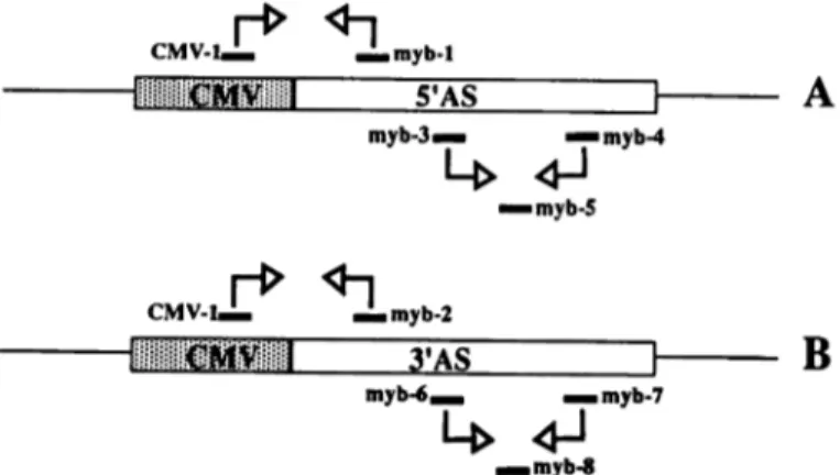
CMV-1 myb-1 S'AS CMV-1 mvb-3. myb-2 i myb-4 •¿myb-S
3'AS
B
mib-65 G. Raschella et al., unpublished observations.
Fig. 2. PCR strategies to detect vector integration and transcription of anti-sense RNA in antianti-sense transfectants. In A, CMV-I and myb-\ are the PCR primers to determine the integration of the 5' antisense (AS) clones; myb-3 and
myb-4 are the primers for RT-PCR; and myb-5 is the probe to detect antisense
RNA transcripts in 5' antisense clones. In B, CMV-1 and myb-2 are the PCR primers to control the integration of the 3' antisense clones; myb-6 and myb-1 are the RT-PCR primers; and myb-S is the probe for antisense RNA detection in 3' antisense clones.
4222
on July 17, 2012
cancerres.aacrjournals.org
1 2 3 456
Fig. 3. Detection of construct integration and antisense transcripts by PCR analysis. A, 3' antisense myb clones. M, size marker. Lane I, pooled transfectant clones; Lanes 2-12, individual clones; Lane 13, transfectant clone containing the pRc/CMV vector (negative control); Lane 14, no DNA; Lane IS, DNA from vector 3' antisense myb (positive control). B, 5' antisense myb clones. M, size marker. Lane 1, pooled transfectant clones; Lanes 2-9, individual clones; Lane 10, transfectant clone containing the pRc/CMV vector (negative control); Lane II, no DNA; Lane 12, DNA from vector 5' antisense myb (positive control). C, hybrid ization analysis of antisense RNA transcripts in transfectanl clones. Lane I, RT-PCR without RNA; Lane 2, RT-PCR of 5' antisense clone in B, lane 9; Lane
3. PCR from RNA of the same clone; Lane 4, RT-PCR without RNA; Lane 5,
RT-PCR of 3' antisense myb clone, in A, Lane 7; Lane 6, PCR from RNA of the same clone. Primers and probes are as described "Materials and Methods."
possibly because of rearrangement
or deletion involving the
antisense insert preventing
the transcription
of the antisense
RNA. Accordingly, the inhibition of LAN-5 cell proliferation
resulting from transfection with the 5' antisense myb construct
might be underestimated.
Clones positive for integration of the
5' antisense construct showed the expected amplified 475-base
pair fragment.
Transcription
of antisense c-myb RNA was confirmed in
RT-PCR experiments
(Fig. 3C). Total
RNA from transfectant
clones was extracted and treated with RNase-free DNase I be
fore the RT-PCR reaction. Primers were designed to amplify a
224-base pair product for 5' antisense myb clones and a
231-base pair product for 3' antisense myb clones (Fig. 2). After size
fractionation
on an agarose gel and transfer to a nylon mem
brane, the amplified products were hybridized to specific probes
for unambiguous
identification;
antisense
c-myb RNA tran
scripts were clearly detected in both 5' and 3' antisense-»y¿>
transfectants
(Fig. 3C). Most of the antisense transfectants
do
not show evident morphological
alterations.
All tested
anti-sense clones retain the capability to differentiate toward a neu
ral phenotype under the effect of retinoic acid (not shown). A
more detailed phenotypic characterization
of the transfectant
clones is now in progress.
Comparison
of growth curves for 3' and 5' antisense-rtryA
clones which were found positive for integration
of the con
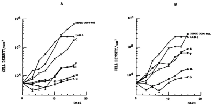
structs (Fig. 4) revealed consistently
slower proliferation
rates
and generally lower growth plateaus than those of the LAN-5
parental cells and sense controls.
Immunocytochemical
analysis using an anti-wye polyclonal
antibody indicated the clear presence of myb protein in the
nucleus of the LAN-5 parental cells and the 5' and 3' sense
controls (Fig. 5, A-C), whereas myb protein was barely detect
able in the 5' and 3' antisense transfectants
(Fig. 5, D and £).
Effect of c-myb Antisense Oligodeoxynucleotides
on Growth
of Neuroblastoma
and Neuroepithelioma
Cell Lines. To fur
ther examine the role of c-myb in the proliferation
of
neuroec-todermal
tumors, we inhibited c-myb expression
using
anti-sense Oligodeoxynucleotides
and analyzed the effects of this
B 10° 10=
to'1
10° SENSE CONTROL 10 20 DAYS IO5to'1
SENSECONTROL LAN 5 10 20 DAYSFig. 4. Growth curves of antisense-m>'¿>transfectants. A, 3' antisense myb; B, 5' antisense myb. Values are the mean of two independent experiments. The sense-m^é control curve in each panel is derived from the mean values using three different 5' (A) and 3' (B) sense clones. Each sense clone had a comparable growth rate. Initial seeding density was 5 x IO3 cells/cm2. Letters, individual clones. Bars, SE.
4223
on July 17, 2012
cancerres.aacrjournals.org
PROLIFERATION AND DIFFERENTIATION OF NEUROBLASTOMA CELLS -'-•-X -.""• -- . * . '•" . * V
Fig. 5. Expression ofc-myh protein in antiscnse clones. Cells were treated with an anti-myb specific polyclonal antibody (A-E) or nonspecific preimmunc serum (F) as described in "Materials and Methods." A, LAN-5; B, 5' sense myb; C, 3' sensemj'e; D, 5' antisense myb; E, 3' antisensc myb; and F. 3' sense myb treated with immune serum, x 200.
Fig. 6. Effect of c-myb anlisense oligodeoxynucleotide on cell growth of neuroblastoma and neuroepithelioma cell lines. SK-N-SH (A), SK-N-MC (B), and LAN-5 (O cells were untreated (O. sense-treated (S) and antisense-treated (AS). Cells were counted after 7 days for SK-N-SH (A) and after 9 days for SK-N-MC (B). LAN-5 (O cells were counted at days 0, 5, 9. and 13 of culture. Cell count values, mean ±SD (bars) of experiment performed in triplicate.
inhibition on the growth of neuroblastoma
cell lines LAN-5 and
SK-N-SH and neuroepithelioma
cell line SK-N-MC (31). In a
typical experiment,
1 x IO4 cells were seeded in the presence of
antisense or sense oligodeoxynucleotides
(80 ßg/m\at 0 h, 40
Mg/ml after 18 h, and 40 tig/ml after 36 h). Cells were counted
after 7 or 9 days. As shown in Fig. 6, antisense c-myb oligode
oxynucleotide
treatment
resulted in almost complete growth
inhibition
in all three cell lines. To determine
whether this
inhibition correlated
with c-myb transcript
levels, total RNA
was extracted from each tumor cell line 24 h after exposure to
120 Mg of c-myb oligodeoxynucleotides
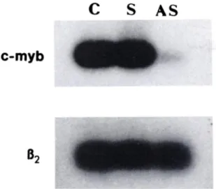
and c-myb expression
was measured by RT-PCR; c-myb mRNA was barely detectable
in antisense-treated
cells, but abundantly
expressed in
sense-treated and unsense-treated cells (Fig. 7).
DISCUSSION
We have shown that down-regulation
of c-myb expression
exerts a strong inhibitory effect on the proliferation
of
neuro-ectodermal
tumor cells. Two different strategies were used in
our work. Neuroblastoma
cell line LAN-5 was transfected with
vectors carrying two different segments of the human c-myb
cDNA in the antisense orientation
and transfection
efficiency
was assayed; the yield of transfectants was dramatically reduced
4224
on July 17, 2012
cancerres.aacrjournals.org
S
AS
c-myb
B2
Fig. 7. Expression of c-myb mRNA in LAN-5 cells exposed to c-myb oligode-oxynucleotides. LAN-5 cells (lOVml) were untreated (O or exposed to ~80 ¿ig/mlof c-myb sense (S) or antisense (AS) oligodeoxynucleotides at time 0. After
12 h a second dose (40 jig/ml) was added. Cells were harvested 12 h later. Total RNA was isolated and divided into two aliquots that were separately amplified by RT-PCR with c-myb- and fo-microglobulin-specific primers as described (44). The resulting cDNAs were hybridized to specific 32P-end-labeled probes as de scribed (29). Results are from a representative experiment.
when either antisense
vector was used as compared
to their
sense controls.
Although
the isolated antisense
transfectants
are heterogeneous
in their growth rate, they show consistently
a slower proliferation
as compared to sense controls. Further
more, they transcribe
antisense
c-myb mRNA
and have a
marked reduction of myb protein synthesis as indicated by
im-munocytochemistry.
Together
these data
indicate
that
the
slower proliferation
of the isolated antisense clones is due to
down-regulation
of c-myb expression caused by antisense RNA
production.
The second strategy involved exposure of neuroblastoma
and
neuroepithelioma
cell cultures to a c-myb antisense
oligodeox-ynucleotide
in order to suppress
c-myb mRNA expression.
Again, the same inhibitory effect on the proliferation
rate of
these cells was observed. The rescue of antisense stable trans
fectants is in apparent contrast with the nearly complete growth
inhibition resulting from the exposure of neuroectodermal
tu
mor lines to c-myb antisense
oligodeoxynucleotides.
Most
likely these findings rest in the low level of antisense c-myb
RNA transcribed
by the transfectant
clones and detectable by
the sensitive RT-PCR technique but insufficient to completely
block the function of c-myb mRNA. In this regard. Gotten et al.
(36) have demonstrated
the requirement for an antisense
RNA-rtarget mRNA ratio of 6:1 to completely abolish the function of
protein
U7. Nevertheless,
the slow proliferation
rate of the
c-myb antisense transfectants
indicates that even incomplete
down-modulation
of c-myb has a readily detectable effect on
cell growth.
In neuroblastoma
several structural abnormalities
such as the
deletion of the short arm of chromosome
1 (dellp32-pter),
double-minute
chromosomes
and homogeneously
staining re
gions are frequent findings (37-39) and have been associated
with the development
and the progression
of this neoplasia
(40). Furthermore,
N-myc gene amplification
has been corre
lated with advanced clinical stages and poor clinical outcome
(27, 41). However, the proliferative
activity of neuroblastoma
has not been clearly associated with a distinct pattern of altered
gene expression.
Two recent reports describe the block of N-myc expression by
means of antisense RNA and oligodeoxynucleotides
(42,43). In
both cases neuroblastoma
proliferation
was only partially af
fected by N-myc down-regulation
and was probably secondary
to the induction of differentiative processes. In addition, a large
percentage of terminal neuroblastomas
do not show amplifica
tion or detectable expression of N-myc. Together,
those data
suggested that other gene activities besides N-myc are involved
in the proliferation of neuroblastoma
cells. The inhibitory effect
on cell growth obtained by abolishing the expression of c-myb
strongly suggests the involvement of this protooncogene
in the
regulation
of neuroblastoma
cell proliferation.
Moreover the
findings reported here, provide direct evidence for the essential
role of c-myb in nonhematopoietic
tissues, perhaps through its
effects on the expression
of genes directly involved in DNA
synthesis and cell cycle progression. The stable transfectant cell
lines that express antisense c-myb RNA should prove useful in
evaluating the possible cooperation of c-myb and other genes in
regulating proliferative and differentiative
processes in neuro
blastoma, which, in turn, may lead to the development
of an
antisense-based
therapy of these neoplastic disorders.
REFERENCES
1. Bister, K., Nunn, M., Moscovici, G., Perbal, B., Baluda, M. A., and Dues-berg, P. H. Acute leukaemia viruses E26 and avian myeloblastosis virus have related transformation-specific RNA sequences but different genetic struc tures. Proc. Nati. Acad. Sci. USA, 72: 3677-3681, 1982.
2. Roussel, M.. Saule, S., Langrow, C., Rommens, C., Beug, H., Graf, T., and Stehelin, D. Three types of viral oncogenes for haematopoietic transforma tion. Nature (Lond.), 281: 452-455, 1979.
3. Moscovici, C., and Gazzolo, L. Transformation of hemopoietic cells and avian leukemia viruses. In: Advances Viral Oncology, Vol. 1, New York: Raven Press, pp. 83-106, 1982.
4. Klempnauer, K. H., Symonds, G., Evan, G. I., and Bishop, J. M. Subcellular localization of proteins encoded by oncogenes of avian myeloblastosis virus and avian leukemia virus E26 and by the chicken c-myb gene. Cell, 37: 537-547, 1984.
5. Boyle, W. J., Lampert, M. A., Lipsick, J. S., and Baluda, M. A. Avian myeloblastosis virus and E26 virus oncogene products are nuclear proteins. Proc. Nati. Acad. Sci. USA, 81: 4265-4269, 1984.
6. Moelling, K., Pfoff, E., Beng, H., Beimling, P., Bunte, T., Scheller, H. E., and Graf, T. DNA-binding activity is associated with purified Myb proteins from AMV and £26viruses and is temperature-sensitive for E26 ts mutants. Cell, ¥0:983-990, 1985.
7. Klempnauer, K. H., and Sippel, A. E. The highly conserved amino-terminal region of the protein encoded by the \-myb oncogene functions as a DNA-binding domain. EMBO J., 6: 2719-2723, 1987.
8. Rosson, D., and Reddy, P. Nucleotide sequence of chicken c-myb comple mentary DNA: implications for myb oncogene activation. Nature (Lond.), 3/9:604-606, 1986.
9. Gonda, T. J., Gough, N. M., Dunn, A. R., and De Blaquere, J. Nucleotide sequence of cDNA clones of the murine myb protooncogene. EMBO J., 4: 2003-2007, 1985.
10. Majello, B., Kenyon, L. C., and Dalla Pavera, R. Human c-myb protoonco gene: nucleotide sequence of cDNA and organization of the genomic locus. Proc. Nati. Acad. Sci. USA, 83: 9636-9640, 1986.
11. Biedenkapp, H., Borgmeyer, U., Sippel, A. E., and Klempnauer, K. H. Viral
myb oncogene encodes a sequence-specific DNA-binding activity. Nature
(Lond), 335: 835-837, 1988.
12. Weston, K., and Bishop, J. M. Transcriptional activation by the V-myb oncogene and its cellular progenitor c-myb. Cell, 58: 85-94, 1986. 13. Ness, S. A., Marknell, A., and Graf, T. The \-myb oncogene product binds to
and activates the promyelocyte-specific MIM-l gene. Cell, 59: 1115-1124, 1989.
14. Ibanez, C. E., and Lipsick, J. S. Structural and functional domains of the
c-myb oncogene: requirements for nuclear transport, myeloid transforma
tion, and colony formation. J. Virol., 62: 1981-1988, 1988.
15. Sakura, H., Kanei-Ishii, C., Nagase, T., Nakagoshi, H., Gonda, T. J., and Ishii, S. Delineation of three functional domains of the transcriptional acti vator encoded by c-myb protooncogene. Proc. Nati. Acad. Sci. USA, 86: 5758-5762, 1989.
16. Crasser, F. A., Graf, T., and Lipsick, J. E. Protein truncation is required for the activation of the c-myb proto-oncogene. Mol. Cell. Biol., //: 3987-3996, 1991.
17. Westin, E. H., Gallo, R. C., Arya. S. K., Eva, A., Souza, L. M., Baluda, M. A., Aaronson, S. A., and Wong-Staal, F. Differential expression of the AMV gene in human hematopoietic cells. Proc. Nati. Acad. Sci. USA, 79: 2194-2198, 1982.
18. Clarke, M. F., Kukowska-Latallo, J. F., Westin, E., Smith, M., and Prochownik, E. V. Constitutive expression of a c-myb cDNA blocks Friend
4225
on July 17, 2012
cancerres.aacrjournals.org
PROLIFERATION AND DIFFERENTIATION OF NEUROBLASTOMA CELLS
murine erylhroleukemia cell differenliation. Mol. Cell. Biol.. *: 884-892. 1988.
19. Gewirtz, A. M., and Calabretta, B. A c-myb antiscnse oligodeoxynucleotide inhibits normal human hematopoiesis in vitro. Science (Washington DC),
242: 1303-1306. 1988.
20. Mucenski. M. L.. McLain. K., Kier. A. B.. Swerdlow. S. H.. Schreiner. C. M.. Miller. T. A.. Pietryga. D. W., Scott. Jr.. \V. J.. and Potter, S. S. A functional
c-myb gene is required for normal murine fetal hepatic hematopoiesis. Cell,
«5:677-689. 1991.
21. Slamon. D. S., deKernion. J. B., Verma, I. M., and Cline, M. S. Expression of cellular oncogenes in human malignancies. Science (Washington DC). 224:256-261, 1984.
22. Calabretta. B.. Sims. R. B.. Valtieri. M.. Caracciolo. D., Szczylik. C., Ven-turelli. D.. Beran. M.. and Gewirtz, A. M. Normal and leukemic hematopoi-etic cells manifest differential sensitivity to inhibitory effects of c-myb anti-sense oligodeoxynucleotides: an in vitro study with relevance to bone marrow purging. Proc. Nati. Acad. Sci. USA, 88: 2351-2355, 1991.
23. Alitalo, K., Winquist. R.. Linn, C. C., de La Chapelle, A., Schwab, M., and Bishop, J. M. Aberrant expression of an amplified c-myb oncogene in two cell lines from a colon carcinoma. Proc. Nati. Acad. Sci. USA, SI: 4534-4538. 1984.
24. Griffin, C. A., and Baylin, S. B. Expression of the c-myb oncogene in human small cell lung carcinoma. Cancer Res., 45: 272-275, 1985.
25. Janssen. J. W. G., Vernole, P.. de Boer, P. A. J., Oosterhuis, J. W., and Collard, J. G. Sublocalization of c-myb to 6q21-q23 by in situ hybridization and c-myb expression in human teratocarcinoma with 6q rearrangements. Cytogenet. Cell Genet.. 41: 129-135. 1986.
26. Thiele. C. J.. McKeon, C., Triche. T. J., Ross, R. A., Reynolds. C. P.. and Israel. M. A. Differential prolooncogene expression characterizes histo-pathologically indistinguishable tumors of the peripheral nervous system. J. Clin. Invest., SO: 804-811, 1987.
27. Seeger. R. C.. Brodeur, G. M.. Sather. H., Dalton, A., Siegel, S., Wong. K. Y., and Hammond. D. Association of multiple copies of the N-mjr oncogene with rapid progression of neuroblastoma. N. I nul. J. Med.. 313: 1111-1116. 1985.
28. Thiele. C. J., Cohen. P. S., and Israel, M. A. Regulation of c-myb expression in human neuroblastoma cells during retinole acid-induced differentiation. Mol. Cell. Biol.. 8: 1677-1688. 1988.
29. Sambrook, J., Fritsch, E. F.. and Maniatis, T. in: Molecular Cloning, A Laboratory Manual. Ed. 2., Cold Spring Harbor. NY: Cold Spring Harbor Laboratory. 1989.
30. Seeger. R. C., Danon. Y. L.. Rayncr. S. A., and Hoover. F. Definition of a THY-1 determinant on human ncuroblastoma, glioma, sarcoma, and ter-atoma cells with a monoclonal antibody. J. luminimi.. 128: 983-989, 1982. 31. Biedler. J. L., Helson. L., and Spengler, B. A. Morphology and growth.
tumorigenicity and cytogenetics of human neuroblastoma cells in continuous culture. Cancer Res.. 33: 2643-2649. 1973.
32. Chomczynski, P., and Sacchi, N. Single-step method of RNA isolation by acid guanidinium thiocyanate-phenol-chloroform extraction. Anal. Bio-chem., 162: 156-159, 1987.
33. Szczylik, C., Skorski, T., Nicolaides, N. C.. Manzella. L., Malaguarnera, L., Venturelli. D.. Gewirtz, A. M., and Calabretta, B. Selective inhibition of leukemia cell proliferation by BCR-ABL antisense oligodeoxynucleotides. Science (Washington DC), 253: 562-565, 1991.
34. Maly, A., and Krchnak, V. Identification of c-myb (chicken), c-myb (mouse) and v-myb (AMV) protein products by immunoprecipitation with antibodies directed against a synthetic peptide. FEBS Lett., 205: 104-108, 1986. 35. Foecking, M. K., and Hofstetter. H. Powerful and versatile enhancer pro
moter unit for mammalian expression vectors. Gene, 45: 101-105. 1986. 36. Cotten, M., Schaffner, G., and Birnstiel, M. L. Ribozyme, antisense RNA,
and antisense DNA inhibition of U7 small nuclear ribonucleoprotein-medi-ated histone pre-mRNA processing in vitro. Mol. Cell. Biol., 9: 4479-4487.
1989.
37. Brodeur. G. M., Green, A. A., Hayes, F. A., Williams. K. J., Williams, D. L.. and Tsiatis, A. A. Cytogenetic features of human ncuroblastomas and cell lines. Cancer Res., 41: 4678-4686, 1981.
38. Franke. F., Rudolph, B., Christiansen. H., Harbott, J., and Lampert, T. Tumor karyotype may be important in the prognosis of human ncuroblas toma. J. Cancer Res. Clin. Oncol.. ///: 266-272, 1986.
39. Gilbert, F.. Feder, M., Balaban. G., Brangman, D., Lurie, D. K., Podolski. R., Rinaldt. V., Vinikoor, N., and Weinsband, J. Human neuroblastomas and abnormalities of chromosomes l and 17. Cancer Res., 44: 5444-5449, 1984. 40. Gilbert. F. Chromosome abnormalities, gene amplification, and tumor pro
gression. In: A. E. Evans, G. J. D'Angio. and R. C. Seeger, (eds.). Advances in Neuroblastoma Research, pp. 151-159. New York: Alan R. Liss. 1985. 41. Dominici, C., Negroni, A.. Romeo, A., Castello, M. A., Clerico, A.,
Scopin-aro, M., Mauro, F., and Raschcllà . G. Association of near-diploid DNA content and N-myc amplification in neuroblastomas. Clin. Exp. Metastasis, 7:201-211. 1989.
42. Whitesell, L., Rosolen. A., and Neckers, L. M. Episome-generatcd n-myc antisense RNA restricts the differentiation potential of primitive neuroecto-dermal cell lines. Mol. Cell. Biol.. //: 1360-1371, 1991.
43. Negroni, A., Scarpa, S.. Romeo, A.. Ferrari, S., Modesti, A., and Raschella. G. Decrease of proliferation rate and induction of differentiation by a MYCN antisense DNA oligomer in a human neuroblastoma cell line. Cell Growth Differ.. 2:511-518, 1991.
44. Venturelli, D.. Mariano, M. T., Valtieri. M., Lange, B.. Crist, W., Link, M., and Calabretta, B. Down-regulated c-myb expression inhibits DNA synthesis of T-leukemia cells in most patients. Cancer Res., 50: 7371-7375, 1990.
4226