DOTTORATO DI RICERCA IN
Scienze e Tecnologie Agrarie, Ambientali e Alimentari
Ciclo XXVII
Settore Concorsuale di afferenza: 07/D1 Patologia Vegetale ed Entomologia
Settore Scientifico disciplinare: AGR12 Patologia Vegetale
TITOLO TESI
Apple latent infection caused by Neofabraea alba: host-pathogen
interaction and disease management
Presentata da: Dott.ssa Irene Cameldi
Coordinatore Dottorato
Relatore
Prof. Giovanni Dinelli
Prof.ssa Marta Mari
Esame finale anno 2015
1
Table of contents
Abstract……….……….2
Introduction………...……4
Chapter one: Pathogenesis of Neofabraea alba in ‘Pink Lady™’ apple and influence of the primary metabolites of fruit on disease development……….13
Chapter two: Susceptibility of five apple cultivars to Bull’s eye rot and
relationship with ambient
pH………..………..42
Chapter three: Bull’s eye rot of ‘Pink Lady™’ management by DA-meter application: practical implications and transcriptional level of ripening, defence involved genes………...…...63
Chapter four: Characterization of Neofabraea alba volatile profile: preliminary study for an early detection of Bull’s eye rot………83
2
Doctoral Dissertation Abstract
Apple latent infection caused by Neofabraea alba: host-pathogen interaction and
disease management
Bull’s eye rot (BER) caused by Neofabraea alba is one of the most frequent and damaging latent infection occurring in stored pome fruits worldwide. Fruit infection occurs in the orchard, but disease symptoms appear only 3 months after harvest, during refrigerated storage. In Italy BER is particularly serious for late harvest apple cultivar as ‘Pink Lady™’. The purposes of this thesis were: i) Evaluate the influence of ‘Pink Lady™’ apple primary metabolites in N. alba quiescence ii) Evaluate the influence of pH in five different apple cultivars on BER susceptibility iii) To find out not chemical method to control N. alba infection iv) Identify some fungal volatile compounds in order to use them as N. alba infections markers. Results regarding the role of primary metabolites showed that chlorogenic, quinic and malic acid inhibit N. alba development. The study based on the evaluation of cultivar susceptibility, showed that Granny Smith was the most resistant apple cultivar among the varieties analyzed. Moreover, Granny Smith showed the lowest pH value from harvest until the end of storage, supporting the thesis that ambient pH could be involved in the interaction between N. alba and apple. In order to find out new technologies able to improve lenticel rot management, the application of a non-destructive device for the determination of chlorophyll content was applied. Results showed that fruit with higher chlorophyll content are less susceptible to BER, and molecular analyses comforted this result. Fruits with higher chlorophyll content showed up-regulation of PGIP and HCT, genes involved in plant defence. Through the application of PTR-MS and SPME GC-MS, 25 volatile organic compounds emitted by N. alba were identified. Among them, 16 molecules were identified as potential biomarkers.
3
Apple latent infection caused by Neofabraea alba: host-pathogen
interaction and disease management
Introduction
Among the wide kinds of plant diseases caused by fungi, latent infections are a very specific category of plant syndromes causing important losses in agriculture, especially during post-harvest phase. According to Verhoeff (1974), latency is defined as “A
quiescent or dormant parasitic relationship where, pathogens spends long periods of time during the host’s life in a quiescent stage, under specific circumstances, can change an active one.” The term quiescent is designed as the period from host infection and the
activation of fungal development and symptoms expression (Prusky et al., 1996). In order to gain the success, a pathogen must overcame plant defense, and to obtain nutrients required to sustain its development. Extensive studies have been carried out to determine the biochemical bases of activation of pathogen quiescence, and three hypotheses have been suggested by Prusky (1996): (i) deficiency of host nutritional resources necessary for pathogen development; (ii) presence of preformed or induced fungistatic compounds; and (iii) unsuitable environment for the activation of fungal pathogenicity factors. In support of the second hypothesis, varieties of fungistatic molecules have been shown to act as major barriers against Colletotrichum spp. in unripe avocado fruits (Prusky et al., 1982). In avocado also phenylpropanoid and flavonoid metabolites, such as epicatechins, have been shown to be crucial in the induction of fungal quiescence in unripe fruits (Ardi et al., 1998). In fruits and vegetables the mechanism of resistance is generally not dictated only by a single gene interaction between host and pathogen, but the interactions maintain a type of dynamic incompatibility (Prusky, 1996). This situation is triggered by the
4 response of the host resistance genes to the pathogens avirulent genes that prevents or retards pathogen growth, under specific host physiological conditions. During maturation and ripening, host physiological changes make the environment more suitable for pathogen colonization. This phenomenon makes the host-pathogen interaction compatible and the symptoms appear. The biological model reported above is the most common condition in postharvest pathogen-host interactions, in fact pathogens such as
Botrytis cinerea, Colletotrichum spp. and N. alba, remaining quiescent during fruit
growth and becoming virulent during senescence of host.
Neofabraea spp. and latent infections of apple fruits
Bull’s eye rot (BER) of stored apples caused by Neofabraea spp. (E.J. Gutrie) is one of the main disease occurring in Europe and North and South America (Maxin et al., 2014, Spolti et al., 2010, Spotts et al. 1999). Fruit are infected in the orchard, but the symptoms appear on the fruits only 3-4 months after harvest, during cold storage. Fruit lesions are flat, brown and often with a lighter brown center (Snowdon, 1990), frequently centered on the lenticels, however they also occurred on wounds and around the stem or calyx. Under humid conditions, on the old lesions are present cream-colored acervuli. Rotted tissue was firm and was not readily separable from healthy tissue (Spotts et al., 2009). BER also known as lenticel rot is caused by four members of Neofabraea genus: N. alba,
N. perennans, N. malicorticis, and Cryptosporiopsis kienholzii. The last species was
found for the first time in 2001 by Jong et al. on apple in Canada and Portugal and it was subsequently described by Spotts et al. (2009). Jong’s work provided a useful study on the phylogenetic relationship among Neofabraea species, making possible a species specific identification of the Neofabraea pathogen complex through a multiplex DNA amplification (Gariépy at al. 2003). The diagnostic technique developed by Gariépy et al.
5 (2003) incorporates five sets of specie-specific primers into a single PCR reaction, and produces distinct bands for each pathogen, making possible an accurate identification of all members of the genus Neofabraea to species. In Germany and UK, the presence of N.
perennans is confirmed by Jong et al., (2001), while N. alba is reported as the major agent
of BER in Italy and in France (Bompeix and Cholodowski-Faivre, 1998; Neri et al., 2009). The presence of N. alba, N. malicorticis and N. perennans was also confirmed in Pacific Northwest of U.S.A. (Gariepy et al., 2005), while in Australia were found N. alba and C. kienholzii (Cunnington, 2004). In Brasil, lenticels rot caused by N. perennans is one of the most important apple’s postharvest disease (Spolti et al., 2010), while N. alba was found in apples cultivar Braeburn, Fuji, Granny Smith, Pink Lady, and Royal Gala.in Chile (Henriquez, 2005).
Biology of Pathogen
Neofabraea sp. causes canker on apple and pear trees (Jong et al. 2001, Garipey et al
2003, Gariepy et al. 2005, Henriquez et al., 2006), however it is not fully elucidated the relationship between Neofabraea sp. and appearance of branch cankers. In order to clarify this association Henriquez et al. (2006) induced cankers in tree branches of pear and apple by artificial inoculation of N.alba and N. perennans. All inoculation showed branch cankers, but some differences were recorded between pear and apple trees. In pear trees, cankers resulted most evident when inoculations were carried out during the autumn and winter months, while in Granny Smith trees, the inoculations successfully induced cankers in summer months. Comparing the two species analyzed N. alba produces smaller cankers than N. perennans. Cankers development began as a blackening of tissue under the inoculation point. Necrosis extended farther in length than in width. Peeling of the bark occurred at the margin of the necrotic area. Branch growth delimited the canker,
6 which acquired a sunken shape with cracked margins, separating from the healthy tissue. Cankers induced by both species produced conidia through most of the year, with the highest amounts at the end of summer and during autumn. These conidia are splash-dispersed to fruit. Henriquez et al. (2006) also observed natural cankers caused by N. alba on D’Anjou pear trees. In addition, the same study revealed as the copper sulfate treatments influenced the conidial production approximately for 1 month after treatment. In general, conidia of pathogens landed on fruit surface, germinate, produce penetration structures and penetrate into the fruit activating pathogenicity factors (Prusky, 1996), but this mechanism remains not understood for Neofabraea spp. fruit infections. The most important challenge of lenticel rot is to understand the mechanism that regulates the end of quiescent stage of pathogen. Neofabraea spp. infects fruits in the orchard, but it does not produce symptoms until a certain stage of fruit ripening, that usually happens 3-4 months after harvest.
In general, once that spores arrive on the fruit surface, the appressoria formation depends on the substances present on the cuticle. Prusky and Plumbley (1992) showed that avocado fruit wax induced the appressoria formation of C. gloeosporioides. According to Kolattukudy et al. (1995), plant surface lipids contain both inducers and inhibitors of spore germination and appressoria formation, and the balance between them might be responsible for the selective activation of the pathogen for initiation of parasitisation. Moreover, fruits and vegetables are provided of numerous biochemical compounds able to delay the fungal infection even if spores reach the internal part of the host. The group of substances preformed or inducible that constitutes the host biochemical barriers is very wide, and it changes depending on vegetable species, cultivar and stage of ripening etc. The preformed compounds are known as phytoanticipins while the inducible compounds
7 are known as phytoalexins and they are synthesized from remote precursors in response to pathogen attack. The distinction between phytoalexins and phitoanticipins is not always clear as some compounds may be phytoalexins in one species and phytoanticipins in another species. The distinction between the two compounds depend on when they are produced, either before or after infection. The preformed compounds tend to be concentrated in the outer layer of plant organs and in general, they are compartmentalized in vacuoles or organelles in healthy plant. Their quantity decreases from unripe to ripe fruit. Biotic or abiotic elicitors could induce the production of phytoalexins that are concentrated around the point of pathogen penetration or abiotic stress. The rate of phytoalexins accumulation determine the outcome of host-pathogen interaction. Susceptible host accumulate phytoalexins slower than resistant host (Kuc, 1995).
N. alba is the main BER pathogen in Italy, and very few are data about the mechanism
that regulate its quiescence and the questions reported by Prusky (1996) remain unacknowledged for this pathogen.
i) What is the nature of preformed barriers to pathogen attack?
ii) How do R genes trigger defense responses and what are the physiological conditions needed for triggering defense responses?
8
Bibliography
Ardi R, Kobiler I, Keen NT, Prusky D, 1998. Involvement of epicatechin biosynthesis in the resistance of avocado fruits to postharvest decay. Physiological and Molecular Plant Pathology 53, 269–85.
Bompeix G., Cholodowski-faivre 1998. The use of natural plant products against scald, Botrytis sp., Gloeosporium sp., Penicillium sp. and Rhyzopus sp. In: Proceedings of the Joint Workshop Non Conventional Methods for the Control of Postharvest Disease and Microbiological Spoilage, Commission of European Communities COST 914-COST915, Bologna, Italy, pp.99-104.
Cunnington J. H. 2004. Three Neofabraea species on pome fruit in Australia. Australasian Plant Pathology. 33:453-454
Gariepy T.D., Rahe J.E., Lévesque C.A., Jong S. N. and Rahe J. E Rahe. 2003. Species specific identification of the Neofabraea pathogen complex associated with pome fruits using PCR and multiplex DNA amplification Mycol. Res. 107(5):528-536
Gariepy T.D., Rahe J.E., Lévesque C.A., Spotts R.A., Sugar D.L., and Henriquez J.L.2005 Neofabraea species associated with bull’s-eye rot and cankers of apple and pear in the pacific Northwest.Ca. J. Plant Pathol. 27: 118-124
Henriquez J. L., 2005. First Report of Apple Rot caused by Neofabraea alba in Chile. Plant Dis. 89:1360
Henriquez J. L., Sugar D., and Spotts, R. A. 2006. Induction of Cankers on Pear Tree Branches by Neofabraea alba and N. perennans, and Fungicide Effects on Conidial Production on Cankers. Plant Dis. 90:481-486
Jong N. S., Lévesque C.A., Verkley G. J. M., Abelin E. C. A., Rahe J.E. and Braun P. G. 2001. Phylogenetic relationships among Neofabraea species causing tree cankers and bull’s-eye rot of apple based on DNA sequencing of ITS nuclear rDNA, mitochondrial rDNA, and the β-tubulin gene. Mycological Research 105(6): 658-669
9
Kolattukudy, P.E., Rogers, L.M., Li, D., Hwang, C.S., Flaishman, M.A., 1995. Surface signaling in pathogenesis. Proc. Natl. Acad. Scie. USA 92, 4080-4087
Kuc, J. 1995. Phytoalexins, stress metabolism, and disease resistance in plants. Annu Rev Phytopathol 33: 275-297.
Maxin, P., Williams, M., Weber, R W. S., 2014. Control of fungal storage rots of apples by hot-water treatments: A Northern European Perspective. Erwerbs-Obstbau 56, 25–34 Neri F., Mari M., Brigati. and Bertolini P. 2009. Control of Neofabraea alba by plant volatile compounds and hot water. Postharvest Biology and Technology 51: 425-430 Prusky and Plumbley, 1992. Quiescent infections of Colletotrichum in tropical and subtropical fruits. Colletotrichum: biology, pathology and control. 1992 pp. 289-307 Prusky D., Keen N.T., Sims J.J., Midland, S.L., 1982. Possible involvement of an antifungal diene in latency of Colletotrichum gloeosporioides on unripe avocado fruits. Phytopathology 72, 1578–82.
Prusky, D., 1996. Pathogen quiescence in postharvest diseases. Annul Rev of Phytopathol 34, 413-434
Snowdon A.L., 1990. Gloeosporium rot A Colour Atlas of Post-Harvest Diseases and Disorders of Fruits and Vegetables. General introduction and fruits, vol. 1 Wolfe Scientific Ltd., London 186–187
Spolti P., Valbenito-Sanhueza R. M. and Medeiros Del Ponte E. 2010. Meio semiseletivo para recuperaçao e quantificaçao de Cryptosporiopsis perennans em maças. Ciencia Rural 40 (3): 661-665
Spotts, R. A., Cervantes, L. A., Mielke, E. A., 1999. Variability in postharvest decay among apple cultivars. Plant Dis. 83,1051-1054
Spotts R. A., Seifert K. A., Wallis K. M., Sugar D., Xiao C. L., Serdani M. and Henriquez J. L., 2009. Description of Cryptosporiopsis kienholzii and species profiles of Neofabraea in major pome fruit growing districts in the Pacific Northwest USA. Mycol. Res. 113:1301-1311
10
Verhoef, K., 1974.Latent infections by fungi. Annual Review of Phytopathology 12, 99-110
11
Aims of the work
This work addresses latent infection caused by Neofabraea alba in apple trough a holistic approach. BER is very important disease for pome production, especially for the economic losses due to the product waste in post-harvest, however the information about its causal agent are very lacking. In order to find out the main factors affecting the N. alba quiescence, and possible alternative method of disease management, different aims were pursued:
- Evaluate the influence of “Pink Lady™” apple primary metabolites in N. alba quiescence
- Evaluate the influence of pH in five different apple cultivars on BER susceptibility - To find out not chemical tool able to limit BER damages
- Identify some fungal volatile compounds in order to use them as N. alba infections markers. This study is still not completed, and results reported in the thesis refer to preliminary studies
12
Chapter one:
Pathogenesis of Neofabraea alba in ‘Pink Lady™’ apple and influence of the
primary metabolites of fruit on disease development
1 Introduction
Bull’s eye rot (BER) caused by Neofabraea spp. is one of the most important diseases of pome fruits, producing notable economic losses during fruit storage. The disease is particularly serious in Europe (Bompeix and Cholodowski-Faivre, 1998; Neri et al., 2009, Kellerhals et al. 2012), North-West of USA (Henriquez et al., 2004, Gariepy et al., 2005), Chile (Soto-Alvear S., 2013) and Brasil (Spolti et al., 2010), while in China BER is classified as quarantine disease (Cao et al. 2013). Fruit lesions are flat to slightly sunken, brown and often with a light brown center (Snowdon, 1990). The rotten tissues are firm, with white acervuli on old lesions. In apple (Spotts et al., 1999) and pear (Henriquez et al. 2004) trees the pathogen can induce canker formation in the branches relating to climate conditions. The fruit, contaminated in the orchard, show the symptoms only three/four months after harvest, during cold storage (Snowdon 1990). A review on pome European fruit genetic resources evaluated the resistance against some pathogens (Kellerhals et al., 2012) including Neofabrea spp. among the main causes of decay during apple storage and commercialization. Four members of the genus Neofabraea are the causal agents of BER of pome fruits (Spotts et al., 2009): N. alba (E. J. Gutrie) Verkley (anamorph Phlyctema vagabunda Desm.), N. perennans Kienholz [anamorph
Cryptosporiopsis perennans (Zeller & Childs) Wollenw.], N. malicorticis H. S. Jacks
13
kienholzii (Seifert, Spott & Lévesque) was described as being associated with BER in the
Pacific Northwest of the United States (Spotts et al. 2009). In Italy, only the presence of
N. alba (anamorph Phlyctema vagabunda) was reported, however there are no molecular
data on the Italian strains of N. alba. In addition, no branch cankers were detected in Italian apple orchards and only BER symptoms on stored fruit were observed.
Despite BER is globally recognized as one of the major fungal disease of stored apple, there are very few information on its biology and host-pathogen interactions. These serious lacks are probably due also to the difficulties of pathogen culturing and conidia obtaining on artificial medium. Methods reported in literature require a very long time of incubation for conidia production on artificial medium and are not reliable. The methodology used by Neri et al. (2009) permits to produce conidia on natural substrate based on apple leaves, but does not allow perpetrating the same isolate during the time because the pathogen loses the vitality once transferred onto Malt Extract Agar (MEA) or Potato Dextrose Agar (PDA). According to Rooney-Latham et al. (2013) the sporulation of N. alba isolates derived from olives was observed in vitro on PDA after 40 days of incubation under near-UV light. Moreover, no molecular data are available for
Neofabreea spp. species present in Italy.
The pathogen infects fruit before harvest and conidia of N. alba germinate on young unripe fruit, sunken into lenticels where remains quiescent for long time (Verhoeff, 1974), until fruit ripening. Among resistance mechanisms of fruit, preformed antifungal compounds present in unripe fruit are considered responsible for fruit disease resistance and their degradation during ripening process could activate the latent infections, making fruit prone to disease appearance (Swinburne, 1983). Among these preformed compounds, in the past, phenols were widely considered liable to the unripe fruit BER
14 resistance; in fact, when their content decreases fruit become quite susceptible to fungal attack. However, a study conducted on the influence of low temperature of fruit storage on N. alba infections, showed that a significant decrease in phenolic content in the skin of the fruit is not correlated with an increase of N. alba susceptibility (Lattanzio et al., 2001). Others mechanisms and compounds are involved in apple resistance to BER that have to be still investigated. In its review on pathogen quiescence in postharvest diseases, Prusky (1996) included an increment of nutritional factors during the ripeness process, among the factor affecting the pathogen quiescence interruption. Previous data available on the primary metabolites in apple fruit focused on a defined lapse of time. Zhang Y. et al. (2013) studied the evolution of primary metabolites in pre-harvest phase, when the fruits were still growing in planta, but did not analyze apple metabolism during apple storage. On the contrary, Hatoum et al. (2014), studied the primary metabolism of ‘Breaburn’ apple from two, until thirty-two weeks after harvest, but did not consider the pre-harvest phase. During the last decade, metabolomics has become a highly valued and widely exploited technology to explore the plant composition (Hall 2011). Among the available technology, Nuclear Magnetic Resonance (NMR) provides a powerful technique for the identification and quantification of metabolites in plant extracts. 1H NMR is a robust technique, highly reproducible, non-selective able to produce structural information and quantitative data (Rolin et al 2013). This technique is the more appropriate for primary metabolites analysis (Hounsome et al., 2008), allowing accurate simultaneous quantification of many compounds in a complex mixture without need for separation. Quantitative 1H NMR have been used to determine formic acid in apple juices (Berregi et al 2007) or epicatechin in apple cider (Berregi et al 2003). Recently 1H
high-15 resolution magic angle spinning (HR-MAS) NMR have been used to explore the metabolome of three different apple cultivars (Vermathen et al 2012).
The aims of this study were to: i) develop a rapid and reliable medium to obtain in vitro
N. alba conidia; ii) identify molecularly Neofabraea spp. present in Italy iii) characterize
the primary metabolites in the skin of ‘Pink Lady™’ apple by proton-NMR analysis, during fruit growth and ripening; iv) evaluate the influence of the main primary metabolites on N. alba development and v) evaluate the N. alba pathogenesis in relationship with the changes in the primary metabolites
2. Materials and method
2.1 Fruits
‘Pink Lady™’ apples were obtained from an experimental orchard of University of Bologna located in Cadriano (BO), Italy. Apple trees were grafted on M9 rootstock and grown according to the integrated production guidelines. Harvest was carried out at commercial maturity which was determined using the starch iodine test scored using a 1-10 points CTIFL scale (Centre technique interprofessionnel des fruits et legumes, association Pink Lady Europe) and firmness. At harvest the starch index was 7 and the value of firmness was 63.7 N·cm-2.
2.2 Isolate collection
The isolates of Neofabraea spp. were obtained from apples stored in packinghouses located in Emilia Romagna region (Italy) showing the typical BER symptoms. Fruits were surface-sterilized by ethanol (70% v/v) and small pieces of rotted tissue from the edges of lesion were placed onto MEA dishes. Subsequently plates were incubated at 20°C for
16 15 days. The developed colonies were evaluated for a preliminary identification by colony morphology and microscopic observations of mycelium. In total, forty-five isolates were obtained. After 15 days, the colonies of Neofabraea spp. morphologically identified, were transferred on fresh MEA plates, and kept at 4 °C until use.
2.3 Molecular identification: DNA extraction and PCR amplification
For pathogen identification, 5 isolates of Neofabrea spp. were randomly selected from the collection obtained as described above. The isolates were grown on MEA at 20° C for 15 days and identified by sequencing of internal transcribed spacer (ITS) regions of ribosomal DNA. For this purpose the DNA extraction was carried out using the CTAB method described by Doyle & Doyle (1987) with slight modifications. Fungal biomass was recovered from dishes, introduced in a 2 ml-Eppendorf tube and grinded manually with blue propylene pestles. After, 1 ml of CTAB-0.04% β-mercaptoethanol solution, previously heated at 65°C for 1 h, was added and the suspension was vortexed for 30 s and 2.5 µl of proteinase-K (10 mg ml-1) were added. The suspension was then incubated at 65°C for 1 h and subsequently, 1 ml of a mixture of chloroform-octanol (24:1) was supplemented to the solution and centrifuged for 5 min at 8000 rpm. After the addition of 5 µl of RNAase (10 mg ml-1) to the supernatant, 1 ml of chloroform-octanol was added again and the solution was centrifuged as described above. DNA precipitation was achieved by adding 0.8 volumes of isopropanol to the supernatant and centrifuging for 20 min at 14000 rpm. Subsequently, the pellet was dried under vacuum, washed with 500 µl of 70% ethanol (conserved at -20°C) and centrifuged for 5 min at 12000 rpm. Finally, the pellet was air dried for 5 min and re-suspended in 50 µl of sterile double distilled water. DNA concentration and A260/A280 ratio were assessed using the Infinite 200
17 NanoQuant spectrophotometer (Tecan® Group Ltd., Grödig, Austria). Molecular identification of the isolates was carried out by the PCR amplification of the Internal Transcribed Spacer (ITS) regions of ribosomal DNA using the universal primers PN23
(5'-CACCGCCCGTCGCTACTACCG-3') and PN34 (5'
TTGCCGCTTCACTCGCCGTT-3') described by Mouyna and Brygoo (1993), supplied by Invitrogen® (Carlsbad, California, USA) and carried out in 25 µl using 2.5 µl of TaKaRa® 10×buffer, 2 µl of 10 mM dNTPs, 1 µl of 10 mM solution of each primer, 0.2 µl of TaKaRa® Taq polymerase (5 U/ml) (Takara® Bio Inc., Otsu, Japan), 17.3 µl of double distilled water and 1 µl of genomic DNA (20-100 ng µl-1). PCR program consisted of an initial denaturation step at 94°C for 5 min, followed by 35 cycles at 94°C for 30 s, 62°C for 30 s and 72°C for 1 min with a final extension step of 5 min at 72°C. To verify amplification reactions, 7 µl of each PCR product were separated on 1.5% agarose gel in 0.5×TAE buffer, stained with GelRedTM (Biotium, Inc., California, USA) and visualized under UV light. Purification of amplification products was obtained using the Gel/PCR Extraction & Purification kit (Fisher Molecular Biology®, Pennsylvania, USA). In order to verify the DNA quality and concentration of the purified products, 5 µl were separated in a 1.5% agarose gel and compared with the MassRuler Low Range DNA ladder (Fermentas®, Vilnius, Lithuania). Purified PCR products were sequenced with both forward and reverse primers at Eurofins MWG Operon (Ebersberg, Germany). Sequences were then clipped and assembled using ContigExpress® Module for Vector NTI Advanced® software (Life Technologies®, California, USA). For each sequence, specie identification was obtained by performing a blast algorithm at the NCBI web site.
18
2.4 Temperature method to produce conidia on artificial substrate
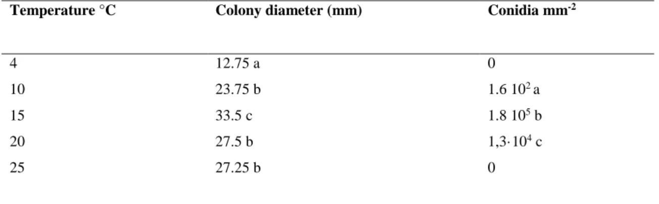
Previously studies conducted inoculating N. alba in different artificial substrate (data not reported), showed that tomato agar (TA) (15 g of agar technical, 500 ml of distilled water and 500 g of tomato sauce) is the best growth media for N. alba development. In order to find out a reliable method to produce conidia, the previously identified N. alba ID02 strain was inoculated on TA and stored under controlled isothermal temperatures for 14 days. The temperatures were 4, 10, 15, 20 and 25°C, selected after a preliminary study. Mycelial growth was determined as the increase in colony diameter after 14 d of incubation at different temperature. Production of conidia was evaluated by adding two mycelium plugs of 6 mm of diameter derived from a tomato agar plate incubated at 15°C for two weeks in 5 ml of distilled water agitated for 3 min by vortex. A sample (20 µl) of washing water was observed at light microscope (400 ×) and the conidial production was estimated counting the conidia with the hemocytometer. The sample unit was represented by 5 plates for each temperature. Once verified the best temperature for ID02 sporulation, five strain of N. alba earlier identified were evaluated for their sporulation attitude by placing TA dishes at 15°C for two weeks, ascertaining the conidia production as described above. The trial was performed in triplicate.
2.5 Study of primary metabolism of ‘Pink lady™’ apple by 1H-NMR
Apple skin of symptoms less fruits was removed using a peeler and immediately frozen in liquid nitrogen before to be freeze dried. Apple peels were sampled from 20 weeks before harvest ( unripe fruits) to 21 weeks after harvest, each 2 or 3 weeks. At each date of sampling three biological replicates were sampled and the peel of four apple fruit represented each replicate. Before 1H-NMR analysis polar and non-polar primary
19 metabolites were extracted according to the method described by Moing et al., (2004) with slight modifications. The frozen powdered samples were lyophilised and polar metabolites were extracted from 50 mg of lyophilised powder successively with 2 ml of ethanol/water mixtures: 80/20, 50/50 (v/v) and pure water (3 ml) for 15 min at 80°C. The supernatants were combined, dried under vacuum and lyophilised. The lyophilized extracts were mixed with 500 µl of 400 mM potassium phosphate buffer pH 6.0, 1mM Ethylene diamine tetraacetic acid disodium salt (EDTA), in D2O, titrated with KOD
solution to pH 6.00 ± 0.02 when necessary, and lyophilized again. The lyophilized titrated extracts were stored in darkness under vacuum at room temperature, and 1H-NMR analysis was completed within one week. For 1H-NMR analysis, dried titrated extracts were solubilised in 0.5 ml D2O, added with sodium salt of (trimethyl)
propionic-2,2,3,3-d4 acid (TSP) in D2O at a final concentration of 0.01% for chemical shift calibration and
transferred into an 5 mm NMR tube. Quantitative 1H-NMR spectra were recorded at
500.162 MHz and 300° K on a Bruker Avance III spectrometer (Wissembourg, France), using a 5 mm inverse probe and an electronic reference for quantification as described previously by Mounet et al. 2007.
2.7 Effect of organic acids and sugars on N. alba conidia germination and mycelial growth
In order to test the effect of apple primary metabolites on N. alba development, major fruit organic compounds were assayed on N. alba conidia germination and mycelial growth. Bioassays were performed using the highest concentration of organic acid and sugar contents found in apple skin by 1H-NMR. Each organic acid diluted in sterile distilled water in order to obtain the desired concentration was sterilized using a filter of 45 µm and added to MEA after autoclave sterilization. For conidial germination test an
20 aliquot of 100 µl of conidia suspension (103 conidia ml-1) was spread onto amended MEA as previously described. For mycelial growth evaluation a mycelial plug (6 mm Ø) taken from the periphery of actively growing pathogen culture was placed in the center of the dishes treated as reported above. The dishes were then immediately wrapped in Parafilm and incubated at 20°C for conidial germination and 15 °C for mycelial growth assays. Control was represented by unamended MEA inoculated with conidia or mycelium of pathogen as described before. The colony forming units (CFUs) and the mycelial growth were recorded after 4 and 15 days of incubation, respectively. For each compound tested five dishes were used and the assays were performed in triplicate. The percentages of the stimulation or the inhibition of conidia germination and mycelial growth caused by organic acid were calculated using the following formula: [(treatment–control)/control)] × 100. Positive values indicated stimulation and negative values indicated inhibition. For the evaluation of sugar effect, each carbohydrate was added in mineral medium agar (NH4H2P04 2.0 g/l, KH2PO4 0.6 g/l, MgSO47H2O 0.5 g/l, K2HP04 0.4 g/l, CaCI2H2O
0.074 g/l, Ferricitrate 0.012 g/l, ZnSO7H2O 0.0066 g/l, MnS044H20 0.005 g/l,
COCl26H2O 0.001 g/l, Thiamine 0.0001 g/l, Technical agar 5.0 g/l) and the experiments
were carried out as described above. The EC50 and EC95 values of organic acids (malic,
quinic and chlorogenic) were calculated as the concentrations that inhibited mycelium growth by 50% and 95%, respectively, compared with the control. EC50 and EC95 values were calculated using the probit analysis applied to the percentage of mycelial growth inhibition (Lesaffre and Molenberghs, 1991).
21
2.8 In-vivo evaluation of BER pathogenesis
In order to evaluate the BER pathogenesis, apple fruits were harvested and stored at 0°C and high humidity (> 92%) in commercial condition for five months followed by 10 days of shelf-life. Every month apples were evaluated for BER incidence. The sample unit was represented by ten replicates of seventy fruits each (total 700 fruits). The experiment was conducted twice.
2.6 Statistical analysis
Data referred to biological effect of temperature and primary metabolites were subjected to one-way analysis of variance (ANOVA) using the statistical package Statistica for Windows (Statsoft Inc.). Separation of means was performed using the least significance difference (LSD) test at P< 0.05. Principal component analysis (PCA) was performed with absolute concentrations of 20 metabolites (centered reduced data) issued from 1 H-NMR analysis of apple peel harvested during apple growth and ripening, using SimcaP+ software (Umetrics, Umea, Sweden). All experiments were carried out in a completely randomized block design.
Results
3.1 Evaluation of conidia production and mycelial growth on TA substrate
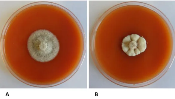
The temperature of incubation influence the pathogen growth, after two weeks from inoculation, important changes in the morphology of the colonies were observed (Fig.1). At 4° and 10°C the mycelium appeared white, soft and woolly with abundant aerial hyphae. At 15°C (Fig.1 A) the mycelium remained soft, woolly, whit aerial hyphae, but the color changed into white/grey. At this temperature was also possible to observe orange acervuli in the middle of the colony. The temperature of 15°C was the optimum for
22 mycelial growth (Tab.2) and conidia production. The highest conidia production was found in colonies grown at 15° C (1.8 105 conidia mL-1), while at 20°C and 10°C the conidia production was lower (1.3 103 and 1.2 102 conidia mL-1 respectively). At the other
tested temperatures, no conidia production was observed. These data obtained with a the strain ID02 were confirmed by the other five strains previously identified as N. alba, and cultivated on TA at 15°C for two weeks. All isolates tested were able to produce conidia on TA at 15° C, conidia production is reported in Tab.2. The most productive strains were ID02, IC10 and ID04, that produced 1.5·105, 1.2·105 and 1.2·105 conidia mL-1
respectively. While the strains ID06 and CR4, appeared less productive with 1.7·104 and 3.6 104 conidia mL-1 respectively.
3.2 DNA extraction and PCR amplification
The universal primers PN23/PN34 successfully amplified a product of 800 bp. After sequencing and sequences assembly, a blast algorithm was performed and all isolates belonged to N. alba. Sequences A.N. for each isolate and % of identity are reported in Table 1.
3.3 Primary metabolism of ‘Pink Lady™’ apple
The primary metabolites of apple epicarp tissue were identified and quantified by 1 H-NMR spectroscopy (Figure 2, 3 and 4). The measurements were performed on apple skin sampled during apple life cycle. Sugars represent the major metabolite group in apple skin (Fig.2). Among them, sucrose and fructose were the two main sugars, while glucose
23 and xylose remain minor fractions. During the maturation process, fructose represents the major apple sugar from 20 weeks before harvest until 5 weeks before harvest.
Sucrose content showed a significant increase 2 weeks before harvest (130.3 mg g-1 DW),
and a stable content until the end of the storage period (153.4 mg g-1 DW). On the contrary, a high content of glucose was observed from 20 (55.5 mg g-1 DW) to 8 (64.2 mg g-1 DW) weeks before harvest, followed by a decrease 1 month before harvest (40.8
mg g-1 DW) and a stabilization of its content until the end of the cold storage (49.3 mg g
-1 DW). Xylose, a minor sugar (from 0.34 to 1.05 mg.g-1 DW) showed a gradual increase
during the whole apple life cycle. Organic acids (Fig.3) represented the second major metabolite group. Among them, malic acid was the most abundant in apple fruit and its content varied from 34.8 to 12.1 mg.g-1 of DW (from 20 to 21 weeks after harvest). During post-harvest life, a general decrease of malic acid content was observed. The first significant reduction of malic acid happened immediately after harvest, decreasing from 24.3 to 16.9 mg.g-1 of DWin 2 weeks. The second decrease in malic acid content was recorded at 18 weeks after harvest, while the content remains stable until the end of the storage period (12 mg.g-1 DW). Quinic and chlorogenic acids showed a similar pattern.
At 20 weeks before harvest, quinic acid reached the highest content at 20 weeks before harvest (9.33 mg.g-1 DW), and its quantity decreased gradually until 5 weeks before
harvest, from this date the content remained stable (under 0.37 mg g-1 DW). Chlorogenic acid showed a decrease from 5.47 (20 weeks before harvest) to 1.92 (21 weeks after harvest) mg.g-1 DW, but no considerable changes were observed during post-harvest life.
Succinic and citric acids showed some changes during the pre-harvest life. The highest content of succinic acid was observed at the harvest time (0.12 mg.g-1DW), also citrate
24 remaining stable during the post-harvest time (0.207 mg.g-1 DW). Citramalic acid showed a very singular distribution pattern, in fact its content increased greatly at the harvest time passing from 0.34 to 1.62 mg.g-1 of DW in only 2 weeks. After this increase, its content
remained almost stable until the end of the storage. Amino acids were the third metabolites group. At the beginning of apple growth, asparagine showed the highest level among amino acids (1098.03 µg g-1 DW), while from the harvest date its content was
similar to aspartate.
3.4 Principal component analysis of biochemical composition of apple during the growth
and ripening
PCA was used to integrate all identified metabolites with centered reduced data. PCA was performed with absolute concentration of 20 metabolites issued from 1H-NMR analysis of sixteen dates of pericarp apple. Peel tissues were sampled from two months after full bloom, until the end of the storage period. The score plots (Fig. 5) showed that the first principal component (PC1), which explains 43.4% of the total variance, separated the developmental stages between growth before the harvest and ripening, whereas the second component (PC2), which explains 16% of the total variance, separated the samples inside each group. The group G showed a large separation on the first component corresponding to the sample harvested during the growth period. The scores plots indicated that during the apple growth, the larger amplitude of metabolic change over time. The T group corresponded to the apple sampled two and five weeks before harvest and the R group to apple sampled at the maturity stage and during cold storage period. The T group showed a similar biochemical composition while the R group showed a separation only on the second component. The corresponding loadings plot (Fig.5) amino
25 acids (aspartate and alanine) and organic acids (citric acid, succinic acid, quinic acid, malic acid and chlorogenic acid) was closely associated with growth phase while sugars as xylose and sucrose and choline were associated with the ripening stage. Fructose have no impact on the separation of these two groups.
3.5 Effect of organic acids and sugars on N. alba conidia germination and mycelial growth
Results on the influence of sugars and organic acids against N. alba growth showed that all sugars had no effect on conidia germination and the mycelial growth (data not reported). Otherwise, all organic acids at highest level, quantified in skin apple, affected
N. alba development. Quinic (9.4 mg g-1 DW), chlorogenic (5,5 mg g-1 DW), and malic
(34.7 mg g-1 DW) acids strongly inhibited both conidia germination and mycelial growth. In particular, malate showed an inhibition of 100 % of pathogen growth. Quinic and chlorogenic acids inhibited mycelial growth of 61.7 % and 50.0 % respectively, whereas the reduction of conidia germination was 73.3 % and 53.4 % for quinic and chlorogenic acids, respectively. Succinic and citric acids, tested at concentration 0.03 mg g-1 DW, stimulated the germination of conidia and mycelial growth of N. alba. of 44 % and 35 % for the mycelial growth and the conidial germination, respectively. In addition, the EC50
value of chlorogenic acid (4.6 mg g-1) revealed as the acid was the most effective against mycelial growth of pathogen followed by quinic acid (6.3 mg g-1) and malic acid (13.9 mg g-1) (Table 3). The lowest EC95 value was attributed to quinic acid (14 mg g-1),
followed by malic acid (28 mg g-1) and chlorogenic acid (29 mg g-1).
26 In stored apple at 0°C the pathogen remained quiescent for eight week, since no BER symptoms appeared until eight weeks from harvest. After the N. alba incidence increased progressively reaching the 29 % at the end of storage (after 21weeks from harvest) (Fig 6)
Discussion
During last decades N. alba was globally recognized as one of the main pome fruit disease in the postharvest phase (Kellerhals et al., 2012; Soto-Alvear et al., 2013, Maxin et al., 2014). Our data showed that the incidence of BER in ‘Pink Lady™’ apple can reach high levels, after 21 weeks of storage it was 29%. BER is a serious problem for apple production however, very little is known about the mechanisms that regulates the host-microbe interaction between N. alba and apple fruit, and no reliable and rapid methods to produce conidia in vitro, on artificial medium are available, making the study of N. alba biology quite difficult. In this study a new based tomato substrate seemed very useful for the in vitro growth of pathogen and for an abundant spore production. Five N. alba isolates, identified by PCR and grown on this substrate, were able to produce abundant conidia with a concentration ranging from 1.5 · 105 conidia ml-1 mm2 (IDO2) to 1.7· 104 conidia ml-1 mm2 (CR4). BER is a typical latent infection, characterized by a long quiescent phase that can overcome 90 days, before of the symptom manifestation. As well known, fruits and vegetables are provided of numerous biochemical compounds able to delay the fungal infection (Prusky, 1996). Among these, phenylpropanoids are usually designated as the main group of defence substances able to delay microbial infection (Naoumkina et al., 2010), nevertheless, our results showed also an important role played by the primary metabolites such as sugars, organic acids and amino acids that are found across all vegetal species within broad phylogenetic groups (Hounsome et al., 2008).
27 Carbohydrates represent the most important energy source for heterotrophic organisms such as fungi, for example, sugars and amino acids can contribute to zygospore formation of Phycomyces blakesleeanus (Leoninan et al., 1940). Organic acids are a group of organic compounds containing carboxylic groups that in solution release protons, that acidify the ambient (Hounsome et al., 2008). Plants contain citric, acetic, malic, oxalic, succinic, fumaric, quinic, tartaric, malonic, shikimic, aconitic, ascorbic, and other organic acids (Heldt 2005). From our data, organic acids can affect N. alba development. Chlorogenic and quinic acids inhibited conidial germination and mycelial growth, however their efficacy difficulty could be associated with postharvest latency of N. alba, since their concentration in planta significantly decreased during apple growth in the field, remained stable during postharvest phase. In addition, quinate inhibition effect was observed only at the concentration of 9.4 and 6.3 mg g-1,a range concentration measured by HNMR at 20 and 17 weeks before harvest, when the fruit is completely unripe. Chlorogenic acid also inhibited N. alba development at concentration of 5.5 mg g-1, a high concentration measured at the beginning of apple life cycle. Otherwise, the malic acid inhibited N. alba also at the concentrations founded in fruit at harvest and during storage. When tested at the maximum concentration of 34.4 mg g-1,malic acidinhibited completely N. alba germination and mycelial growth. The EC50 value referred to malic
acid (13.9 mg g-1), is very close to malic content measured from 8 weeks after harvest (14.9 mg g-1) until 18 and 21 weeks after harvest (12.1 mg g-1). These results suggest that malic acid, at concentration measured during storage, does not inhibit completely the fungal infection, but can delay the host colonization by N. alba. In the interaction between N. alba and ‘Pink Lady™’ apple, chlorogenic and quinic acids play a defense role like feeding deterrent (Ikonen at al., 2001) in the first part of apple life cycle, but
28 probably their protection function is minor during postharvest phase. Not only malic acid influenced N. alba development, but all tested fruit organic acids affected significantly the pathogen growth. Whilst malic acid inhibited conidia germination and the mycelial growth, when tested at the detected concentration of 0.03 mg g-1,succinic and citric acids significantly stimulated the pathogen development. Organic acids are essential in the metabolism of post-harvest produce; as reported by Kays & Paull (2004), some of them are important components of the respiratory tricarboxylic acid cycle and phosphoglyceric acid playing a critical role in photosynthesis. Their presence is fundamental to impart flavor, taste and odor to fruits and vegetables, but they can also represent a readily available source of stored energy (Kays & Paull, 2004). Biochemical results obtained in this work showed that during fruit postharvest, only the malic acid significantly decrease, and became stable 18 weeks after harvest, when the symptoms of BER rise considerably (Fig. 3 and Fig. 6)) . Higher plants are rich in pectic substances, high molecular mass glycosidic macromolecules. They are present in the primary cell wall as major component of the middle lamella and are the responsible for the structural integrity and cohesion of plant tissues (Pedrolli et. Al, 2009). In order to express their virulence, plant pathogens must to degrade pectic substances, and colonize the host tissue. This process requires a rich enzymatic pool displayed by fungal pathogens able to overcome this pectic barrier. Pectinase are an enzyme group that catalyzes pectic substances degradation through depolymerization and deesterification reactions. Pathogenic fungi work through depolymerization by hydrolases, and they are more active in a range of pH between 4 (Endo-PG I from Aspergillus carbonarius) to 5.3 (Endo PG II from Fusarium
moliniforme) (Pedrolli et al., 2009). Recently, pH influence on the pathogenicity of
29
Colletotrichum gloesporoides (Alkan et al., 2013), Botrytis cinerea (Manteau et al., 2013)
and Penicillium digitatum (Zhang et al., 2013), but for N. alba no data are provided. Our results showed that malic acid strongly affect N. alba conidial germination and mycelial growth, however a pH ambient reduction caused by the acid may be also considered. The outcomes described above suggest that the decreasing of malic acid can be involved in the interruption of N. alba latency. In fact, the optimal condition for N. alba growth could depend by the different organic acid balancing formed during storage that permits the recrudescence of N. alba pathogenicity.
In conclusion, our data can hypothesize that malic acid may be involved in the interaction between N.alba and apple fruit since BER symptoms appeared on apple only when malic content decreased, moreover results obtained on fruits are supported by in vitro trials. On artificial substrate N. alba is strongly inhibited by malic acid and EC95 value is very close
to the concentration detected at the beginning of apple life cycle, while EC50 value (1.39
30
Bibliography
Alkan N., Meng X., Friedlander G., Reuveni E., Sukno S., Sherman A., Thon M., Fluhr R., Prusky D. 2013. Global Aspects of pacC Regulation of Pathogenicity Genes in Colletotrichum gloeosporioides as Revealed by Transcriptome Analysis. MPMI 26, 1345–1358.
Berregi, I., Santos, J.I., Del Campo, G., Miranda J.I., 2003. Quantitative determination of (-)-epicatechin in cider apple juices by 1H NMR. Talanta 61, 139-145
Berregi, I., Santos, J.I., Del Campo, G., Caracena R., Miranda J.I., 2007. Quantitative determination of formic acid in apple juices by 1H NMR spectrometry. Talanta 72, 1049-1053
Bompeix, G., and Cholodowski-faivre 1998. The use of natural plant products against scald, Botrytis sp., Gloeosporium sp., Penicillium sp. And Rihyzopus sp. In: Proceedings of the Joint Workshop Non Conventional Methods for the Control of Postharvest Disease and Microbiological Spoilage, Commission of European Communities COST 914-COST915, Bologna, Italy, pp.99-104.
Cao, D., Li, X., Cao, J., Wang W., 2013. PCR Detection of the Three Neofabraea Pathogenic Species Responsible for Apple Bull’s Eye Rot. Advances in Microbiology, 3, 61-64
Cunnington J. H., 2004. Three Neofabraea species on pome fruit in Australia. Australasian Plant Pathology. 33, 453-454
Doyle, J.J. and Doyle, J.L. 1987. A rapid DNA isolation procedure for small quantities of fresh leaf tissue. Phytochemical Bulletin. 19, 11-15
31 Gariepy, T.D., Rahe, J.E., Lévesque, C.A., R.A., Spotts, D.L, Sugar. J.L. Henriquez 2005. Neofabraea species associated with bull’s-eye rot and cankers of apple and pear in the pacific Northwest.Ca. J. Plant Pathol. 27, 118-124
Hall, R., Robert H., (Ed.) 2005. Biology of plant metabolomics. Chichester, West Sussex, UK: Wiley-Blackwell. Annual plant reviews, 43, 420.
Hatoum, D., Annaratone, C., Hertog, M.L.A.T.M., Geeraerd, A.H., Nicolai, B.M, 2014. Targeted metabolomics study of ‘Braeburn’ apples duringlong-term storage. Postharvest Biol. Tec. 96, 36-41
Heldt, HW., 2005. Plant biochemistry. 3rd ed. London, U.K.: Elsevier Academic Press. P. 656 p
Henriquez J. L., Sugar D., Spotts R. A., 2004. Etiology of Bull’s Eye Rot of Pear Caused by Neofabraea spp. in Oregon, Washington, and California. Plant Dis. 88,1134-1138 Hounsome, N., Hounsome, B., Tomos, D., Edwards-Jones, G., 2008. Plant metabolites and nutritional quality of vegetables. JouRnal of food science 73, 48-65
Ikonen A.,Tahvanainen J. and Roininen H., 2001. Chlorogenic acid as an antiherbivore defence of willows against leaf beetles. Entomol. Exp. Appl. 99, 47–54.
Lattanzio, V., Di Venere, D., Linsalata, V., Bertolini, P., Ippolito, A., Salerno, M., 2001. Low Temperature Metabolism of Apple Phenolics and Quiescence of Phlyctaena vagabunda. J. Agric. Food Chem. 49, 5817-5821
Leonian L. H., and Lilly, V. G., 1940. Studies on the nutrition of fungi. V. Factors affecting zygospore formation in Phycomyces blakesleeanus. American Journal of Botany 27, 670-675
32 Lesaffre, E., Molenberghs, G., 1991. Multivariate probit analysis: a neglected procedure in medical statistics. Stat. Med. 10, 1391–1403
Kays J.K. and Paull R. E. 2004. Secondary metabolic process and products. Postharvest Biology 4, 196-201
Kellerhals, M., Szalatnay, D., Hunziker, K., Duffy, B., Nybom, H., Ahmadi-Afzadi, M., Ho¨fer, M., Richter, K., Lateur, M., 2012. European pome fruit genetic resources evaluated for disease resistance. 26:179–189
Manteau, S., Abouna, S., Lambert, B., Legendre, L., 2003. Differential regulation by ambient pH of putative virulence factor secretion by the phytopathogenic fungus Botrytis cinerea. FEMS Microbiology Ecology 43-359,366
Maxin, P., Williams, M., Weber, R. W. S., 2014. Control of fungal storage rots of apples by hot-water treatments: a northern European perspective.
Moing, A., Maucourt, M., Renaud, C.a, Gaudillère, M., Brouquisse, R., Lebouteiller, B., Gousset-Dupont, A.,Vidal, J., Granot, D., Denoyes-Rothan, B., Lerceteau-Köhler, E., Rolin, D., 2004. Quantitative metabolic profiling by 1-dimensional 1H-NMR analyses: Application to plant genetics and functional genomics. Functional Plant Biology 31, 889-902
Naoumkina, M. A., Zhao, Q., Gallego-Giraldo, L., Dai, X., Zhao P. X., Dixon, A., 2010. The phenylpropanoid pathway and plant defence—a genomics perspective. Mol. Plant Pathol. 11, 829–846
Neri, F., Mari, M., Brigati, S., Bertolini, P., 2009. Control of Neofabraea alba by plant volatile compounds and hot water. Postharvest Biology and Technology 51, 425-430 A.L.
33 Pedrolli D.B., Costa M. A., Gomes E. and Cano C. E. 2009. Pectin and pectinases: production, characterization and industrial application of microbial pectinolytic enzymes. TOBIOTJ 3, 9-18 Prusky D., 1996. Pathogen quiescence in postharvest diseases. Annu. Rev. Phytopathol. 34, 413-434
Rolin, D., Deborde, C., Maucourt, M., Cabasson, C., Fauvelle, F., Jacob, D., Canlet, C., Moing, A. 2013. High-Resolution 1H-NMR Spectroscopy and Beyond to Explore Plant Metabolome
Advances in Botanical Research, 67, 1-66
Rooney-Latham, S., Gallegos, L. L., Vossen, P. M., Gubler W. D., 2013. First Report of Neofabraea alba Causing Fruit Spot on Olive in North America. Plant Dis. 97, 1384 Snowdon A.L., 1990. Gloeosporium rot A Colour Atlas of Post-Harvest Diseases and Disorders of Fruits and Vegetables. General introduction and fruits, vol. 1 Wolfe Scientific Ltd., London 186–187
Soto-Alvear, S., Lolas, M., Rosales I. M., Chávez, E. R., Latorre B. A., 2013. Characterization of the Bull’s Eye Rot of Apple in Chile 2013. Characterization of the Bull’s Eye Rot of Apple in Chile
Spolti, P., Valbenito-Sanhueza, R. M., Medeiros Del Ponte E., 2010. Meio semiseletivo para recuperaçao e quantificaçao de Cryptosporiopsis perennans em maças. Ciencia Rural 40, 661-665
Spotts, R. A., Cervantes L. A., Mielke E. A., 1999. Variability in Postharvest Decay Among Apple Cultivars. Plant Dis. 83, 1051-1054
34 Swinburne, T.R., 1983. Quiescent infections in post- harvest diseases. In Post-Harvest Pathology of Fruits and Vegetables, in C Dennis (ed) Academic press, London 1–21 Spotts, R. A., Seifert, K. A., Wallis, K. M., Sugar, D., Xiao, C. L., Serdani, M., Henriquez J. L., 2009. Description of Cryptosporiopsis kienholzii and species profiles of Neofabraea in major pome fruit growing districts in the Pacific Northwest USA. Mycological Research 113, 1301-1311
Tilburn, J., Sarkar, S., Widdick, D.A., Espeso, E.A., Orejas, M., Mungroo, J., Peñalva, M.A., Arst Jr., H.N. 1995 . The Aspergillus pacC zinc finger transcription factor mediates regulation of both acid- and alkalineexpressed genes by ambient pH. EMBO J. 14, 779-790
Verhoeff, K., 1974 Latent Infections by Fungi. Annual Review of Phytopathology 12, 99-110
Vermathen M., Marzoratia M., Vermathenb P., 2012. Exploring High-resolution Magic Angle Spinning (HR-MAS) NMR Spectroscopy for Metabonomic Analysis of Apples. Chimia 66, 747–751
Zhang T., & Sun X., Xu Q., Candelas L.G.and Li H., 2013. The pH signaling transcription factor PacC is required for full virulence in Penicillium digitatum. Appl Microbiol Biotechnol 97, 9087–9098
35 Figures
Figure 1: Influence of temperature on Neofabrea alba mycelium growth at 15°C (A) and 25°C (B).
Figure 2: Sugars identified and quantified by HNMR from 20 weeks before harvest untill 21 weeks after
36
Figure 3: Organic acids identified and quantified from 20 weeks before harvest until 21 weeks after
harvest. Bars represent the SE.
0 5 10 15 20 17 15 12 10 8 5 2 0 2 5 8 12 15 18 21 Quinate
37
Figure 4: Amino acids identified and quantified from 20 weeks before harvest until 21 weeks after
38
Figure 5: Principal component analysis (PCA) of 20 metabolites measured by quantitative 1H NMR
spectroscopy in apple epicarp tissue at three stages of development (growth, turning and ripening). The PCA was performed with averaged data expressed on DW basis. (A) PCA scores plot of the first two principal components (PC1 and PC2) showing the distribution of the samples at three stages of development. (B) PCA loadings plot showing two sets of metabolites associated with the growth and ripening stage -4 -2 0 2 4 6 8 -6 -4 -2 0 2 4 6 8 G R R T PC2 16,0% A) glucose saccharose fructose xylose chlorogenate citrate citramalate formate malate quinate succinate alanine asparagine aspartate isoleucine choline unkS5,56 unkM5,00 unkM0,90 unkD0,86 -0,1 -0,05 0 0,05 0,1 0,15 0,2 0,25 0,3 0,35 0,4 -0,4 -0,3 -0,2 -0,1 0 0,1 0,2 0,3 0,4 B)
39
Figure 6: Evolution of Bull’s eye rot in ‘Pink Lady™’ apple stored at 0°C for 21 weeks. Each datum is the
average of 700 fruits + SE
Tables
Table 1 Molecular identification and determination of conidial production
Strains A.N. (GeneBank) % identity Conidia mm-2
ID02 KJ396074 100 1.5·105a
IC10 KJ396076 99 1.2 ·105a
ID04 KJ396077 99 1.2·105a
ID06 KJ396078 99 1.7·104b
CR4 KJ396075 100 3.6·104b
*Within the same column data followed by the same letters are not statistically different for LSD test (P<0.05).
** Data are the mean of 5 plates for each medium+ SE
0 10 20 30 40 0 2 5 8 12 15 18 21 % o f infe cte d f ru its
40
Table 2 Influence of temperature on mycelial growth and conidia production of Neofabea alba ID02
isolate
Temperature °C Colony diameter (mm) Conidia mm-2
4 12.75 a 0
10 23.75 b 1.6 102 a
15 33.5 c 1.8 105 b
20 27.5 b 1,3·104 c
25 27.25 b 0
*Within the same column data followed by the same letters are not different for LSD test (P<0.05).
**Data are the mean of 5 plates for each medium+ st.er
Table 3 Determination of EC50 and EC95 values of inhibiting organic acids
Acknowledgments
The 1H-NMR analyses were performed at the Metabolome Facility-MetaboHUB of
Bordeaux Functional Genomics Center. This work was supported by ANR MetaboHUB (ANR-11-INBS-0010 project).
mg g-1 mg g-1
Malic acid 13.9 28
Chlorogenic acid 4.6 29
41
Susceptibility of five apple cultivars to Bull’s eye rot and relationship with ambient pH
1. Introduction
Neofabraea spp. causes the lenticel rot of apple and pear fruits, well known as Bull’s eye
rot (BER). The disease is one of the main important postharvest diseases of pome fruit, producing notable economic losses during fruits storage. BER is particularly serious in Europe (Bompeix and Cholodowski-Faivre, 2000, Neri et al., 2009), North-West of United States of America (Gariepy et al., 2005, Henriquez et al., 2004) Australia (Cunnington, 2004), Chile (Soto-Alvear et al., 2013) and Brasil (Spolti et al., 2010). In China BER has classified as quarantine pest disease (Cao et al. 2013). Fruit lesions are flat to slightly sunken, brown and often with a light brown center (Snowdon, 1990), the rotten tissues are firm and white acervula can be present in old lesions. Common cultivars of apple (Spotts et al., 1999) and pear (Henriquez et al. 2004) are subjected to disease relating to climate conditions. The fruit contamination occurs in the orchard, but the symptoms appear only three/four months after harvest, during cold storage (Snowdon 1990). Four members of the genus Neofabraea are causal agents of lenticel rot of pome fruits: N. alba, N. perennans, N. malicorticis and Cryptosporiopsis kienholzii, (Jong et al., 2001, Spotts et al. 2009) however, in Italy only the presence of N. alba (E. J. Gutrie) Verkley (anamorph Phlyctema vagabunda Desm.) was detected. ‘Golden Delicious’ apple and ‘Bosc’ pear are highly susceptible cultivar and in certain years favorable to pathogen development high incidences of fruit infections (over 40%) can occur. Few are the scientific data on apple cultivars susceptibility to N. alba. The scarce information derive from packaginghouse surveys however, they are not specific on BER susceptibility and apple cultivar. A review on pome European fruit genetic resources evaluated for
42 disease resistance (Kellerhals et al., 2012), included BER among the main causes of decay during apple storage, but no information on cultivar resistance or tolerance to BER disease was reported. Jönsson et al. (2012) assert that also in Sweden BER is one of the most common problem during apple storage.
The latent infections produced by Neofabrea spp. can be influenced by many factors, such as phenolic content (Naoumkina et al., 2010), maturity stage (Guidarelli et al., 2011, Neri et al., 2014), and insufficient nutritional requirements for pathogen expression of virulence factors (Prusky, 1996). Previously, the studies of Bateman and Beer (1965) on the close relationship between pH and pathogenicity, were resumed by Prusky et al. (2001) that suggested as some fungal pathogens may enhance their virulence by locally modulating the host's ambient pH. Therefore the importance of ambient pH for the expression of some enzymes as cell-wall degrading enzymes (CWDE), represent a key factor in specific enzymes secretion (Wubben et al., 2005) allowing to pathogen the penetration in host tissue (Akimitsu et al., 2004). Each enzyme can be expressed only at a precise range of pH and for this reason the pH value modulation performed by fungi, can result crucial for host invasion and disease appearing (Akimitsu et al., 2004, Prusky et al., 2001). Molecular studies demonstrated that genes encoding CWDE are expressed, and their products secreted, only under optimal pH conditions (Eshel et al., 2002; Prusky et al., 2001). The ability to modify pH may be differentially expressed by fungi, that are able to raise (‘alkalinising fungi’) or reduce the ambient pH (‘acidifying fungi’) in view of the pathogenic process (Prusky and Lichter, 2008). Some pathogens such as
Colletotrichum gloeosporioides (Prusky et al., 2001) and Alternaria alternata (Eshel et
al., 2002), alkalinise their host tissues by producing significant amounts of ammonia. Other fungi, such as Penicillium expansum, P. digitatum, P. italicum (Prusky and
43 Yakoby, 2003), Botrytis cinerea (Manteau et al., 2003) and Sclerotinia sclerotiorum (Bateman and Beer, 1965), utilize tissue acidification to support their attacks via the secretion of organic acids.
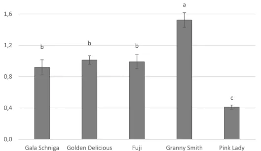
The aims of the present work were to i) evaluate the susceptibility of five common Italian apple cultivars; ii) study the relationship between natural ambient pH of apple cultivar and the N. alba development iv) investigate the N. alba capability to affect the pH of host tissue, in order to define it as ‘alkalinising‘ ’ or ‘acidifying’fungus’.
44
2. Materials and methods
2.1. Fruits
‘Gala Schniga’, ‘Golden Delicious’, ‘Fuji’, ‘Granny Smith’ and ‘Pink Lady™’ apples were harvested at commercial maturity according to specific parameters for each ‘ cultivar in five different orchards located in Emilia Romagna region.Fruits were selected for uniform size, without physical injuries or apparent infections.
2.2. Quality parameters at harvest and during storage
In order to characterized the stage of maturity the following parameters: starch content, size, color, firmness, total soluble solid (TSS), IAD were determined. Starch content at harvest was determined visually using the standard iodine test as described by Nyasordzi et al., 2013. The number 1 indicates maximum starch content (maximal dark stain) and index 10 represents maximum starch hydrolysis (clear stain) (CTFL starch conversion chart EURFRU). Apple size was measured at harvest by caliber and expressed in mm. Color was measured on two opposite sides of each fruit using a tristimulus colorimeter (Chromameter CR-200,Minolta, Japan) able to determine the values of lightness (L*), red-greenness (a*) and yellow-blueness (b*) parameters. Flesh firmness (FF) was evaluated on the two opposite sides of each fruit, after eliminating a thin layer of the epicarp, using an automatic pressure tester (FTA-GUSS, South Africa) fitted with an 11 mm plunger. The TSS was determined with an Atago digital refractometer (Optolab, Modena, Italy) by squeezing a part of the mesocarp in order to obtain a freshly prepared juice from each apple cultivar. The IAD value was measured using a 17 portable DA-Meter (TR-Turoni, Forlì, Italy), that gives the ripening index in relation with the chlorophyll
45 content. The TA was determined on 20 mL of flesh juice (titration with 0.25N NaOH) using a semiautomatic instrument (Compact-S Titrator, 31 Crison, Modena, Italy). With the exception of starch content and apple size, the evaluation of each quality parameter for each cultivar was repeated at harvest, after 60, 90, 120 days of storage at 0°C and after two weeks of shelf life at 20°C.
2.3 Fungal cultures
N. alba ID02 isolate, deposited in GenBank and belonging to our collection, was
maintained onto tomato agar (TA: 500 g of tomato sauce, 15 g of agar technical and 500 ml of distilled water) at 15°C. Conidial suspensions were prepared by adding 10 mL of sterile water with 0.01% (w/v) Tween-80 on the surface of 15-day-old cultures grown on TA and rubbing the surface of the agar with a sterile glass rod. Conidia were counted in a haemocytometer and diluted to the desiderated concentration.
2.3 Apple inoculation by wounds
Each apple was wounded by a sterile nail, then inoculated with 20 μL of a conidia suspensions of N. alba 104 conidia/mL. Apple inoculated with distilled water were used as control. Apples were stored at 0 °C and 85% RH for 60 and 90 days. The severity and the incidence of disease were recorded by measuring the diameter of lesion and by calculating the percentage of infected wound respectively. The sample unit was represented by four replicates of five fruits each per variety. The experiment was repeated once.
2.4. pH measurement in decayed apples
In order to measure the pH of mesocarp, a micro-pH electrode was introduced into the wound (pH & Ion-Meter GLP 22+ Model 5033 pH electrode, Crison). The sample unit