Università degli Studi di Pisa
Facoltà di Medicina e Chirurgia
Scuola di Specializzazione in Oncologia
Tesi di Specializzazione
EZH2 polymorphisms and outcome of metastatic colorectal
cancer patients
Relatore:
Chiar.mo Prof. Alfredo Falcone
Candidato:
Dr. Lorenzo Fornaro
Index
List of Tables iii
List of Figures iv
Summary and Key words v
Chapter 1. Introduction 1
1.1. Treatment of metastatic colorectal cancer: state of the art 1 1.1.1. Role of chemotherapy in metastatic colorectal cancer 2 1.1.2. Role of bevacizumab in metastatic colorectal cancer 12 1.1.3. Role of anti-EGFR agents in metastatic colorectal cancer 16 1.2. Prognostic and predictive factors in metastatic colorectal cancer 25
Chapter 2. Cancer stem cells: essentials of biology 31
2.1. Cancer stem cells and colorectal cancer 31
2.2. Preliminary clinical implications of the cancer stem cell hypothesis
in colorectal cancer 35
2.3. Epigenetics and cancer stem cells: role of Polycomb Repressor
Complexes 38
Chapter 3. EZH2 polymorphisms and outcome of metastatic colorectal
cancer patients 45
3.1. Rationale 45
3.2. Materials and methods 48
3.2.1. Oncomine analysis 48
3.2.3. Evaluation of activity and efficacy 50 3.2.4. Sample collection, DNA and RNA isolation 50 3.2.5. SNP genotyping and EZH2 mRNA expression 51
3.2.6. Statistical analysis 52
3.2.7. In silico characterization of the 626-394C>T SNP 54
3.3. Results 55
3.3.1. Polycomb targets are specifically silenced in FOLFIRI
non-responders 55
3.3.2. Patient characteristics and treatment outcome 55
3.3.3. Genotype information 56
3.3.4. Correlation between EZH2 SNPs and outcome 57
3.3.5. Cox model and interaction test 58
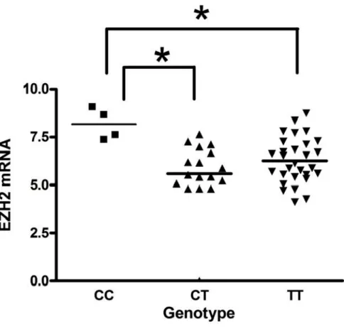
3.3.6. In silico and in vivo characterization of 626-394C>T SNP 59
Chapter 4. Discussion and conclusions 75
4.1. Discussion 75
4.2. Conclusions and future development 79
LIST OF TABLES
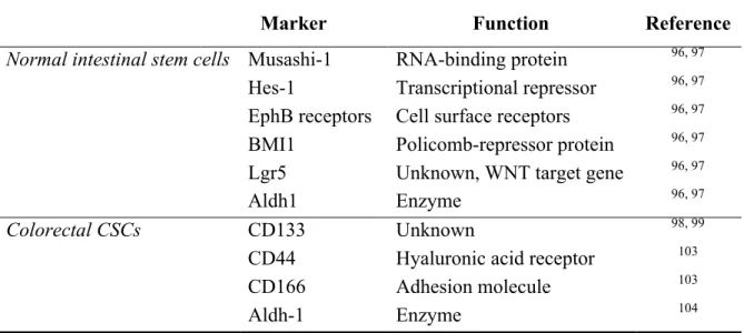
2.1. Markers of normal intestinal stem cells and colorectal CSCs 43
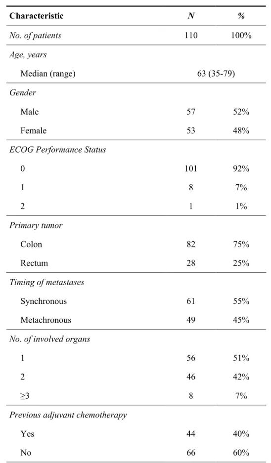
3.1. Patient characteristics 61
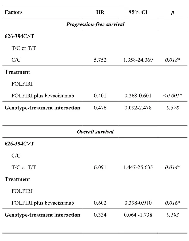
3.2. EZH2 polymorphisms: association with RR, PFS and OS 63 3.3. Multivariate analysis: FOLFIRI plus bevacizumab cohort 64 3.4. Multivariate analysis: FOLFIRI with or without bevacizumab 67
LIST OF FIGURES
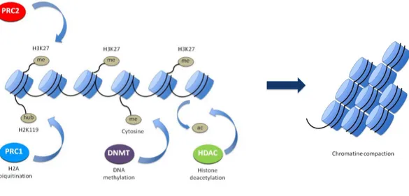
2.1. Simplified view of PcG function 44
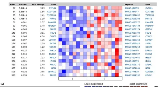
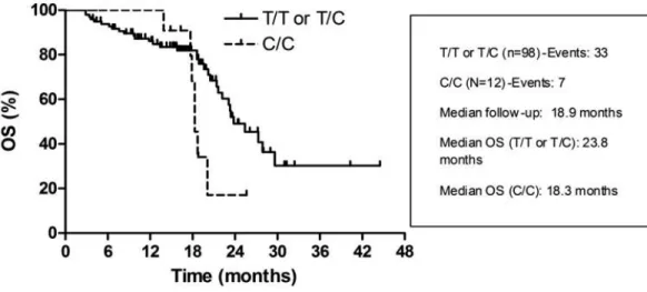
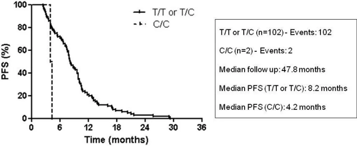
3.1. PcG targets are down-regulated in FOLFIRI non-responders 68 3.2. PFS and EZH2 genotype in the FOLFIRI plus bevacizumab cohort 69 3.3. OS and EZH2 genotype in the FOLFIRI plus bevacizumab cohort 70 3.4. PFS and EZH2 genotype in the FOLFIRI cohort 71
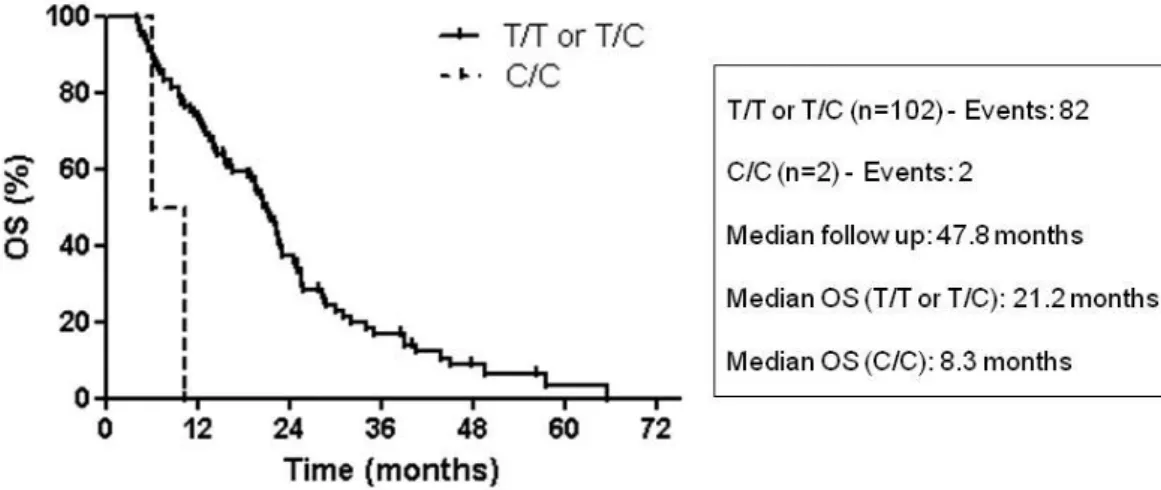
3.5. OS and EZH2 genotype in the FOLFIRI cohort 72
3.6. Characterization of TF binding sites affected by rs3757441 SNP 73
SUMMARY
Despite therapeutic innovations, metastatic colorectal cancer (mCRC) is still characterized by poor prognosis. Few molecular markers are available to predict progression risk and to help therapeutic decisions. Polycomb group genes (PcGs) are epigenetic modifiers involved in tumor suppressor gene silencing. EZH2 is a PcG member that mediates gene silencing through histone-H3 lysine-27 methylation. In CRC, EZH2 over-expression is associated to shorter survival. Recently, 4 EZH2 single nucleotide polymorphisms (SNPs) have been characterized: the present study aimed at evaluating the correlation between EZH2 SNPs and outcome parameters in mCRC patients.
DNA was extracted from blood samples of 110 mCRC patients treated with first-line FOLFIRI plus bevacizumab, and 104 mCRC patients treated with FOLFIRI. Genotyping was performed by Real-Time PCR. Allelic variant distribution was used to predict objective response, progression-free survival (PFS) and overall survival (OS). EZH2 mRNA levels were evaluated on lymphocytes of a parallel cohort of 50 radically resected stage II or III CRC patients.
One allelic variant (rs3757441 C/C vs. C/T or T/T) was significantly associated to shorter PFS and OS in both mCRC patient cohorts receiving first-line FOLFIRI (with or without bevacizumab). At multivariate analysis, the same variant resulted an independent predictor of both PFS and OS (p<0.05). Among the 50 patients analysed for EZH2 expression and genotyped for EZH2 rs3757441 SNP, mRNA levels were significantly higher in patients harbouring the C/C genotype with respect to C/T and T/T (p<0.05), with no difference between C/T and T/T genotypes.
Our results indicate that an EZH2 SNP may be useful to predict PFS and OS in mCRC patients treated with first-line FOLFIRI (with or without bevacizumab).
KEY WORDS
Bevacizumab, Cancer stem cell, Colorectal cancer, EZH2, FOLFIRI, Polycomb, Single nucleotide polymorphisms
CHAPTER 1. INTRODUCTION
1.1. Treatment of metastatic colorectal cancer: state of the art
Colorectal cancer (CRC) still represents the third most common malignancy and the second leading cause of cancer death in Western countries1: about 30.000 new cases and over than 15.000 deaths have been recorded in Italy in 20002. Even if
in the 90% of the cases a radical resection of the primary tumor is possible, 25% of patients present with metastatic disease at diagnosis and 50% die from systemic disease3.
In the last decade, great advances have been achieved in the treatment of metastatic CRC (mCRC): thanks to the availability of a greater number of effective cytotoxic and targeted agents and to the higher percentage of patients who undergo radical resection of metastases, overall survival (OS) has approached 24 months in most recent trials, and definitive cure of the disease is a realistic aim of treatment in at least 10-15% of the patients4,5. Moreover, together with the introduction of new drugs into clinical practice, new strategies have been developed, and therapeutic approach toward mCRC patients is growingly individualized according to the goal of treatment itself (palliation, long-term OS or cure)6,7. In the following paragraphs we will discuss the current data supporting the use of cytotoxic and targeted agents in the treatment of mCRC and review the role of the available prognostic and predictive biomarkers in individualising treatment algorithm in different patient subsets.
1.1.1. Role of chemotherapy in metastatic colorectal cancer
For over than 30 years 5-fluorouracil (5-FU) has been the only available treatment for which reliable data supported a role in mCRC: a meta-analysis8 of
various randomized trials has demonstrated that 5-FU improves OS and quality of life (QoL) of mCRC patients compared to best supportive care (BSC) alone and a randomized trial demonstrated that the advantage is greater when chemotherapy is started early in the course of the disease, before the onset of symptoms9.
The introduction of irinotecan and oxaliplatin improved the antitumor activity and the efficacy of chemotherapy in this disease7. The combinations of irinotecan plus either infusional or bolus 5-FU/leucovorin (LV) (FOLFIRI and IFL, respectively) and oxaliplatin plus infusional 5-FU/LV (FOLFOX) have demonstrated increased activity and efficacy compared with 5-FU/LV alone in randomized studies10,11,12,13. Of interest, phase III studies comparing irinotecan plus 5-FU/LV
with 5-FU/LV alone suggested that a more active treatment administered upfront can prolong OS, even if active second-line therapies are offered to patients progressing on 5-FU/LV. Furthermore, studies with oxaliplatin plus 5-FU/LV have indicated that a highly active first-line chemotherapy regimen may permit, in a small subgroup of initially unresectable mCRC patients, a secondary radical surgical approach on metastases after response to chemotherapy, and that approximately 30% to 40% of operated patients will survive without evidence of disease for more than 5 years14,15. Therefore, taken together these data indicate that in mCRC a more active first-line treatment can be more effective, and a meta-analysis of 25 randomized trials of
first-line treatment also supports the relationship between tumor response to first-first-line chemotherapy and OS16.
More recently, a randomized study by the GERCOR group17 assigned 220 untreated mCRC patients to receive first-line FOLFIRI followed by FOLFOX-6 at progression (arm A), or the reverse (arm B). Both sequences achieved similar activity and efficacy, and, of interest, median OS was 21.5 months in arm A and 20.6 months in arm B, which are among the highest survival times reported up to now in any randomized study of chemotherapy alone in mCRC. This study suggests that the exposure of mCRC patients to all the three most active agents (i.e. 5-FU/LV, irinotecan and oxaliplatin) is associated with best survival outcome. In addition, a study by Goldberg et al.18 demonstrated the superiority of the FOLFOX-4 regimen to IFL and confirmed the importance of the three-drug exposure, considering that in the IFL arm only 24% of patients could receive oxaliplatin as second-line treatment, while in the FOLFOX-4 arm 60% of patients were able to receive salvage treatment with irinotecan. These data are in line with the results of a recent pooled analysis of seven phase III trials demonstrating that OS is correlated with the proportion of patients who received all the three active drugs in the course of their disease, but not with the proportion of patients who received any second-line therapy19.
Another relevant advance in the management of mCRC is the availability of the oral fluoropyrimidine capecitabine. A meta-analysis of the studies comparing capecitabine plus oxaliplatin (CAPOX) with FOLFOX demonstrated that capecitabine is non-inferior to 5-FU in terms of PFS and OS, but results in a significantly lower RR20. Conversely, the results reported with capecitabine plus irinotecan (CAPIRI) are controversial, underlining that oral chemotherapy needs
active patient management. In fact, the EORTC 40015 trial21 was prematurely suspended due to alarming safety concerns: CAPIRI reported an unacceptably higher rate of grade 3-4 diarrhoea compared to FOLFIRI (37% vs. 13%), with an increased incidence of treatment-related deaths (5 vs. 2 patients). As a consequence, both PFS (5.9 vs. 9.6 months) and OS (14.8 vs. 19.9 months) were shorter with CAPIRI. In the BICC-C study22,23 CAPIRI reported shorter median PFS when compared with FOLFIRI (5.8 vs. 7.6 months; HR=1.36, p=0.015) and higher rates of severe events, particularly diarrhoea (47.5% vs. 13.9%) and dehydration (19.1% vs. 5.8%). On the contrary, the CAIRO trial24 completed accrual and demonstrated an acceptable rate
(26%) of grade 3-4 diarrhoea. All these trials tested the same CAPIRI schedule, so such differences in tolerability cannot be merely explained by differences in the dose intensity of chemotherapy. One possible confounding factor in the EORTC 40015 and BICC-C studies is represented by a second randomisation to either celecoxib or placebo, since coxibs have been associated with an increased risk of toxicities (particularly cardiovascular thrombotic events) in CRC patients: however, a causative role of celecoxib in explaining the observed high rate of severe diarrhoea appears unlikely since this agent seems to protect against irinotecan-induced mucosal injury25.
In order to expose all patients to 5-FU/LV, irinotecan and oxaliplatin (thus overcoming the risk not to receive active cytotoxics in second-line in case of rapid disease progression and patient conditions deterioration) and in order to improve the activity of treatment (thus potentially increasing the rate of secondary surgery of metastases26), the Gruppo Oncologico Nord Ovest (G.O.N.O.) developed the triplet regimen named FOLFOXIRI which proved to be feasible with acceptable toxicities
and promising activity and efficacy in phase I-II studies27,28. When compared with FOLFIRI in a randomized phase III trial29, FOLFOXIRI confirmed to be feasible and resulted in higher RR (66% vs. 41%, p=0.0002) and radical resection rate of metastases (15% vs. 6% of patients, p=0.033). Moreover, at a median follow-up of 18.4 months 216 patients have progressed and median PFS and OS were significantly longer in the FOLFOXIRI arm (9.8 vs. 6.9 months, p=0.0006 and 22.6 vs. 16.7 months, p=0.032, respectively) with a HR of 0.60 in favour of FOLFOXIRI. Finally, the rate of early progressions (within 6 months from treatment onset) was significantly lower in the FOLFOXIRI arm (18% vs. 45%, p<0.0001). A recent update of the trial30 confirmed the PFS (median: 9.8 vs. 6.8 months, HR=0.59, p<0.0001) and OS (median: 23.4 vs. 16.7 months, HR=0.74, p=0.026; 5-year survival rate: 15% vs. 8%) benefit for the triplet at a median follow up of 60.6 months. The authors concluded that the FOLFOXIRI regimen is feasible with manageable toxicities also in a multicenter setting: the incidence of grade 3-4 neutropenia and grade 2-3 peripheral neurotoxicity is increased with FOLFOXIRI, but febrile neutropenia, diarrhoea and other toxicities are comparable to FOLFIRI. As recently reported by the same group, the benefit derived from the triplet is not entirely due to the increased rate of secondary radical surgery: in fact, FOLFOXIRI retained its superiority also when resected patients were excluded from the analysis, confirming a better palliative effect in unresectable mCRC than FOLFIRI. Moreover, such an aggressive strategy does not harm the feasibility and the efficacy of salvage therapies with the same agents used in first-line. The G.O.N.O. group conducted a phase I-II trial in order to evaluate capecitabine instead of 5-FU in a triplet regimen (XELOXIRI), but the change from infusional to oral fluoropyrimidine resulted in
increased rate of severe diarrhoea, confirming that capecitabine is not a preferable alternative to 5-FU in both irinotecan-containing doublet and triplet regimens.
Another phase III trial by the Hellenic Oncology Research Group (H.O.R.G.) evaluated the triple combination in the first-line treatment of mCRC patients31.
Results demonstrated a non-significant trend for superiority for the FOLFOXIRI regimen compared to FOLFIRI, both in terms of activity (RR: 33.6% vs. 43%; secondary R0 metastasectomy: 4% vs. 10%) and efficacy (median TTP: 6.9 vs. 8.4 months; median OS: 19.5 vs. 21.5 months). These apparent discrepancies between the G.O.N.O. and H.O.R.G. trials can be explained by differences in treatment schedules and study populations. In fact, the G.O.N.O. regimen allows to avoid the 5-FU bolus administration and to administer a higher dose intensity of 5-FU, irinotecan and oxaliplatin, compared with the H.O.R.G. schedule (1600 mg/sqm/week vs. 1000 mg/sqm/week; 82.5 mg/sqm/week vs 75 mg/sqm/week; and 42.5 mg/sqm/week vs. 32.5 mg/sqm/week, respectively). As regards patient characteristics, patients aged more than 75 years were not included in the G.O.N.O. trial and if aged 70-75 an ECOG PS of 0 was required, while the H.O.R.G. study had no upper age limit (age range reached 82 years in the FOLFOXIRI arm) and elderly (more than 70-year old) patients with poor PS (1-2) were eligible: indeed, study population in the H.O.R.G. trial was older (median age: 66 vs. 62 years, respectively) and with a poorer performance status (ECOG PS 0/1/2: 36/53/11% in the H.O.R.G. trial vs. 61/37/2% in the G.O.N.O. trial). All these factors could have contributed to the different results of the triple regimens and underlined the need for adequate selection of fit patients when intensive chemotherapy is administered. Indeed, the potentials of the upfront triple strategy have been confirmed by the meta-analysis of
the G.O.N.O. and H.O.R.G. trials reported by Golfinopoulos et al.32, who demonstrated significant advantages for FOLFOXIRI compared to FOLFIRI both in terms of disease progression and survival.
Masi et al.33 recently published the results of a retrospective pooled analysis on
196 patients treated with first-line FOLFOXIRI in order to evaluate the long-term outcome of patients radically resected after chemotherapy. Thirty-seven (19%) could undergo a secondary R0 surgery on metastases (complete pathological response has been achieved in 4 patients) after a median of 5.5 months of treatment. The authors reported no intra-operative or post-operative mortality and a low rate of peri-operative complications (27% of cases), all of which were transient and resolved completely. After a median follow-up of 67 months, 5-year and 8-year OS were 42% and 33%, respectively. At 5 years, 29% of the resected patients were free of progression. Such results demonstrated the feasibility and the efficacy of FOLFOXIRI as conversion therapy and were consistent with the observation of Folprecht et al.26 that more active first-line treatments result in a higher secondary resection rate of metastases.
If the abovementioned results support the role of a more intense first-line approach, several randomized phase III trials addressed the question whether all mCRC patients should receive upfront combination chemotherapy. In the CAIRO study24, 820 patients were randomized to first-line capecitabine, second-line irinotecan, and third-line CAPOX or first-line CAPIRI and second-line CAPOX. The trial was powered to detect a superiority of the combination over single-agent based on an anticipated median OS of 14.0 vs. 17.5 months, but did not demonstrate any difference in OS, the primary end point (16.3 months for the sequential treatment
group vs. 17.4 months for the combination group; HR 0.92, 95%CI 0.79-1.08, p=0.3281), despite increased RR (41% vs. 20%, p<0.0001) and PFS (median: 7.8 vs. 5.8 months; HR 0.77, 95%CI 0.67-0.89; p=0.0002) being reported with the upfront combination. The FOCUS trial34 randomized over 2100 patients to three different
strategies: 5-FU/LV followed by irinotecan (strategy A); 5-FU/LV followed by FOLFIRI/FOLFOX (strategy B); first-line FOLFIRI/FOLFOX followed by the reverse regimen at progression (strategy C). FOCUS aimed to show non-inferiority of the single-agent approach. Again, the authors did not report OS differences among study arms (strategy C vs. B, median: 15.9 vs. 15.1 months; HR 1.06, 90%CI 0.97-1.17), while higher RR (5-FU/LV vs. FOLFIRI or FOLFOX: 28% vs. 49% or 57%, p<0.001) and PFS (5-FU/LV vs. FOLFIRI or FOLFOX: 6.3 vs. 8.5 or 8.7 months, p<0.001) were achieved with combination therapy, at the price of increased but acceptable toxicity: data from FOCUS therefore exclude a reduction of more than 5% in 2-year survival or a difference in median OS of more than 2.3 months with the upfront use of single-agent chemotherapy. Even though both CAIRO and FOCUS reported median OS in the range of 13.9-17.4 months, which is less than expected in patients treated with a sequence of doublet regimens (approximately 20 months), several factors contribute to explain this finding: the poor prognosis of the patients enrolled (in fact, potentially resectable patients were excluded in the FOCUS trial) and the small proportion of patients who received all the three active agents after initial treatment with monotherapy (in the range of 19-55%).
Among elderly or unfit patients, the recently published FOCUS-2 trial35 demonstrated that the addition of oxaliplatin to fluoropyrimidine increases RR, with a trend toward higher PFS and equal OS, compared with single-agent
fluoropyrimidine. After comprehensive health assessment, 459 patients were randomly assigned to one of the following arms: 5-FU/LV (group A); oxaliplatin and 5-FU (group B); capecitabine (group C); or oxaliplatin and capecitabine (group D). The trial investigated whether the addition of oxaliplatin ([A vs. B] + [C vs. D]) to fluoropyrimidine is beneficial in terms of PFS and whether the substitution of 5-FU with capecitabine ([A vs. C]+[B vs. D]) improves QoL, as assessed by change from baseline to 12 weeks in global QoL. Final results demonstrate that a non-significant improvement in median PFS can be achieved with the upfront combination regimen (5.8 vs. 4.5 months; HR 0.84, 95%CI 0.69-1.01, p=0.07), while replacing 5-FU with capecitabine does not result in improved global QoL (56% patients receiving 5-FU reported improvement in global QoL compared with 56% receiving capecitabine): however, the incidence of grade 3 or worse toxicities, while not significantly increased with oxaliplatin (38% vs. 32%, p=0.17), was higher with capecitabine than with 5-FU (40% vs. 30%, p=0.03). Again, even in FOCUS-2 OS did not differ between monotherapy and combination groups (HR 0.99, 95%CI 0.81-1.18, p=0.91).
Taken together, these data underline a key issue in mCRC patient management, i.e. the importance to target the intensity of first-line chemotherapy on the basis of both patient and disease characteristics and the aim of treatment. In fact, if comorbidities or advanced age might impair the feasibility of more intense chemotherapy in some patients, disease burden might require (or not) the need for a rapid tumor response in others. Again, in selected cases a more active treatment might be the best choice to achieve enough tumor shrinkage and allow a radical secondary resection of metastases. We have convincing evidence that an upfront combination regimen achieves superior RR, secondary surgery of metastases rate and
PFS at the price of acceptable toxicity: for these reasons, patients with potentially resectable disease or with high tumor burden or disease-related symptoms may benefit from a more active combination regimen. On the other hand, data from phase III trials support the use of sequential single-agent chemotherapy in those cases with more indolent disease course, when the objective is prolonging OS without impairing QoL with consistent toxicities.
If the intensity of the upfront strategy may differ in different patient subgroups, however, several randomized trials convincingly demonstrated that scheduled treatment duration or pauses after a predefined period of treatment do not impair results in terms of OS and might contribute to improve treatment tolerability. The OPTIMOX-1 trial36 was the one of the first large study suggesting that prolonging combination chemotherapy is not necessary in mCRC: investigators randomized 620 patients to FOLFOX-4 until progression or FOLFOX-7 for 12 weeks followed by 5-FU/LV (with oxaliplatin reintroduced at progression). The median duration of disease control (DDC, the primary end point, defined by the authors as PFS, or, if FOLFOX was reintroduced, addition of the initial PFS and the PFS of the reintroduction, except in case of progression at the first evaluation after FOLFOX reintroduction) did not differ between the two arms (9.0 vs. 10.6 months, HR 0.99, 95%CI 0.81-1.15, p=0.89) and also PFS and OS times were comparable between the continuous and the maintenance treatment arms (median PFS: 9.0 vs. 8.7, HR 1.06, 95%CI 0.89-1.20, p=0.47; median OS 19.3 vs. 21.2, HR 0.93, 95%CI 0.72-1.11, p=0.49), as was RR (58.5% vs. 59.2%, p=NS). Among patients allocated to maintenance 5-FU treatment, there was a trend toward reduced incidence of grade 3 oxaliplatin-related neurotoxicity (13.3% vs. 17.9%, p=0.12).
After this pivotal experience, two trials tested chemotherapy-free intervals during first-line therapy. The randomized phase II OPTIMOX-2 study37 compared stop-and-go FOLFOX-7 (as tested in OPTIMOX-1) with FOLFOX-7 for 3 months followed by an observation period of 3 months (with the same regimen reintroduced at progression): the median DDC (primary end point) and median PFS were significantly longer in the maintenance arm (13.1 and 8.6 months, respectively) than in the chemotherapy-free interval arm (9.2 and 6.6 months, respectively) (HR=0.71, p=0.046 for DDC comparison; HR=0.61, p=0.0017 for PFS comparison), and a trend toward decreased OS was observed in the experimental arm (median: 19.5 vs. 23.8 months, HR 0.88, p=0.42; 2-year survival rate: 39.4% vs. 50%). Even though with several limitations due to the study design and sample dimension, OPTIMOX-2 seems to suggest that a maintenance treatment with a fluoropyrimidine may help in maximizing the benefit of upfront combination regimens, while a complete interruption of chemotherapy may be detrimental.
Similar data have been reported by Maughan et al.38 who have recently published the final results of the COIN trial. Two of the three arms of the trial answered the question whether intermittent combination chemotherapy (XELOX or mFOLFOX for 12 weeks, than stopped and restarted at disease progression for a further 12 weeks of treatment) is non-inferior in terms of OS to continuous combination chemotherapy until progression. Investigators randomized 1630 patients and demonstrated that median OS achievable with intermittent chemotherapy is non-inferior to the one reported by continuous treatment (14.4 vs. 15.8 months; HR 1.084, 80%CI 1.008-1.165) (according to the study design, however, results reliably exclude a detriment larger than 10 weeks in median OS). Intermittent chemotherapy
allowed a median time interval free from chemotherapy of 3.7 months and significantly reduced toxicity, particularly grade 3 or more peripheral sensory neuropathy.
More convincingly, results of the recently published phase III GISCAD trial39
suggest that comparable outcome can be achieved with continuous and intermittent (2 months on, 2 months off) FOLFIRI in first-line: among the 337 randomized patients, author reported equal OS results (primary endpoint) (18 months in the intermittent chemotherapy arm vs. 17 months in the continuous chemotherapy arm, HR 0.88, 95%CI 0.69-1.14). Also PFS was comparable in the two groups (6 months in both, HR 1.03, 95%CI 0.81-1.29), and no difference was reported even in terms of RR (34% vs. 42%, p=0.192). The median chemotherapy-free period in the intermittent arm was 3.5 months, in line with the data reported by COIN investigators with an oxaliplatin-based regimen. In the Italian trial the incidence of grade 3-4 toxicity was similar between arms, but this was not unexpected since main adverse events were represented by acute toxicities such as myelosuppression, fever and diarrhoea, rather than cumulative toxicity (such as oxaliplatin-induced neurotoxicity).
1.1.2. Role of bevacizumab in metastatic colorectal cancer
Vascular endothelial growth factor (VEGF) plays a crucial role in the development of new blood vessels in both healthy tissues and tumors. Its effect is mainly mediated through binding to the VEGF receptor-2, found predominantly on
the surface of the vascular endothelial cells. Induction of the intracellular tyrosine kinase activity of receptor by VEGF binding triggers the phosphorylation of a multitude of proteins with a subsequent cascade of intracellular signalling pathways40. VEGF plays a number of key roles in the pathogenesis of cancer:
through excessive and deregulated angiogenesis, not only it allows the tumor to embark upon its exponential growth phase, but also provides an exit route for haematogenous metastases and allows them to establish themselves in distant organs41.
Bevacizumab is a recombinant humanized monoclonal IgG1 antibody that binds to and inhibits the biologic activity of human VEGF in in vitro and in vivo assay systems. Bevacizumab contains human framework regions and the complementarity-determining regions of a murine antibody that binds to VEGF42.
Hurwitz et al.43 conducted a randomized phase III trial evaluating the addition of bevacizumab to first-line irinotecan-based treatment of mCRC patients. A total of 813 patients were randomly assigned to receive bolus IFL plus placebo, bolus IFL plus bevacizumab, or 5-FU/LV plus bevacizumab. Enrolment in the latter arm was discontinued, as pre-specified, when the toxicity profile of bevacizumab in combination with the bolus IFL regimen was deemed acceptable. The addition of bevacizumab to IFL significantly improved RR (44.8% vs. 34.8%; p=0.004), PFS (10.6 months vs. 6.2 months; HR 0.54, p<0.001) and OS (20.3 months vs. 15.6 months; HR=0.66, p<0.001), the primary end point of the trial. In terms of toxicity, adding bevacizumab to chemotherapy significantly increased the incidence of grade 3 hypertension (11% vs. 2.3%, p<0.01), but interestingly it did not impact significantly upon the rates of proteinuria, thrombosis and bleeding. Six
gastrointestinal perforations occurred in patients receiving IFL plus bevacizumab. The results of this study demonstrated a significant improved activity and efficacy of the combination of bevacizumab with IFL in comparison to chemotherapy alone with manageable toxicities.
In the NO16966 study44 about 1400 mCRC patients were randomized to receive chemotherapy (FOLFOX or XELOX) plus bevacizumab or chemotherapy (FOLFOX or XELOX) plus placebo as first-line treatment. The primary end point of this phase III study was PFS: the addition of bevacizumab to oxaliplatin-based regimens significantly increased PFS in comparison to chemotherapy alone (9.4 months vs. 8.0 months; HR 0.83, 97.5%CI 0.72-0.95, p=0.0023). Median OS was 21.3 months in the bevacizumab group and 19.9 months in the placebo group: this difference did not reach statistical significance (HR 0.89, 97.5%CI 0.76-1.03, p=0.077). RR was also superimposable in the two groups. The magnitude of the effect of bevacizumab seemed relatively less impressive if compared with that reported by Hurwitz and colleagues. As specified by the authors and suggested by Giantonio at the 2007 ASCO Annual Meeting45, the main explanation could be the frequent discontinuation of bevacizumab plus chemotherapy before disease progression (in 71% of patients), which seems not to be related to unexpected adverse events. In fact, the toxicity profile of bevacizumab was in line with the data obtained in previous trials: the incidence of grade 3-4 thromboembolic events, hypertension and bleeding was 10%, 4% and 2% respectively, while grade 3-4 gastrointestinal perforations, proteinuria and wound healing complications were rare (<1%). Treatment discontinuation because of adverse events was reported in 30% of patients receiving bevacizumab.
The importance of maintaining bevacizumab until disease progression even if chemotherapy is stopped has been recently underlined by the MACRO trial46. Investigators randomized 480 patients to receive 6 cycles of XELOX plus bevacizumab followed by the same regimen or bevacizumab alone until disease progression. The trial suggested that bevacizumab could be non inferior to maintenance XELOX plus bevacizumab in terms of PFS (primary end point), since a detriment larger than 3 weeks with bevacizumab alone can be excluded, even though non-inferiority was not formally demonstrated (median PFS: 11.0 vs. 10.3 months, HR 1.07, 95%CI 0.84-1.36; upper non inferiority limit according to study design: 1.32). As reported by the studies evaluating optimal duration of chemotherapy alone, even in the MACRO trial patients in the single-agent bevacizumab arm experienced less grade 3-4 neurotoxicity and hand-and-foot syndrome.
The G.O.N.O. group conducted a phase II trial47 in order to evaluate the combination of bevacizumab with the FOLFOXIRI regimen repeated every 2 weeks, for a total of 12 cycles, followed by a maintenance treatment with bevacizumab with or without 5-FU/LV. A total of 57 unresectable mCRC patients were enrolled. Objective response was obtained in 77% of patients and radical surgery of metastases was performed in 26% of patients (43% in patients with liver-only metastases). After a median follow-up of 18.4 months, median PFS was 13.4 months. The most common grade 3-4 bevacizumab-related toxicities were deep venous thrombosis (5%) and hypertension (11%). Results of this study are very promising and suggest that bevacizumab can be safely combined with the G.O.N.O. FOLFOXIRI regimen with manageable toxicities: in order to confirm these preliminary results and compare a triplet regimen plus bevacizumab with a standard doublet chemotherapy
(FOLFIRI) plus the same antiangiogenic agent, a phase III trial is currently ongoing and has recently completed accrual.
1.1.3. Role of anti-EGFR agents in metastatic colorectal cancer
Epidermal growth factor receptor (EGFR) is a transmembrane glycoprotein with an intracellular tyrosine kinase domain. Binding of specific ligands, such as epidermal growth factor (EGF) or transforming growth factor alpha (TGF-α), to the receptor causes the dimerization of single-chain EGFR and subsequent activation of receptor autophosphorylation through tyrosine kinase activity48. These molecular events initiate a cascade of intracellular signalling pathways, which ultimately regulate cancer cell proliferation and differentiation, apoptosis and survival, invasion and metastatic potential and tumor-induced neovascularisation.
Considering that deregulation of EGFR-controlled pathways is a common phenomenon in human epithelial carcinogenesis, EGFR was the first growth factor receptor to be proposed as a target for cancer therapy. Up today, two different classes of EGFR-inhibitors have been developed and successfully tested in clinical trials for malignancies of different origin: anti-EGFR monoclonal antibodies and small-molecule EGFR tyrosine kinase inhibitors49.
Two anti-EGFR monoclonal antibodies are currently approved for the treatment of mCRC: cetuximab and panitumumab. Cetuximab is a chimeric IgG1 monoclonal antibody that competitively inhibits endogenous EGF/TGF-α binding targeting the EGFR extracellular domainwith a consequent inhibition of cancer cell
proliferation and induction of apoptosis. Panitumumab is a fully humanized IgG2 monoclonal antibody that elicits a minimal immunogenic response, thus reducing the risk of hypersensitivity reactions. However, IgG2 antibodies are not able to induce antibody-dependent cell-mediated cytotoxicity (ADCC): the clinical impact of ADCC is not fully understood, but it is thought to contribute to the antitumor activity of IgG1 antibodies49.
Recently, deeper insights into EGFR biology have led to the identification of KRAS mutational status as a key determinant of sensitivity to anti-EGFR treatment in mCRC50 (see paragraph 1.2. for details). In fact, it is now well established that
benefit from both cetuximab and panitumumab is limited to the KRAS wild-type population, while KRAS mutant tumors are resistant to anti-EGFR agents. This is particularly true for the most common mutations at codons 12 and 13, while for less frequent mutations the available evidence is less conclusive.
Several trials established the role of anti-EGFR agents in different lines of treatment in mCRC. The CRYSTAL study51,52, a randomized phase III study evaluating FOLFIRI with or without cetuximab, demonstrated significant improvement for the experimental arm in RR (57.3% vs. 39.7%; p<0.0001), PFS (9.9 vs. 8.4 months; HR 0.696, 95%CI 0.558-0.867, p=0.0012) and OS (23.5 vs. 20.0 months; HR 0.796, 95%CI 0.670-0.946, p=0.0093) among KRAS wild-type patients. Interestingly, among patients with liver-limited disease RR increased from 44.4% to 70.6% with the addition of cetuximab to FOLFIRI. As regards secondary resection rate53, cetuximab had a positive impact in patients unselected for KRAS mutations, since 7.0% of patients in the anti-EGFR arm were resected compared to 3.7% with FOLFIRI alone and R0 resection rates were 4.8% and 1.7%, respectively (p=0.002).
Among KRAS wild-type patients, radical surgery was achieved in 5.1% of patients treated with cetuximab compared with 2.1% of patients treated with chemotherapy alone (p=0.027), and for patients with liver-only metastases the percentages raise to 13.2% and 5.6%, respectively (p=0.15). The toxicity profile of the combination treatment, cetuximab plus FOLFIRI, was in line with that expected: the incidence of grade 3 skin reactions, mainly acne-like rash, was significantly higher in patients receiving the anti-EGFR antibody in comparison with those receiving FOLFIRI alone (skin reactions: 19.7% vs. 0.2%, p<0.001; acne-like rash: 16.2% vs. 0.0%; p<0.001). None of the skin-related toxicities reported were grade 4 and, in the cetuximab-treated group, grade of rash was shown to be associated with PFS. Also the incidence of grade 3-4 diarrhoea (15.7% vs. 10.5%; p=0.008) and infusion-related reactions (2.5% vs. 0.0%; p<0.001) was significantly increased in the cetuximab-FOLFIRI group. However, these toxicities were manageable and the combination treatment appeared feasible.
With regard to oxaliplatin-based chemotherapy, the most interesting results are those reported by the investigators of the randomized phase II OPUS trial54,55: among the 337 patients enrolled, there was a trend in favour of the cetuximab-containing arm compared to the FOLFOX-4 arm in terms of RR (46% vs. 36%; p=0.064), with super-imposable outcome in terms of PFS (median: 7.2 vs. 7.2 months; HR 0.931, 95%CI 0.705-1.230, p=0.62). When analysis was limited to the 179 patients with KRAS wild-type tumor, results achieved statistical significance in favour of cetuximab both in terms of RR (57% vs. 34%, p=0.0027) and PFS (median: 8.3 vs. 7.2 months; HR 0.567, 95%CI 0.375-0.856, p=0.0064). Interestingly, in contrast with the CRYSTAL trial, OPUS showed significantly poorer results in terms of activity
(RR: 34% vs. 53%, p=0.0290) and efficacy (median PFS: 5.5 vs. 8.6 months; HR 1.720, 95%CI 1.104-2.679, p=0.0153) for the cetuximab-containing arm among the 136 patients with a KRAS mutant tumor. Again, OPUS confirmed that a cetuximab-based treatment may be interesting in increasing the percentage of KRAS wild-type patients converted to resectability over chemotherapy alone (7.3% vs. 3.1%, p=0.22; in patients with liver-only metastases: 16% vs. 4.3%, p=0.35), even though data from such limited subgroups should be interpreted with caution.
Folprecht et al.56 reported the results of the phase II CELIM trial, which randomized 111 patients with unresectable liver metastases to receive cetuximab with either FOLFOX-6 or FOLFIRI. In contrast with the unselected populations enrolled in CRYSTAL and OPUS, CELIM investigators excluded patients with extra-hepatic disease and precisely defined criteria for non-resectability (i.e. five or more liver metastases or liver metastases judged technically non-resectable by the local liver surgeon and radiologist on the basis of inadequate future liver remnant, infiltration of all hepatic liver veins, infiltration of both hepatic arteries or both portal vein branches). It should be noted that the trial did not enrol patients with potentially resectable liver metastases only: in fact, expected resectability was not considered an inclusion criterion and patients with extensive liver involvement were eligible. Resectability was re-assessed by a local multidisciplinary team using CT scans after 8 cycles of treatment and, if not feasible, every 2 months thereafter. As regards the primary endpoint, FOLFOX-6 and FOLFIRI achieved comparable RR in combination with cetuximab in the KRAS unselected population (68% vs. 57%, p=0.23), resulting in similar R0 resection rates (38% vs. 30%, respectively). A retrospective review by independent surgeons was made on the scans performed
baseline and after treatment by 68 patients and clearly indicates that resectability rates increased from 32% to 60% (p<0.0001) as a consequence of treatment activity.
Despite these intriguing results, treatment with anti-EGFR agents still has some dark sides that question the role of cetuximab and panitumumab in mCRC patients. The MRC COIN study57, a large phase III trial conducted in the United Kingdom, randomized 2445 unselected mCRC into three different arms: continuous fluoropyrimidine (5-FU/LV or capecitabine) and oxaliplatin, intermittent fluoropyrimidine and oxaliplatin or continuous fluoropyrimidine and oxaliplatin plus cetuximab. The trial tried to answer two different questions, i.e. whether intermittent chemotherapy is non-inferior in terms of OS to continuous chemotherapy and whether the addition of cetuximab improves OS compared to chemotherapy alone. As regards the latter question, among the 729 KRAS wild-type patients investigators failed to demonstrate a significant difference in favour of the cetuximab-containing arm compared to chemotherapy alone in terms of OS (median: 17.0 vs.19.9 months; HR 1.04, 95%CI 0.90-1.20; p=0.68) and PFS (median: 8.6 vs. 8.6 months; HR 0.959, 95%CI 0.84-1.09; p=0.60), despite a marginally significant improvement in RR was proved (64% vs. 57%, p=0.049). Interestingly, a test for interaction suggested a differential effect for cetuximab on PFS according to the fluoropyrimidine administered (p=0.10), indicating a potential benefit in patients receiving 5-FU/LV and a detrimental effect with capecitabine: these data are confirmed by the authors by pooling the data of first-line trials with cetuximab both for PFS and OS. These results could possibly reflect the higher toxicity registered in the cetuximab arm when combined with XELOX, particularly in terms of severe mucosal injury (mainly grade 3 or more diarrhoea: 15% vs. 26%, p=0.0001) that led to a reduction in the
dose of capecitabine during the study. Therefore, even in molecularly selected patients the addition of cetuximab to an oxaliplatin-based chemotherapy in a large trial failed to demonstrate significant advantage, with the exception of a marginal improvement in RR. As regards secondary resection of metastases, no increase in potentially curative liver surgery was identified, with resection rates among KRAS wild-type patients who had liver-only metastases at baseline of 13% (12 out of 91 patients) in the control group and 15% (13 out of 87 patients) in the cetuximab group (p=0.74).
In line with these results are those from the NORDIC VII trial58, which
randomized 566 unselected mCRC patients to first-line therapy with a combination of bolus 5-FU and oxaliplatin (FLOX regimen), FLOX plus cetuximab until disease progression, or FLOX intermittently plus continuous cetuximab. The three arms achieved similar OS results, with no differences between continuous chemotherapy with or without cetuximab (19.7 vs. 20.4 months; HR 1.06; 95%CI, 0.83-1.35; p=0.67). Surprisingly, even among KRAS wild-type patients OS did not differ between these arms (20.1 vs. 22.0 months; HR 1.14, 95%CI 0.80-1.61, p=0.66), as did PFS (7.9 vs. 8.7 months, HR 1.07, 95%CI 0.79-1.45, p=0.66). It should be underlined however that sample dimension in NORDIC trial was not adequately powered for subgroup analysis. Anyway, NORDIC results stress the need for optimal companion chemotherapy with anti-EGFR agents in mCRC, and bolus 5-FU such as in the FLOX regimen is probably a not preferable schedule for combination with oxaliplatin.
As previously discussed, triplet chemotherapy plus anti-EGFR may be a promising approach to maximize the activity of first-line treatment and possibly
increase the conversion to resectability rate. Up to now, data from phase II trials with different chemotherapy schedules are available. Garufi et al.59 recently reported the results of a triplet regimen with chrono-modulated irinotecan, 5-FU/LV and oxaliplatin (named chrono-IFLO) administered as neoadjuvant chemotherapy to increase the resectability of colorectal liver metastases in 43 initially unresectable mCRC patients due to extensive liver involvement or presence of extra-hepatic disease. Patients were not selected for KRAS status, but mutations were retrospectively evaluated in 37 out of 43 patients: 81% of analyzed samples were KRAS wild-type. The chrono-IFLO plus cetuximab combination achieved an interesting 79.1% RR, allowing radical resection of metastases in 26 patients (60%) after a median of 6 cycles before surgery, suggesting that a rapid shrinkage with a very active combination may shorten the duration of preoperative chemotherapy.
A possible concern with the combination of triplet chemotherapy and anti-EGFR agents is represented by the increased risk of mucosal toxicity: the POCHER trial reported a high rate of grade 3-4 diarrhoea (93% of patients), requiring dose reduction for all the cytotoxic agents. After dose reduction, incidence of diarrhoea decreased, but still occurred as grade 3-4 in 36% of treated patients, with an additional 26% of patients experiencing grade 2 diarrhoea: these results suggest that chrono-IFLO plus cetuximab is feasible, but requires adequate patient selection to avoid unacceptable toxicity.
A French group60 tested cetuximab in combination with a more suitable triplet schedule for outpatient management (FOLFIRINOX) in a phase II trial in clinically and molecularly unselected mCRC patients aged 75 years or less and with good performance status, with complete RR as the primary end point. Preliminary
interesting results have been presented: among 22 evaluable patients, 4 complete and 18 partial responses were reported (RR, 82%). Gastrointestinal tolerability was greater than that reported by POCHER investigators: however, grade 3-4 diarrhoea and neutropenia (even despite primary G-CSF prophylaxis) still occurred in 35% and 28% of the patients, respectively. Taken together, these two trials confirm that combining cetuximab with a three-drug regimen may result in consistent increase in activity, but efforts should be made in order to improve the feasibility of these regimens and better select patients on the basis of optimal staging techniques and molecular characterization.
As regards panitumumab, final results of the phase III PRIME trial61 have been recently published. PRIME randomized 1183 unselected mCRC patients to first-line FOLFOX-4 with or without panitumumab and results of the study were prospectively analyzed by tumor KRAS status. In the wild-type KRAS population, adding panitumumab to chemotherapy significantly improved PFS (primary endpoint) compared with chemotherapy alone (median, 9.6 vs. 8.0 months; HR=0.80, 95%CI 0.66-0.97, p=0.02), while a non-significant increase in OS was observed (median OS, 23.9 vs. 19.7 months; HR=0.83, 95% CI 0.67-1.02, p=0.072). As regards RR, despite a strong trend the increase in activity by the addition of panitumumab to chemotherapy did not reach statistical significance (55% vs. 48%; p=0.068). As a result, panitumumab plus FOLFOX-4 did not improve secondary resection rate over FOLFOX-4 alone: in fact, metastasectomy of any site was attempted in 10.5% of patients treated with the anti-EGFR agent and in 9.4% of patients treated with chemotherapy alone, while R0 resection was achieved in 8.3% and 7.0% of patients, respectively. As in other series, the addition of the anti-EGFR
antibody to oxaliplatin-based chemotherapy resulted in worse outcome in terms of PFS (median, 7.3 vs. 8.8 months; HR 1.29, 95%CI 1.04-1.62, p=0.02) and OS (median, 15.5 vs. 19.3 months; HR 1.24, 95%CI 0.98-1.57, p=0.068) among KRAS mutant patients.
Globally considered, first-line trials testing anti-EGFR antibodies in mCRC demonstrate that the combination of such agents with chemotherapy may improve activity and, to a lesser extent and with less consistent results across trials, increase the efficacy of chemotherapy. If KRAS status evaluation is recognized as an essential element for treatment allocation, molecular refinement of patient selection may help to further improve the results of anti-EGFR agents. Moreover, companion chemotherapy is probably more relevant in order to maximize the benefit from these agents when compared to bevacizumab, as confirmed by COIN, PRIME and NORDIC trials which suggest oxaliplatin-based doublets as a not preferable alternative to irinotecan-containing ones. If the results of the OPUS and CELIM trials partially reassure about the possibility of adding an anti-EGFR antibody to an infusional 5-FU/LV and oxaliplatin (FOLFOX) regimen, results presented by the COIN and NORDIC investigators indicate that oxaliplatin plus either oral fluoropyrimidine (XELOX) or bolus 5-FU/LV (FLOX) should be avoided when patients are considered for a first-line cetuximab-containing combination.
1.2. Prognostic and predictive factors in metastatic colorectal cancer
Together with improvements in the medical management of mCRC, deeper insights into the molecular basis of colorectal carcinogenesis have provided additional targets for new drug development and potential (or validated) predictive and prognostic biomarkers62,63. Despite these advances, however, our ability to predict disease course and patient response to treatments is relatively poor, and new hypothesis are being tested in order to optimize treatment decision in single cases.
A comprehensive description of all the molecular variables evaluated as prognostic and predictive factors in mCRC is beyond the scope of this Introduction: we will therefore focus on the already validated biomarkers which have entered routine clinical practice and on the most promising alternatives which are still passing through the hard challenge of validation.
The relevance of KRAS mutational status evaluation in mCRC patient candidate to anti-EGFR treatment has been already mentioned in the previous paragraph. Since the evaluation of EGFR expression by immunohistochemistry was not demonstrated as a useful tool to predict treatment efficacy64,65, many efforts have been made to identify potential predictors of benefit from anti-EGFR monoclonal antibodies. Attention has been therefore focused on intracellular mediators involved in the transduction of EGFR signal and both the KRAS/BRAF/MAPKs and the PTEN/PI3K/pAKT pathways have been investigated66.
KRAS belongs to the RAS family of genes (comprising KRAS, NRAS, and HRAS) that encode guanosine-5´-triphosphate (GTP)-binding proteins. KRAS mainly acts as an effector of the ligand-bound EGFR through BRAF and the MAPK axis, but
can also activate PI3K through direct interaction with its catalytic subunit67. About 32-40% of CRC cases harbour a KRAS mutation: most (85-90%) of these mutations occur in codons 12 or 13, with the remaining mainly occurring in codons 61 (5%) and 146 (5%)68. These mutations abolish the intrinsic GTPase activity of Ras protein,
leading to the constitutive activation of the RAS/RAF/MAPKs cascade69: signalling events are thus independent from EGFR control, since KRAS accumulates in the active GTP-bound conformation.
Several retrospective experiences70,71,72,73,74, then corroborated by the results of post-hoc analyses of large phase III randomized studies52,75,76, have evidenced the
role of KRAS codon 12-13 activating mutations as predictors of resistance to anti-EGFR antibodies. The abovementioned results of first-line trials are confirmed by the analysis of phase III trials that randomized heavily pretreated mCRC patients to anti-EGFR monotherapy or BSC alone: notably, results of these trials are not affected by the potential confounding effect of the associated chemotherapy regimens. When compared to BSC, both cetuximab and panitumumab demonstrated a survival benefit only for patients with KRAS wild-type tumors. No responders were identified among patients with KRAS mutated disease treated with panitumumab in comparison with the 17% of patients with KRAS wild-type tumors. Similar findings were reported in terms of PFS: the treatment effect in the KRAS wild-type (HR 0.45, 95%CI 0.34-0.59) was significantly greater (p<0.0001) than in the mutant group (HR 0.99, 95%CI 0.73-1.36)76. On the basis of these results, panitumumab was initially approved by regulatory authorities for the treatment of mCRC patients with KRAS wild-type disease77. Analogous results were obtained by the analysis of KRAS mutational status in samples from patients enrolled in the CO.17 trial that
randomized fluoropyrimidine-, irinotecan- and oxaliplatin-refractory mCRC patients to cetuximab or BSC. The anti-EGFR antibody significantly improved PFS (3.7 months vs. 1.9 months, HR 0.40, p<0.001) and OS (9.5 months vs. 4.8 months, HR 0.55, p<0.001) only among patients with KRAS wild-type tumors75. As a result of the
above reported results, demonstrating the negative predictive value of KRAS codon 12 and 13 mutations, the use of cetuximab is now restricted to patients with KRAS wild-type disease78.
Unfortunately, although the specificity of KRAS testing as predictor of resistance to anti-EGFR monoclonal antibodies is quite high, the sensitivity is less satisfactory79, so that while patients bearing such alterations do not benefit from treatment, a percentage of patients with KRAS wild-type status does not achieve benefit from anti-EGFR antibodies. Additional predictive biomarkers are therefore eagerly awaited in order to refine molecular selection for anti-EGFR treatment allocation.
KRAS activating mutations, occurring in codons other than 12 and 13, have been described in mCRC. Codon 61 and 146 mutations80 determine the constitutive activation of RAS protein, by reducing its intrinsic GTPase activity or increasing its affinity for GTP. It has been recently reported that, among 87 patients with KRAS codon 12 and 13 wild-type disease, none of the patients bearing codon 61 or 146 mutations responded to cetuximab plus irinotecan, compared to 22 out of 68 wild-type patients (p=0.096) and KRAS rarer mutations were also associated with shorter PFS (HR 0.46, p=0.028)81. More recently, a European retrospective study82 over 1022 samples of cetuximab-treated patients showed that codon 61 mutations (but not codon 146 mutations) had an adverse effect similar to codon 12 mutations: in fact,
patients harbouring codon 61 mutant tumors had a significantly lower RR than did patients with wild-type disease (0% vs. 35.7%, p=0.0055), but this difference was not observed among patients with codon 146 mutant tumors (18.2% vs. 36.9%, p=0.34). Moreover, authors found that codon 146 mutations co-occurred with other KRAS mutations, suggesting that this might not be an important oncogenic site. Moreover, in a large retrospective pooled exploratory analysis of chemotherapy-refractory patients83, a positive association between KRAS G13D mutations and cetuximab treatment was seen in regard to better OS and PFS: compared to patients with other KRAS-mutated tumors, the 32 patients with G13D-mutated tumors treated with cetuximab had longer median OS (7.6 vs. 5.7 months; HR 0.50, 95%CI 0.31-0.81, p=0.005) and longer median PFS (4.0 vs. 1.9 months; HR 0.51, 95%CI 0.32-0.81, p=0.004). There was a significant interaction between KRAS mutation status (G13D vs. other KRAS mutations) and OS benefit with cetuximab treatment (HR 0.30, 95%CI 0.14-0.67, p=0.003). Tejpar et al. have recently presented data regarding G13D mutation for first-line OPUS and CRYSTAL trials: heterogeneous treatment effects were seen for all endpoints across the mutation types with significant treatment interaction by KRAS mutation status for RR, PFS and OS and among the 83 patients harbouring a G13D mutant tumor the addition of cetuximab improved the activity and the efficacy of chemotherapy, even though to a lesser extent, compared to wild-type patients84. However, other confirmatory results from prospective randomised trials are needed before definitive conclusions about the predictive role of the G13D mutation can be inferred.
Among the other molecular determinants with already validated usefulness in the clinics is BRAF mutational status. BRAF is a member of the RAF gene family
(comprising BRAF, ARAF1 and RAF1) which encodes a serine-threonine protein kinase that is a downstream effector of activated KRAS. The most frequent (over 95% of the cases) BRAF mutation is the V600E mutation within the kinase activation domain of the B-RAF protein82. Functional consequences of the V600E mutation are
not completely understood, but it is conceivable that it results in increased MAPK1/3 activation, as seen for mutant KRAS, since BRAF acts downstream of KRAS to activate MAP2K. BRAF mutations occur in about 10-15% of CRC cases85, with higher percentages reported in earlier lines of therapy. Interestingly, KRAS and BRAF mutations are mutually exclusive in CRC82, suggesting that they could identify
different tumor subtypes: in fact, BRAF mutant tumors are clinically and histologically different from KRAS-mutant ones86. Moreover, from a biological perspective BRAF mutations are associated with the CpG island methylator phenotype (CIMP) and microsatellite instability, whereas KRAS mutations are more common in CIMP-low and microsatellite-stable tumors86. Nowadays V600E-mutant
BRAF is recognized as probably the most important negative prognostic determinant in mCRC, while the prognostic role of KRAS mutations (beyond their proved predictive effect for anti-EGFR agents) has not been convincingly demonstrated85. All retrospective and post-hoc analyses of BRAF mutations in mCRC series clearly showed that the prognosis of mutant tumor is worse than that of wild-type cases. Even in the first-line setting, the CRYSTAL trial reported poor PFS and OS results among patients with BRAF mutant tumor both in the control (5.6 and 10.3 months, respectively) and experimental arm (8.0 and 14.1 months, respectively)52.
On the other hand, great debate is ongoing about the additional predictive role of BRAF V600E mutation in KRAS wild-type mCRC patients receiving anti-EGFR
agents. In most studies, no response was seen with the use of cetuximab or panitumumab in patients with BRAF mutant mCRC in the chemotherapy refractory setting81,82,87,88,89 and the available data strongly suggest that this mutation confers resistance to anti-EGFR monoclonal antibodies. Among previously untreated patients enrolled onto the CRYSTAL study, the results are limited but the small sample size prevents any conclusion: however, no clear benefit in terms of RR (15.2% vs. 19.2%, p=0.91) and only a modest gain in efficacy (median PFS: 5.6 vs. 8.0 months; HR 0.934, 95%CI 0.425-2.056, p=0.87) from the addition of cetuximab to chemotherapy were evident at subgroup analysis52.
CHAPTER 2. CANCER STEM CELLS: ESSENTIALS OF BIOLOGY
1.1. Cancer stem cells and colorectal cancer
The classical model of cancer development and progression (i.e. the stochastic model) holds that most or all cancer cells have inherent tumorigenic potential and have the same possibility to develop oncogenic mutations: in such a model, tumor heterogeneity is caused by subclones of tumor cells that result from a combination of different microenvironments and random genetic changes90.
In the last few years an alternative approach to cancer modelling (the so called cancer stem cell [CSC] model) has been proposed and is now supported by growing evidences90. Stem cells are defined as cells that have two key properties, i.e. the
ability to perpetuate themselves (or self-renewal) and to generate all the differentiated cells of the tissue of origin through differentiation (multipotency)91. Self-renewal is the result of asymmetric division, which generates a quiescent stem cell (with extensive proliferative capacity and the same developmental potential as its parent) and a committed progenitor. Normal stem cells and CSCs share similar signalling pathways (such as Wnt, Sonic Hedgehog and Notch) and epigenetic modulators (such as Polycomb genes) for regulation of self-renewal92. According to the CSC model, human malignancies are hierarchically organized, with CSCs at the apex, because they are the only cancer-initiating cells within a tumor. CSCs display three essential characteristics93: 1) the expression of a repertoire of markers common
to stem and progenitor cells; 2) unlimited growth in vitro using media optimized for normal stem cell cultures; 3) ability to reproduce the parental tumor upon injection in immunocompromised mice. Through asymmetric division, CSCs generate also non-self-renewing cells that finally give rise to tumor heterogeneity. Importantly, the stochastic model and the CSC model should not be seen as mutually exclusive90. In fact, CSCs (as other malignant cells) likely exhibit genomic instability and in the course of tumor progression different subclones of CSCs may develop: these distinct subsets may then give rise to genomically distinct non-tumorigenic cells. Intriguingly, CSCs may arise from normal tissue stem cells, but recent studies suggest that they can also develop from progenitor cells, i.e. non self-renewing cells that acquire the capacity for self-renewal90.
Models of carcinogenesis have not only theoretical implications, but also relevant practical significance. According to the stochastic model, effective treatment of cancer must involve eradication of all cancer cells, since all cancer cells are tumorigenic. On the contrary, if we interpret cancer as a CSC-driven disease, effective treatment modalities will be based on direct targeting of CSCs, rather than non-tumorigenic cells, which can not further sustain cancer growth90. Moreover, since CSCs represent only a small fraction of the tumor mass, it is conceivable that objective response, as evaluated by conventional radiological imaging may be not the best way to evaluate strategies targeting the CSC compartment94. Finally, the CSC model prompts us to rethink the way we investigate the mechanisms of resistance to anticancer treatments and we identify potential predictive factors through pharmacogenomic analyses: indeed one possible pitfall of these studies could reside in the underestimation of the contribution of CSCs to drug response95.
Even though the CSC model may not apply to all cancer types, recent literature data provide reliable evidence that cells with stem cell properties may be identified in most of solid malignancies90,92. As regards CRC, convincing experimental evidence supporting the existence of colonic CSCs has been recently provided96,97.
Several authors have independently demonstrated that the CRC tumorigenic cell population can be isolated by means of the expression of different cell surface biomarkers: such experiments used flow cytometric analysis and spheroid culture formation to identify cells through specific surface markers that might enrich for the tumorigenic stem cell compartment of the tumor mass. Most of these studies used CD133 to identify a colon cancer-initiating cell population in human tumors98,99, but a plethora of putative CSC markers have been proposed (Table 2.1). CD133, also known as prominin-1, is structured as a five transmembrane domain molecule and is located in the apical plasma membrane protrusions of embryonic epithelial structures100: despite some intriguing hypothesis suggesting a role in the regulation
of plasma membrane structure and function, the exact role of CD133 remains unknown. CD133+ cells account for approximately 2.5% of the bulk tumor cells and do not express the cytokeratin (CK) 20 epithelial marker (which identifies terminally differentiated cells), while they express the epithelial adhesion molecule BerEp4 (also known as EpCAM)98,99. In clonogenic assays, cultured CD133+ cells are
capable of colonies and crypt-like structures formation. When cultured in serum-free liquid medium, they organize in spheres which can be transplanted into immunosuppressed non-obese diabetic/severe combined immunodeficiency (NOD/SCID) mice resulting in tumor formation, whereas the CD133− cell population was unable to generate tumors98,99. Some authors calculated that the frequency of
CRC-initiating cells in an unfractionated population of cancer cells is approximately one out of 5.7×10⁴: however, when enriched for CD133+ cells, frequency rises up to one out of 262 cells96. Further supporting the existence of a real CSC population in CRC, tumor xenografts generated by CD133+ cells have the same morphologic
features of the parental tumor (with CD133+ and CD133− cells present at similar ratios to the original tumor) and can be maintained upon serial transplantation. Since CD133 is expressed at lower frequency also in the normal colonic tissue, it can be hypothesized that CSCs in CRC arise from malignant transformation of normal colonic stem cells96,97. Some argued against the role of CD133 as optimal marker for
CSC identification, since CD133 mRNA and protein are expressed by both differentiated cancer cells and CSCs in CRC specimens101. Methodological issues should however be taken into account, since it has been recently shown that only one CD133 epitope (AC133) is specifically expressed by CSCs due to differential glycosilation of the protein102.
Alternatively, other groups employed additional (or alternative) markers for CSCs isolation from CRC samples. Dalerba et al.103 proved that, as previously seen for CD133+ cells, cells expressing both CD44 (which acts as a receptor for the extracellular matrix component hyaluronan, thus regulating cell survival, motility and chemoresistance) and EpCAM (i.e. CD44+/EpCAMhigh) resulted in the
generation of tumor xenograft with high frequency when injected subcutaneously into NOD/SCID mice, whereas CD44−/EpCAMlow cells lack tumor-initiating activity. Further subfractionation of the CD44+/EpCAMhigh cell population has been attempted by using the mesenchymal stem cell marker CD166: the tumorigenic potential of the CD44+/EpCAMhigh/CD166+ (as measured by xenograft formation)
increased, suggesting that a combination of markers might be a more reliable indication of stemness than CD133 alone. However, even with the use of these apparently more robust markers uncertainties exist, since CD44 expression may be demonstrated by immunohistochemistry not only in the stem cell compartment at the crypt bottom but also in cells within the proliferative compartment of normal colonic epithelium96.
More recently, aldehyde dehydrogenase 1 (ALDH1) has been proposed as a promising new marker for both normal and malignant human colonic stem cells104. Flow cytometric isolation of cancer cells based on enzymatic activity of ALDH1 and subsequent injection into NOD/SCID mice not only resulted in the formation xenograft tumors, but also proved highly effective in generating cancer (since as few as 25 cells were sufficient) in animals. Interestingly, further refinement of CSC isolation by the use of a second marker (CD44+ or CD133+ serially) only modestly increased enrichment based on tumor-initiating ability. Thus, in the experience by Huang et al. ALDH1 seems to be a specific marker for identifying, isolating, and tracking human colonic stem cells during CRC development.
1.2. Preliminary clinical implications of the cancer stem cell hypothesis in colorectal cancer
Intriguing despite somewhat conflicting results in colorectal CSC isolation have prompted the way toward the investigation of clinical implications of CSC markers in patient with CRC. Results are still limited, but available preliminary