1
U
NIVERSITY OFN
APLESF
EDERICOII
P
H.D.
P
ROGRAM INC
LINICAL ANDE
XPERIMENTALM
EDICINEC
URRICULUMI
NT
RANSLATIONALP
EDIATRICS
CIENCESXXXI Cycle
(Years 2015-2018)
Chairman: Prof. Francesco Beguinot
P
H.D.
T
HESIST
ITLEP
ROSPECTIVE DEVELOPMENTS TOWARDS NEW THERAPIESIN
W
ILSON’
SD
ISEASETUTOR PH.D.STUDENT
2
Index
Chapter I : Wilson’s Disease……….………….…pag.3
1.1 Genetics and Pathogenesis……….………..………...pag.4 1.2 Disease Modeling………...……..pag.11 1.3 Epidemiology………...pag.13 1.4 Clinical Features……….………...………….pag.15 1.5 Genotype-Phenotype Correlation……...pag.19 1.6 Diagnosis………...………...pag.21 1.7 Molecular Analysis and Sibiling Screening……..………....pag.26 1.8 Wilson Disease Mimic Disorders………...pag.27 1.9 Treatment & Outcome……….……….…pag.34 1.10 New therapeutic strategies………..pag .37
Chapter II : Aims of research project...pag.45
Chapter III : A new in vitro platform derived from patients
cells………pag.4
Chapter IV : To search new therapeutic options using modern
strategy for drug discovering : the hypothesis driven
strategy……….…..pag. 54
Chapter V : To search new therapeutic options using modern
strategy for drug discovering : the high throughput screening
strategy……….…..pag. 63
Chapter VI : From Bench to Bedside : To develop a clinical
trial……….pag. 69
Chapter VII : Conclusion………..pag. 78
Acknowledgments………...pag. 80
References………pag. 81
3
CHAPTER 1
WILSON’S DISEASEWilson’s disease (WD) is a human copper (Cu) storage disorder caused by mutations in the ATP7B gene located on chromosome 13. 1 Also known as hepatolenticular degeneration, its clinical presentation can vary widely, but the key features of WD are liver disease and neuropsychiatric disturbances.2-3 The
hepatic clinical presentation ranges widely from asymptomatic hypertransaminasemia and/or hepatomegaly to cirrhosis and acute liver failure. The clinical neuropsychiatric symptoms are multiple and for their aspecificity sometimes misinterpreted. They include sudden behavioral changes, worsening in school performances, inability to carry out activities that need hand-eye coordination and modification in handwriting. If WD is not recognized and adequately treated, the progression of hepatic and neurologic damage can be very rapid. Therefore the prompt detection of this condition is vital. Unfortunately, diagnosis remains a challenging patchwork involving clinical, laboratory, histological and molecular tools.2 But the first essential step in
making the diagnosis is to think of it. All WD patients, also pre-symptomatic ones, need treatment. The therapeutic success using oral copper chelating agents and zinc therapy makes WD one of the few treatable metabolic liver diseases. None of the available drugs is side-effect-free. The patient education, adherence to therapy and early detection of possible side effects of drugs are the cornerstones for a successful treatment.2
4
1.1 GENETICS AND PATHOGENESIS
Copper is an essential metal that is an important cofactor for many proteins in metabolic processes acting as a reactive cofactor in proteins (e.g., connective tissue formation, tryptophan synthesis, iron bioavailability, respiration, free radical scavenging, pigmentation, and neurotransmitter synthesis)
However, the same redox properties that make Cu an essential element also make it dangerous and potentially damaging to cells. The average diet provides typically 2-5 mg/day (Figure 1).
5 The total daily requirement is about 0.9 mg, so that most dietary copper ends up being excreted. In contrast with the iron turnover, which is regulated by increasing absorption in deficiency conditions, copper metabolism is regulated by increasing the excretion in conditions of overload. Excretion of the majority (98%) of copper from the body is mediated by biliary export, indicating that the liver plays a central role in the regulation of body copper homeostasis4. Copper
is absorbed by enterocytes mainly in the duodenum and proximal small intestine and transported in the portal circulation in association with albumin and the aminoacid histidine to the liver. Her it is avidly removed from the circulation. Although copper is virtually absent in the cell under physiological conditions, accumulation of the free copper ion is highly toxic due to its capacity to react with H2O2 and catalyze the formation of highly reactive hydroxyl radicals that can potentially target and destroy any biological molecule.6 Furthermore, excess Cu is also toxic because of its ability to displace zinc and other metal cofactors, as well as its capacity to ectopically bind to and damage proteins.
The liver utilizes some copper for metabolic needs, synthesizes and secretes the copper-containing protein ceruloplasmin, and excretes excess copper into bile (Figure 2).5
6 Copper is imported in the cell via Copper Transporters (CTR) 1 and 2 and in the cytoplasm, important scavenger proteins such as metallothionein and glutathione protect hepatocytes from its toxic effects. A specific copper chaperone is ATOX-1, that delivers copper to ATP7B on the Trans Golgi Network (TGN) in the hepatocyte.
ATP7B maintain a strong interaction with ATOX-1, that influence strongly its function (Figure 3).
7 ATP7B, mutated in WD, plays two main roles: one is the incorporation of the copper into apoceruloplasmin to synthesize ceruloplasmin at the sinusoidal pole, being the protein localised in the TGN; the other is addressing the metal to the bile pole, in conditions of excess copper. One of the most specific properties of the copper-transporting ATPase ATP7B is its copper-dependent localization. Under physiological Cu conditions, Atp7b is synthesized in the endoplasmic reticulum and then transported to and retained in the TGN to facilitate Cu acquisition by proteins, such as ceruloplasmin, that move through the TGN. An increase in hepatocyte Cu levels causes the release of Atp7b from the TGN and its translocation to the site of Cu excretion. Therefore, the elimination of excess Cu by the liver is tightly linked to the Cu regulated translocation of Atp7b in the
8 polarized hepatocyte. So all those mutations that affect synthesis of ATP7B, its stability, correct localization in the trans-Golgi region and its capacity of traffic in condition of intracellular Cu excess, determine toxic accumulation of Cu in the hepatocyte with cell damage and consequent release of Cu in the blood that hamper other organs.
The protein structure of ATP7B, with high homology with the Menkes disease gene ATP7A, is shown in Figure 46.
Several conserved motifs are required for ATP catalysis: the nucleotide binding domain (N-domain), the phosphorylation domain (P-domain) and the actuator domain (A-domain). Highly conserved residues are present in these motifs: SEHPL in the N-domain, DKTG in the P-domain, and TGE in the A-domain. The N-terminal contains six metal-binding domains (MBD1-MBD6, cyan) with
9 conserved CxxC Cu-binding motifs. The transmembrane domain encompasses eight transmembrane helices and position of residues that are predicted to be involved in Cu-coordination within the membrane. The two leucine motifs (1459LL and 1487LL) located at the C-terminal have been shown to be essential for endocytosis and/or TGN relocalization from plasma membrane.6
A sequence of events is needed to the ATP-driven translocation of the copper: 1) the binding of the ion, 2) the binding of ATP to the N-domain, 3) ATP hydrolysis and phosphorylation of the P-domain, 4) translocation of the target ion, and 5) dephosphorylation of the P-domain by the A-domain. ATP7B gene mutations can lead to the WD phenotype in different ways.6
To date, over 800 mutations in ATP7B gene have been reported in the Human Genome Mutation Database (http://www.hgmd.org). 7 The sensitivity of
molecular genetic testing for WD was initially reported as 80%, but subsequent studies using more sensitive DNA sequencing methods have raised the sensitivity to greater than 98%. Furthermore, cases with a confirmed clinical and biochemical diagnosis of WD in whom two ATP7B mutations could not be identified have been reported. The possibility of a second WD gene has been discussed, but causative mutations in other genes involved in copper homeostasis have not yet been identified. 8 WD is caused by mutations which
can impede every step of the catalytic cycle of ATP7B and the final impact on the protein can range from mild to severe depending on which residues are affected. The three most-frequent mutations, are p.H1069Q (50-60% in
10 European and North American patients), p.R778L (40% in East Asian patients), and c.2299insC among Caucasians. Mutations with a more-restricted distribution include the prevalent c.2007_2013del mutation in Iceland, p.M645R in Spain, a distinct 5′ untranslated region c.129_125del mutation in Sardinia, c.3402delC in Southern Brazil, p.C271STOP in eastern, western, and southern India, and p.Q1399R in the Middle East. It is worth noting the recent report of the failure to identify mutant ATP7B alleles in 1 in 10 of a large cohort of clinically and biochemically confirmed WD patients. 9
The most frequent mutations in Italy (that cover 43% of total population) are summarized in the following Table 1. (courtesy of Dr. G. Loudianos).
Mutation Eson Domain %
p.H1069Q 14 SEHPL 14.8 c.2532delA 10 Tm4 4.9 p.R1319X 19 Tm8 4.2 p.G591D 5 Cu5 4.2 p.R969Q 13 Tm6 3.8 c.2304-2305insC 8 Tm4 3.5 p.G626A 6 Cu6 1.9 c.3648-3653del 17 Tm6 1.9
c.-441/-427del Promoter Promoter 1.9
p.T977M 13 Tm5 1.9
A number of studies, both biological/biochemical and biophysical, characterized WD-causing mutations which showed that these mutations may have various effects on ATP7B function. The most commonly observed effect is “protein misfolding” where ATP7B is retained in endoplasmic reticulum with a marked decrease in protein stability and thus results in the loss of Cu-transport in cells.
11 Some ATP7B variants (comprising most frequent H1069Q, D765N, R778L) retain the ability to pump Cu, but fail to achieve right conformation and tend to aggregate. As a consequence these ATP7B mutants do not pass the quality control, remain held inside the endoplasmic reticulum (ER) and undergo rapid degradation, instead of being transported to the sites of excretion of Cu. 10
Normal ATP7B localization is showed in Figure 5.
1.2 DISEASE MODELING
The three animal modelpromoters oprf WD—the Long-Evans Cinnamon (LEC) rat, the toxic milk mouse and t13he Atp7B knockout (Atp7b–/–) mouse—lose ATP7B function. As a result, these animals manifest hepatic Cu accumulation due to impaired biliary Cu excretion and low circulating ceruloplasmin levels.5
12 The LEC rat suffers extensive liver damage owing to the Cu accumulation, which results from a deletion in the 3′ region of Atp7b that causes loss of hepatobiliary Cu excretion capacity. Surprisingly, neither the LEC rat nor the Atp7b–/– mice exhibit susceptibility to neurological disease.
Other animal models mimic Wilson’s disease like the white perch, the fresian horse and the north ronaldsay sheep (Figure 6).
The availability of authentic animal models that recapitulate hepatic WD in humans, especially the LEC rat, has advanced research in WD. Yet, no mice models carrying the most frequent missense mutations of ATP7B (H1069Q or R778L) are available. On the other hand, the metabolic pathways of rodents are frequently vastly different from those of man, and this is the pivotal reason that efforts persist to develop a platform of human cells.
Hepatocytes obtained via liver biopsy cannot be considered as a good model system for WD studies. Indeed, it is extremely difficult to keep human hepatocytes from biopsies in culture, as they do not proliferate and rapidly
13 undergo apoptosis. In addition, these cells are generally already damaged at the moment of liver biopsy execution.11
1.3 EPIDEMIOLOGY
The real prevalence of WD is still debated. The widely cited prevalence figure of 1:30,000 with a carrier frequency of 1:90 pre-dates the discovery of ATP7B as the disease-causing gene defect and has been questioned. Moreover, based on the mass screening of ceruloplasmin levels in dried blood samples from children, higher prevalence rates have been found in Japan (1/1,394) and Korea (1/3,667), suggesting a significantly higher frequency, ranging from 1:500 to 1:3000 .13
Regional clustering of mutations has been very well established. The highest incidence of WD reported within a single population is in a mountainous area of Crete, in which six out of 90 births were diagnosed as WD patients.14 In France recently have been conducted an observational pupulation based study to investigate the prevalence WD patients in the nation, concluding that in 2013 906 prevalent cases were identified, yielding a crude prevalence of 1.5 cases per 100,000.15 This prevalence is comparable to that reported in other population-based studies in European countries and to a study using a similar method. In a recent UK population-based genetic study, recent sequencing of the ATP7B coding region and adjacent splicing sites in 1,000 apparently healthy neonates revealed a heterozygous ATP7B mutation carrier frequency of 1 in 40 and predicted a 1 in 7,026 genetic prevalence for WD in the United Kingdom, which
14 is considerably higher than the estimated prevalence of WD. 13 Even if updated Italian epidemiological data are missing, WD incidence in the Sardinian population (1:2707 live births) remains one of the highest in the world and six mutations account for 85% of all WD cases.14
In Italy an association of patients with WD was founded in 2009 by Salvatore Di Lorenzo and Giuditta Scalpi to support research activities on this disorder (http://www.malattiadiwilson.org/associazione-chi-siamo.html). Nowadays this association accounts for more than 500 affected patients (total extimation from drug consumers of 1200 total Italian patients). In the last few years a group of scientists involved in WD research field have supported this association activities (photo).
15
1.4 CLINICAL FEATURES
Clinical features associated with symptomatic WD are protean, and it is an ultimate great imitator requiring a high index of suspicion for timely diagnosis. Although the failure to excrete biliary copper is present from birth, WD symptoms generally do not develop until about two years of age, and rarely become evident before age of five. Unfortunately, symptoms at any age are frequently non-specific. Most of the pediatric WD patients present with liver disease, whereas neuropsychiatric symptoms are more common after the age of 18 years.2
The hepatic clinical presentation ranges widely from asymptomatic hypertransaminasemia and/or fatty liver at ultrasound until cirrhosis or less commonly acute liver fail- ure (ALF).1-3 In childhood, the percentages of WD patients with hepatic, neurologic or neuropsychiatric presentation can vary widely according to expertise of care units and health policy. For example, the percentage of WD children presenting with isolated elevated serum aminotransferases ranges from 14 to 88%.16 In particular, in Italy where aminotransferases serum levels are evaluated in the context of check-up, it is more common that WD patients are iden- tified during the first decade of life, when liver disease is mild.17,18 There is evidence that copper silently accumulates in the liver during childhood, so that alterations in liver function tests may precede the onset of symptoms for a considerable time. The acute
16 hepatic presentation is characterized by the presence of liver failure and Coombs negative hemolytic anemia. WD is the identified etiology in about 5% of ALF patients worldwide.19
In childhood, presenting symptoms of WD can be sudden behavioral changes, worsening in school performances, inability to carry out activities that need good hand-eye coordination and modifications in handwriting as the micrographia. In approximately 10—25% of WD patients, a psychiatric disturbance is the initial clinical presentation, even before the appearance of any movement disorder.20 Diagnosis of WD is rarely made during the period in
which psychiatric symptoms predominate. Although neuropsychiatric symptoms are considered secondary to liver damage, neurologic and psychiatric manifestations without hepatic involvement have been described also in children.20,21
Nuerologic presentation is more common in adulthood, but it may develop insidiously or precipitously, even with a stroke-like presentation. 3 Difficulty
with speech is the most common initial symptom, usually preceding any movement disorder. Bradykinesia, facial grimacing, dystonia, tremor, and rigidity occur, the latter being of ‘lead-pipe’ rather than clasp-knife or cog-wheel type. The intention tremor may be initially unilateral then becomes coarse generalized, and incapacitating ("wing-beating" tremor). Features which are not usually present in WD are: cortico-spinal or cerebellar signs; abnormalities of peripheral nerves, skeletal muscle, or cranial nerves. Neurological features are
17 presenting signs in 40–50% of patients with Wilson’s disease. They tend to present later than hepatic manifestations, in childhood but frequently in the third decade of life. They can be extremely subtle or may develop rapidly with complete disability apparent within a few months. Neurological manifestations can be classified as: (1) a dystonic syndrome,(2) akinetic-rigid syndrome akin to Parkinson’s disease, (3) pseudosclerosis dominated by tremor, and (4) ataxia. Other neurological features include dysarthria, drooling, dysphagia (pseudobulbar palsy with a risk of aspiration), a proximal wing-beating tremor, dystonia leading to severe contractures, migraines and insomnia, spasticity and lack of coordination. Untreated patients continue to deteriorate eventually becoming bedbound and dependent in activities of daily living. In patients with advanced liver disease, neurological manifestations can be misdiagnosed as hepatic encephalopathy. A Global Assessment Scale for Wilson's Disease has been devised and evaluated as a tool for assessment and monitoring of adult neurological patients.22
Haemolysis may be the initial presentation, sometimes apparently precipitated by infection or drugs. There may be a history of a previous undiagnosed haemolytic episode in cases presenting with hepatic or neurologic features, and haemolysis is prominent in fulminant Wilson’s. 2,3
Ophthalmic findings include Kayser-Fleischer (KF) rings and sunflower cataracts. Both findings are reversible with medical therapy or after liver transplantation. It should be pointed out that KF, although very specific for WD,
18 is rarely described in early pediatric ages and in WD children pre- senting with clinically asymptomatic hypertransaminasemia, being more frequent (up to about 50% of cases) in adolescents and young adults with more severe liver disease and/or with neurologic symptoms.18
Renal tubular abnormalities are frequently found in WD if sought, comprising glycosuria, aminoaciduria, renal tubular acidosis, impaired phosphate reabsorption, or a full-blown renal Fanconi syndrome, and are the presumed consequence of tubular copper deposition. Glomerular dysfunction is less frequent, but proteinuria may be exacerbated by penicillamine. Recurrent hypokalaemic muscle weakness, hyperoxaluria, renal calculi and nephrocalcinosis are uncommon features. Renal abnormalities may become manifest post-transplantation as a hepato-renal syndrome.
Copper-mediated oxidative damage to collagen probably underlies the arthritis which occurs in a small number of patients with Wilson’s disease. The secondary effects of renal tubular phosphate leak and hepatic osteodystrophy are likely to be the cause of the radiologic abnormalities such as rickets or osteoporosis which occur in a larger percentage. Skeletal complications appear to be more frequent in Asian/Indian patients.
Infrequent presentations include cardiomyopathy and arrhythmias, gigantism, hypoparathyroidism,infertility and repeated miscarriages.
19
1.5 GENOTYPE-PHENOTYPE CORRELATION
Genotype-phenotype correlation in WD remains a hotly debated topic.16
Although the high intrafamilial concordance of the WD phenotype suggests that mutations and genetic background are the main causes of variability in the WD phenotype, the emerging mutation/WD phenotype correlation is poor. Indeed, reports linking hepatic and neurologic forms of WD and their early or late onset to specific mutations are doubted because of frequent differences between the phenotypes of patients who carry the same homozygous mutation, including siblings and twins, and by the high variability of the same heterozygous mutation. Based on the best evidence, deletions, frame-shift, nonsense, and splicing mutations that produce truncated forms of the protein are often associated with severe phenotypes and early disease onset. This can be explained by the predicted copper transport impairment caused by such mutations that disrupt protein transcription. So fulminant hepatic failure is more likely in patients with truncating mutations On the other hand, there is in vitro evidence that some missense mutations could allow a residual capacity to export copper to the Golgi lumen, where the metal binds to apoceruloplasmin forming its stable form ceruloplasmin. Their disease-causing effect on ATP7B protein would be the loss of bile pole redistribution capability. The meaning of these genotype-linked features can be of interest. So missense mutation seems to produce a mild phenotypes with a late presentation. In particular patients with common H1069Q mutation tend to present WD later and with neurological
20 disease As previously mentioned, the well known presence of a subset of patients with normal or border-line ceruloplasmin could be explained, at least in part, with the role played by the genotype. Anyway, one cannot exclude that the differences observed among the patients could be related to factors other than the ATP7B genotype, as dietary copper intake and its bioavailability, endocrine factors as the influence of the sex hormones, allelic variants of protective genes as metallothioneins (Figure 7). Furthermore an additional role of unknown modifier genes has been hypothesized. These include polymorphisms in genes: apolipoprotein E, prion protein, methylenetetrahydrofolate reductase, copper metabolism gene Murr1, antioxidant 1, inhibitor of apoptosis linked to chromosome X, as well as those related to iron metabolism, inflammatory processes, oxidative stress, and even gender.2 A genetic polymorphism in
rs738409, in the patatin-like phospholipase domain (PNPLA3), seems to have a role in NAFLD and a recent paper also supports its influence in steatosis in WD patients.23
21
1.6 DIAGNOSIS
An accurate diagnosis of WD always requires a careful evaluation of the clinical symptoms, genetic data, and a combination of tests detecting Cu metabolism disruption.2
The asymptomatic initiation and false unimportance of mild hepatitis and moderate psychiatric symptoms and movement disorders that compound the first manifestations of WD often delay diagnosis. Consequently, early treatment, which is crucial for preventing extensive damage to the liver, the major Cu sequestering organ, and irreversible damage to the brain, a principal Cu user, is delayed. In 2003, Ferenci et al21 proposed a diagnostic score for WD, including
clinical, biochemical, histologic and molecular findings.24 Table 1 shows scoring
system clarifying for each item the diagnostic validated cutoff in pediatric population. It has been confirmed that WD scoring system may be a reliable tool also in children with a mild liver disease.18
The first step in WD investigation is the measurement of ceruloplasmin serum level, which is reduced because of its impaired biosynthesis. Ceruloplasmin is an acute-phase reactant, so in presence of histologically active hepatitis, it may therefore be falsely normal. Up to 20% of pediatric and adult WD patients show normal ceruloplasmin level.2,19 On the contrary, low levels of ceruloplasmin are not always indicative of a copper storage disorder because both heterozygotes for WD and patients with other disorder may share this feature.16 In particular,
22 ceruloplasmin deficiency has been observed in decompensated liver failure and in other pathological conditions, including congenital disorder of glycosylation.25 It has been recently demonstrated that also in children the best WD diagnostic threshold of ceruloplasmin is 20 mg/dL.18,26
Serum non-ceruloplasmin bound copper (improperly named serum free copper), has been proposed as a diagnostic test but false negatives values are often encountered.
Basal urinary copper (CuB) is another test useful for recognizing WD. The level taken as diagnostic of WD in symp- tomatic patients is commonly > 100 μg/24 hours.2,18 Anyway, in several pediatric series up to 19% of WD patients showed urine copper values below the mentioned cutoff. CuB seems to be directly correlated with the age at diagnosis of WD, suggesting an accumulation of the metal over time. It has been suggested that diagnosis of WD in chil- dren should be considered when this test produces the value >40 μg /24/h.18
Urine copper excretion measurement after a penicillamine challange (CuPCT) is considered a diagnostic tool for WD, and it has been commonly considered diagnostic when >1600 μg /24 h.3 As suggested by our recent study, CuPCT should not be performed in children without symptomatic liver disease, because only patients with severe liver damage due to WD had a positive CuPCT.18
For diagnostic purposes, liver biopsy is only required if the clinical signs and non-invasive tests do not provide a final diagnosis or if there is suspect of additional liver pathologies.2,3 Hepatic copper accumulation is the hallmark of
23 WD. The liver in WD may demonstrate a wide range of damage patterns. Some patients may present almost no detectable microscopic pathology, while others display lesions consistent with fulminant hepatitis or acute liver failure. Most liver biopsy specimens show moderate to severe steatosis, variable degree of portal and/or lobular inflammation, and fibrosis eventually progressing to cirrhosis. Additional findings include liver cell degeneration and ballooning, Mallory hyaline bodies, liver cell necrosis, and glycogenation of periportal hepatocytic nuclei. None of the above lesions are specific for Wilson disease and should be interpreted in a wider medical context and particular clinical setting. 27
Qualitative measurements of hepatic copper content, such as stains for copper or copper-associated proteins (e.g. orcein, rhodanine, rubeanic acid stain), are useful but unreliable tools for diagnosis or exclusion of WD. Liver copper content greater than 250 μg /g dry weight is considered diag- nostic for WD.3 It is well known that in long standing cholestatic disorders, hepatic copper content may also be increased above this level. Values < 40—50 μg /g dry weight exclude diagnosis of WD.2 Hepatic copper threshold value has been criticized as being too high. The problem to apply a lower threshold is linked mainly to the fact that the concentration of hepatic copper in heterozygotes is frequently higher than normal.3,18
Establishing a diagnosis of fulminant WD can be difficult because KF rings may not be present and parameters of copper metabolism are neither specific nor
24 diagnostic.28 Serum alkaline phosphatase levels are disproportionately low, while total bilirubin is disproportionately high due to concomitant rise of indirect bilirubin from copper induced haemolysis. Some series supported that values of less than 2.0 for the alkaline phosphatase (AP)-total bilirubin (TB) ratio and greater than 4.0 for the aspartate (AST)-alanine transaminase (ALT) ratio provide a good sensitivity and specificity in identifying fulminant hepatic failure caused by WD from other etiologies.29 However, a pediatric study showed that differentiation from other causes of fulminant liver failure in children on the basis of these biochemical parameters was not sufficient.28 In
particular, in children, AP levels are higher as they originate also from the bone component.
Finally in Wilson’s disease, computed tomography of the brain will show increased density around the basal ganglia, whereas magnetic resonance (MR) imaging may be more sensitive and will usually reveal hyperintensity on T2-weighted MR imaging of the basal ganglia.3 Other features include the ‘face of
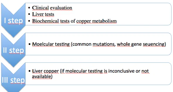
the giant panda’ sign and hyperintensities in the tectal-plate, central pons and the brainstem. Abnormal brain imaging may even be present in some individuals prior to the onset of symptoms.3 Figure 8 summarize diagnostic approach for
25
Table 2: Diagnostic score for Wilson Disease. (Modified from Ferenci et al) Laboratory tests Clinical symptoms and signs
Serum ceruloplasmin Kayser-Fleisher rings
Normal (>20 mg/dl) 0 Present 1
10-20 mg/dl 1 Absent 0
< 10 mg/dl 2
Urinary copper (in absence of acute hepatitis)
Neurologic involvement
Normal (< 40 μg/24h) 0 Severe 2 1-2 x ULN* (40-80 μg/24 h) 1 Mild 1 >2 x ULN* (> 80 μg/24 h) 2 Absent 0 Normal, but > 5 x ULN*
(>200 μg/24 h after penicillamine challenge)
2
Liver copper (in absence of cholestasis)
Coombs negative hemolytic anemia
Normal (<50 μg/g dry weight) -1 Present 1 < 5 x ULN* (50-250 μg/g dry weight) 1 Absent 0 < 5 x ULN* (>250 μg/g dry weight) 2
Rhodanine stain Mutation analysis
Absent 0 2 chromosome mutations 4 Present 1 1 chromosome mutation 1 No mutations detected 0 Assessment of the WD-diagnostic score: 4 or more=affected by WD; 2-3= WD likely, do more investigations; 0-1= WD unlikely; *ULN: upper limit of normal
26
1.7 MOLECULAR ANALYSIS AND SIBILING SCREENING
From a genetic point of view, the diagnosis of WD is based on the identification of two disease causing mutations. It should be performed for individuals in whom the diagnosis is difficult to establish by clinical and biochemical testing or to screen asymptomatic sibling of patient with WD.3 A further indication is in
case of WD-related fulminant hepatitis, when the conventional diagnostic tools are unre- liable. Detection of mutations on both chromosomes allows a definitive diagnosis of WD, whereas the diagnosis cannot be excluded because of their absence.
Sibling screening is mandatory when a new case of WD is diagnosed.30 This should include clinical examination, liver function tests, biochemical tests of copper metabolism and mutational analysis if the proband’s mutations are known. If mutational analysis is not available, biochemical testing should be delayed until after six months of age, because ceruloplasmin levels are low in the newborn. In definitive, despite the availability of multiple tools, diagnosis of WD is very challenging in childhood. The first essential in making the diagnosis is to think of it and an high suspicion index is required.
Newborn and childhood population screening has been considered by various groups. WD does fulfill some criteria for population screening: it is sufficiently common, serious, and treatable. There are 2 problems: first, the uncertain prognosis of the genetically diagnosed neonate, and second, the robustness of available methodology. The low neonatal ceruloplasmin in neonates makes
27 detection of WD very difficult. On the other hand mutation screening would only be effective in populations with a single common mutation.2,30 Such
screening programs pre-suppose knowledge of the prognosis of the genotypically affected individual, which we do not have. Whilst it is appropriate to test the siblings of a child with active disease, it is not appropriate to bestow a diagnosis whose implications we are unsure about. This is a topic which requires further research and pilot studies.
1.8 WILSON DISEASE MIMIC DISORDERS
There are some inherited disorders other than Wilson disease that impair directly or indirectly hepatic copper metabolism and look-alike Wilson disease. These conditions determine disease phenotypes and/or biochemical abnormalities that may mimic Wilson disease, making the appropriate diagnosis extremely challenging.
Congenital disorders of glycosilation (CDG) are inherited metabolic disease caused by abnormalities in proteins glycosilation.
Some CDG subtypes share with Wilson Disease not only liver and brain involvement, but also partial or complete Wilson phenocopy (low serum copper and/or ceruloplasmin, copper accumulation on liver biopsy, increased urinary copper excretion). Liver involvement is present in about 22 % of CDG types. The CDG types with predominant or isolated liver involvement comprise MPI-CDG, CCDC115-MPI-CDG, TMEM199-MPI-CDG, and ATP6AP1. Coagulopathy may
28 occur independently from the severity of liver dysfunction and is caused by abnormal glycosilation of clotting factors. CDG testing (transferrin and apoCIII IEF, CDG panel genetic testing) should be performed in patients diagnosed as affected by Wilson Disease but in whom ATP7b mutations are not found. The main CDG subtypes, that could be misdiagnosed as Wilson Disease, are the TMEM199-CDG, CCDC115-CDG, ATP6AP1-CDG, PMM2-CDG, and COG2-CDG. In particular, for TMEM199-CDG and CDG type IIx a correct differential diagnosis of WD may be challenging because the patients may show only an isolated liver disease in the absence of typical CDG phenotype.31,32
Sometimes further investigations for CDG can be triggered by the presence of coagulopathy not explained by the liver disease. In CDG it has been hypothesized that copper abnormalities are linked to disturbed biosynthesis of the glycoprotein ceruloplasmin, as observed in aceruloplasminemic mice model. MEDNIK syndrome (Mental retardation, Enteropathy, Deafness (sensorineural hearing loss), peripheral Neuropathy, lamellar and erythrodermic Ichtyosis, Keratodermia) is a severe neurocutaneous disorder. It is a genetic disorder, included in the family of adaptinopathies, caused by mutations to the AP1S1 gene, on chromosome 7q22.1. 33,34 It determines an improper function and trafficking of copper ATPases resulting in less retention in the trans-Golgi and excess copper in the plasma membrane. As for disease phenotype some of the neurological, cutaneous and skeletal symptoms are similar, but milder than those of Menkes disease. 33 On the other hand, hepatopathy and brain MRI features are
29 reminiscent of Wilson disease. In particular, liver involvement in this disorder, not clearly considered as main hallmark of disease phenotype, is a common finding in affected patients. They show hepatomegaly, intractable itching, liver steatosis, elevation of transaminases and bile acids. As for copper metabolism parameters, patients show, as Wilson’s disease patients, low levels of ceruloplasmin, increased urinary copper excretion and liver copper content.34 At brain MRI, patients have basal ganglia involvement with bilateral symmetrical hyperintensities in caudate nuclei and putamina at T2-weighted images, a pattern similar but milder than in Wilson’s disease. Differently from Wilson’s disease cerebral atrophy is a common finding. No patients have Kayser-Fleisher ring. Considering similarities to Wilson’s disease, one patient has been treated with zinc acetate that successfully reduces liver copper overload and cholstasis, with clear improvement of the patient’s behaviour.34 Manganese overload could determine a neurologic and liver dysfunction resembling Wilson’s disease. 35 The first inborn error of Mn metabolism has
been identified by Tuschl et al and called hypermanganesemia. It is an autosomal recessive disorder, with a pleomorphic phenotype, characterized by an early-onset generalized dystonia (before 10 years old), polycythemia (with depleted iron stores and low ferritin), characteristic brain MRI findings in the basal ganglia and chronic liver disease (hepatic disfunction may vary among members of the same family).36 It is due to mutations in one of the solute carrier
30 the liver and brain, including in the basal ganglia. Patients with mutations in SLC30A10 have not abnormalities in copper urinary excretion, but high blood levels of manganese (exceed 2000 nmol/L; normal <320 nmol/L). 37,38 Recently, it has been described a Mn/zinc transporter defect caused by mutations in SLC39A14 (ZYP 14), also linked to a progressive childhood-onset parkinsonism dystonia, probably linked to an alterated hepatic uptake (and hence biliary excretion) of manganese, leading to symptoms caused by excessively high manganese levels in the brain.39 Additionally, in a recent paper, Nagappa et al
described an Indian cohort of patients with the so called “non-Wilsonian hepatolenticular degeneration” and a brain MRI that showed hyperintensity on T1 and hyper-hypointensyty on T2, in the “absence of an “acquired” liver disease, assuming a defect of manganese metabolism, also in absence of polycitemia.40 Therefore, as for Wilson's disease, manganese blood levels should be routinely tested alongside copper and ceruloplasmin in the initial diagnostic work-up of patients with young-onset generalized dystonia.
Aceruloplasminemia could be as a link between copper and iron metabolism. Firstly described in 1987 by Miyaima et al, it is an autosomal recessive disorder of iron homeostasis due to loss-of function mutations in the ceruloplasmin gene.
41 Ceruloplasmin determines the rate of iron efflux from cells with
mobilizable iron stores. Affected individuals may present in adulthood with evidence of hepatic iron overload, diabetes, peripheral retinal degeneration, dystonia, dementia or dysarthria. Laboratory studies demonstrate: the triad
31 microcytic anemia, elevated serum ferritin and a complete absence of serum ceruloplasmin ferroxidase activity. Consistent with the observed neurologic findings, MRI reveals iron accumulation within the basal ganglia. Histologic studies detect abundant iron in reticuloendothelial cells of the liver and spleen, in beta cells of the pancreas, and in astrocytes and neurons throughout the central nervous system.41,42
Finally in chronic cholestatic conditions the normal physiological secretion of copper into the bile is blocked leading to lysosomal accumulation of copper, as in late WD. Furthermore, patients with MDR3 deficiency (previously known as progressive familial intrahepatic cholestasis 3 or PFIC 3), primary sclerosing cholangitis (PSC), and primary biliary cholestasis (PBC) have been found to have significantly elevated hepatic copper concentrations and may share some histopathological features with WD.43 Generally, however in these conditions, liver copper content does not reach levels as high as that observed in WD. Occasional patients with MDR3 deficiency have been described as having increased liver copper content and also abnormal urinary copper excretion, and sometimes low serum ceruloplasmin levels.43 Therefore, the diagnosis of MDR3 deficiency may be challenging in the absence of genetic analysis to exclude WD. While some of the histologic findings in MDR3 deficiency (mild chronic hepatitis, steatosis, glycogenated nuclei and Mallory-Denk bodies) are seen in WD, none of them is specific to either disease. Immunohistochemical staining for the MDR3 on bile canalicular membranes is negative in MDR3 deficiency
32 but intact in WD. 44,45 Liver biopsy preceding therapy, both in WD and MDR3 deficiency, can show dense lysosomal deposits due to aggregates of copper and copper binding proteins such as metallothioneins. Moreover, it has been recently found that similar to individuals with WD, patients with bile salt export pump (BSEP) deficiency or MDR3 deficiency can have elevated hepatic copper concentrations sufficient to impair nuclear receptor activity.46
33
1.9TREATMENT&OUTCOME
The overall therapeutic aim for WD is the generation of a negative copper balance. Today this can be achieved either by liver transplantation, which phenotypically corrects the gene defect in the liver, or by medical therapy. Obviously liver transplantation is a treatment option for patients with severe life-threatening conditions in whom the window for medical treatment is not wide enough. It cannot be proposed as a therapeutic strategy, given the high rate of complications and the need for immunosuppressive therapy for life. For all other WD patients, lifelong medical therapy is indicated. Conventional medical therapies for children comprise treatment with either copper chelators (penicillamine or trientine) and zinc (Table 2).2,3 Chelators mobilize intracellular
copper into the circulation and enhance urinary excretion of copper, while zinc acts inducing copper-binding metallothione in both in enterocytes, reducing metal intestinal absorption into portal circulation, and in hepatocytes, reducing the damaging effects of free liver copper.
Today, it has been recently supported that, regardless of the drug used, appropriately treated patients with WD have an excellent long-term prognosis, with a survival probability not differing from the general age and sex-matched population (Figure 4).47 It has been described that more than 30% of WD patients adequately treated since childhood with the available drugs do not exhibit complete normalization of liver enzymes but have a good quality of life
34 and a favourable outcome.47 On the other hand, regardless of type of treatment, it has been described that a poor compliance or discontinuation of medical therapy is associated with high risk of hepatic decompensation requiring even liver transplantation.2,3
American3 and European48 guidelines provide an useful therapeutic support
even if many points remain unclarified for pediatric patients. There is a lack of high quality evidence to compare the relative treatment effects of the available drugs in children.
For pre-symptomatic patients, who include the subjects identified following family screening before the onset of symptoms, the recommended approach is therapy with zinc, considered its proven efficacy and safety profile.49
Initial treatment for patients with significant chronic liver disease (portal hypertension, cirrhosis etc.) should include always a chelating agent.50 Penicillamine and trientine have many beneficial effects but also multiple potential toxicities, which may require discontinuation in up to 30% of cases (Figure 5).2,3 In particular in patients with neurological symptoms chelators should be introduced gradually over time to avoid rapid deterioration of neurological function.3 In contrast to copper chelators, zinc has a lower toxicity
and rarely leads to worsening in neurological symptoms. Its efficacy is contested especially in adult patients with liver disease, when applied for very long periods.50 Pediatric experience is totally different from that concerning adult
35 childhood, zinc monotherapy seems to be effective in controlling WD related liver disease both as first-line and as maintenance treatment, with a low rate of adverse events. 49 In patient presenting with neurologic or psychiatric signs, zinc may have a role as a first line therapy as it is not associated with neurological deterioration.2,3 As maintenance therapy after the induction phase
with chelators, the patients with a good control of the disease may be treated with lower doses of chelators or be shifted to zinc.3
Initial old studies by George Brewer with another drug tetrathiomolybdate, showed a very rapid reduction of free copper levels and significantly less neurological deterioration than with use of a chelator, were a substantial step forward. 51 Tetrathiomolybdate, a strong decoppering agent, recently has been
licensed in Europe. Consisting of a molybdenum molecule surrounded by four sulfhydryl groups, TTM forms a tripartite complex with almost any protein. Tetrathiomolybdate increases the Cu excreted in the stool by binding the metal in food and in gastric juices, and at the same time by slowing the entrance of Cu into tissues and organs that enables the formation of a complex between the metal and albumin in the blood, which is slowly metabolized by the liver and eliminated through the bile. It was succesfully proposed the use of bis-choline tetrathiomolybdate (WTX101), a stable form of tetrathiomolybdate, a for patients with Wilson’s disease who cannot be adequately treated with chelators or zinc. This phase 2 study shows that after 24 weeks of treatment, patients also had a large improvement in neurological symptoms. In this study, the dosage of
36 WTX101 was variable and needed to be individually titrated. Before this drug can be used more generally, more specific guidelines are needed.52
The adherence to a life-long therapeutic regimen may be poor, mainly in the teenage age. Therefore, the patients should be checked periodically to supervise the occurrence of side effects and to assess the adherence to the prescribed regimens.
Table 3: Drugs used in the treatment of children with Wilson Disease in Italy
Drug Dosage and timing Efficacy parameters Penicillamine - 20mg/kg/die divided in 2-4 divided
dosages
- in young adults 1000 mg/die (maximum 1500 mg/die) in 2-4 divided dosages - maintenance dose 10-20mg/kg/die up
to 750-1000 mg/day in 2 divided dosages
- 1 h before or 2-3 hours after meals - Supplemental pyridoxine should be
provided (25-50 mg/day) - Reduce dose during pregnancy - Reduce dose for surgery to promote
wound-healing
Urinary copper: around 1000 μg (16 μmol) after starting
treatment
Urinary copper: 200-500 μg/24 h (3-8 μmol/24 h) on maintenance treatment
Trientine - 20mg/kg/die divided in 2-3 doses - maintenance dose 900-2700 mg/day - 1 h before or 2-3 h after meals - Reduce dose during pregnancy - Reduce dose for surgery to promote
wound-healing
Urinary copper: around 1000 μg (16 μmol) after starting
treatment
Urinary copper: 200-500 μg/24 h (3-8 μmol/24 h) on maintenance treatment
37 Zinc salts - Dosage:
Elemental zinc (acetate)
Age < 6 yrs: 25 mg twice daily
6 – 16 yrs with weight < 50 kg: 25 mg three times daily
Age > 16 yrs or weight > 50 kg: 50 mg three times daily
Zinc sulphate
Age < 6 yrs: 100 mg twice daily
6 – 16 yrs with weight < 50 kg: 100 mg three times daily
Age > 16 yrs or weight > 50 kg: 200 mg three times daily
- 1 h before or 2-3 h after meals - No dosage reduction for surgery and pregnancy Urinary copper: > 75 μg/24 h (1.2 μmol) on maintenance treatment Urinary zinc: >2 mg/24 h on maintenance treatment Serum zinc: > 1250 μg/l on maintenance treatment
1.10 NEW THERAPEUTC STRATEGIES
New therapeutic strategies are based on: correction of dysfunctional ATP7B, gene therapy and cell therapy. 53
Correction of ATP7B is obtained mainly manipulating their translocation from the ER to ensure correct localization in the cell. This could be beneficial for a sizable portion of WD patients affected by mutations resulting in aberrant protein products that frequently exhibit residual Cu-transporting activity but which undergo strong degradation due to misfolding and retention within the endoplasmic reticulum (ER). It is worth noting that most of ATP7B mutations have this property, belonging to missense (58%) or small deletion/insertion (27%) categories.10
Considering these similarities, several labs have tested the potential of ΔF508-cystic fibrosis correctors, such as curcumin and 4-phenylbutyrate (4PBA), for
38 ATP7B mutant rescue. Both correctors were demonstrated as capable of reducing degradation of ATP7B mutants expressed in HEK293 cells 14;
however, curcumin failed to do so in HLCs expressing the R778L variant of ATP7B 11. It remains unclear to what extent these drugs rescue localization and function of ATP7B mutant 14. Curcumin has been reported to correct
localization of ATP7B-R778L and to facilitate Cu efflux from patient-derived HLCs. 54,55
On the other hand, a recent analysis of global gene expression in hepatic HepG2 cell lines expressing either wild type ATP7B or ATP7B-H1069Q mutant revealed that suppression of p38 and JNK reduces retention of H1069Q (Figure 8) and a few other ATP7B mutants in the ER, inhibits their degradation, and facilitates Cu excretion from mutant-expressing cells (Figure 9).56
39 Therefore, p38 and JNK have become attractive targets for exploring approaches to mutant correction. Indeed, it has been recently reported57 that p38 and JNK control a cluster of ER quality control genes that promote ATP7B-H1069Q degradation. This cluster comprises a well-known chaperone, heat shock protein 70 (HSP70), whose silencing protects the ATP7B mutant from degradation and facilitates its export from the ER to the Golgi complex where ATP7B normally works.
Finally the current pharmacological treatments have largely failed in rescue of Cu homeostasis in WD patients with acute liver failure, leaving LT as the only viable option for treatment. A recent study using the lec rat model of WD proffered an extra option for such patients.58 The authors investigated the therapeutic value of methanobactin (MB), a peptide produced by Methylosinus trichosporium which has an extremely high affinity for Cu. Short-term MB
40 treatment efficiently reversed liver damage in the acute stages of liver Cu accumulation, as compared with that seen in untreated ATP7B-deficient rats. This beneficial effect was associated with depletion of Cu from sites of intracellular deposition, in particular from the mitochondria. Interestingly, the regular Cu chelators penicillamine and tetrathiomolybdate failed to clean toxic metal from the mitochondrial stores. As a consequence, MB treatment prevented hepatocyte death and the subsequent liver failure, elongating the life span of the rodent model. These findings suggest that MB has potential as a new therapeutic agent for the treatment of acute WD.
Findings from another recent study have suggested that liver X receptor (LXR)/retinoid X receptor agonist may be used to combat Cu toxicity in WD.59
This approach does not require Cu chelation. Careful investigation of the transcriptional and metabolic changes in samples from WD patients and Atp7b-/- mice revealed dysregulation of LXR as one of the key events in the pathogenesis of WD. Treatment with the LXR agonist T0901317 improved disease manifestations in the Atp7b-/- mice despite substantial Cu overload. Moreover, liver fibrosis and inflammation significantly decreased in LXR agonist-treated animals, while lipid profiles normalized and liver function and histology improved. Thus, the potential of T0901317 for WD cure is likely to be further explored.
Cell therapy, as well as gene therapy, targets the liver, which does not express functional ATP7B protein. The fundamental purpose of cell/gene therapy in WD
41 is to restore ATP7B-mediated hepatobiliary Cu excretion.59 Cell therapy in WD seems feasible because transplanted hepatocytes can integrate in liver parenchyma and restore deficient functions, including the transport of Cu into bile. The availability of authentic animal models that recapitulate hepatic WD, especially the LEC rat, has advanced cell transplantation research in WD. It was through this animal model that it was found necessary to repopulate at least 30%–40% of the liver with healthy hepatocytes in order to achieve sufficient Cu clearance and therapeutic benefits. Extrahepatic cell therapy using engineering applications (i.e. transplantation of liver tissue into small intestine or the abdominal cavity) is currently considered insufficient because Cu removal requires an intact bile excretion system. 60 Thus, in the case of either cell or gene
therapy in WD, the rationale for targeting the liver first and foremost is based on the physiological restriction of ATP7B expression to hepatocytes as well as the availability of mechanisms required for the transfer of Cu ions by ATP7B into the bile canaliculus, the entry of Cu into bile ducts and intestines, and the elimination of Cu from the body.61 Fortunately, transplantation studies using donor cells have confirmed the ability for biliary excretion, providing the first clue that biliary Cu transport is a feasible target for cell therapy in WD.62
IPSC-derived HLCs can repopulate the liver but they support only some hepatocyte functions, which are restricted to fetal-like stages.11 Therefore, transplanted cells can proliferate in the presence of native cells, having a low rate of proliferation. This happens when native liver cells have extensive DNA
42 damage (as induced by toxins, ischemia and/or hepatectomy). As such, the nature of preconditioning regimens suitable for clinical applications in people will require substantial additional intervention to address chemical exposure (e.g. retorsine) or ablative procedures (e.g. partial hepatectomy), will be undesirable in the setting of existing liver injury in WD. The ability of transplanted HLCs to express ATP7B was evaluated in liver of LEC mice, in which the transplanted cells did not increase in number over an observational period of several months.63 Therefore, despite adequate engraftment of
transplanted cells into liver, the gradual onset of liver repopulation over a very long period implies that therapeutic correction in WD will require significant time.60
Gene therapy aims to correct the defect in native hepatocytes by providing healthy copies of ATP7B via introduction of a transgene by vectors capable of indefinitely integrating and/or persisting in cells. The proof-of-principle for gene therapy in WD came from adenoviral and lentiviral vector expression of ATP7B in the liver of LEC rats and murine models of WD, which achieved transient correction of Cu excretion and incorporation of Cu into ceruloplasmin.64 Recently, these initial observations were confirmed with an adeno-associated vector serotype 8 (AAV8) encoding the human ATP7B cDNA placed under control of the liver-specific α1-antitripsin promoter (AAV8-AAT-ATP7B). Expression of AAV8-AAT-ATP7B in ATP7B-deficient mice resulted in reductions of serum transaminases and urinary Cu excretion, normalization of
43 serum ceruloplasmin levels, and recovery of physiological biliary Cu excretion.64 This study showed a potential of AAV8-AAT-ATP7B-mediated
gene therapy in a clinically relevant animal model of WD, providing a solid background for future translational studies. AAV-mediated gene therapy should allow ATP7B to be permanently expressed in WD patient liver and, hence, would eliminate a need for lifelong intake of Cu-reducing drugs. On the other hand, such risks as immune response, tumor biogenesis and poor integration into damaged cellshave to be weighted before AAV-mediated gene transfer therapy
65and will be a critical component of the repertoire of WD treatments.
Furthermore, studies investigating the fate of genetically modified native cells within the liver in WD are still required. It has yet to be determined whether targeting will be effective for subsets of undamaged native cells or putative endogenous stem/progenitor cells that have not been affected by Cu-induced damage.60
44
CHAPTER 2
AIMS OF RESEARCHPROJECTAs we previously descuss, while available drugs allow for sufficient control of the symptoms, they do not cure WD. Thus, there remains a clear need for novel treatment strategies aimed at curing WD through correction of the cellular defect.
Aims of our study was:
1. To create a new in vitro WD platform derived from patients cells to explore pathogenic mechanism linked to H1069Q-ATP7B mutants
2. To found new active compounds using the hypothesis driven strategy 3. To found new active compounds using the the high- throughput screening 4. From bench to bedside: to develop a clinical trial
45
CHAPTER 3
A NEW IN VITRO WD PLATFORM DERIVED FROM PATIENTS CELLS TO EXPLORE PATHOGENIC MECHANISM LINKED TO H069Q-ATP7B MUTANTS
The recent development of stem cell-derived hepatocyte-like cells (HLCs) opens new avenues for gene- and/or drug-based therapy to treat liver diseases. This new “in vitro model” better mimics patient cell biology. HLCs can be fairly easily derived from induced pluripotent stem cells (iPSCs) that are generated through the reprogramming of human fibroblasts. iPSCs can be indefinitely expanded in vitro and differentiated in all cells types derived from the three germ layers. Thus, iPSC technology features the potential benefits of embryonic stem cells while addressing their major ethical and scientific concerns: embryo destruction and immune-incompatibility. In this manner, human iPSCs represent an unlimited source of human hepatocytes for translational research and hold great promise for drug screening and liver disease modeling in particular.
In this context, it seems particularly helpful to obtain HLCs from WD patients. It is important to underline that in addition to their HLC differentiation capacity, iPSCs can differentiate into neurons, providing a serious advantage in studying whether and how certain ATP7B mutations contribute to neurologic dysfunction in WD patients. But in this project we don’t consider neurons differentiation.
46 GENERATION, CHARACTERIZATION AND HEPATIC DIFFERENTIATION OF WD AND CONTROL IPSCs
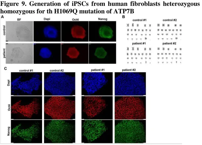
To generate iPSCs we obtained primary fibroblasts from skin biopsies of two WD patient carrying the H1069Q mutation (H1069Q/H1069Q, patient # 1 and # 2) and his mother and brother (respectively) as a control (WT/H1069Q) (henceforth referred to as patient and control). iPSCs were generated using an integration-free method by transfecting episomal plasmid vectors into primary fibroblasts, thus avoiding genetic integration in the reprogrammed cells. iPSC-like colonies emerged three weeks after transfection and their stemness was assessed by immunofluorescence for Nanog and Oct4 positivity
Figure 9. Generation of iPSCs from human fibroblasts heterozygous and homozygous for th H1069Q mutation of ATP7B
Two clones of each WD and control iPSCs were subjected to cytological analysis and showed a normal karyotype (Fig. 9B). These clones were adapted
47 to grow in feeder-free conditions to avoid feeder contamination during differentiation and then analyzed for the maintenance of stemness after multiple passages (≥15). The expression of pluripotent stem cell marker, Oct4 and Nanog, was assessed by immunofluorescence microscopy (Fig. 9C).
Next, we differentiated patient and control iPSCs into HLCs using a previously established protocol that mimics three developmental stages of liver development: definitive endoderm, hepatic progenitors and hepatocyte-like cells. The expression of markers corresponding to the three phases was verified by immunofluorescence microscopy and quantitative real-time PCR.
ATP7B EXPRESSION IN HLCS FROM PATIENT AND CONTROL iPSCs
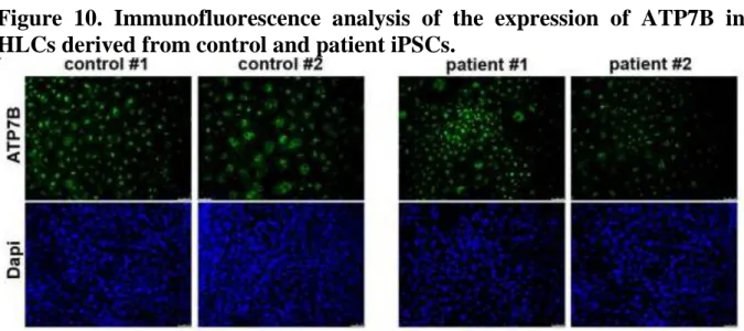
We analyzed the expression of ATP7B in HLCs derived from patient and control iPSCs in all differentiation experiments by immunofluorescence and found that both in control and patient HLCs 30–40% of the total cell population was positive for the protein (Figure 10).
Figure 10. Immunofluorescence analysis of the expression of ATP7B in HLCs derived from control and patient iPSCs.
48 LOCALIZATION OF ATP7B
To assess the localization of ATP7B in patient and control HLCs, we first used confocal immunofluorescence analysis. In HLCs from the patient significant amounts of ATP7B-H1069Q also were found in the Golgi and at moderate levels in the ER (Fig. 11). This was surprising considering that when the ATP7B-H1069Q mutant is expressed in heterologous as well as in hepatic cells it resides almost exclusively in the ER, while only very small amounts of this mutant protein can be found in the distal compartments of biosynthetic pathway.
Figure 11. Confocal immunofluorescence analysis of HLCs immunolabeled for ATP7B and Golgin 97 (upper panels) or KDEL containing proteins (lower panels) as markers of Golgi complex and ER, respectively
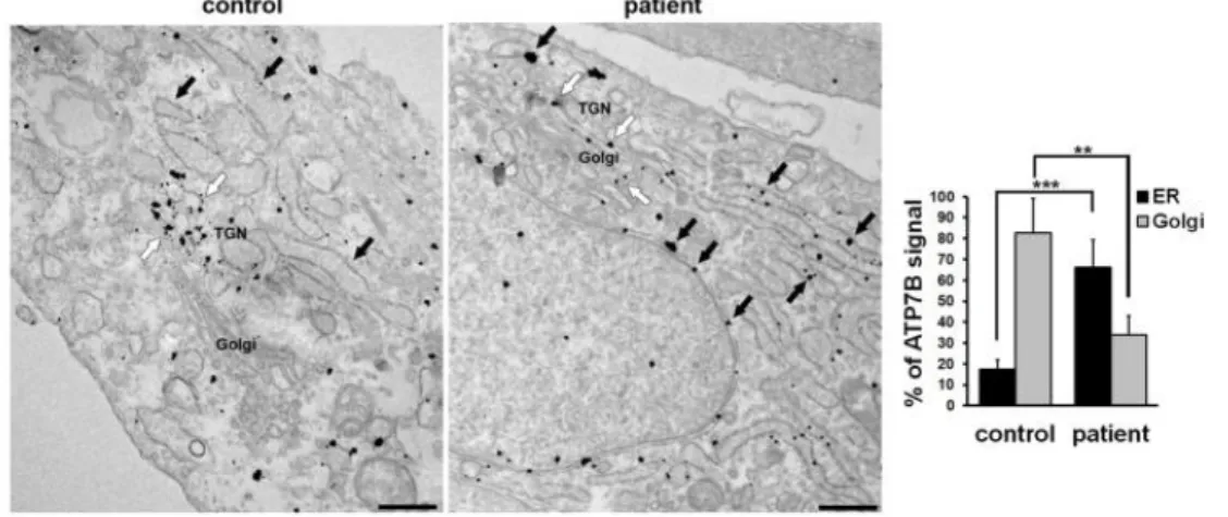
49 The subcellular distribution of ATP7B was therefore further investigated with immuno-gold electron microscopy (EM). As expected, in control HLCs ATP7B was preferentially localized along the membranes of the TGN, at the trans-side of the Golgi stack (Fig. 12, white arrows). On the other hand, patient HLCs showed that both cisternae of the ER (Fig. 12 black arrows) and the Golgi membranes (Fig. 12, white arrows) contained mutant ATP7B protein. The graph in Fig. 12 shows that 35% of gold particles representing ATP7B signal were found in the Golgi and 65% in the ER. These results indicate that trafficking of ATP7B-H1069Q from the ER to the Golgi is hampered, but that the mutant maintains about 50% of the ability of the wt protein in reaching the Golgi complex.
Figure 12. Confocal immunofluorescence analysis of HLCs immunolabeled for ATP7B and Golgin 97 (upper panels) or KDEL containing proteins (lower panels) as markers of Golgi complex and ER, respectively
50 DEGRADATION RATE OF ATP7B
HLCs derived from patient iPSCs showed lower expression of ATP7B-H1069Q (20% of control, graph in Fig. 13A). Furthermore, when HLCs obtained from control and patient iPSCs were incubated in the presence of the protein synthesis inhibitor cycloheximide, a clear reduction of ATP7B-H1069Q level was observed after 4 hours in patient HLCs, while no decrease was seen in the control (Fig. 13B). These results, taken together, strongly suggest that endogenously expressed ATP7B-H1069Q accumulates at lower level in patient HLCs because undergoes a faster degradation than the wt protein.
Figure 13. Relative accumulation and degradation of ATP7B in HLCs obtained from control and patient iPSCs
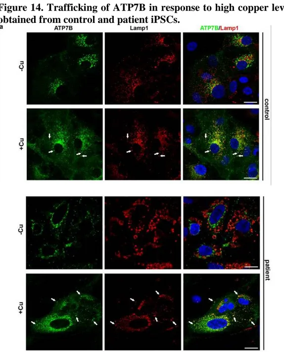
51 ATP7B TRAIFFIC IN RESPONSE TO COPPER
The ability of ATP7B to regulate copper levels heavily relies on copper-dependent trafficking of the protein. In hepatic cells, elevated copper induces redistribution of ATP7B from the TGN to the endo/lysosomal compartments, where the transporter supports transient sequestration of excess copper into the organelle lumen. Mutations that disrupt copper-dependent trafficking of ATP7B prevent elimination of toxic copper from the cytosol. Therefore, to characterize the sensitivity of endogenous ATP7B-H1069Q to copper, we exposed both control and patient HLCs to CuSO4 for 2 h. As expected, confocal
immunofluorescence analysis showed that in control cells copper triggers a redistribution of ATP7B from the Golgi to peripheral structures containing the endo/lysosomal marker LAMP1 (Fig. 14, arrows). Interestingly, patient HLCs exhibited identical relocation of the Golgi ATP7B pool towards endo/lysosomal compartments (Fig. 14, arrows). In summary, our observations suggest that ATP7B-H1069Q isogenically expressed in patient HLCs is functional in terms of copper-dependent trafficking.
52
Figure 14. Trafficking of ATP7B in response to high copper level in HLCs obtained from control and patient iPSCs.
53
CHAPTER 4
TO SEARCH NEW THERAPEUTIC OPTIONS USING MODERN STRATEGY FOR DRUG DISCOVERING: THE HYPOTHESIS DRIVEN STRATEGY
Hypothesis 1
In WD both serum lipoprotein alterations and fatty liver are common and share many pathophysiological mechanisms with lysosomal lipase activity deficiency (LAL-D). The earliest characteristic alterations of the liver pathology in WD include steatosis, which is sometimes indistinguishable from fatty liver due to metabolic syndrome. Recently it has been supposed that a genetic polymorphism in rs738409, in the patatin-like phospholipase domain (PNPLA3), may have a role in steatosis in WD patients. We could suppose that, as PNPLA3 polymorphism, also LAL deficiency may be a contributing factor that influences progression of WD in a subgroup of patients.
Study design 1
A) To understand if lipid metabolism is compromised in Wilson disease, we employed ATP7B knockout (ATP7B-KO) HepG2 cell line. ATP7B-KO and control HepG2 cells were incubated with Cu for 24h and their transcriptional responses to the metal were analyzed using QuantSeq 3’ mRNA sequencing, which allows very precise measurements of quantitative difference in gene involved in lipid metabolism expression.