UNIVERSITA’ DELLA CALABRIA
Dipartimento di Farmacia e Scienze della Salute e della Nutrizione
Dottorato di Ricerca in
“Biochimica Cellulare ed Attività dei Farmaci in Oncologia”
CICLO XXVIIPhD THESIS
Study of the expression of a Small Leucin-Rich Proteoglycan,
Asporin, in normal human osteoblasts and regulation by breast
cancer cells
Settore Scientifico Disciplinare
(SSD: MED/46 SCIENZE TECNICHE DI MEDICINA DI LABORATORIO)
Internship made at Metastases Research Laboratory, University of Liege, Belgium Director: Professor Vincent Castronovo Supervisor: Professor Akeila Bellahcène
Coordinatore Supervisore/Tutor esterno Prof. Diego Sisci Professoressa Akeila Bellahcène
Supervisore/Tutor interno Professoressa Marilena Lanzino
Dottoranda: Dottoressa Giovanna Elvi Trombino
Scientific Publications:
1. Sisci D, Maris P, Cesario MG, Anselmo W, Coroniti R, Trombino GE, Romeo F, Ferraro A, Lanzino M, Aquila S, Maggiolini M, Mauro L, Morelli C, Andò S.
The estrogen receptor α is the key regulator of the bifunctional role of FoxO3a transcription factor in breast cancer motility and invasiveness. Cell Cycle. 2013
Sep 17;12(21).
2. Ortensia Ilaria Parisi, Catia Morelli, Francesco Puoci, Carmela Saturnino,Anna Caruso,Diego Sisci, Giovanna Elvi Trombino, Nevio Picci and Maria Stefania Sinicropi. Magnetic molecularly imprinted polymers (MMIPs) for carbazole
derivative release in targeted cancer therapy. Journal of Materials Chemistry B.
2014 August 7; 12:23:33
3. Pamela Maris, Arnaud Blomme, Ana Perez Palacios, Akeila Bellahcène, Elettra,Giovanna Elvi Trombino, Sylvie Maweja ,Eric Lifrange, Philippe Delvenne ,Guy Jerusalem, Vincent Castronovo ,Andrei Turtoi. Asporin is a
fibroblast-derived TGF-ß inhibitor and a tumor suppressor associated with good prognosis in breast cancer. Submitted in PLOS MEDICINE.
Ai miei Nonni,
Un immenso GRAZIE da chi porterà sempre nel cuore
il vostro insegnamento e l’amore verso la vita……..
E’ veramente bello battersi con persuasione, abbracciare la
vita e vivere con passione. Perdere con classe e vincere
osando…..perchè il mondo appartiene a chi osa! La vita è
troppo bella per essere insignificante.
Study of the expression of a Small
Leucin-Rich Proteoglycan, Asporin,
in normal human osteoblasts and
regulation by breast cancer cells.
INDEX
I.
SUMMARY ………1
II.
INTRODUCTION
II.1 Breast cancer ………. 3
II.2 Staging of Breast Cancer
II.3 Breast cancer heterogeneity: MCF7 and MDAMB231 cell lines
II.4 Breast Cancer Progression and Bone Metastasis Development
II.5 Bone tissue ………..7
II.5.1 Osteoclasts.
II.5.2 Osteoblasts.
II.6 Osteoblast differentiation.
II.7 Regulation of osteoblast differentiation and bone metabolism.
II.7.1 TGFbeta signaling pathway.
II.7.2 Wnt signaling pathway.
II.7.3 The cyclic AMP signaling pathway.
II.8 Mineralization process
II.9 Sclerostin: an inhibitor of osteoblast differentiation.
II.10 Asporin: a member of SLRPs family …………..………17
II.10.1 The role of Asporin in TGFbeta pathway and osteoblast
differentiation.
III. MATERIALS AND METHODS ……….20
IV. RESULTS ………..23
V. DISCUSSION ……….38
LIST OF ABBREVIATIONS
ALP = alkaline phosphatase ASPN = asporin
BMP = bone morphogenetic protein BSP = bone sialoprotein
CM = conditioned medium DLX-5 = distal less homeobox-5 DMP-1 = dentin matrix protein-1 ECM = extracellular matrix ER = estrogen receptor
GSK3β = glicogen sintase kinase 3β HER2 = human epidermal growth factor IL-11 = interleuchin 11
MEPE = matrix extracellular phosphoglycoprotein MSX-2 = msh homeobox homologue-2 MV = matrix vesicles OB = osteoblasts OC = osteocalcin OPG = osteoprotegerin OPN = osteopontin PGE2 = prostaglandin E2 PgR = progesteron receptor
RANK/RANKL = receptor activator of nuclear factor Kappa-B/ligand RUNX2 = runt-related transcription factor 2
SLRPs = small leucin rich proteins SOST = sclerostin
SP7 = osterix
TGF-β = transforming growth factor β TNF = tumor necrosis factor
1
I. SUMMARY
Introduction:
Asporin (ASPN) is an extracellular matrix protein that belongs to the Small Leucine Rich Repeat proteoglycan (SLRP) family. Asporin is abundantly expressed in the articular cartilage of individuals with osteoarthritis. In the context of osteoarthritis, several studies have shown that asporin regulates cartilage matrix gene expression and cartilage formation by modulating the transforming growth factor-β (TGF- β) signaling pathway. Asporin directly binds to TGF‐β and inhibits TGF-β-mediated expression of cartilage matrix genes. Previous studies in our laboratory, showed that Asporin inhibits TGF- β-1-mediated SMAD2 phosphorylation in breast cancer cells as well as migration and epithelial to mesenchymal transition in A549 human lung cancer cells. The present study was undertaken to investigate whether asporin secretion could indirectly mediate the ability of metastatic breast cancer cells to regulate osteoblastic differentiation. The Wnt antagonist sclerostin (SOST) is a potent inhibitor of bone formation. We considered the possibility that the balance between ASPN and SOST present in the ECM may create a specific environment favorable to aggressive breast cancer cell growth.
Results:
Breast cancer cells do not produce ASPN themselves but they regulate its expression in osteoblasts. Normal human osteoblasts have been cultured in presence of MCF7 and MDA-MB-231 serum-free conditioned medium. Immunoblot analysis and real time PCR, revealed a significant increase in ASPN expression and secretion in osteoblasts treated with MCF7-conditioned medium, while the opposite effect was observed with
2
MDA-MB-231-conditioned medium. We investigated the role of MCF7 and MDAMB231 conditioned media in osteoblast differentiation and mineralization through alkaline phospatase and Runx2 expression. Our results showed the ability of MCF7 conditioned medium to induce the osteoblast differentiation and mineralization compared to the MDA-MB-231 conditioned medium treatment. Osteoblasts treated with MCF7 conditioned medium and challenged with recombinant SOST showed a significant reduction in their differentiation potential through the decrease of ASPN expression.
Conclusion:
Contrarily to non-metastatic MCF-7 breast cancer cells, MDA-MB-231 metastatic breast cancer cells inhibited the secretion of ASPN by osteoblasts through the overexpression of SOST. The result is the reduction of osteoblast differentiation and mineralization that can create a specific environment favorable to aggressive breast cancer cell growth.
3
II. Introduction
II.1 Breast cancer
Breast cancer is the most common cancer among women and its estimated incidence represents 23% among all types of cancer. More than half of the reported cases occur in industrialized countries (about 361,000 cases in Europe and 230,000 in North America, In part, the high incidence in more developed regions of the world is due to the availability of screening programs that allow to more easily diagnose invasive cancers. Population-based cancer screening is a much more complex public health undertaking than early diagnosis and is usually cost-effective when done in the context of high-standard programs that target all the population at risk in a given geographical area with high specific cancer burden, with everyone who takes part being offered the same level of screening, diagnosis and treatment services. Incidence rates vary greatly worldwide from 19.3 per 100,000 women in Eastern Africa to 89.7 per 100,000 women in Western Europe. In most of the developing regions the incidence rates are below 40 per 100,000 (GLOBOCAN 2008). In Italy, it is estimated that approximately 300,000 new cases each year with a distribution of the incidence among the Italian regions that revealed a marked north-south gradient, higher in the North than in the rest of Italy, like other cancers linked to the styles of Western life. The frequency of cancer according to age shows a gradual increase up to 50 years, or age at menopause; from 60 to 65 there is a lull, while after age 65, the incidence increases again with age.
II.2 Staging of breast cancer
The staging of breast cancer is important in planning the therapeutic strategy. The TNM system is universally accepted to estimate prognosis, to define the best treatment and evaluate the results. It allows a description of the extension of the neoplastic disease at a given time, using three parameters:
• The extension of the primary tumor (T factor); • The extent of lymph node involvement (N); • Distant metastases (M-factor).
4
The combination of the three elements allows us to assign a single tumor at a specific stage with a defined prognosis and treatment. The stage 0 defines the non-invasive carcinoma or carcinoma in situ. Stages I and II are attributed to early forms of breast cancer, but potentially invasive. Stage I cancer cells have not spread beyond the breast and the tumor has a diameter not exceeding 2cm. Stage II may include any of the following cases: breast cancer has about 2cm in diameter and has spread to the axillary lymph nodes; the tumor size from 2 to 5 cm in diameter with or without invasion of the lymph nodes; or the tumor is more than 5 cm but the lymph nodes are not invaded. Stage III defines the metastatic breast cancer: the malignant tumor has spread from the breast to other organs. Invasive carcinomas of the breast initially spread via lymphatic vessels, first giving metastases to axillary lymph nodes, with different frequencies depending on the area of the breast in which they occured. The neoplastic cells initially spread to the lymph nodes of the armpit, grow and disseminate to the other parts of the body (Figure 1). The organs most frequently affected by metastasis are the bones, lungs and liver.
Figure 1. Stages of breast cancer progression from early to invasive lesion.
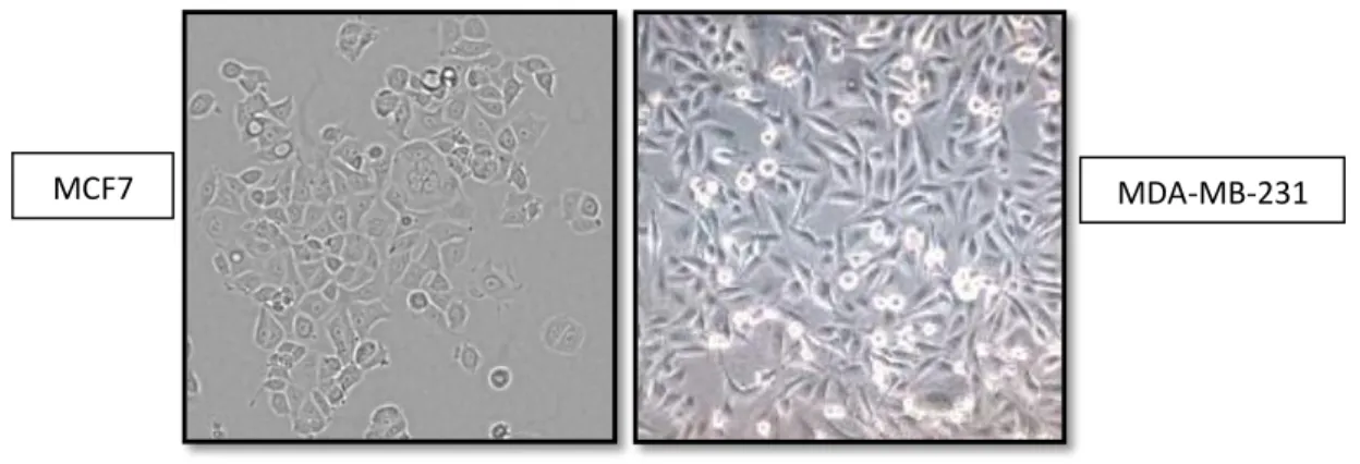
II.3 Breast cancer heterogeneity : MCF7 and MDA-MB-231 cell lines
Breast cancers show extensive heterogeneity despite their common tissue of origin. Breast tumors are categorized into three main groups based on markers that reflect available treatment options: estrogen receptor (ER) or progesterone receptor positive, erbB2 amplified (HER2 positive) with and without ER/progesterone receptor, and triple negative breast cancer (TNBC) defined by the absence of ER/progesterone receptor expression and HER2 amplification. MCF7 and MDA-MB-231 are human cell lines5
derived from adenocarcinomas, cancers of the breast epithelium tissue that originated in the mammary gland. The MCF7 breast cancer line was derived from an in situ carcinoma, meaning that the cancerous cells had not yet invaded surrounding tissues. It is hormone responsive in the sense that it expresses estrogen and progesterone receptors. Estrogen is a known tumor promoter. Mammary gland epithelium expresses estrogen receptor and the presence of estrogen stimulates the non-specific proliferation of cells while opposite effects are shown by blocking the estrogen receptor. On the other hand, the MDA-MB-231 breast cancer line is derived from the pleural effusion of a metastatic breast cancer patient (Figure 2). MDA-MB-231 are triple-negative breast cancer, so called because it lacks expression of the estrogen receptor, progesterone receptor, and HER2, is often, but not always, a basal-like breast cancer. [1]. Relatively to MCF7 cells, MDA-MB-231 cells grow faster and are more resistant to drug therapies
in vitro.
Figure 2. Representative pictures of MCF7 and MDA-MB-231 human breast cancer cells in in vitro
culture.
II.4 Breast cancer progression and bone metastasis development
Bone is the most common site for distant spread of breast cancer. Once the cancer cells have entered the lymphatic system, it can and usually spread to other areas of the body after completing a complex cascade of events to form secondary tumors called metastases (Figure 3). Bone metastasis occurs when cancer cells spread from their original site to bone. Nearly all types of cancer can metastasize to the bones. However, some types of cancer are particularly likely to spread to the skeleton, such as breast cancer and prostate cancer, and are called osteotropic cancers. Bone metastasis can occur in any bone but more commonly occurs in the pelvis and spine. Eighty percent of
6
bone metastases in breast cancer are osteolytic, although the two components, osteolytic and osteoblastic , can coexist.
Figure 3. General mechanism of tumor metastasis to the bone. Multiple steps involved in tumor cells
metastasis from a primary site to the skeleton. Each of these steps represents a potential therapeutic target for the development of drugs designed to prevent or treat bone metastases. (Guise and Mundy, Cancer
and Bone. Endocrine Reviews, 1998).
The presence of osteolytic areas is associated with increased risk of skeletal related events (SREs), defined as pathological fractures, spinal cord compression, bone pain requiring palliative radiotherapy and/or orthopedic surgery. Breast cancer patients with estrogen receptor (ER) positive develop bone metastases more frequently than those with negative hormone receptors and have a generally longer survival. A Canadian study evaluated the characteristics of metastatic disease in 180 patients from breast cancer triple-negative (lack of expression of ER, of the receptor for progesterone (PgR) and Human Epidermal growth factor Receptor 2 (HER2 /neu) -negative) compared to other subtypes of breast cancer (N = 1428) [2]. The risk of developing bone metastases within 10 years after the diagnosis was 7-9% for all subgroups. Bone metastasis may be the first sign of breast cancer but it may also occur several years after treatment. Available treatments such as Bisphosphonates can help to reduce pain and the other negative effects associated with bone metastatic lesions.
The bone microenvironment is unique and provides a special milieu that metastatic cancer cells can colonize. The mineralized bone matrix is embedded with abundant growth factors and cytokines during the bone formation phase, such as transforming growth factor-β (TGF-β), activins and insulin-like growth factors, which are released
7
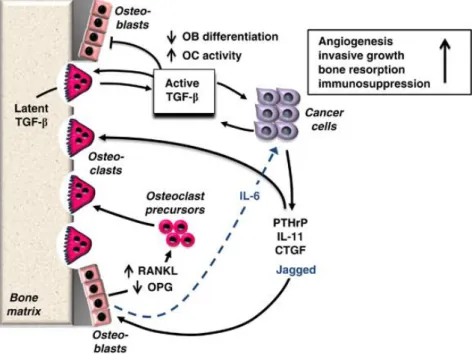
and activated upon tumor-induced osteoclastic bone resorption. High local levels of active TGF-β cause increased invasion, chemotaxis, angiogenesis and immunomodulation. In addition, TGF-β stimulates tumor production of osteolytic factors that further stimulate bone resorption [3-4]. As such TGF-β is a crucial factor responsible for driving the feed-forward vicious cycle of tumor growth in bone (Figure 4). Therefore blocking TGF-β release, its production and/or signaling is a promising strategy to treat bone metastasis.
Figure 4. The skeleton is a preferred site for cancer metastasis. In skeletal metastases, TGF-β is
released by osteoclasts from the bone matrix and acts on cancer cells to stimulate the production of osteolytic factors, such as PTHrP, connective tissue growth factor (CTGF) and IL-6 and -11. These factors increase the RANKL/OPG expression ratio in bone stromal cells such as osteoblasts, resulting in osteoclastogenesis. (BoneKEy Reports 2012).
TGF-β itself has also direct effects on bone cells by stimulating osteoclast activity and inhibiting osteoblast differentiation.
II.5 Bone tissue
Bone is a living organ that serves both mechanical and physiological functions. The adult human skeleton is composed of 80% cortical bone and 20% trabecular bone [5]. Bone is a crucial tissue that provides internal skeletal support for every organ as well as forming and structuring the entire human frame. In addition, bone is the home for the formation of haematopoietic cells and the regulation of blood calcium. Due to its
8
importance in the human body, bone needs to be continuously replenished in order to maintain its strength and structural integrity. This replenishment, also known as bone remodeling, is controlled by two equal but opposing forces: bone formation by osteoblasts and bone destruction or resorption by osteoclasts. Intimate communication between these cells is an integral element in maintaining bone homeostasis. The bone tissue is populated by four different cell types: osteoclasts, osteoblasts, osteocytes and bone lining cells (Figure 5).
Figure 5. Illustration of osteoblasts, osteoclasts and osteocytes in bone tissue.
II.5.1 Osteoclasts
Osteoclasts are multinucleated cells derived form haematopoietic progenitors and responsible for degradation of bone and are differentiated from hemopoietic cells of the monocyte/macrophage lineage (Figure 5). During bone resorption osteoclasts attach to the bone surface isolating the area to be degraded. The acidification of this area is responsible for dissolving the inorganic ECM part while the subsequent action of different proteases guarantees the disassembling of the organic matrix [5]. RANKL and macrophage CSF (M-CSF) are two cytokines that are critical for osteoclast formation. Both RANKL and M-CSF are produced mainly by marrow stromal cells and osteoblasts in membrane-bound and soluble forms. RANKL belongs to the TNF superfamily and is critical for osteoclast formation. M-CSF is required for the proliferation, survival, and differentiation of osteoclast precursors, as well as osteoclast survival and cytoskeletal rearrangement required for bone resorption.
9
II.5.2 Osteoblasts
Osteoblasts are specialised stromal cells that are exclusively responsible for the formation, deposition and mineralisation of bone tissue (Figure 5). Specifically, the mature osteoblasts synthesise and deposit calcium phosphate crystals, mainly hydroxyapatite, and various constituents of extracellular matrix, such as type 1 collagen and proteoglycans, osteocalcin, osteonectin, osteopontin, bone sialoprotein [6]. The formation of hydroxyapatite crystals in osteoid is also regulated by osteoblasts. Therefore, the expression of a number of bone-related extracellular matrix proteins, high enzyme activity of alkaline phosphatase (ALP), and responses to osteotropic hormones and cytokines are believed to be major characteristics of osteoblasts. Pre-osteoblastic cells share the same alkaline phosphatase, collagenase 3 and osteopontin markers as osteoblasts, although the expression of these markers is usually much higher in mature osteoblasts [7] but they do not have the capacity to form bone matrix and have limited capability of dividing. Mature osteoblasts possess several unique biochemical markers and growth receptors including osteocalcin, bone sialoprotein, parathyroid hormone (PTH) receptors, insulin-like growth factor, integrins and cytokines. At the end of the bone-forming phase, the majority of osteoblasts enter apoptosis. The remainder either survives entrapped in the mineralized ECM as terminally differentiated osteoblasts, now designated osteocytes, or covering the resting bone surface as bone lining cells [8].
II.6 Osteoblast differentiation
A large number of signaling pathways have been implicated in the control of bone formation by the osteoblasts. The osteoblast differentiation process is controlled by osteoblast specific transcription factors, runt-related transcription factor 2 (RUNX2)
[10-11] and osterix (SP7) [12], in coordination with other transcription mediators,
including distal-less homeobox-5 (DLX5) and msh homeobox homologue-2 (MSX2)
[13]. Osteoblast development begins with the proliferation and commitment of MSCs
residing in the bone marrow and periosteum [14] into osteoprogenitor cells. Little is known about osteoprogenitor cells identity and what regulates their cellular fate. In the subsequent phase of development the osteoprogenitor cells become pre-osteoblasts and start the ECM synthesis expressing first collagen type I a1 and bone sialoprotein. The pre-osteoblasts differentiate further into mature osteoblasts with increasing levels of
10
alkaline phosphatase (ALP) activity. At this point the ECM produced by the osteoblasts is mature and mineralization is soon initiated within specialized vesicles. In the final stages of osteoblasts differentiation only a fraction of mature osteoblasts survive, as osteocytes or as bone lining cells (Figure 6). The latter are usually localized at the interface of bone with bone marrow but their exact function is not known [15]. Entombed in the mineralized ECM, osteocytes represent the most abundant cellular component in mammalian bone, representing up to 95% of the total cells. Osteocytes, which are by far the most numerous of the bone cells, are derived from osteoblasts that are buried by the growing bone. Osteocytes are physically smaller in size and as a consequence have smaller organelles than osteoblasts. This means that osteocytes usually have significantly lower bone matrix protein secretion, lower metabolic and alkaline phosphatase activity and generally do not divide, unlike osteoblasts which may divide further [9]. The osteocytes are not able to form or resorb bone, but they do appear to play a critical role in bone remodeling by modulating the activity of the osteoblasts and osteoclasts. Osteocytes may thus play a key role in bone remodeling by regulating the balance between bone formation and resorption by the osteoblasts and osteoclasts, respectively. They are characterized by their long cytoplasmatic processes that keep them in communication with the surrounding cells and the expression of a new set of genes, including dentin matrix protein-1 (DMP-1) and matrix extracellular phosphoglycoprotein (MEPE) [16-17]. The nature of the osteocyte signals that are used to communicate with the other bone cells is uncertain. Given the apparent importance of the gap junctions, it is conceivable that small messengers such as Ca2+ or cyclic AMP might be passed directly to the osteoblasts and osteoclasts. Alternatively, the osteocyte might release factors such as prostaglandin E2 (PGE2), ATP or nitric oxide (NO). The osteocytes respond to parathyroid hormone (PTH) by decreasing the release of sclerostin (SOST) that acts as an inhibitor of the Wnt pathway [18]. In terms of function, osteocytes have long been known by the mechano-sensing properties, translating mechanical strain into biochemical signals of resorption or formation. Signals for resorption were recently highlighted in osteocytes with the expression of high levels of receptor activator of NF-kB ligand (RANKL) an important osteoclastogenic cytokine [19].
11
Figure 6. Osteoblast differentiation. Osteoblasts originate from mesenchymal stem cells in a sequential
order of events governed by different transcription factors, cytokines, growth factors and ECM proteins. The different stages of osteoblast differentiation are accompanied by the expression of specific genes/proteins and matrix vesicles (in green). See text for details.
II.7 Regulation of osteoblast differentiation and bone metabolism
Osteoblast differentiation is controlled by morphogens, growth factors, cytokines and hormones all contributing to an intricate network of signaling cascades, from autocrine/paracrine to endocrine and bone is exposed to these factors from circulation. For the sake of clarity, we will next develop the two main signaling pathways implicated in the control of osteoblastic differentiation.
II.7.1 TGF-β signaling pathway
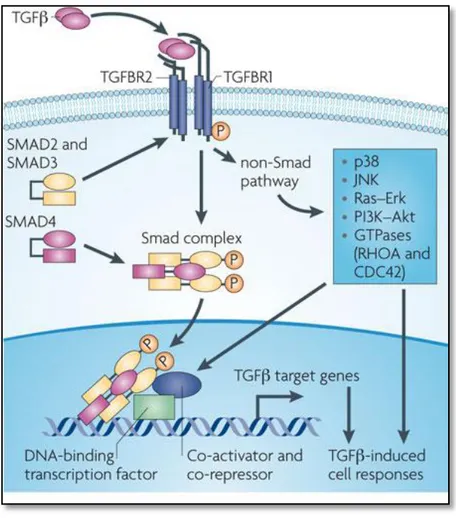
The transforming growth factor beta (TGF-β) pathway includes TGF-β superfamily members, including TGF-β itself, BMPs and activins. Members of the TGF-beta superfamily interact with a conserved family of cell surface serine/threonine-specific protein kinase receptors to activate Smad signaling cascades. Activated TGF-beta superfamily receptors induce a series of phosphorylation cascade, from receptor phosphorylation to subsequent phosphorylation and activation of downstream signal transducer R-Smads (receptor-activated Smads). Phosphorylated R-Smads form a heteroligomeric (often trimeric) complex with Smad4 (Co-Smad). The Smad complex is imported into the nucleus and regulates the expression of target genes by direct binding to their promoters and/or through the interaction with transcriptional cofactors in a cell-type-specific manner (Figure 7).
12
Figure 7. TGFbeta signaling. Canonical Smad-dependent TGFbeta signaling first binds to receptor type
2 and receptor type 1, and then signaling trasduces to their Smads. Activated Smads form a complex with Smad4 and then translocate into the nucleus where they interact with other transcription factors to trigger target gene expression (Hiroaki Ikushima et al., Nature Reviews Cancer 2010).
In the early stages of differentiation the pro-osteoblastic effect of BMPs is reinforced by the action of TGF-β stimulating the expansion of the osteoprogenitor cell pool. However, later in osteoblast development TGF-β counteracts the effects of BMPs by acting as an inhibitor of osteoblast differentiation and mineralization [21-22]. Another member of the TGF-β superfamily are the activins. These proteins bind to type I and II serine/threonine kinase receptors located on the cell surface. Upon ligand binding the type II receptor phosphorylates serine residues of the type I receptor, activating the receptor complex. The signal is transmitted further through Smad proteins, Smad2/3 and Smad4 in the case of activin [23-24]. Once phosphorylated, the active Smad proteins translocate to the nucleus,bind to transcription factors and modulate the expression of target genes
13
II.7.2 Wnt signaling pathway
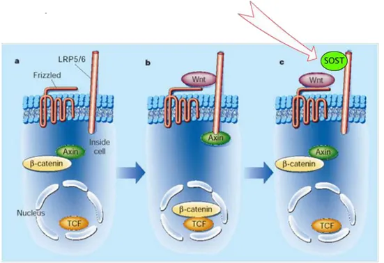
Another major signaling pathway involved in osteoblast differentiation is the Wnt signaling pathway. The real function of Wnt signaling in osteoblast differentiation remains unclear and needs further investigation. Commitment of mesenchymal stem cells to the osteoblast lineage requires the canonical Wnt/catenin pathway and associated proteins. The Wnt system is also important in chondrogenesis and hematopoiesis and may be stimulatory or inhibitory at different stages of osteoblast differentiation [27]. The canonical Wnt signaling pathway acting through β-catenin has a primary role in driving osteoblast proliferation and differentiation. β-catenin is a cytoplasmic phosphoprotein under resting conditions and the key mediator of the Wnt pathway. In the nucleus, β-catenin interacts with transcription factors and controls the transcriptional activation of specific genes. Activating mutations in the human β-catenin gene have been found in human colon cancer and melanomas [28]. Activation of Wnt signaling is induced by a number of factors (Figure 8).
Figure 8. The Wnt canonical cascade. A. The Wnt canonical cascade along with LRP5 regulate the
level of β-catenin in the nucleus. B. The TGF/BMP pathway promotes osteoblastogenesis through interaction with the heterodimer Smad/Runx2. C. The Wnt cascade activates Src/ERK, PI3K/ AKT proteins and promotes anti-apoptotic action. D. The Wnt induced Runx-2 transcription regulates the expression of RANKL and OPG. E. The Ca2+ mediated non-canonical Wnt pathway promote osteoblast
14
Wnt signaling up-regulates Runx2 transcription in both committed osteoprogenitor cells and embryonic mesenchymal cells prior to the induction of osteoblast phenotypic genes, promoting the entry of mesenchymal cells into the osteogenic lineage for both endochondral and intramembranous bone formation. The Wnt pathway, through an elaborate interaction of various components, regulates the expression of certain transcription factors, such as Runx2, Osterix and dlx5, and strongly stimulates osteoblastogenesis. At the same time, it inhibits the expression of adipogenic transcription factors C/ERB and PPARγ and blocks preadipocyte differentiation
[29-30]. Recently, the evolving concept of the role of LRP5/6 and its antagonist sclerostin,
as a point of mechanotransduction between osteocytes and osteoblast progenitors, focused attention again on the Wnt cascade. Mutations in genes associated with Wnt pathway are associated to changes in bone mass and skeletal fragility [31] demonstrating the importance of this pathway in bone metabolism. Osteoprotegerin (OPG) is a cytokine receptor, and a member of the tumor necrosis factor (TNF) receptor superfamily and RANKL is a “receptor activator of nuclear factor kappa-B ligand”.
RANKL/RANK signaling regulates osteoclast formation, activation and survival in normal bone modeling and remodeling and in a variety of pathologic conditions characterized by increased bone turnover. OPG protects bone from excessive resorption by binding to RANKL and preventing it from binding to RANK. OPG binding to RANKL on osteoblast/stromal cells, blocks the RANKL-RANK ligand interaction between osteoblast/stromal cells and osteoclast precursors. This has the effect of inhibiting the differentiation of osteoclast precursors into mature osteoclasts.
II.8 The mineralization process
Osteoblasts are able to secrete and to mineralize the bone matrix that is mainly composed of: type I collagen, osteocalcin (OC), osteopontin (OPN), bone sialoprotein (BSP), BMPs, TGF-β and the inorganic mineral hydroxyapatite. The initial hydroxyapatite crystal formation occurs within extracellular membrane-invested vesicles highly enriched in ALP activity called matrix vesicles (MV) [32]. Osteoblast differentiation is characterized by three major stages: (a) cell proliferation, (b) matrix maturation, and (c) matrix mineralization
[33]. Initially, calcium and phosphate accumulate inside the vesicles favoring
precipitation and crystal formation. In a second phase mineral grows further, leading to the disruption of the MV membrane and exposure of the crystals to the extracellular fluid forming a mineral nucleation site [34]. This mechanism is dependent on several factors such as calcium/phosphate concentrations and pH (Figure 9). ALP is an enzyme
15
involved in the mobilization of phosphate from substrates like pyrophosphate. The increase or decrease of ALP plays a critical role during osteoblasts differentiation. The reduction of ALP activity can inhibit mineralization.
Figure 9. The mineralization process. Bone remodeling begins when osteoclasts resorb bone mineral
and matrix. Mononuclear cells prepare the resorbed surface for osteoblasts, which generate newly synthesized matrix as they differentiate. Matrix mineralization and the differentiation of some osteoblasts into osteocytes complete the remodeling cycle.
II.9 Sclerostin: an inhibitor of osteoblast differentiation and
mineralization
Sclerostin (SOST) is a glycoprotein produced by osteocytes. It is a monomeric glycoprotein containing a cysteine knot-like domain that has homology to some BMP antagonists [35]. In humans, a lack of expression or reduced expression of sclerostin, results in the sclerosing bone dysplasia, sclerosteosis and van Buchem disease that are associated with osteoclastic hyperactivity, progressive skeletal overgrowth, a high bone mass, and cranio-facial abnormalities. These data suggest that sclerostin is a regulator of bone mass in humans. Sclerostin may play a role in inhibiting bone formation and occurs at late stages of differentiation, presumably when osteocytes are buried in the bone matrix. Sclerostin acts, in part, by binding to the extracellular domain of the low-density lipoprotein receptor-related protein 5 (LRP-5) and its closely related co-receptor LRP-6 on osteoblast-lineage cells, thereby antagonizing the canonical Wnt signaling
16
BMPs are ligands that activate pathways involved in cell proliferation and differentiation. Sclerostin binds BMP receptor and activates the Smad 1-5-8 phosphorylation and their nucleus translocation (Figure 10). In the nucleus, this complex induces the transcription of cell regulatory factors including p21, p53, and Bcl-2. Recent studies have demonstrated that Runx2 regulates sclerostin expression to inhibit osteoblast differentiation. Inhibition of osteoblast differentiation is a feature of breast cancer tumour-induced osteolysis. Runx2 and CBFβ are required for the expression of genes that mediate the ability of metastatic breast cancer cells to directly modulate both osteoclast (GM-CSF, IL-11) and osteoblast (sclerostin) function. Runx2-dependent inhibition of osteoblast differentiation, by MDA-MB-231 cells, is mediated through the Wnt antagonist, sclerostin [37].
Figure 10. Wnt pathway (a). Activation of Wnt pathway (b). Sclerostin is made primarily by osteocytes, and it inhibits bone formation and enhances apoptosis of osteoblasts. One of the
well-know actions of Sclerostin is that it inhibits the canonical Wnt pathway by antagonizing Wnt protein for the binding to LRP-5/6 receptor, and thus it leads to beta-catenin degradation (c).
II.10 Asporin : a member of SLRPs family
Asporin is a small leucine-rich proteoglycans (SLRPs) family. In the first time, the SLRPs, were grouped into three distinct classes but more recently, the SLRP gene family has expanded to encompass 18 genes classified into five distinct subfamilies
17
(Figure 11). The name asporin reflects the unique aspartate-rich N terminus and the overall similarity to decorin. Higher levels are present in osteoarthritic articular cartilage, aorta and uterus. Moderate expression in small intestine, heart, liver, bladder, ovary, stomach, and in the adrenal, thyroid, and mammary glands. Low expression is found in trachea, bone marrow and lung.
Figure 11. Phylogenetic analysis and chromosomal organization of various human SLRP classes.
The color-coded dendrogram (left) shows the presence of five distinct families of SLRP and related LRR proteins. The consensus for the N-terminal Cys-rich cluster is also shown. The chromosomal arrangement of the various SLRP genes is shown in a telomeric orientation (right). Transcriptional direction is shown by the arrows above the color-coded boxes. The horizontal distance between genes is not to scale. (Henry
et al., J.Biol. Chem, 2001).
This protein was identified for the first time in human articular cartilage [38-39]. The biological role of asporin has been unclear, but recent genetic studies have demonstrated an association between asporin and various bone and joint diseases, including osteoarthritis, rheumatoid arthritis and lumbar disc disease [40]. Variation in the poly-Asp region of asporin is associated with osteoarthritis susceptibility type 3 (OS3) ; also known as osteoarthritis of knee/hip. Osteoarthritis is a common disease that produces joint pain and stiffness together with radiologic evidence of progressive degeneration of joint cartilage.
II.10.1 Role of Asporin in TGF-β pathway and osteoblast differentiation
Various growth factors and transcriptional factors have been reported to be involved in the formation and differentiation of bone tissue. TGF-β is the one of the most important
18
growth factors in the development of this process. Asporin is abundantly expressed in the articular cartilage of individuals with osteoarthritis [41]. In the context of osteoarthritis, several studies have shown that asporin regulates cartilage matrix gene expression and cartilage formation by modulating the transforming growth factor-‐β (TGF-‐ β) signaling pathway [41]. Asporin is able to bind different growth factors, including TGF-β and BMP-2, and negatively regulates their activity. Asporin directly binds to TGF‐β and inhibits TGF‐β‐mediated expression of cartilage matrix genes. In 2010, Kou et al. [42] demonstrated that amino acids 159–205 of asporin mediate its interaction with TGF‐β and effectively repress TGF‐β‐induced cartilage matrix gene expression (Figure 12).
Figure 12. Asporin inhibits TGF‐β‐mediated expression of cartilage matrix genes. The amino acids
159–205 of asporin mediate its interaction with TGF‐β and effectively repress TGF ‐β-induced cartilage matrix gene expression (Kou et al., J. Biol. Chem. 2010).
Asporin also has a binding ability to type II collagen in vitro, but its binding pattern is different from that of TGF‐β. These findings suggest that asporin is one of the important cartilage matrix proteins that binds to the ECM and TGF‐β and thereby modulates interactions between TGF‐β and its signaling receptors. Overexpression of Asporin blocks the TGF‐β activity and induces bone formation and osteoblasts differentiation. Asporin is the only SLRP able to regulate collagen calcification. This function is similar
19
to other polyacid domain proteins, including osteopontin and bone sialoprotein that can bind calcium and influence hydroxyapatite crystal formation in collagen [43-44-45-46]. Asporin-collagen binding and Asporin-induced mineralization can be inhibited by decorin. This suggest that Asporin, but not other SLRPs, induces collagen mineralization and triggers increased levels of osteoblastic transcription factor, such as RUNX2 and ALP important to start the differentiation process. To date, the expression and role of asporin in cancer is almost unknown. Our Laboratory has recently identified asporin as an accessible biomarker in human pancreas adenocarcinoma by ex vivo tissue biotinylation [47]. In this proteomic study, we reported the overexpression of asporin in pancreas tumors and we were the first to identify this protein as an accessible biomarker that bears potential clinical value for diagnostic and therapeutic applications.
20
III. MATERIALS AND METHODS
Normal human osteoblasts were isolated from trabecular bone specimens obtained from patients undergoing knee replacement surgery. The age of the patients ranged from 66 to 83 years. Bones were cut into small fragments, washed with Dulbecco’s modified Eagle’s medium, and then submitted to enzymatic digestion . Small pieces of bone were sequentially incubated with 0,5 mg/ml hyaluronidase type IV S (Sigma Aldrich) for 20 minutes at 37 °C and 0,6 mg/ml clostridial collagenase IA (Sigma Aldrich) for 30 minutes and 240 minutes successively at 37° C. The digested bone pieces were plated in T75 flasks and cultured in Dulbecco’s modified Eagle’s medium supplemented with 15% fetal calf serum, 10 mM HEPES, 100 units/ml penicillin and 100 µg/ml streptomycin until osteoblasts migrated out of bone explants. At this point the medium was replaced with fresh medium containing 10% fetal calf serum, 10 m M HEPES, 100 units/ml penicillin , and 100 µg/ml streptomycin [48]. All tissues used in this study were obtained from the Laboratory of cartilage (Prof. Y Henrotin) University of Liege with Medicine Faculty Ethics Committee approval. To validate the osteoblasts isolated, we excluded the presence of adipocytes using Oil Red staining and the presence of osteoclasts using TRAP staining. Osteoblasts cells were confirmed by ALP activity.
The activity of ALP in the membrane was visualized usingnaphtol AS-MX phosphate/fast red method.
III.2 Human cancer cell lines culture
The human epithelial breast cancer cell lines MCF-7, MDA-MB-231, the human osteosarcoma MG63, the human prostate carcinoma LNCaP, the human prostate adenocarcinoma PC3, were obtained from American Type Cell Collection (ATCC, Manassas, VA, USA). MCF-7, MDA-MB-231, and human osteoblasts were maintained in DMEM supplemented with 10% FBS (Lonza). MG63 cells were maintained in MEM supplemented with 10% FBS, 1mM Sodium pyruvate, 0,1 mM NEAA (Lonza). LNCaP were maintained in RPMI 1640 supplemented with 10% FBS, 1mM Sodium pyruvate, 10 Mm Hepes (Lonza). PC3 were maintained in HAM’S F12K supplemented with 7% FBS, (Invitrogen). Sub-confluent cultures (70–90% confluence) of low passages (until passage 9) were utilized for all experiments.
21
III.3 Osteoblast differentiation
Osteoblasts were maintained for 14 days in Ob differentiation media composed of DMEM containing 100 U/ml penicillin, 100 µg/ml streptomycin, 10 mM HEPES, 2 mM glutamine, 2% Ultroser G, a serum substitute, 10-8 M 1,25(OH)2 vitamin D3, 50µg/ml ascorbic acid and 20 µg/ml proline (Sigma-Aldrich, Belgium) for mineralization assessment. For the mineralization assessment, 10 mM beta-glycerophosphate were added 72h before the end of the culture.
III.4 Gene expression analysis
Total RNA was isolated with High Pure RNA Isolation Kit (Roche). One microgram of total RNA was reverse transcribed using the Transcriptor First Strand cDNA Synthesis Kit (Roche) according to the manufacturer’s instructions. The cDNAs (100ng) were mixed with primers, human UPL-probe system (Roche) and 2X Fast Start Universal Probe Master mix (Roche) and analyzed in triplicate. qRT-PCR was performed using the LightCycler480 system and the corresponding manufacturer software (Roche). The following primers were used:
1- Asporin: forward 5’- GGTGGATAACTTCTACTTTTAGGAGGA -3’ and reverse 5’ AAGAAGGGTTTGGCAGAGC-3’ and UPL probe #72.
2- Sclerostin: forward 5’- AGCTGGAGAACAACAAGACCA -3’ and reverse 5’ GCTGTACTCGGACACGTCTTT -3’ and UPL probe #77.
3- Alkaline phosphatase: forward 5’- CCTGCCTTACTAACTCCTTAGTGC -3’ Runx2: forward 5’- CAGTGACACCATGTCAGCAA -3’ and reverse 5’ GCTCACGGTCGCTCATTTTG -3’ and UPL probe #41.
The relative gene expression levels were normalized using 18S rRNA content (Life Technologies, Carlsbad, CA, USA; Cat. # 4310893E).
III.4 Statistics
The results are reported as means with standard deviation (s.d.) as descrived in legends. Statistical analysis are performed using one-way ANOVA depending on the number of grouping factors. Groups means are compared by a Student T-test or Bonferroni’s post-test according to the group number. P≤0,05 are considered as statistically significant. The experiments are performed as several independent biological replicates.
22
III.5 Western Blotting analysis
Human Osteoblasts cells were lysed with RIPA buffer containing 50mM Tris-HCl, 150 mM NaCl, , 0.5% sodium deoxycholate, 2 mM sodium fluoride, 1% Triton X-100, 0.2% SDS, and a mixture of protease inhibitors. The following monoclonal (m) and polyclonal (p) antibodies (Ab) were used: (m) p-smad 2/3 D27F4 (Cell Signaling), (m) smad 2/3 D7G7 (Cell Signaling), (p) Asporin HPA008435 (SIGMA Aldrich), Anti-Active Beta Catenin Clone 8E7 # 05-665 (Millipore), (m) Beta Catenin sc-59897 (Santa Cruz Biotechnology), (m) HSC70 sc-7298 (Santa Cruz Biotechnology). Conditioned media were concentrated 10-fold using Amicon Ultra centrifugal filters (Millipore, Cat#UFC500324) and dissolved in Leamli buffer containing protease and phosphatase inhibitors (Roche). 20 μg of cell lysates or concentrated conditioned cell culture media were separated on a 10% polyacrylamide denaturing gel and transferred to nitrocellulose membranes.
III.6 Calcified nodule formation in coltures of osteoblastic cells
The MG63 cell line were cultured in MEM supplemented with 10% FBS, 1mM Sodium pyruvate, 0,1 mM NEAA (Lonza). To induce the osteoblastic phenotype and formation of calcified bone-like nodules, the cells were grown to confluence and supplemented with 100 n M dexamethasone and 6 m M Calcium Cloride, replenished each day. For the assay, the cells were also supplemented with conditioned media MCF7, conditioned media MDAMB 231, SOST recombinant protein (200ng/mL). Nodules were quantified on day 2 of colture : after PBS washes, Alizarin Red was applied for 10 minutes and then rinsed with water. Asporin recombinant protein was used as a positive control (3µg/mL) to induce mineralization.
23
IV RESULTS
IV.1 ASPORIN EXPRESSION IS MODULATED BY
BREAST CANCER CELLS
IV.1.1 Cancer cells do not express asporin
We evaluated asporin expression in breast (MDA-MB-231 and BO2) and prostate (PC3 and LNCaP) cancer cells (Figure 13). Total protein extracts from cells stably overexpressing asporin, previously generated in our Laboratory, and human osteoblasts were used as positive controls.
Figure 13. Cancer do not express asporin. Breast and prostate cancer cells do not produce asporin at the
protein (A) and mRNA levels (B). Cells stably overexpressing asporin and human osteoblasts were used as a positive control.
A
24
IV.1.2 Breast cancer cells and prostate cancer cells modulate in
different way asporin expression in osteoblasts
We evaluated the effect of different breast and prostate cancer cell conditioned media on asporin expression in human osteoblasts (Figure 14).
a) Protocol of osteoblasts treatment;
To evaluate the effect of these different cancer cell conditioned medium on asporin expression, we treated a confluent monolayer of pre-osteoblasts with serum-free medium previously preincubated with MDA-MB-231, BO2, PC3, LNCaP cells for 48h. All the cells were maintained in a unique medium (DMEM).
b) Breast and prostate cancer cells modulate asporin expression in osteoblasts.
Figure 14. Conditioned mediem from Breast and prostate cancer cells modulate in different way asporin expression in osteoblasts. Cancer cells do not produce asporin but their conditioned media
(CM) are able to modulate asporin expression in osteoblasts at the protein (A) and mRNA level (B). Immunoblot analysis revealed a significant decrease in asporin level in osteoblasts treated with MDA-MB-231 and BO2 conditioned medium, while the no effect was observed with PC3 and LNCaP conditioned medium compared to the control.
A
25
IV.1.3 Conditioned media from metastatic and non metastatic human
breast cancer cells differentially modulate the expression of asporin in
osteoblasts
We have next focused our attention on 2 specific breast cancer cell lines and we used in one hand MDA-MB-231 metastatic cells and on the other hand MCF7 non metastatic human breast cancer cells. We treated a confluent monolayer of pre-osteoblasts with MCF7 and MDA-MB-231 conditioned media for 48h. Immunoblot analysis and real time PCR, revealed a significant increase in asporin level in osteoblasts treated with MCF7-conditioned medium, while the opposite effect was observed with MDA-MB-231-conditioned medium. Asporin is a secreted protein and its detection in the medium of osteoblasts treated with MCF7 or MDA-MB-231 cells conditioned media showed the same results (Figure 15).
Figure 15. MCF7 conditioned medium increases Asporin level in osteoblasts. MCF7 and
MDA-MB-231 breast cancer cells do not express asporin (A). MCF7-conditioned medium is able to induce asporin expression and secretion, while the opposite effect was observed with MDA-MB-231-conditioned medium. Cell lysates (B), protein secretion (C) and Real Time PCR (D), showed the same results. Results are expressed as mean ± s.d. *** P≤ 0,001.
B A
D
26
IV.1.4 CM MCF7 but not CM MDA-MB-231 is able to induce
mineralization in osteoblasts
Next, we investigated the role of MCF7 and MDA-MB-231 conditioned media on osteoblast differentiation and mineralization.
a) ALP and RUNX2 expression in osteoblasts treated with MCF7 and MDA-MB-231 conditioned media ;
To evaluate the effects of MCF7 and MDAMB231 conditioned media in the differentiation process, we checked Alkaline phosphatase (ALP) and “runt-related transcription factor 2” (RUNX2) expression in osteoblasts. We found that MCF7 CM, but not MDAMB231 CM, is able to induce ALP and RUNX2 expression in osteoblasts suggesting that these cells have a pro-differentiation effect on osteoblasts (Figure16).
Figure 16. CM MCF7 but not CM MDA-MB-231 is able to induce ALP and RUNX2 expression in osteoblasts: MCF7-conditioned medium (CM) increased ALP (A) and RUNX2 (B) level in osteoblasts
after 48 hours of treatment.. The significant decrease was observed with MDA-MB-231 conditioned medium. Results are expressed as mean ± s.d. *** P≤ 0,001.
B A 0 0,5 1 1,5 2 2,5 3 Control CM MCF7 CM MDAMB231 R el a ti ve R UNX2 m R NA l ev el
27
b) MCF7 but not MDA-MB-231 conditioned medium induces smad2/3 phosphorilation;
Following TGF-β induction, both the smad converge at the RUNX2 gene to control mesenchymal precursor cell differentiation. TGF-β1 promotes matrix production and osteoblast differentiation and reduces the ability of osteoblasts to secrete osteoclasts differentiation factors such as RANKL. In the last stage of osteoblast differentiation, TGF-β represses RUNX2 transcription reducing the mineralization. We are using pre-osteoblasts in the first stage of differentiation and we investigated the smad 2/3 phosphorilation after MCF7 and MDA-MB-231 conditioned media treatment for 30 minutes. Our result showed the ability of MCF7 conditioned medium to induce smad 2/3 phosphorilation compared to the control and to MDA-MB-231 conditioned medium treatment (Figure 17). This induction activates RUNX2 transcription, osteoblast differentiation and matrix production by osteoblasts.
Figure 17. CM MCF7 but not CM MDA-MB-231 is able to induce smad 2/3 phosphorilation.
Immunoblot analysis showed that osteoblasts, treated with TGFbeta1 (2,5 ng/ml) and MCF7 conditioned medium, induce smad 2/3 phosphorilation. TGF-β1 was used as a positive control. CM MDA-MB-231, differently, is able to reduce smad 2/3 phophorilation and to block TGF-β a signaling pathway.
c) OPG and RANKL expression in osteoblasts treated with MCF7 and MDA-MB-231 conditioned media;
When osteoblasts are treated with MCF7 conditioned medium, we observed that RANK-L basal level is decreased while OPG expression is increased. An inverse regulation is observed upon MDA-MB-231 conditioned medium addition (Figure 18). These results suggest that next to their opposite effects observed on osteoblasts (Runx2
28
and ALP expression), these cell lines also exert opposite indirect effects on osteoclastic maturation and activation through the regulation of RANK-L and OPG expression in osteoblasts.
Figure 18. CM MB-231 but not CM MB-231 stimulates osteoclasts formation :
MDA-MB-231-conditioned medium (CM) increased RANKL level and decrease OPG level in osteoblasts. The opposite effects were observed with MCF7 conditioned medium treatment.
d) MCF7 CM, but not MDA-MB-231 CM, induces osteoblastic mineralization;
In the end, we evaluated the effects of MCF7 CM and MDA-MB-231 CM on the mineralization process (Figure 19). Osteoblasts produce a calcium and phosphate-based mineral that is deposited, in a highly regulated manner, into the organic matrix forming a very strong and dense mineralized tissue - the mineralized matrix. We used human osteosarcoma cells MG63 as an osteoblastic model that induces mineral formation that can be visualized using Alizarin red staining. To induce the osteoblastic phenotype and formation of calcified bone-like nodules, the cells were grown to confluence and supplemented with 100 nM dexamethasone and 6 mM calcium cloride, replenished each day. For the assay, the cells were also supplemented when indicated with MCF7 and MDA-MB-231 conditioned media. Asporin recombinant protein was used as a positive control for the mineralization. We controlled asporin expression levels after MDA-MB-231 and MCF7 conditioned media treatment in MG63 cells after 48 hours of treatment. Real time PCR showed the same asporin modulation as previously obtained with osteoblasts (Figure 15). 0 0,5 1 1,5 2 2,5 3 3,5 4 4,5 5 OB C CM MCF7 CM MDAMB231 R el a ti ve R AN KL a n d OP G m R N A le ve
l RANKL and OPG Real Time PCR
RANKL OPG
29
Figure 19. MCF7 CM ,but not MDA-MB-231CM, is able to induce mineralization.
MCF7-conditioned medium (CM) induced the mineralization in MG63 after 48 hours of treatment (A). Asporin recombinant protein was used as a positive control in the mineralization process.Treatments with MCF7 and MDA-MB-231 conditioned media in MG63 cells showed the same asporin modulation as observed in osteoblasts (B).
IV.1.5 The absence of asporin is able to reduce the mineralization
Previously we have demonstrated the different capacity of MCF7 and MDAMB231 conditioned media to induce or inhibit, respectively, the osteoblast differentiation and mineralization. We next investigated the role of asporin in this process.a) Asporin expression increases during osteoblasts differentiation;
We checked asporin expression at different stages of osteoblastic differentiation (Figure 20). The cells were maintained for 14 days in an osteoblast differentiation mediumas described in the Material and Method section. Real time PCR showed a significant increase of asporin expression in the last stage of the osteoblastic differentiation (Figure 20). A B -0,5 0 0,5 1 1,5 2 2,5 C CM MCF7 CM MDAMB231 R el a ti ve A SP N m R NA le ve l
30
Figure 20. Asporin expression increases during osteoblastic differentiation. Real time PCR showed
the increase of asporin expression in the stage 2 osteoblasts when compared with starting osteoblasts (stage 0). Results are expressed as mean ± s.d. *P≤0,05.
b) MCF7 conditioned medium increases the mineralization through asporin expression;
Previous results showed the ability of MCF7 conditioned medium to increase asporin expression and osteoblast differentiation and mineralization through the increase of the expression of different important factors. To evaluated if the presence of asporin play a critical role in these processes, we used a transient transfection with siASPN RNA to deplete its expression in osteoblasts (Figure 21A). Asporin depleted and control osteoblasts were treated with MCF7 conditioned media for 48h. Our results showed that the role of MCF7 conditioned medium in the mineralization process is dependent by asporin expression (Figure 21B).
31
Figure 21. Asporin depletion in osteoblasts using transient transfection. We used a transient
transfection with siASPN RNA to decrease its expression in osteoblasts. After 48 hours of transfection, the cells were collected and asporin expression was checked by Real Time PCR (A). Results are expressed as mean ± s.d. *** P≤ 0,001, **P≤ 0,01, *P≤0,05. Asporin depleted MG63 were not able to induce the mineralization compared to the control and we observed the decrease of calcification after treatment with MCF7 conditioned medium (B).
32
IV.2 MOLECULAR MECHANISMS OF ASPORIN
EXPRESSION REGULATION
IV.2.1 SOST is overexpressed in metastatic breast cancer cells
Sclerostin (SOST) is a cystein-knot protein secreted mostly by osteocytes, bone cells that respond to mechanical stress applied to the skeleton and appear to play an important role in the regulation of bone remodeling. Consistently with previous reports, we found that SOST is overexpressed in metastatic breast cancer cells and not in non metastatic breast cancer cells (Figure 22).
Figure 22. SOST is overexpressed in MDA-MB-231 metastatic breast cancer cells. Real time PCR
and immunoblot analysis revealed a significant increase of SOST expression (A) and secretion (B) in aggressive breast cancer cell lines MDA-MB-231 compared with MCF7. Results are expressed as mean ± s.d. **P≤ 0,01.
IV.2.2 MCF7, but not MDA-MB-231, conditioned medium activates
Wnt pathway in osteoblasts
SOST, secreted by mature osteocytes, is a subject of much interest as it pertains to bone disorders. Recent studies have shown that SOST gene is also expressed by articular chondrocytes and that modulation of its activity may have effects on articular cartilage and subchondral bone. The role of SOST in the pathogenesis of osteoarthritis in humans has not yet been defined, and the potential utility of treating osteoarthritis with interventions that alter SOST is not known. SOST is a potent inhibitor of bone growth and inhibits beta catenin signaling via its interaction with the Low Density Lipoprotein Receptor-related Proteins-5/6 (LRP5/6) receptor. Under resting condition, the
B A
33
cytoplasmic beta-catenin is bound to its destruction complex, consisting of APC, axin/conductin, and GSK-3beta. Upon binding to the receptors (LRP5/6), GSK3beta releases beta catenin to translocate to the nucleus (active beta catenin) thus inducing the transcription of Wnt target genes . We investigated the effects of MCF7 and MDA-MB-231 conditioned media on Wnt pathway through beta catenin and GSK3beta expression in osteoblasts after 48 hours of treatment (Figure 23). The results reveled the significant increase of active beta catenin expression after MCF7 conditioned medium treatment compared with MDA-MB-231 conditioned medium and SOST treatment in osteoblasts.
Figure 23. MCF7, but not MDA-MB-231, conditioned medium activates Wnt pathway in osteoblasts. Immunoblot analysis showed that osteoblasts, treated with recombinant SOST (200ng/mL)
and MDA-MB-231 conditioned medium, reduced their active beta catenin expression (B) through GSK3beta phosphorylation (A). MCF7 CM, on the inverse, is able to induce active beta catenin expression (B) and to reduce GSK3beta phosphorylation (A) after 48 hours of treatment in osteoblasts.
IV.2.3 SOST competes with MCF7 conditioned medium in the
mineralization process
In the previous results, we showed the capacity of MCF7 conditioned medium comparing with MDA-MB-231 and SOST, to activate the Wnt pathway. We investigated the capacity of SOST to compete with MCF7 conditioned media in the mineralization process. In order to evaluate this effect, we treated MG63 with MCF7 conditioned medium and SOST together for 48 hours, and we checked active beta catenin expression. The same conditions of treatment was used to perform an Alizarin red assay. Our results showed the significant capacity of SOST to reduce active beta catenin expression and the mineralization process after MCF7 conditioned medium treatment in osteoblasts (Figure 24).
34
Figure 24. Sclerostin competes with MCF7 conditioned medium in the mineralization process.
Immunoblot assay (A) revealed that osteoblasts treated with recombinant SOST and challenged with MCF7 conditioned medium showed a significant reduction in active beta catenin expression and in the mineralization potenzial ( B ) using Alizarin Red assay.
IV.2.4 Wnt pathway modulates Asporin expression in osteoblasts
Thorfve et all. [49], showed the capacity of Wnt pathway to modulate asporin
expression. To confirm the results obtained with MCF7 conditioned medium on Wnt pathway and asporin expression, we investigated the role of TGF-β (Wnt activator) and SOST (Wnt inhibitor) on asporin expression in osteoblasts.
a) TGF-β activates Wnt pathway and increases asporin expression in osteoblasts;
We treated a confluent monolayer of osteoblasts with TGF-β (5ng/mL) and we checked the beta catenin and asporin expression. Immunoblot analysis and Real Time PCR confirmed an activation of beta catenin and an increase of asporin expression after TGF-β treatment (Figure 25).
A
35
Figure 25. TGF-β activates Wnt pathway and increases asporin expression in osteoblasts. TGF-β
induces active beta catenin (A) and asporin expression (B-C) in osteoblasts after 48 hours of treatment. The results are confirmed by immunoblot analysis and Real Time PCR. Results are expressed as mean ± s.d. *** P≤ 0,001.
b) SOST reduces asporin expression in osteoblasts;
We treated a confluent monolayer of osteoblasts with SOST recombinant protein (200ng/mL) and we checked asporin expression. Immunoblot analysis and Real Time PCR confirmed the decrease of asporin expression in osteoblasts after 48 hours of treatment (Figure 26).
Figure 26. SOST reduces asporin expression in osteoblasts. The results was confirmed by immunoblot
analysis (A) and Real Time PCR (B). Results are expressed as mean ± s.d. *P≤0,05.
A B
C
36
c) SOST blocks asporin expression induction by both MCF7 conditioned medium and TGF-β1 treatment;
To study the mechanism mediating SOST blocking effects on the mineralization process in presence of MCF7 conditioned medium, we checked for asporin expression. We treated the osteoblasts with MCF7 conditioned medium and with different concentrations of SOST recombinant protein (50,100,200 ng/mL). Real Time PCR showed a significant decrease of asporin basal level after these treatments (Figure 27). We considered also the possibility for SOST to decrease asporin expression upon TGF-β1 treatment. Our results revealed a significant reduction of asporin expression in any condition after SOST treatment.
Figure 27. SOST blocks asporin expression induction by both MCF7 conditioned medium and TGF-β1 treatment. SOST treatment in osteoblasts is able to reduce asporin expression. The reduction
was also showed after MCF7 conditioned medium treatment (A). Upon SOST treatment (200ng/mL) ,osteoblasts showed a reduced asporin expression. The reduction was also observed upon TGF-β1 (5ng/mL) treatment after 48 hours (B).
d) Reduction of RUNX2/SOST in MDA-MB-231 re-induces osteoblastic mineralization;
Mendoza et al. [49] showed that the expression of SOST is dependent by RUNX2
expression. To evaluate the role of SOST depleted MDAMB231 conditioned medium in
0 0,5 1 1,5 2 2,5 3 3,5 4 4,5 ob c CM MCF7 CM MCF7/ SOST TGFbeta TGFbeta SOST Re lat iv e A SP N m RN A le ve l
ASPN Real time PCR
A
37
osteoblast mineralization, we performed a transient transfection using siRNAs directed against RUNX2 in MDA-MB-231. Our results showed the significant decrease of RUNX2 expression in MDA-MB-231 after the transfection with siRUNX2 (Figure 28A) and the consequent SOST decrease (Figure 28B). Conditioned medium from depleted RUNX2/SOST-MDA-MB-231 cells, was used to treat MG63 cells to evaluate its effect on mineralization after 48 hours. Reduction of RUNX2/SOST in MDA-MB-231 re-induces mineralization in MG63 cells (Figure 28C).
Figure 28. Reduction of RUNX2/SOST in MDA-MB-231 re-induces mineralization in MG63. We
used a transient transfection with siRUNX2 RNA to decrease its expression in MDA-MB-231(A). SOST expression is dependent by RUNX2 expression (B). After 48 hours of transfection, MDA-MB-231 conditioned medium depleted RUNX2/SOST was collected and was used to evaluate the mineralization in MG63. Reduction of RUNX2/SOST in MDA-MB-231 re-induces mineralization in MG63 (C).
A B
38
V. DISCUSSION AND PERSPECTIVES
Asporin is a protein of the extracellular matrix identified for the first time in human cartilage [38]. The biological role of asporin has not been clearly described, but recent genetic studies have shown an association between asporin and other human bone diseases, including osteoarthritis, rheumatoid arthritis and lumbar disk disease. In the context of osteoarthritis, several studies have shown that asporin regulates gene expression and the subsequent formation of the cartilage matrix, essentially by interfering with the signal induced by TGF-β. Asporin directly binds to TGF-β [41] blocking its activity and inducing consequently, the transcription factors involved in osteoblast differentiation and bone formation. These results have led to investigate the role of asporin as a possible therapeutic target in osteoarthritis. It is well admitted that bone metastatic cancer cells are able to influence bone cells in order to favor their settling and colonization of bone. Aggressive cancer cells produce factors such as parathyroid hormone-related peptide (PTHrP) that promote the formation and activation of osteoclasts [51]. Osteoclast activation results in bone resorption and thus release of factors by the bone matrix, such as TGF-β, which stimulate tumour cell proliferation
[52].
The present study was undertaken to investigate whether cancer cells are able to modulate asporin expression in human osteoblasts in order to inhibit their differentiation and favor their own colonization of bone. Our results demonstrate that tumor cells do not produce directly asporin but the conditioned media collected from different breast and prostate cancer cell lines, have shown an interesting ability to modulate asporin expression in human pre-osteoblasts. In particular, a relevant result was obtained with MDA-MB-231 and BO2 osteotropic breast cancer cells that induced a strong reduction of asporin basal level in osteoblasts. PC3 and LNCaP prostate cancer cell lines are respectively derived from a bone and a lymph node metastasis. However, there was no marked difference between them in the context of the regulation of asporin expression in osteoblasts. In fact, the expression of asporin was not altered after treatment with conditioned media obtained from both PC3 and LNCaP suggesting that our observations are more relevant to breast cancer biology. These results have allowed us to focus our attention on the role played by breast cancer cells on the modulation of asporin