INDEX
Summary ………... pag. 4
Introduction
……….... 5
Materials and Method ……… 11
¾ Materials ……… 11
¾ Cell cultures ... 11
¾ His6-arom plasmid construction ... 12
¾ Site-directed mutagenesis ... 13
¾ Transient transfection ... 14
¾ Aromatase Activity Assay ... 15
¾ Total RNA Extraction and ReverseTranscription-PCR Assay ... 15
¾ Immunoblotting and Immunoprecipitation Analysis ... 16
¾ Detection of His6-tagged aromatase protein by Western Blotting 18
¾ Statistical Analysis ... 18
Results ………. 19
¾ Short time estradiol exposure increases aromatase activity in MCF-7 cells ... 19
¾ P450arom mRNA and protein expression in E2-treated MCF-7 cells ... 21
¾ E2 increases tyrosine phosphorylation levels of aromatase protein 23 ¾ Aromatase activity is enhanced by tyrosine phosphatases inhibitor 25 ¾ Role of the protein tyrosine phosphatase, PTP1B, in E2-stimulated aromatase activity ... 27 ¾ Aromatase activity is up-regulated by E through PI3K/Akt
¾ A protein complex involving Akt, P450arom and PTP1B activates
aromatase ... 31 ¾ Role of tyrosine residues in the E2-activation of aromatase ... 34 ¾ A cross-talk between GF receptors and ER-α is involved in the
increased aromatase activity in MCF-7 cells ... 37
Discussion ... 39
References ... 45
Scientific Publication
¾ Evidences that leptin upregulates E-cadherin expression in breast
cancer: effects on tumor growth and progression
¾ Fas Ligand Expression in TM4 Sertoli Cells is Enhanced by
Estradiol ‘‘In situ’’ Production
¾ Human sperm express a functional androgen receptor: effects on
PI3K/AKT pathway
¾ Molecular Mechanism through Which Leptin Upregulates
E-cadherin Expression in Breast Cancer.In vitro and In Vivo Effects on Tumor Cell Growth and Progression
¾ Evidence that PI3K/AKT pathway is involved in the short non
genomic loop between 17β-estradiol and aromatase activity
¾ Fas Ligand Expression in TM4 Cells is Upregulated by Estradiol
through Estrogen Receptor α Interaction with SP-1
¾ A new role of leptin as amplifier of estrogen signaling in breast
SUMMARY
Estrogens are strongly associated with breast cancer development and progression. The intratumoral conversion of androgens to estrogens by aromatase within the breast may be an important mechanism of autocrine stimulation in hormone-dependent breast cancer.
The aim of this study was to investigate if E2/ER can modulate aromatase activity in human breast cancer cells. In MCF-7 cells we examined, by tritiated water release assay, aromatase activity. Immunoprecipitation studies, using a vector containing aromatase gene with polyhistidine tags, were performed to evaluate phosphorylation status of aromatase protein.
Our results demonstrated that 17-β estradiol is able to enhance aromatase activity, at short time, through PI3K/Akt pathway in MCF-7 breast cancer cells. These up-regulatory effects involved rapid changes in tyrosine phosphorylation status of aromatase purified protein. Indeed, a tyrosine phosphatases inhibitor enhanced basal and E2-induced aromatase activity. The overexpression of the tyrosine phosphatase PTP1B reduced enzymatic activity while E2-induction was completely reversed in MCF-7 cells cotransfected with PTP1B and dominant negative of Akt supporting the involvement of PI3K/Akt pathway. It should be taken into account that a cross-talk between tyrosine kinase growth factor receptors and estrogen receptor signaling may influence aromatase activity.
Our data indicate the existence of a short non genomic autocrine loop between E2 and aromatase in MCF-7 cells, giving a great emphasis to the role of aromatase in promoting breast cancer cell growth.
Introduction
INTRODUCTION
Estrogens play a crucial role in the development and progression of breast cancer. Approximately 60% of premenopausal and 75% of postmenopausal patients have estrogen-dependent carcinomas (Chen, 1998). The biosynthesis of estrogens from androgens is catalyzed by the enzyme complex termed aromatase (also estrogen synthase or P450 AROM), which is composed of two polypeptides, an ubiquitous nonspecific flavoprotein, NADPH-cytochrome P450 reductase and a specific microsomial form of cytochrome P450arom encoded by the CYP19 gene (Fig. 1)(Simpson et al, 1994).
Fig. 1 Biosynthesis of estrogens from androgens by aromatase.
Aromatase is mainly expressed in the ovaries of premenopausal women. A very high level of aromatase is found in placenta in pregnant women. In postmenopausal women and men, adipose tissue and skin cells are the major sources of estrogen production, but the aromatase activity in these tissues is significantly lower than that
Androgens + O
2Aromatase cytochromeP450 enzyme complex
NADPH P450 reductase
Estrogens + H2O
cytochrome P450aromatase
Introduction
in ovaries and the level of circulating estrogen is much lower in postmenopausal women and men than in premenopausal and pregnant women (Chen, 1998).
Interestingly, P450arom is found to be expressed at higher levels in breast cancer tissue than normal breast tissue (Bulun et al, 1993; Miller, 1997; Harada, 1997). Aromatase presence in breast cancer tissue as well as in breast cancer cell lines has been shown by enzyme activity measurement, immunocytochemistry and RT-PCR analysis (Sasano et al, 1994; Sourdaine et al, 1996; Maggiolini et al, 2001). Cell culture (Santner et al, 1993; Sun et al, 1997) and nude mouse experiments (Yue et al, 1994) using aromatase-transfected MCF-7 cells have shown that aromatase expressed in breast cancer cells can promote breast cancer growth in both an autocrine and a paracrine manner. In addition, overexpression of aromatase in mammary gland of transgenic mice causes premalignant lesions, such as atypical ductal hyperplasia (Tekmal et al, 1996; Gill et al, 2001). These results indicate that
in situ estrogen production, due to overexpressed aromatase in breast cancer cells,
plays a more important role than circulating estradiol in breast tumor promotion. Indeed, intratumoral aromatase of breast carcinoma has been extensively studied for its potential clinical significance as a target for endocrine therapy using aromatase inhibitors (Brodie, 1991; Santen et al, 1999; Altundag & Ibrahim, 2006).
A complex mechanism is involved in the control of human aromatase expression. The human aromatase gene contains nine translated exons (II-X) and at least ten untranslated exon I's (Fig. 2).
Introduction
Fig. 2 Scheme of alternative utilization of tissue-specific exon Is and promoters of the human aromatase gene.
The translation start site is positionedin exon II and one of the 5'-untranslated exon I's of aromatase mRNA is spliced onto a common splicing junction of exon II, upstream of the translation start site (Chen et al, 1986; Means et al, 1991). It has been found that the various untranslated exon I's are present at different levels in different aromatase-expressing tissuesand cells, providing tissue-specific controls of aromatase protein expression (Harada et al, 1993; Zhou et al, 1996; Simpson et al, 1997). In particular, it has been revealed that exons I.3 and PII are the majorexon I's in aromatase mRNA isolated from breast cancer cells, indicating that promoters I.3 and II are the major promotersdriving aromatase expression in breast cancer (Harada
et al,1993; Zhou et al, 1996).Promoter I.4 is the major promoter expressed in breast stromalcells (Harada et al, 1993; Zhou D et al, 1996).On the other hand, there are only few studies regarding post-transcriptional regulation of aromatase protein. For instance, it has been documented for different members of the P450 enzyme family in vertebrates and insects post-transcriptional modulation of enzymatic activity. In
Introduction
human P450c17 (CYP17), phosphorylation of serine and threonine residues by a cAMP-dependent protein kinase is essential to acquire 17,20-lyase activity (Zhang et
al, 1995; Miller et al, 1997; Biason-Lauber et al, 2000). Bovine P450scc (CYP11A1)
has been identified as an active form phosphorylated by a protein kinase C (Defaye
et al, 1982; Vilgrain et al, 1984). Similar activation of P450s through
phosphorylation has been found in human liver enzymes such as CYP2E1 and CYP2B1 (Oesch-Bartlomowicz & Oesch, 1990; Oesch-Bartlomowicz et al, 1998). The steroid metabolizing enzymes of insects, ecdysone 20-monooxygenase (Hoggard & Rees, 1988) and ecdysone 26-hydroxylase (Williams et al, 2000) are also activated by phosphorylation.
Bellino and Holben reported that aromatase activity in placental microsomes was more stable in phosphate buffer than in either Tris–HCl or Hepes buffer. In addition, stability of the enzyme was enhanced by either of the phosphatase inhibitors, tartaric acid and EDTA (Bellino & Holben, 1989). Besides, Balthazart et al. (2001, 2003) have demonstrated that phosphatases modulate, in a significant manner, the activity of brain aromatase and hence bio-availability of estrogens in quail. Notably, they have provided several evidences that the phosphorylation status of the enzyme is critical for its activity, identifying 15 potential consensus phosphorylation sites on aromatase sequence. They have shown in hypothalamic homogenates two different modes of regulation of aromatase activity that result from changes in the concentration of the enzyme or from changes in its conformation and phosphorylation status. The involvement of protein phosphorylation in the regulation of aromatase activity was suggested by other authors in long-term estrogen deprived MCF-7 cells (Yue et al, 2003) and in bone-derived cells (Shouzu et al, 2001). All these reports suggests that aromatase P450 activity is acutely regulated by
Introduction
serine/threonine/tyrosine kinase and the corresponding phosphatase (or serine/threonine/tyrosine phosphatase).
It has been reported that estradiol is able to modulate aromatase expression in other vertebrates (Tsai et al, 2001) and a recent report evidenced that estradiol up-regulates aromatase expression by a nongenomic action of ERα via cross-talk with growth factor-mediated pathways in breast cancer cells (Kinoshita & Chen, 2003). The actions of E2 are traditionally thought to be mediated by the nuclear estrogen receptor (ER), through the regulation oftarget gene transcription (Budhram-Mahadeo
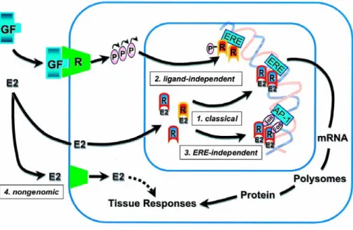
et al, 1998). This occurs when ER either binds estrogen response elements on the promoters of target genes, or acts through protein-protein interactions involving a variety of coactivators,corepressors and the basal transcriptional machinery protein complex. Emerging evidence, however, has implicated a second distinctmechanism of E2 action, where this steroid binds a putativeplasma membrane ER (Pietras R & Szego, 1980) and it elicits rapid non transcriptional effects involving activation of signal transducing pathways, such as protein kinase A, MAPK signaling and phosphotidylinositol 3-kinase/Akt pathway (Fig. 3) (Migliaccio et al, 1996; Simoncini et al, 2000; Hall et al, 2001; Simoncini et al, 2003).
Introduction
Fig. 3 The multifaceted mechanisms of estradiol and estrogen receptor signaling.
Akt (protein kinase B or PKB) has been identified as a downstream target of growth factor receptor (GF-R) activation, including insulin like growth factor-1 (IGF-1), epidermal growth factor (EGF), basic fibroblast growth factor (bFGF) (Datta et al, 1999; Downward, 1998). In addition, ERα has been also shown to interact with GF and induce GF-R and extracellular signal-regulated kinase activation, resulting in synergistic tumoral growth stimulation (Kahlert et al, 2000; Levin, 2005; Stoll, 2002; Surmacz & Bartucci, 2004).
In the present study, we investigate whether 17-β estradiol (E2) is able to induce rapid effects on aromatase activity in estrogen dependent MCF-7 breast cancer epithelial cells. Our results, for the first time, demonstrate that E2/ERα, through a non transcriptional event, can activate PI3K/Akt signaling pathway and then increase aromatase enzymatic activity. Besides, it emerges that aromatase is a target of a cross-talk between ERα and GF-R transductional pathways. Local estrogen production, through a short autocrine loop, sustains aromatase enzymatic activity in breast cancer cells and furthemore gives a great emphasis to
Materials and Methods
MATERIALS AND METHODS Materials
Dulbecco’s Modified Eagle’s Medium/Nutrient Mixture F-12 Ham (DMEM/F12), Aprotinin, leupeptin, phenylmethylsulfonyl fluoride (PMSF), sodium orthovanadate, Tamoxifen, 17-β estradiol, LY294002, IGF-1, EGF, AG 1478 and AG1024 (Sigma, Milan, Italy).
L-Glutamine, penicillin, calf serum (CS), streptomycin, bovine serum albumine (BSA), phosphate-buffered saline (PBS) (Eurobio, Les Ullis Cedex, France).
Triazol Reagent, 100 bpDNA ladder, Ni-NTA Agarose, Lipofectamine 2000 Reagent (Invitrogen life technologies, Carlsbad, California).
FuGENE 6 (Roche, Indianapolis, Indiana).
GoTaq DNA polymerase (Promega, Madison, Wisconsin). The RETROscript kit (Ambion, Austin, Texas).
Protein A/G-agarose plus (Santa Cruz Biotechnology , Santa Cruz, California) . ECL System (Amersham Pharmacia, Buckinghamshire, UK).
[1β-3H]androst-4-ene-3,17-dione (PerkinElmer Life Science, Wellesley, MA, USA). Letrozole (Novartis).
ICI 182,780 (Tocris chemical, Bristol, UK).
Cell Cultures
Wild-type human breast cancer ERα-positive MCF-7 cells were gifts from E. Surmacz (Philadelphia, PA). This cell line was cultured in DMEM/F-12 medium
Materials and Methods
containing 5% calf serum, 1% L-glutamine and 1mg/ml penicillin-streptomycin. The cells were cultured in Phenol Red-free DMEM containing 0.5% bovine serum albumine and 2 mM L-glutamine for 24 h before each experiment.
His6-arom plasmid construction
His6-arom plasmid (Fig. 4) constructed was used to express the C-terminus 6xHis-tagged form of human aromatase. The plasmid pUC19-arom containing the complete coding sequence of human aromatase (CYP19), kindly provided by Dr. E. R. Simpson and Dr. C. D. Clyne (Prince Henry’s Institute of Medical Research, Clayton, Australia), was used as template.
The 6xHis epitope tag was inserted by two PCR reactions using the following primers:
forward 5’-ATATAAGCTTATGGTTTTGGAAATGCTGA-3’ reverse 5’-ATGATGATGGTGTTCCAGACACCT-3’
5’-ATATTCTAGACTAATGATGATGATGATGATGGTGTTCCAGA-3’. PCR product was sub-cloned into HindIII/XbaI sites of pcDNA3.1 and Hys6-arom sequence was confirmed by nucleotide sequence analysis. We proved that the enzymatic activity of polyhistidine-containing recombinant protein was well preserved by measuring aromatase activity in MCF-7 cells transiently transfected with Hys6-arom vector.
Materials and Methods
Fig. 4 Schematic map of the His6-arom construct used in this study
Site-directed mutagenesis
This step was performed with the QuickChange™ Site-Directed Mutagenesis method from Stratagene. Briefly, this was based on a PCR reaction with two complementary oligonucleotide primers containing the mutation. The PCR was performed with the Pfu DNA polymerase during 16 cycles (30 s at 95 °C, 30 s at 55 °C and 7 min at 68 °C), using the templates and the specific oligonucleotides summarized in Table 1. The PCR products were then digested with DpnI which only digests the parental methylated cDNA. Nicked vector DNA with the desired mutations was then transformed into Escherichia coli XL1-Blue supercompetent cells and the constructed mutated expression vector was confirmed by DNA sequencing.
Materials and Methods
Table 1
Mutants Template Primer Sequence
YsbdF YhemeF Yheme/sbdF CYP19 CYP19 YhemeF Forward Reverse Forward Reverse Forward Reverse 5’-GGAAAACTTCATTtttGAGAGCATGCGGTACCAGCCTGTCG-3’ 5’-CGACAGGCTGGTACCGCAGCTCTCaaaAATGAAGTTTTCC-3’ 5’-GGCTGTGCAGGAAAGttcATCGCCATGGTGATG-3’ 5’-CATCACCATGGCGATgaaCTTTCCTGCACAGCC-3’ 5’-GGAAAACTTCATTtttGAGAGCATGCGGTACCAGCCTGTCG-3’ 5’-CGACAGGCTGGTACCGCAGCTCTCaaaAATGAAGTTTTCC-3’ Transient transfection
Transient transfection was performed using the FuGENE 6 reagent as recommended by themanufacturer with the mixture containing 3 µg/well of CYP19 vector or YhemeF, YsbdF or Yheme/sbdF mutants. A set of experiments was performed cotransfecting3 µg/well of CYP19 and 3 µg/well of the kinase-inactive dominant negative Akt mutant (AktK179M) a gift from Dr. T. Simoncini (University of Pisa, Italy). Another set of experiments was carried out using PTP1B-WT kindly provided by Dr MJ Quon (National Institute of Health, Bethesda, Meryland, USA).
Materials and Methods
transfection. 24 hours after transfection aromatase activity was determined under different treatments.
To perform immunoblotting assay, MCF-7 cells were plated in 10 cm dishes and then transfectedwith 10 µg of Hys6-arom or CYP19 using FuGENE 6 reagent and then exposed to different treatment. For the siRNA studies, Stealth Select RNAi annealed duplexes (25pb double stranded RNA) against human PTP1B was obtained from Invitrogen and used according to the manifacturer’s instructions.
Aromatase Activity Assay
The aromatase activity in subconfluent MCF-7 cells culture medium was measured by the tritiated water release assay using 0.5 µM [1β-3 H]androst-4-ene-3,17-dione assubstrate (Lephart & Simpson, 1991). The incubations were performed at 37°C for 5 h under an air/CO2 (5%) atmosphere. The results obtained were expressed as fmol/h and normalized to mg of protein(fmol/h/mg protein).
Total RNA Extraction and Reverse Transcription-PCR Assay
Total cellular RNA was extracted from MCF-7 cells using TRIzol reagent as suggested by the manufacturer. The purity and integrity of the RNA were checked spectroscopically and by gel electrophoresis before carrying out the analytical procedures.
Materials and Methods
Aromatase mRNA was analyzed by the reverse transcription-PCR method. cDNA was synthesized by oligo(dT) using a RETROscript kit as suggested by the manufacturer. The cDNAs obtained were further amplified by a PCR using the following primers: 5’-CAAGGTTATTTTGATGCATGG-3’ (forward, nucleotides 776–796) and 5’-TTCTAAGGTTTGCGCATGA-3’ (reverse, nucleotides 1261– 1241) for human P450arom and CTCAACATCTCCCCCTTCTC-3’ and 5’-CAAATCCCATATCCTCGT-3’ for the internal control gene 36B4. The PCR was performed for 35 cycles (94 °C for 1 min, 55 °C for 1 min, and 72 °C for 2 min) for P450arom and 15 cycles (94 °C for 1 min, 59 °C for 1 min, and 72 °C for 2 min) to amplify 36B4 in the presence of 5 µl and 1 µl of first strand cDNA, respectively, 1 µM each of the primers mentioned above, 0.5 mM dNTP, Taq DNA polymerase (2 units/tube) in a final volume of 25 µl. To check for the presence of DNA contamination, a reverse transcription-PCR was performed on 1µg of total RNA without Moloney murine leukemia virus reverse transcriptase (negative control). The PCR products were resolved on 2% agarose gels and bands were visualized by staining with ethidium bromide.
Standard DNA (100-bp DNA ladder) was run to provide the appropriate size marker.
Immunoblotting and Immunoprecipitation Analysis
For western blot analysis MCF7 cells, grownin 10 cm dishes to 70% to 80% confluence, were treated with 100nM of E2 at different timesbefore lysisin 500 µL of lysis buffer [50 mmol/L HEPES (pH 7.5),150 mmol/L NaCl, 1.5 mmol/L MgCl2, 1 mmol/L EGTA, 10% glycerol,1% Triton X-100, a mixture of protease inhibitors (aprotinin,phenylmethylsulfonyl fluoride, and Na-orthovanadate)]. Equal amounts of
Materials and Methods
total protein were resolved on 11% SDS-PAGE. The proteins were transferred onto a nitrocellulose membrane, probed with rabbit polyclonal antiserum directed against the human placental P450arom (Hauptman-Woodward, Medical Research Institute, Inc, Buffalo, NY) or GAPDH (FL-335, Santa Cruz Biotechnology, Santa Cruz, CA). Immunoprecipitation studies were carried out with cellular extracts from MCF-7 cells transiently transfected with empty vector or CPY19 plasmid for 24 hours or transfected with siRNA against PTP1B for 72 hours before lysis. The antibodies used for immunoprecipitation are rabbit polyclonal anti-PTP1B (H-135, Santa Cruz Biotechnology) or anti-Akt 1/2 (H-136, Santa Cruz Biotechnology). 600 µg of protein extracts were incubated at 4°C overnight under slow rotation with 1 µg of corresponding antibody and 20 µl of protein A/G. Immunoprecipitates were collected by centrifugation at 12,000 x g for 10 minutes, followed by washing three times with HNTG (immunoprecipitation) buffer [50 mmol/L HEPES (pH 7.4), 50 mmol/L NaCl, 0.1% Triton X-100, 10% glycerol, 1 mmol/L phenylmethylsulfonyl fluoride, 10 µg/mL leupeptin, 10 µg/mL aprotinin, 2 µg/mL pepstatin]. Following the final wash, supernatant was removed. Samples were resuspended in the Laemmli sample buffer, subjected to SDS-polyacrylammide gel electrophoresis (11% gel [w/v]) and then transferred onto a nitrocellulose membrane. The immunoprecipitated proteins were detected by Western blot using a mouse monoclonal antibody against human cytochrome P450arom (Serotec, Oxford, UK), a rabbit polyclonal antibody against PTP1B and Akt 1/2. Microsomal extracts from placenta were used as positive control. Negative control was performed by incubation of MCF-7 lysates with protein A/G agarose and normal rabbit serum. The immunocomplexes were detected with an enhanced chemiluminescence detection kit.
Materials and Methods
Detection of His6-tagged aromatase protein by Western Blotting
MCF7 cells were transiently transfected with His6-arom vector and exposed to different treatmentsbefore lysis. 300µg of cellular proteins were incubated for two hours with Ni-NTA agarose beads a 4°C under slow rotation. Ni-NTA resin was used to isolate P450 aromatase tagged with six tandem histidine residues from cellular lysates. The beadscontaining bound proteins were washed thrice by centrifugationin PBS buffer added with a mixture of protease inhibitors, then denatured by boiling in Laemmli sample buffer and analyzed by Western blot to identify aromatase protein content and its phosphorylation status. Membranes were probed with a mouse monoclonal antibodies directed against P450 human aromatase or phosphotyrosine-containing proteins (pY99, Santa Cruz Biotechnology) and a rabbit polyclonal antiserum directed against phosphoserine-containing proteins (pSer, Bioreagents, Canada). Two set of controls were done in parallel: surnatant removed after the first centrifugation was added to one control and vector-transfected cell lysates plus Ni-NTA agarose beads was included in the other control. Microsomal extracts from placenta were used as positive control.
Statistical Analysis
Each datum point represents the mean ± S.E. of three different experiments. Data were analyzed by ANOVA test using the STATPAC computer program.
Results
RESULTS
Short time estradiol exposure increases aromatase activity in MCF-7 cells
We first aimed to evaluate the effects of estrogens on aromatase activity by tritiated water assay in MCF-7 cells incubated for 10, 30 and 120 minutes in the presence of 100nM of E2. As reported in Fig. 5A, E2 enhanced significantly enzymatic activity at all times investigated and a maximal stimulatory effect was achieved at 120 minutes. The same induction by E2 was also observed in MCF-7 cells transiently transfected with the aromatase gene (CYP19 MCF-7), that displayed a 6-fold increase in enzymatic activity (95.36 ± 0.92 fmol/h/mg protein) compared to wild-type MCF-7 cells (15.16 ± 0.47 fmol/h/mg protein) (Fig. 5B).
The aromatase inhibitor letrozole and ER antagonists, ICI 182,780 and Tamoxifen (TAM) at a concentration of 1µM were able to abrogate the maximal enhancement induced by E2 (Fig. 5C).
This latter result indicates that estrogen can increase aromatase activity by binding to estrogen receptors.
Results
Fig. 5 Effects of E2 on aromatase activity in cultured 7 cells: Wild type (A) or CYP19
MCF-7 cells (B) were treated with vehicle or E2 100nM for 10, 30 and 120 minutes. C, CYP19 MCF-7 cells were pretreated with 1µM letrozole, ICI 182,780 and Tamoxifene (TAM) for 30 minutes and then exposed to 100nM of E2 for 120 minutes. Aromatase activity was evaluated by measuring the tritiated water released from MCF-7 cell cultures after incubation with 0.5 µM [1β-3 H]androst-4-ene-3,17-dione at 37 °C for 5 h. The results obtained were expressed as fmol [3H]/H
2O released and were normalized for mg of protein(fmol/h/mg protein). Empty vector: aromatase activity measured in cells transfected with pUC19 vector. The values represent the means ± S.E. of three different experiments, each performed with triplicate samples. * p<0.01 compared to vehicle; ** p<0.01 compared to E2
-Results
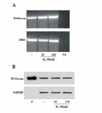
P450arom mRNA and protein expression in E2-treated MCF-7 cells
In MCF-7 cell line we investigated the effects of E2 on P450arom mRNA levels by reverse transcription-PCR. As shown in Fig. 6A, the expected transcript of 465 bp was clearly detected using primers designated to amplify the highly conserved sequence of P450arom, which includes the helical and aromatase regions. The treatment with E2 100nM for 30 and 120 minutes did not induce any change on P450arom mRNA. mRNA expression of the aromatase gene was normalized by the human housekeeping gene 36B4 (Fig. 6A).
Next, we performed western blotting analysis using a rabbit polyclonal antiserum against human placental P450arom. A 55 kDa specific protein in MCF-7 cells comigrated with microsomal extracts from human placenta used as positive control. The intensity of the band in E2-treated MCF-7 samples was not modified compared to the control (Fig. 6B).
These results evidence that the changes in E2-induced aromatase enzymatic activity were not correlated with any variations in the transcription of P450arom mRNA and thus in concentration of the enzyme.
Results
Fig. 6 P450arom mRNA and protein expression in E2 treated MCF-7 cells: MCF-7 cells were
treated with vehicle or E2 100nM for 30 and 120 minutes. A, total RNA was isolated from MCF-7 cells and reverse transcribed. cDNA was subjected to PCR using specific primers for P450arom (35 cycles) or 36B4 (15 cycles). NC: negative control, RNA sample without the addition of reverse transcriptase. B, MCF-7 cells were immunoblotted with a rabbit polyclonal antiserum against human placental P450arom. Microsomal extracts from human placenta (P) were used as positive control. GAPDH served as loading control.
Results
E2 increases tyrosine phosphorylation levels of aromatase protein
It is well known that the activity of many enzymes can be modulated rapidly by phosphorylation processes inducing conformational changes in the enzyme molecule (Albert et al, 1984; Daubner et al, 1992). Previous analyses of the aromatase gene in a variety of mammalian and avian species had demonstrated several consensus sites of phosphorylation on aromatase cDNA and deduced aminoacid sequence (Harada, 1988; McPhaul et al, 1988; Shen et al, 1994).
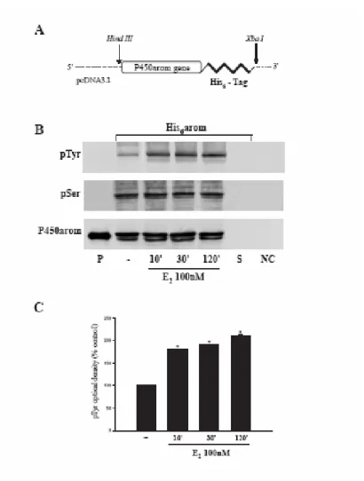
To evaluate carefully the phosphorylation status of aromatase protein, we constructed a plasmid (His6-arom) containing full length of human aromatase gene bearing six tandem histidine residues on the carboxyl terminus, as described in Materials and Methods (Fig. 7A). Indeed, this His6-tagged protein had the advantage to allow a higher yield of purified aromatase due to the specificity of Ni-NTA agarose beads and avoid interference with the band of 55kDa from heavy chains of antibodies used for immunoprecipitation. Thus, MCF-7 cells were transiently transfected with His6-arom and treated with E2 100nM for 10, 30 and 120 minutes. Equal amounts of proteins were incubated with Ni-NTA agarose beads for isolation of recombinant P450arom. Western blotting with antibodies directed against phospho-tyrosine and phospho-serine residues showed that E2 was able to increase phospho-tyrosine levels of purified aromatase protein, while no changes were detectable on serine phosphorylation status. P450 human aromatase antibody was used as loading control (Fig. 7B and C).
These data indicate, for the first time, how tyrosine phosphorylation processes may play a key role in the modulation of human aromatase enzymatic activity by E2.
Results
Fig. 7 Enhancement of tyrosine phosphorylation of aromatase protein induced by E2 : A,
Schematic map of the His6-arom construct used in this study. B, MCF-7 cells transiently transfected with His6-arom were treated with vehicle or E2100nM for 10, 30, 120 minutes. Aromatase was purified using Ni-NTA agarose beads and then the complexes were resolved in SDS-PAGE. Immunoblotting was performed using the anti-phosphotyrosine (pTyr) and anti-phosphoserine (pSer) antibodies. To verify equal loading, the membrane was probed with an antibody against P450 human aromatase. Microsomal extracts from placenta (P) served as positive control. As negative controls we used the surnatant removed after incubation with Ni-NTA agarose beads (S) and vector-transfected MCF-7 cell lysates incubated with Ni-NTA agarose beads (NC). C, The histograms represent the means ± S.E. of three separate experiments in which band intensities were evaluated in terms of optical density arbitrary units and expressed as percentages of the control, which was assumed to be 100%. * p<0.01 compared to vehicle.
Results
Aromatase activity is enhanced by tyrosine phosphatases inhibitor
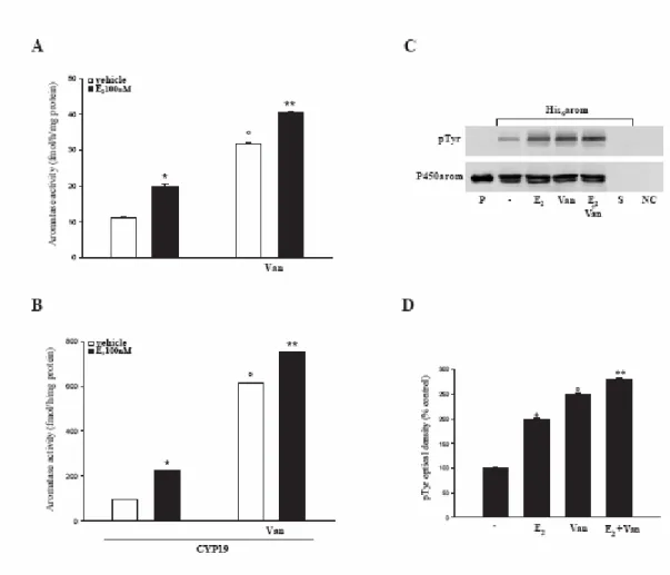
To support the idea that aromatase activity may be modulated through changes in protein tyrosine phosphorylation status, we performed enzymatic assay and immunoblot analysis in the presence of a specific inhibitor of tyrosine phosphatases, sodium orthovanadate (Van).
Treatment of MCF-7 cells with Van (10µM) increased basal levels of aromatase activity compared to the control (vehicle) and potentiated the up-regulatory effects induced by E2 (Fig. 8A). Both events are amplified in MCF-7 cells transiently transfected with CYP19 vector (Fig. 8B).
It is worth to underline that the enzymatic changes induced by Van well fit with a specific enhancement of the tyrosine phosphorylated status in the His6-tagged purified aromatase protein (Fig. 8C and D).
Results
Fig. 8 Tyrosine phosphatase inhibitor, sodium orthovanadate, increases aromatase activity: Wild
type (A) or CYP19 MCF-7 cells (B) were pretreated with 10µM sodium orthovanadate (Van) for 30 minutes and then exposed to E2 100nM for 120 minutes. Aromatase activity was evaluated by measuring the tritiated water released from MCF-7 cell cultures after incubation with 0.5 µM [1β-3H]androst-4-ene-3,17-dione at 37 °C for 5 h. The results obtained were expressed as fmol [3H]/H
2O released and were normalized for mg of protein(fmol/h/mg protein). The values represent the means ± S.E. of three different experiments, each performed with triplicate samples. * p<0.01 E2 vs vehicle; ° p<0.01 Van vs vehicle; ** p<0.01 E2+Van vs Van. C, MCF-7 cells transiently transfected with His6 -arom were treated with vehicle or E2100nM for 120 minutes, with or without Van. Aromatase was purified using Ni-NTA agarose beads and then the complexes were resolved in SDS-PAGE. The membrane was probed with anti-phosphotyrosine (pTyr) antibody. To verify equal loading, the membrane was probed with an antibody against P450 human aromatase. Microsomal extracts from placenta (P) served as positive control. As negative control we used the surnatant removed after incubation with Ni-NTA agarose beads (S) and vector-transfected MCF-7 cell lysates incubated with Ni-NTA agarose beads (NC). D, The histograms represent the means ± S.E. of three separate experiments in which band intensities were evaluated in terms of optical density arbitrary units and expressed as percentages of the control, which was assumed to be 100%. * p<0.01 E2 vs vehicle; ° p<0.01 Van vs vehicle; ** p<0.01 E2+Van vs Van.
Results
Role of the protein tyrosine phosphatase, PTP1B, in E2-stimulated aromatase activity
The phosphorylation of proteins on tyrosine residues is modulated by the competing activities of protein tyrosine kinases and protein tyrosine phosphatases (PTPs), a large family of enzymes that serves as key regulatory components in signal transduction pathways. Since PTP1B is the PTPase highly expressed in several human breast cancer cell lines (Wiener et al, 1994; Bjorge et al, 2000), we focused our attention on the potential role of this phosphatase in modulating aromatase activity in MCF-7 cells.
Here, by western blot analysis, we demonstrated the expression of PTP1B in MCF-7 cell extracts and the specific association between PTP1B and aromatase at protein-protein level using cell lysates from wild-type and CYP19 MCF-7 cells immunoprecipitated with anti-PTP1B (Fig. 9A).
To directly evaluate the role of PTP1B in E2-stimulated aromatase activity, we performed tritiated water release assay in MCF-7 cells overexpressing wild type human PTP1B.
PTP1B overexpression reduced basal and E2-induced levels of aromatase activity by about 50% compared to the control (empty vector), even though the estradiol stimulatory effects still persist (Fig. 9B).
Results
Fig. 9 Physical and functional interaction of P450arom and PTP1B: A, In vitro association of
P450arom and PTP1B was revealed by co-immunoprecipitation analysis. Protein extracts (600µg) from MCF-7 cells transiently transfected with empty vector or CYP19 vector were immunoprecipitated with an antiserum against PTP1B (IP: anti-PTP1B) and then the immunocomplexes were resolved in SDS-PAGE. The membrane was blotted with P450arom antibody. To verify equal loading, the membrane was probed with an antibody against PTP1B. Lys: lysates from wild-type MCF-7 cells. NC: negative control was performed by incubation of MCF-7 cell lysates with protein A/G agarose and normal rabbit antiserum. B, MCF-7 cells were transiently transfected with 3 µg of empty-vector or PTP1BWT and treated with E2 100nM. After 120 minutes the cells were assayed for aromatase activity. The values represent the means ± S.E. from triplicate assays. * p<0.01 compared to cells transfected with empty vector; ** p<0.01 compared to E2 treated cells transfected with empty vector.
Results
Aromatase activity is up-regulated by E2 through PI3K/Akt pathway
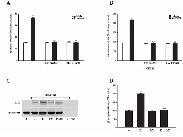
It has been demonstrated that the tyrosine phosphatase PTP1B is a substrate for Akt and the phosphorylation at Ser50 by Akt impairs the ability of the enzyme to engage and dephosphorylate its substrates (Ravichandran et al, 2001). On the other hand, it is well known that PI3K/Akt pathway is activated by non genomic estradiol signal in MCF-7 cell line (Castoria et al, 2001; Stoica et al, 2004). Thus, it is reasonable to assume that the same pathway may be a crucial intermediate of E2 induction on aromatase activity. To support this assumption, we performed enzymatic assay in MCF-7 cells treated with the PI3-kinase inhibitor LY294002 or transfected with a dominant negative of Akt-expressing construct (Akt K179M). As shown in Fig. 10A and B, our results revealed that either LY294002 (10µM) or Akt K179M were able to abrogate the E2 stimulatory effects in wild-type as well as in CYP19 MCF-7 cells. In agreement with these results, LY294002 reduced the E2-associated tyrosine phosphorylation level of the protein aromatase purified from MCF-7 cells transfected with Hys6-arom construct, as revealed by western blot analysis (Fig. 10C and D).
These data lead us to hypothesize that E2-activation of aromatase is a phosphorylation dependent event mediated by PI3K/Akt pathway.
Results
Fig. 10 PI3K/Akt pathway mediates E2–induction on aromatase activity: Wild-type (A) or CYP19
MCF-7 cells (B) were transiently transfected with empty vector or AktK179M, or pretreated with LY294002 (10µM) 30 minutes before E2 stimulation. After 120 minutes aromatase activity was performed. The values represent the means ± S.E. from triplicate assays. *p<0.01compared to vehicle; **p<0.01 compared to E2 treated samples. C, MCF-7 cells transiently transfected with His6-arom were treated with vehicle or E2 100nM, LY294002 (10µM) or E2 + LY. Aromatase recombinant protein was purified and then resolved in SDS-PAGE. The membrane was probed with anti-phosphotyrosine (pTyr) antibody. To verify equal loading, the membrane was probed with an antibody against P450 human aromatase. Microsomal extracts from placenta (P) served as positive control. As negative controls we used the surnatant removed after incubation with Ni-NTA agarose beads (S) and vector-transfected MCF-7 cell lysates incubated with Ni-NTA agarose beads (NC). D, The histograms represent the means ± S.E. of three separate experiments in which band intensities were evaluated in terms of optical density arbitrary units and expressed as percentages of the control, which was assumed to be 100%. *p<0.01compared to vehicle; **p<0.01 compared to E2 treated samples.
Results
A protein complex involving Akt, P450arom and PTP1B activates aromatase
After previously demonstrating the physical association of PTP1B with P450arom (Fig. 9A), we identified a protein complex involving Akt, P450arom and PTP1B by immunoprecipitation of Akt and detection of coimmunoprecipitated PTP1B and P450arom under basal conditions in MCF-7 cell lysates (Fig. 11A). We confirmed the formation of the protein complex by using a reciprocal order of immunoprecipitation of PTP1B and then detection of both P450arom and Akt on Western blot (data not shown).
To test whether PTP1B is required for the physical interaction between P450arom and Akt, we transfected MCF-7 cells with a specific siRNA against human PTP1B and 72 hours after transfection we documented a marked decrease of PTP1B expression compared with levels of control proteins. Our results, using cell lysates immunoprecipitated with anti-Akt, revealed that down-regulation of PTP1B blocked P450arom/Akt interaction (Fig. 11A), suggesting that this tyrosine phosphatase may play an important role in MCF-7 breast cancer cells by linking the serine/threonine kinase Akt to P450 aromatase.
Moreover, to corroborate the involvement of PI3K/Akt pathway through PTP1B in E2-activation of aromatase, we performed tritiated water release assay in MCF-7 cells cotransfected with PTP1B-WT and Akt K179M. The presence of the kinase-inactive dominant negative Akt mutant completely reversed the E2-induction on enzymatic activity (Fig. 11B).
Results
Fig. 11 Physical and functional interaction of P450arom, PTP1B and Akt: A, In vitro association
of P450arom, PTP1B and Akt was revealed by co-immunoprecipitation analysis. Protein extracts (600µg) from MCF-7 cells untransfected or transfected with a specific siRNA against human PTP1B for 72 hours were immunoprecipitated with an antiserum against Akt (IP: anti-Akt) and then the immunocomplexes were resolved in SDS-PAGE. The membrane was blotted with P450arom and PTP1B antibody. To verify equal loading, the membrane was probed with an antibody against Akt. Lys: lysates from wild-type 7 cells. NC: negative control was performed by incubation of MCF-7 cell lysates with protein A/G agarose and normal rabbit antiserum. B, Another set of experiments was carried out cotransfecting MCF-7 cells with PTP1BWT and AktK179M. 24h after transfection MCF-7 cells were treated with E2 100nM for 120 minutes and then assayed for aromatase activity. The values represent the means ± S.E. from triplicate assays. *p<0.01compared to vehicle; **p<0.01 compared to E2 treated samples.
Results
In summary, the data obtained suggest that the rapid E2 up-regulation of aromatase activity in MCF-7 cells occurs through the activation of PI3K/Akt pathway leading to a block of PTP1B and an enhanced tyrosine phosphorylation status of aromatase protein (Fig. 12)
Fig. 12 Hypothesized model of estradiol signalling in modulating aromatase activity in breast cancer
Further investigations are necessary to asceirtain whether PTP1B/Arom complex may be the effector of other mitogenic-stimulated signaling, that contributes to the regulation of the intrinsic aromatase enzymatic activity in breast cancer cells.
PTP1B Aromatizable Androgens EE E E E E ER E E E E Akt PI3K AROM AROM E P P P P -+ Cytosol Breast Tumor Progression
Results
Role of tyrosine residues in the E2-activation of aromatase
Site-directed mutagenesis experiments were performed to explore the role of the residues specifically involved in the E2 induction. Consensus phosphorylation sites were analysed using the public domain software (NETPHOS 2.0 PREDICTION server) available on the web site of the Center for Biological Sequence Analysis at
http://www.cbs.dtu.dk. Based on a deduced amino acid sequence (in the present case, the
human aromatase) and on a previously encoded database of potential phosphorylation sites (PHOSPHOBASE 2.0), this program identified all serine, threonine and tyrosine residues in the protein that could potentially be phosphorylated (Kemp & Pearson, 1990; Kennelly & Krebs, 1991). The program also provided for each residue a phosphorylation score ranging from 0 to 1.0 whose value was proportional to the probability that the residue could in fact be phosphorylated in vivo. A score equal or larger to 0.5 was considered to predict a likely phosphorylation consensus site (see Blom et al, 1999 for more detail). The NETPHOS 2.0 program (http://www.cbs.dtu.dk/services/NetPhos/) identified four of the 17 tyrosine residues. These
sites correspond to the Tyrosine residues located at positions 77, 184, 361 and 441 of the human aromatase sequence. Notably, the positions at 361 and 441 correspond to the two residues present in important functional domains of human aromatase (Fig. 13).
Results
Fig. 13 Amino acid sequence of human aromatase: in red are indicated the different functional
domains of the enzyme and in bold case Y the potential phosphorylation consensus site.
Therefore, we created three different constructs in which the tyrosine residues (Y) were mutated to phenylalanine (F):
1. YsbdF plasmid was mutated in Y361 in steroid-binding domain; 2. YhemeF plasmid was mutated in Y446 in heme-binding domain; 3. Yheme/sbdF plasmid was mutated in Y361 and Y446.
As shown in Fig. 14, E2 as well as Van were not able to up-regulate aromatase activity in MCF-7 cells transfected with these mutant expression vector. These results further support that tyrosine phosphorylation may represent the molecular mechanism that rapidly changes aromatase activity following estradiol-treatment.
MVLEMLNPIHYNITSIVPEAMPAATMPVLLLTGLFLLVWNYEGTSSIPGPGYCMGI GPLISHGRFLWMGIGSACNYYNRV 80 YGEFMRVWISGEETLIISKSSSMFHIMKHNHYSSRFGSKLGLQCIGMHEKGIIFNNN PELWKTTRPFFMKALSGPGLVRM 160 VTVCAESLKTHLDRLEEVTNESGYVDVLTLLRRVMLDTSNTLFLRIPLDESAIVVKI QGYFDAWQALLIKPDIFFKISWL 240 YKKYEKSVKDLKDAIEVLIAEKRRRISTEEKLEECMDFATELILAEKRGDLTRENV NQCILEMLIAAPDTMSVSLFFMLF 320 LIAKHPNVEEAIIKEIQTVIGERDIKIDDIQKLKVMENFI
Y
ESMRYQPVVDLVMRKA LEDDVIDGYPVKKGTNIILNIGR 400 MHRLEFFPKPNEFTLENFAKNVPYRYFQPFGFGPRGCAGKY
IAMVMMKAILVTLL RRFHVKTLQGQCVESIQKIHDLSLH 480 PDETKNMLEMIFTPRSSDRCLEHSTEROID BINDING DOMAIN
HEME BINDING DOMAIN AROMATIC REGION
TRANSMEMBRANE
Results
Fig. 14 Tyrosine residues involvement in aromatase activation: MCF-7 cells were transfected with
CYP19 vector or YsbdF or YhemeF or Yheme/sbdF mutants and then treated with 100nM E2 or 10µM Van for 120 minutes. Aromatase activity was evaluated by measuring the tritiated water released from MCF-7 cell cultures after incubation with 0.5 µM [1β-3H]androst-4-ene-3,17-dione at 37 °C for 5 h. The results obtained were expressed as percentages of the control, which was assumed to the 100%. The values represent the means ± S.E. of three different experiments, each performed with triplicate samples. * p<0.01 compared with vehicle; ** p<0.01 compared with E2 or Van treated CYP19 cells.
Results
A cross-talk between GF receptors and ER-α is invoved in the increased aromatase activity in MCF-7 cells
PI3K/Akt pathway mediates the action of several growth factors (Toker & Cantley, 1997). In addition, it has been reported, in breast cancer, a cross-talk between endogenous membrane ER and growth-factor-signaling pathways, including EGF and IGF-I (Kahlert et al, 2000; Levin, 2005; Stoll, 2002; Surmacz & Bartucci, 2004).
In order to determine whether IGF-1R or EGF-R stimulation leads to an increased production of estrogen, both wild type and CYP19 MCF-7 cells were treated with IGF-1 and AG1024, a monoclonal antibody specific to IGF-1R or EGF and a selective inhibitor of the EGF-R kinase, AG1478. Our results demonstrated that IGF-1 or EGF IGF-100ng/ml for IGF-120 minutes significantly increased aromatase enzymatic activity reproducing the same up-regulatory effects induced by E2 (Fig. 15A, B and C, D, respectively). Pretreatment with AG1024 or AG1478 10 µM for 30 minutes completely abrogated the growth-factor as well as the E2 induction of aromatase activity (Fig. 15A, B and C, D). It is worth to evidence how in the presence of short exposure to ICI 182,780 or LY294002 , IGF-1 and EGF are not able to up-regulate aromatase activity. The latter result addresses how the induction of aromatase enzymatic activity may involve the cross-talk between estradiol/ERα and GF-R signaling, both converging on the activation of the downstream PI3K/Akt pathway.
Results
Fig. 15 Induction of aromatase activity by IGF-1 and EGF: Wild-type (A, C) or CYP19 MCF-7
(B, D) were pretreated with AG1024 or AG1478 or LY294002 10µΜ or ICI 182,780 1 µΜ for 30 minutes and then exposed to E2 100nM or IGF-1 or EGF 100 ng/ml. After 120 minutes aromatase activity was performed.
The values represent the means ± S.E. from triplicate assays. *p<0.01 compared to vehicle; **p<0.01 compared to E2 treated samples; °p<0.01 compared to IGF-1/EGF treated samples.
Discussion
DISCUSSION
Locally estrogen production plays a more important role than circulating estradiol in breast tumor promotion (Miller, 2006). Several experimental systems have been used to determine the biological significance of in situ estrogen production by breast tissue. For instance, in aromatase transgenic mice an increased risk of developing neoplasia and a major susceptibility to environmental carcinogens have been observed (Tekmal et al, 1996; Gill et al, 2001). On the other hand, aromatase inhibitors appears to enhance the survival outcome in breast cancer patients (Altundag & Ibrahim, 2006).
The present study shows, for the first time, that E2 is able to modulate aromatase activity by PI3 kinase/Akt signaling cascade in estrogen dependent breast cancer epithelial cells. Of note, our results demonstrate that short exposure to E2 induces an increase on aromatase enzymatic activity of the same extent in wild-type as well as in CYP19 MCF-7 cells. This up-regulatory effect was specifically mediated by the interaction of E2 with ERs, since it was abrogated in the presence of ER antagonists, such as Tamoxifen and ICI 182,780.
Estrogen signaling has traditionally been identified with the transcriptional control of target genes via the binding of nuclear estrogen receptors to genomic consensus sequences (Green et al, 1990). Nonetheless, in the past few years, a large spectrum of biological actions of estrogen has been shown as too rapid to be compatible with transcriptional mechanisms (Hall et al, 2001). These “non classical” estrogen effects are attributed to the cell surface ERs which may activate several intracellular signal transduction pathways such as PI3 kinase/Akt, as reported in human vascular endothelial cells, human breast cancer cell lines (MCF-7) and rat
Discussion
primary cortical neurons (Honda et al, 2000; Castoria et al, 2001; Simoncini et al, 2003). For instance, the most striking example is the direct association between the ER and PI3K in cells stimulated by estrogen (Simoncini et al, 2000).
Our findings demonstrate a direct involvement of PI3K/Akt activation in E2-stimulated aromatase activity by the evidence that the PI3K/Akt inhibitor LY 294002 or a dominant negative of Akt completely reversed the increase of aromatase activity.
It is well known that PI3K/Akt pathway could be activated by several growth factor receptors (Toker & Cantley, 1997) and that ER and growth factor signaling pathways intersect and directly interact at many levels of signal transduction (Kahlert et al, 2000; Levin, 2005; Stoll, 2002; Surmacz & Bartucci, 2004). Our cell culture study shows that the treatment of ER positive MCF-7 cells with IGF-1 or EGF increased, at short time, aromatase activity. It is worth to evidence how the two GF-R tyrosine kinase inhibitor or the pure antiestrogen ICI 182,780 or LY 294002 were able to abrogate the GF as well as the E2 induction of aromatase activity. These results suggest that the upregulation of aromatase activity in MCF-7 cells by E2 may involve a cross-talk between ERα and the GF/ PI3K/Akt pathway.
The E2-enhanced aromatase activity was not correlated with any increase in the level of aromatase mRNA and protein content, suggesting a post-transcriptional modulation of aromatase protein.
Post-translational modification of enzymatic activity has been demonstrated for different members of the P450 enzyme family in vertebrates. For instance, cAMP-dependent protein kinase was essential for the activation of human and rat cholesterol 7α-hydroxylase (CYP7A) (Nguyen et al, 1996) as well as for phosphorylation of serine and threonine residues in human P450c17 (CYP17) (Zhang
Discussion
et al, 1995; Miller et al, 1997; Biason-Lauber et al, 2000). Bovine P450scc
(CYP11A1) has been identified as an active form phosphorylated by a protein kinase C (Defaye et al, 1982; Vilgrain et al, 1984) and similar activation of P450s through phosphorylation has been found in human liver enzymes such as CYP2E1 and CYP2B1 (Oesch-Bartlomowicz et al, 1990; Oesch-Bartlomowicz et al, 1998). Besides, several studies uncovered evidences of a rapid regulation of P450 aromatase via processes such as protein phosphorylation in the brain (Balthazart et al, 2001; Balthazart et al, 2003). A rapid increase of aromatase activity was reported in human ejaculated spermatozoa through the activation of cAMP/PKA signaling pathway (Aquila et al, 2002).
In the current study, we demonstrate, in MCF-7 cells, that E2 up-regulatory effects on aromatase activity involve rapid changes in phosphorylation status of this protein. Indeed, we observed after E2 treatment a specific enhancement of tyrosine phosphorylation levels in recombinant aromatase protein purified by Ni-NTA agarose beads. Additional support to the idea that tyrosine phosphorylation processes may affect the enzymatic activity of aromatase protein was provided by experiments utilizing inhibitor of tyrosine phosphatases. Sodium orthovanadate increased basal and E2 induced enzymatic activity as well as tyrosine phosphorylation of aromatase purified protein. In addition, site-directed mutagenesis experiments evidenced that phosphorylation of tyrosine residues present in important functional domains of human aromatase were involved in the up-regulation of enzymatic activity. Taken together, these data demonstrate that the rapid changes in aromatase activity result from a direct phosphorylation of the enzymatic protein itself.
Tyrosine phosphorylation is a reversible and dynamic process controlled by the activities of the protein tyrosine kinases (PTKs) and the competing actions of the
Discussion
protein tyrosine phosphatases (PTPs). In this respect, our findings documented a protein-protein interaction between the protein tyrosine phosphatases, PTP1B, and aromatase. PTP1B, the primary phosphatase in human breast cancer cell lines, is an endoplasmic reticulum-associated protein tyrosine phosphatase (Wiener et al, 1994; Bjorge et al, 2000), implicated in the regulation of a variety of cellular processes such as cell growth, differentation and transformation (Dube & Tremblay, 2005). To better define the role of this phosphatase in modulating aromatase activity, we performed enzymatic assay in MCF-7 cells overexpressing PTP1B. In this condition, we observed a reduction of aromatase activity both in untreated as well as E2-treated MCF-7 cells. It is worth to note that E2-induction of enzymatic activity was completely reversed in MCF-7 cells cotransfected with PTP1B and the dominant negative of Akt. These data further support the involvement of PI3K/Akt pathway in E2-enhanced aromatase activity through the regulation of PTP1B activity. Our findings well fit with previous results demonstrating that PTP1B is a substrate of the ser/thr kinase Akt (Ravichandran et al, 2001). Indeed, PTP1B contains the consensus phosphorylation motif (RYRDVS50) in a region that forms important stabilizing contacts with aminoacids immediately upstream from the phosphotyrosine residue of PTP1B substrates. Thus, phosphorylation of Ser50 in this region by Akt negatevely modulates its tyrosine phosphatase activity. In the current work, we identified by immunoprecipitation studies a ternary protein complex involving Akt, P450arom and PTP1B. Using a specific siRNA against human PTP1B, we revealed that down-regulation of this phosphatase blocked P450arom/Akt interaction, suggesting that PTP1B may play an important role in MCF-7 breast cancer cells by linking the serine/threonine kinase Akt to P450 aromatase.
Discussion
In summary, this study supports data suggesting that the rapid up-regulatory effects of E2 on aromatase activity in MCF-7 cells involve phosphorylation of the aromatase protein. This occurs through the activation of PI3K/Akt pathway leading to a block of PTP1B and an enhanced tyrosine phosphorylation status of aromatase protein.
From the present findings, it emerges, for the first time, that aromatase is a target of a cross-talk between ERα and GF-R transductional pathways. Local estrogen production, through a short autocrine loop, further sustains aromatase enzymatic activity in breast cancer cells (Fig. 15). These data demonstrate how aromatase may be functionalized by different signaling involved in tumor cell growth and progression and provide rational to design novel combinatory therapeutic strategies for breast cancer treatment.
Discussion
Fig. 16 Cross-talk between ERα and IGF-1R leads to downstream signaling and changes in aromatase activity in MCF-7 breast cancer cells.
References
REFERENCES
Albert KA, Helmer-Matyjek E, Nairn AA, Muller TH, Haycock JW, Greene LA, Goldstein M, Greengard P (1984) Calcium/phospholipids-dependent
protein kinase (protein kinase C) phosphorylates and activates tyrosine hydroxylase. Proc Natl Acad Sci USA 81: 7713-7717
Altundag K, Ibrahim NK (2006) Aromatase inhibitors in breast cancer: an
overview. Oncologist 11: 553-562
Aquila S, Sisci D, Gentile M, Middea E, Siciliano L, Andò S (2002) Human
ejaculated spermatozoa contain active P450 aromatase. J Clin Endocrinol Metab
87: 3385-3390
Balthazart J, Baillien M, Ball GF (2001) Phosphorylation processes mediate
rapid changes of brain aromatase activity. J Steroid Biochem Mol Biol 79: 261-277
Balthazart J, Baillien M, Charlier TD, Ball GF (2003) Calcium-dependent
phosphorylation processes control brain aromatase in quail. Eur J Neurosci 17:
1591-1606
Bellino FL, Holben L (1989) Placental estrogen synthetase (aromatase):
evidence for phosphatase-dependent inactivation. Biochem Biophys Res Commun
References
Biason-Lauber A, Kempken B, Werder E, Forest MG, Einaudi S, Ranke MB, Matsuo N, Brunelli V, Schonle EJ and Zachmann M (2000)
17alpha-hydroxylase/17,20-lyase deficiency as a model to study enzymatic activity regulation: role of phosphorylation. J Clin Endocrinol Metab 85: 1226–1231
Bjorge JD, Pang A, Fujita DJ (2000) Identification of protein-tyrosine
phosphatase 1B as a major tyrosine phosphatase activity capable of dephosphorylating and activating c-Src in several human breast cancer cell lines.
J Biol Chem 275: 41439-41446
Blom N, Gammeltoft S, Brunak S (1999) Sequence- and structure-based
prediction of eurkaryotic protein phosphorylation sites. J Mol Biol 294: 1351– 1362.
Brodie A (1991) Aromatase and its inhibitors: an overview. J Steroid Biochem Mol Biol 40: 255-261
Bulun SE, Sebastian S, Takayama K, Suzuki T, Sasano H, Shozu M
(2003)The human CYP19 (aromatase P450) gene: update on physiologic roles and genomic organization of promoters. J Steroid Biochem Mol Biol 86:219–24
Bulun SE, Price TM, Aitken J, Mahendroo MS, Simpson ER (1993) A link
between breast cancer and local estrogen biosynthesis suggested by quantification of breast adipose tissue aromatase cytochrome P450 transcripts
References
using competitive polymerase chain reaction after reverse transcription. J Clin
Endocrinol Metab 77: 1622–8
Budhram-Mahadeo V, Parker M, Latchman DS (1998) POU transcription
factors Brn-3a and Brn-3b interact with the estrogen receptor and differentially regulate transcriptional activity via an estrogen response element. Mol Cell Biol
18:1029–1041
Castoria G, Migliaccio A, Bilancio A, Di Domenico M, De Falco A, Lombardi M, Fiorentino R, Varricchio L, Barone MV, Auricchio F (2001)
PI3-kinase in concert with Src promotes the S-phase entry of oestradiol-stimulated MCF-7 cells. EMBO J 20: 6050-6059
Chen S (1998) Aromatase and breast cancer. Front Biosci 3: 922-933
Chen S, Shively JE, Nakajin S, Shinoda M, Hall PF (1986) Amino terminal
sequence analysis of human placenta aromatase. Biochem Biophys Res Commun
135: 713–9
Chong YM, Colston K, Jiang WG, Sharma AK, Mokbel K (2006) The
relationship between the insulin-like growth factor-1 system and the estrogen metabolising enzymes in breast cancer tissue and its adjacent non-cancerous tissue. Breast Cancer Res Treat advance online publication 5 June 2006; doi:10.1007/s10549-006-9215
References
Datta SR, Brunet A, Greenberg ME (1999) Cellular Survival: A play in three
Akts. Genes Dev 13: 2905-2927
Daubner SC, Lauriano C, Haycock JW, Fitzpatrick PF (1992) Site-directed
mutagenesis of serine 40 of rat tyrosine hydroxylase. J Biol Chem 267: 12639-12646
Defaye G, Monnier N, Guidicelli C, Chambaz EM (1982) Phosphorylation of
purified mitochondrial cytochromes P450 (cholesterol desmolase and 11 beta-hydroxylase) from bovine adrenal cortex. Mol Cell Endocrinol 27: 157–168
Downward J (1998) Mechanisms and consequences of activation of protein
kinase B/Akt. Curr Opin Cell Biol 10: 262-267
Dube N, Tremblay ML (2005) Involvement of the small protein tyrosine
phosphatases TC-PTP and PTP1B in signal transduction and disease: from diabetes, obesity to cell cycle, and cancer. Biochim Biophys Acta 1754: 108-117
Gill K, Kirma N, Tekman RR (2001) Overexpression of aromatase in
transgenic male mice in the induction of gynecomastia and other biochemical changes in mammary glands. J Steroid Biochem Mol Biol 77: 13-18
Hall JM, Couse JF, Korach KS (2001) The multifaceted mechanisms of
References
Harada N (1988) Cloning of a complete cDNA encoding human aromatase:
immunochemical identification and sequence analysis. Biochem Biophys Res
Commun 156: 725-732
Harada N, Utsumi T, Takagi Y (1993) Tissue-specific expression of the human
aromatase cytochrome P-450 gene by alternative use of multiple exons 1 and promoters, and switching of tissue-specific exons 1 in carcinogenesis. Proc Natl
Acad Sci U S A 90:11312–6
Harada N (1997) Aberrant expression of aromatase in breast cancer tissues. J Steroid Biochem Mol Biol 61: 175–84
Hoggard N, Rees HH (1988) Reversible activation–inactivation of
mitochondrial ecdysone 20-mono-oxygenase: a possible role for phosphorylation–dephosphorylation. J Insect Physiol 34: 647–653
Honda K, Sawada H, Kihara T, Urushitani M, Nakamizo T, Akaike A, Shimohama S (2000) Phosphatidylinositol 3-kinase mediates neuroprotection
by estrogen in cultured cortical neurons. J Neurosci Res 60: 321-327
Kahlert S, Nuedling S, van Eickels M, Vetter H, Meyers R, Grohé C (2000)
Estrogen receptor α rapidly ativates the IGF-1 receptor. J Biol Chem 275: 18447-18453
References
Kemp BE, Pearson RB(1990) Protein kinase recognition sequence motifs.
Trends Neurosci 15: 342–346.
Kennelly PJ, Krebs EG (1991) Consensus sequences as substrate specificity
determinants for protein kinases and protein phosphatases. J Biol Chem 266: 15555–15558
Kinoshita Y, Chen S (2003) Induction of Aromatase (CYP19) expression in
breast cancer cells through a nongenomic action of estrogen receptor α. Cancer
Res 63: 3546-3555
Lephart ED, Simpson ER (1991) Assay of aromatase actiity. Methods Enzymol 206: 477-483
Levin ER (2005) Integration of the extranuclear and nuclear actions of estrogen Mol Endocrinol 19: 1951-1959
Maggiolini M, Carpino A, Bonofiglio D, Pezzi V, Rago V, Marsico S, Picard D, Andò S (2001) The direct proliferative stimulus of dehydroepiandrosterone on
MCF-7 breast cancer cells is potentiated by overexpression of aromatase. Mol
Cell Endocrinol 184: 163-171
McPhaul MJ, Noble JF, Simpson ER, Mendelson CR, Wilson JD (1988) The
References
(aromatase) that catalyzes the formation of estrogen from androgen. J Biol Chem
263: 16358-16363
Means GD, Kilgore MW, Mahendroo MS, Mendelson CR, Simpson ER
(1991) Tissue-specific promoters regulate aromatase cytochrome P450 gene expression in human ovary and fetal tissues. Mol Endocrinol 5:2005–13
Migliaccio A, Di Domenico M, Castoria G, De Falco A, Bontempo P, Nola E, Auricchio F (1996) Tyrosine kinase/p21 ras/MAP-kinase pathway activation by
estradiol-receptor complex in MCF-7 cells. EMBO J 15: 1292-1300
Miller WL, Auchus RJ, Geller DH (1997) The regulation of 17,20-lyase
activity. Steroids 62: 133–142
Miller WR (1997) Uptake and synthesis of steroid hormones by the breast. Endocrin Relat Cancer 4: 307-311
Miller WR (2006) Aromatase and the breast: regulation and clinical aspects. Maturitas 54: 335-341
Nguyen LB, Shefer S, Salen G, Chiang JY, Patel M (1996) Cholesterol
7alpha-hydroxylase activities from human and rat liver are modulated in vitro post-translationally by phosphorylation–dephosphorylation. Hepatology 24: 1468–1474
References
Oesch-Bartlomowicz B, Oesch F (1990) Phosphorylation of cytochrome P450
isoenzymes in intact hepatocytes and its importance for their function in metabolic processes. Arch Toxicol 64: 257–261
Oesch-Bartlomowicz B, Padma PR, Becker R, Richter B, Hengstler JG, Freeman JE, Wolf CR, Oesch F (1998) Differential modulation of CYP2E1
activity by cAMP-dependent protein kinase upon Ser129 replacement. Exp Cell
Res 242: 294–302
Pietras R, Szego CM (1980) Partial purification and characterization of
oestrogen receptors in subfractions of hepatocyte plasma membranes. Biochem J
191:743–760
Ravichandran LV, Chen H, Li Y, Quon MJ (2001) Phosphorylation of PTP1B
at Ser50 by Akt impairs its ability to dephosphorylate the insulin receptor. Molr
Endocrinol 15: 1768-1780
Santen RJ, Yue W, Naftolin F, Mor G, Berstein L (1999) The potential of
aromatase inhibitors in breast cancer prevention. Endoc-Relat Cancer 6: 235-243
Santner SJ, Chen S, Zhou D, Korsunsky Z, Martel J, Santen RJ (1993)
Effect of androstenedione on growth of untransfected and aromatase-transfected MCF-7 cells in culture. J Steroid Biochem Mol Biol 44: 611-616
References
Sasano H, Nagura H, Harada N, Goukon Y, Kimura M (1994)
Immunolocalization of aromatase and other steroidogenic enzyme in human breast disorders. Hum Patol 25: 530-535
Shen P, Campagnoni CW, Kampf K, Schlinger BA, Arnold AP, Campagnoni AT (1994) Isolation and characterization of a zebra finch
aromatase cDNA: in situ hybridization reveals high aromatase expression in brain. Mol Brain Res 24: 227-237
Shozu M, Sumitani H, Murakami K, Segawa T, Yang HJ, Inoue M (2001)
Regulation of aromatase activity in bone-derived cells: possible role of mitogen-activated protein kinase. J Steroid Biochem Mol Biol 79: 61-65
Simoncini T, Hafezi-Moghadam A, Brazil DP, Ley K, Chin WW, Liao JK
(2000) Interaction of oestrogen receptor with the regulatory subunit of phosphatidylinositol-3-OH kinase. Nature 407: 538-541
Simoncini T, Rabkin E, Liao JK (2003) Molecular basis of cell membrane
estrogen receptor interaction with phosphatidylinositol 3-kinase in endothelial cells. Arterioscler Thromb Vasc Biol 23: 198-203
Simpson ER, Mahendroo MS, Means GD, Kilgore MW, Hinshelwood MM, Graham-Lorence S, Amarneh B, Ito Y, Fisher CR, Michael DM, Mendelson CR, Bulun SE (1994) Aromatase cytocrome P450, the enzyme responsible for
References
Simpson ER, Michael MD, Agarwal VR, Hinshelwood MM, Bulun SE, Zhao Y (1997) Expression of the CYP19 (aromatase) gene: an unusual case of
alternative promoter usage. FASEB J 11: 29-36
Sourdaine P, Mullen P, White R, Telford J, Parker MG, Miller WR (1996)
Aromatase activity and CYP19 gene expression in breast cancers. J Steroid
Biochem Mol Biol 59: 191-198
Stoica GE, Franke TF, Wellstein A, Czubaiko F, List HJ, Reiter R, Morgan E, Martin MB, Stoica A (2004) Estradiol rapidly activates Akt via the ErbB2
signaling pathway. Mol Cell Endocrinol 17: 818-830
Stoll BA (2002) Oestrogen/insulin-like growth factor-I receptor interaction in
early breast cancer: clinical implications. Ann Oncol 13: 191-6
Sun XZ, Zhou D, Chen S (1997) Autocrine and paracrine actions of breast
tumor aromatase. A three-dimensional cell culture study involving aromatase transfected MCF-7 and T-47D cells. J Steroid Biochem Mol Biol 63: 29-36
Surmacz E, Bartucci M (2004) Role of estrogen receptor alpha in modulating
IGF-1 receptor signaling and function in breast cancer. J Exp Clin Cancer Res
23:385-394.
Tekmal RR, Ramachandra N, Gubba S, Durgam VR, Mantione J, Toda K, Shizuta Y, Dillehay DL (1996) Overexpression of int-5/aromatase in mammary