Head and neck
Head and neck reconstruction with pedicled flaps
in the free flap era
Ricostruzioni del distretto testa collo con lembi peduncolati nell’era dei lembi liberi
R. MAHIEU1 2, G. COLLETTI3, P. BONOMO4, G. PARRINELLO1, A. IAVARONE1, G. DOLIVET5, L. LIVI4, A. DEGANELLO1 1 Department of Surgery and Translational Medicine, Azienda Ospedaliero-Universitaria Careggi, University of Florence,Italy; 2 University of Groningen, University Medical Center Groningen, the Netherlands; 3 Department of Maxillo-facial
Surgery, University of Milan, Milan, Italy; 4 Department of Radiation Oncology, Azienda Ospedaliero-Universitaria Careggi,
University of Florence, Italy; 5 Department of Oncologic Surgery l’institut de Cancérologie de Lorraine, Nancy, France
SUMMARY
Nowadays, the transposition of microvascular free flaps is the most popular method for management of head and neck defects. However, not all patients are suitable candidates for free flap reconstruction. In addition, not every defect requires a free flap transfer to achieve good functional results. The aim of this study was to assess whether pedicled flap reconstruction of head and neck defects is inferior to microvas-cular free flap reconstruction in terms of complications, functionality and prognosis. The records of consecutive patients who underwent free flap or pedicled flap reconstruction after head and neck cancer ablation from 2006 to 2015, from a single surgeon, in the AOUC Hos-pital, Florence Italy were analysed. A total of 93 patients, the majority with oral cancer (n = 59), were included, of which 64 were pedicled flap reconstructions (69%). The results showed no significant differences in terms of functional outcome, flap necrosis and complications in each type of reconstruction. Multivariate regression analysis of flap necrosis and functional impairments showed no associated factors. Multivariate regression analysis of complicated flap healing showed that only comorbidities remained an explaining factor (p = 0.019). Sur-vival analysis and proportional hazard regression analysis regarding cancer relapse or distant metastasis, showed no significant differences in prognosis of patients concerning both types of reconstruction. In this retrospective, non-randomised study cohort, pedicled flaps were not significantly inferior to free flaps for reconstruction of head and neck defects, considering functionality, complications and prognosis.
KEY WORDS: Head and neck reconstruction • Oral cavity reconstruction • Pedicled flap • Free flap • Head and neck cancer
RIASSUNTO
La trasposizione di lembi liberi microvascolari rappresenta oggi la procedura maggiormente diffusa nelle ricostruzioni del distretto testa-collo. Tuttavia, non tutti i pazienti sono candidati ideali per ricostruzioni microvascolari, né tutti i difetti richiedono necessariamente lembi microvascolari per ottenere buoni risultati funzionali. Lo scopo di questo studio è quello di valutare se la ricostruzione di difetti del distretto testa-collo mediante lembi peduncolati sia inferiore alle ricostruzioni microvascolari in termini di complicanze, outcome funzio-nale e prognosi. In una coorte di pazienti consecutivi che sono stati sottoposti a resezione maggiore per carcinomi del distretto testa collo, abbiamo confrontato i dati delle ricostruzioni mediante lembi peduncolati con quelli delle ricostruzioni microvascolari. Tutti gli interventi sono stati eseguiti da un unico chirurgo dal 2006 al 2015. Sono stati inclusi un totale di 93 pazienti, la maggior parte dei quali affetti da carcinoma del cavo orale (n = 59), di cui 64 hanno subito ricostruzione tramite lembo peduncolato (69%). Nei due gruppi non si sono registrate differenze significative in termini di necrosi del lembo, complicanze ed outcome funzionale. L’analisi multivariata ha mostrato che le comorbidità preoperatorie rappresentano l’unico fattore significativo per il rischio di complicanze nella guarigione del lembo (p = 0,019). Nei due gruppi l’analisi di sopravvivenza e l’analisi di regressione proporzionale al rischio di recidiva di malattia o metastasi a distanza non hanno mostrato differenze significative. In questo studio retrospettivo di coorte, non randomizzato, i lembi peduncolati non sono risultati significativamente inferiori rispetto ai lembi liberi in termini complicanze, outcome e prognosi.
PAROLE CHIAVE: Chirurgia ricostruttiva della testa e del collo • Chirurgia ricostruttiva del cavo orale • Lembo peduncolato • Limbo libero • Neoplasie della testa e del collo
Acta Otorhinolaryngol Ital 2016;36:459-468
Introduction
The head and neck area is a particularly complex region providing very important functions: respiration, voice pro-duction, articulation and swallowing. Head and neck cancer resection results in loss of functioning tissue, which can lead to a broad range of functional impairments and in some cases
to disfigurement. Only small defects in this region are ame-nable for primary closure and in general medium sized and large or complex defects require reconstruction 1. Currently,
tumour resection and reconstruction are conducted as a sin-gle stage procedure; optimal reconstructive outcomes aim at enhancing residual functions and allowing good mobility
of the preserved structures around the resected area 2. The
inevitable substitution of dynamic structures by static ones has obvious limitations and thus a thoughtful analysis of the anticipated defect and impairment is mandatory.
Nowadays, the most popular method for the management of defects in the head and neck area is represented by the transposition of microvascular free flaps. The introduction of free flaps in reconstructive surgery has provided the head and neck surgeon with a broad variety of available tissues, such as skin, muscle and bone, for optimal restoration of form and function 3-5. This reconstructive method represents a major
evolution in the management of head and neck cancer, with a success rate, as defined by flap survival, of approximately 94% 4 5, resulting in a reduced utilisation of pedicled flaps.
Overall, there are no validated contraindications for micro-vascular reconstruction in head and neck surgery, and in high volume institutions around the world the indications for free flaps are extended to even fragile patients or patients presenting with disadvantageous anatomical conditions (e.g. previous vessel depleted neck or previous chemo-radiation). However, not every defect requires a microvascular free flap reconstruction in order to achieve good functional results 6.
Moreover, surgeons frequently deal with both elderly pa-tients suffering from severe medical comorbidities 7 and
pre-treated patients presenting recurrent disease or second pri-mary malignancies 8 9, which may preclude or overburden a
microvascular procedure 10.
The surgeon must be cautious with the application of ad-vanced reconstructive techniques and should always care-fully evaluate the general status and regional anatomy of each patient, in order to select and propose the most appropriate reconstructive solution 11 12; this calls for the
evaluation of valid alternatives.
Several reports have indicated the reliability and good func-tional results of alternative pedicled flaps 6 7 11-15, which may
still have an important role even in the free flap era. The primary goal of this study was to investigate whether pedicled flap reconstruction in head and neck cancer treat-ment is inferior to microvascular free flap reconstruction in terms of healing results (flap necrosis and complicated healing of the flap) and functional outcome (deglutition and speech). Additionally, survival and follow-up status of patients were documented in an effort to assess whether the type of flap employed for their reconstruction was as-sociated with a different prognosis.
Materials and methods
This study is retrospective in nature and is therefore dis-charged from the local institutional review board; none-theless, the study abided the guidelines of the Declaration of Helsinki.
The clinical and pathological data of patients who under-went microvascular free flap reconstruction or pedicled flap reconstruction following cancer ablation, treated by
the senior author (AD), between July 2006 until Decem-ber 2015 at the Department of Surgery and Translational Medicine, University of Florence, Italy, were reviewed. In all cases the reconstruction restored a separation between different compartments, created by the surgical approach for tumour resection 1 (upper aerodigestive tract and neck
contents, oral cavity and nasal/sinonasal cavities, orbital and cranial contents). This aspect represented the first in-clusion criteria.
Patients were excluded from this study in case of simulta-neous free flap and pedicled flap reconstruction; in case of overlay pectoralis myofascial flap transposition for pharyn-geal suture enforcement during salvage total laryngectomy after chemo-radiation failure; in case of flap transposition during the postoperative course of a non-flap surgery for healing problems (e.g. fistula formation 16); in case of
re-construction by local flaps or skin grafts. The remaining exclusion criterion was inadequate follow-up data.
Out of 143 reconstructive procedures reviewed, 93 pa-tients met the inclusion criteria. Information about age, gender, date of procedure, tumour status (first primary, re-currence or second primary), anatomical site and subsite of cancer, TNM-stage, previous treatments in the head and neck region, vessel depleted neck status, comorbidities, type of tissue defect resulting from resection, type of flap used for reconstruction, surgical margins status, presence of extracapsular tumour spread in positive lymph nodes, adjuvant therapy, functional assessment, length of hospi-tal stay and flap outcome were obtained. Finally, follow-up time and follow-follow-up status were acquired from the last outpatient consultation; the survival status was recovered from the records or checked through telephonic survey.
Functional assessment of swallowing
In order to assess postoperative swallowing function, di-etary status was evaluated at the last follow-up consulta-tion of each patient; with regular diet indicating normal swallowing function; moist or soft diet indicating mod-erate swallowing impairment; liquid diet indicating se-vere swallowing impairment and tube-dependent intake indicating inability to swallow. Not being able to assess the swallowing function (e.g. passing away of the patient rapidly after surgery) led to a ‘not recordable’ status. Any impairment regarding swallowing was noted as a ‘swal-lowing disorder’ in the functional impairment assessment.
Functional assessment of speech
In order to assess postoperative speech function, the in-telligibility was evaluated at the last follow-up consulta-tion; with always intelligible indicating normal speech function; usually intelligible, but frequent repetition or face-to-face contact required, indicating moderate speech impairment; difficult intelligibility, even with face-to-face contact, indicating severe speech impairment; and never intelligible, written communication required, indicating
the inability to speak. Not being able to assess the speech function (e.g. total laryngectomy) led to a ‘not recordable’ status. Any impairment regarding speech function was noted as a ‘speech disorder’ in the functional impairment assessment.
Statistical analysis
All data were analysed with professional statistics soft-ware (IBM Corp. Released 2011. IBM SPSS Statistics for Windows, Version 20.0. Armonk, NY: IBM Corp.). Data were expressed as mean ± SD, unless otherwise indicated, for continuous variables. Number of cases and percent-ages were represented for categorical variables. Univari-ate analysis by means of the Students t-test was applied for parametric continues values, while the χ2-test was
applied for categorical variables, for comparing pedicled flap and free flap outcome measures. Also, for comparing functional outcome in pedicled flap and free flap recon-structions per anatomical site the χ2-test was used, if the
number of cases was small (n ≤ 5) Fisher’s exact test was used. Patients with a ‘not recordable’ status were exclud-ed from this analysis.
Subsequently, binary logistic regression analysis was applied to assess independent correlations of existing functional impairments after reconstruction, complicated healing of the flap and flap necrosis. Target variables were converted into binary variables, in order to appropriately fit into a logistic regression model. The co-variates used in this model were age, type of flap used for reconstruc-tion, tumour status (first primary, recurrence or second primary), anatomical site of cancer, previous treatment for cancer, type of tissue defect resulting from resection, and existing comorbidities.
Furthermore, to compare the survival distribution of pa-tients who received either pedicled flap or free flap re-construction, a Log-Rank test was applied and a Kaplan-Meier survival curve was computed regarding patients with a follow-up time over three years, but inferior to the 95th-percentile of the follow-up time. In addition, due to their important prognostic role, the presence of extra-capsular tumour spread in positive lymph nodes and the status of surgical margins were analysed in both types of reconstruction by the χ2-test. Patients who did not receive
a neck dissection (previous vessel depleted neck) were ex-cluded from the analysis for extracapsular tumour spread. Finally, with the purpose to assess an estimate of the effect regarding the type of flap used for reconstruction on the development of recurrences, distant metastases or second primaries, after adjustment for explanatory variables, a Cox-regression was applied. Covariates were age, type of flap used for reconstruction, tumour status (first primary, recurrence or second primary), T-stage, N-stage, surgical margin status and the presence of extracapsular tumour spread in positive lymph nodes. A p value of < 0.05 was regarded as statistically significant.
Results
The rate of total flap necrosis in this series was 1 of 93 (1%), while 7 patients (8%) required further surgery for flap healing complications. Analysis of the pathological reports regarding tumour resection showed that there was no significant difference in surgical margin status between both types of reconstruction, nor concerning extracapsu-lar tumour spread in positive lymph nodes. Table I and Table II display the characteristics of the population com-paring pedicled flap with free flap reconstructions. Patients who underwent a pedicled flap reconstruction were older than patients who underwent a free flap
re-Table I. Cohort characteristics.
N = 93 Pedicled Flap reconstruction (n = 64) Free Flap reconstruction (n = 29) P-value Age: 64.5 (SD ± 9.7) 58.2 (SD ± 10.4) 0.005* Gender: 0.874† Male (n = 62) 43 (67%) 19 (65%) Female (n = 31) 21 (33%) 10 (35%) Anatomical site: 0.073† Oral Cavity (n = 59) 39 (61%) 20 (69%) Oropharynx (n = 17) 9 (14%) 8 (28%) Larynx (n = 6) 6 (9%) 0 (0%) Hypopharynx (n = 4) 4 (6%) 0 (0%) Oesophagus (n = 3) 3 (5%) 0 (0%) Other (n = 4) 4 (6%) 1 (3%) Previous treatment: 0.006† None (n = 52) 29 (45%) 23 (79%) Previous RT (n = 3) 1 (2%) 2 (7%) Previous surgery (n = 15) 11 (17%) 4 (14%) Previous surgery + RT (n = 15) 15 (23%) 0 (0%) Previous CT+RT (n = 3) 3 (5%) 0 (0%)
Previous surgery and
CT+RT (n = 5) 5 (8%) 0 (0%)
Vessel Depleted Neck: 0.004†
None (n = 69) 41 (64%) 28 (97%) Unilateral (n = 12) 11 (17%) 1 (3%) Bilateral (n = 12) 12 (19%) 0 (0%) Comorbidity: < 0.001† None (n = 55) 28 (44%) 27 (94%) Diabetes (n = 2) 2 (3%) 0 (0%) Neurological disease (n = 1) 0 (0%) 1 (3%) Severe cardiovascular disease (n = 19) 18 (28%) 1 (3%) Multiple (n = 16) 16 (25%) 0 (0%)
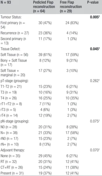
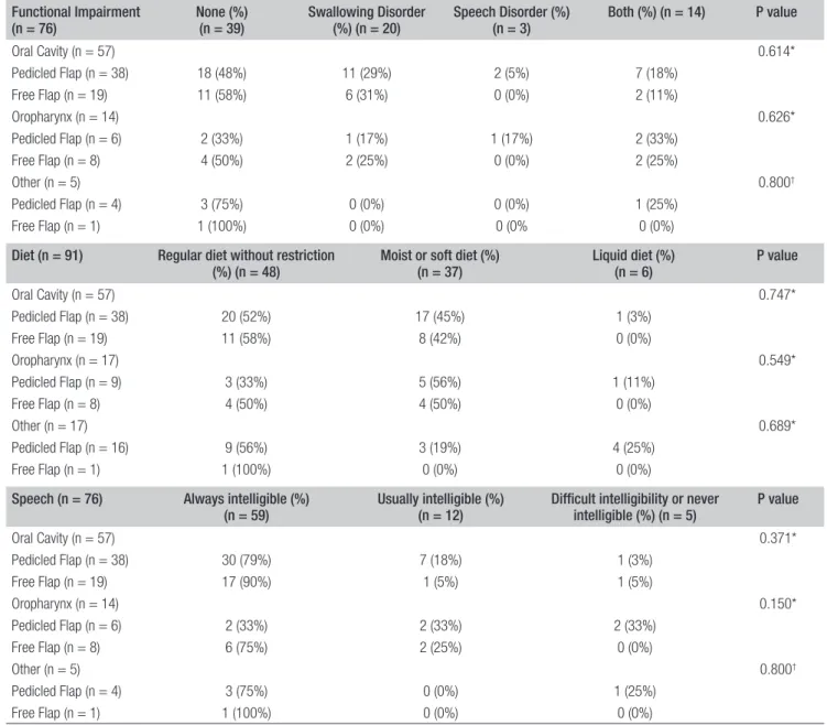
construction (mean 64.5 vs 58.2, p = 0.005), and more of-ten presented a recurrence or a second primary (36% and 17% vs 14% and 3%, p = 0.005). Furthermore, patients who underwent a pedicled flap reconstruction were more often identified with a unilateral or bilateral vessel depleted neck (17% and 19% vs 3% and 0%, p = 0.004) and suffered more often from comorbidities (56% vs 6%, p < 0.001) than pa-tients who underwent free flap reconstruction. Finally, the resulting defect more frequently involved soft tissue or soft tissue and marginal mandibular resection in patients who un-derwent pedicled flap reconstruction (88% vs 69%); while reconstruction for segmental bony tissue defects (which also include soft tissue defects) was more frequently achieved by means of a free flap reconstruction (12% vs 31%, p = 0.045). Table III shows that, when corrected for the anatomical site of cancer resection, the degrees of impairment for swallow-ing function and speech between pedicled flap and free flap reconstructions were not statistically significant.
Table IV shows that the differences in healing results and flap related complications were not statistically significant between the two groups, although patients who under-went a free flap reconstruction were admitted for a longer period of time than those who underwent a pedicled flap reconstruction (mean 21.1 days vs 17.6 days, p = 0.028). In multivariate regression analysis regarding the develop-ment of functional impairdevelop-ments, no associated factors were found; also, no associated factors were found in multivari-ate regression analysis regarding necrosis of the flap after reconstruction. However, a considerable trend towards significance concerning comorbidities was found regard-ing the probability of facregard-ing a flap necrosis (p = 0.057); and the multivariate regression analysis showed that only the presence of comorbidities remained an explaining fac-tor for complicated flap healing (p = 0.019, OR = 2.018), Table V. Of note, a pedicled flap reconstruction provided uneventful healing process in 81% of fragile patients who suffered from severe comorbidities.
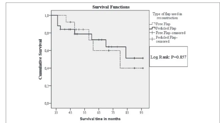
Figure 1 displays the overall survival of patients with a follow-up time over 36 months, receiving pedicled flap and free flap reconstructions; the differences were not sta-tistically significant (Log Rank: p = 0.857). Figure 2 dis-plays the development of recurrences, distant metastasis or second primary cancers, overtime, after pedicled flap and free flap reconstructions. Although the odds (hazard ratio = 0.665) for developing a recurrence, distant metasta-sis or second primary cancer were higher for patients who underwent pedicled flap reconstruction, as compared to patients who underwent a free flap reconstruction, this dif-ference was not statistically significant corrected for age, tumour status (first primary, recurrence or second primary), T-stage, N-stage, surgical margin status and the presence of extracapsular tumour spread in positive lymph nodes.
Discussion
In our study, comparison of outcomes in patients who underwent a pedicled flap or a free flap reconstruction showed no statistically significant differences in terms of functional outcomes, flap necrosis, or complications. Furthermore, multivariate regression analysis showed that only the existence of comorbidities remained an explain-ing factor for complicated flap healexplain-ing. In addition, there was no significant differences in terms of overall survival between patients who underwent a pedicled flap recon-struction and those who underwent a free flap reconstruc-tion. Finally, proportional hazard regression analysis, regarding the development of recurrences, distant metas-tases or second primary cancers, showed no significant differences between patients who underwent a pedicled flap reconstruction and patients who underwent a free flap reconstruction. In our cohort, pedicled flap reconstruc-tion did not seem be inferior to free flap reconstrucreconstruc-tion in terms of complications, functional outcome, survival,
Table II. Tumour-defect characteristics.
N = 93 Pedicled Flap reconstruction (n = 64) Free Flap reconstruction (n = 29) P-value Tumour Status: 0.005† First primary (n = 54) 30 (47%) 24 (83%) Recurrence (n = 27) 23 (36%) 4 (14%) Second primary (n = 13) 11 (17%) 1 (3%) Tissue Defect: 0.045† Soft Tissue (n = 56) 39 (61%) 17 (59%)
Bony + Soft Tissue
(n = 17) 8 (12%) 9 (31%) Soft Tissue + marginal (n = 20) 17 (27%) 3 (10%) pT-stage (grouping): 0.262† T1-T2 (n = 21) 15 (23%) 6 (21%) T3 (n = 19) 10 (16%) 9 (31%) T4 (n = 26) 16 (25%) 10 (35%) rT1-rT2 (n = 8) 7 (11%) 1 (3%) rT3 (n = 5) 4 (6%) 1 (3%) rT4 (n = 14) 12 (19%) 2 (7%) pN-stage (grouping): 0.075† N0 (n = 28) 20 (31%) 8 (28%) N+ (n = 38) 21 (33%) 17 (58%) rN0 (n = 17) 15 (23%) 2 (7%) rN+ (n = 10) 8 (13%) 2 (7%) Adjuvant therapy: 0.070† None (n = 35) 29 (45%) 6 (21%) RT (n = 32) 20 (31%) 12 (41%) CT+RT (n = 26) 15 (24%) 11 (38%) Present (n = 31) 19 (37%) 12 (41%)
Table III. Functional assessment outcome in different types of reconstruction per anatomical site. Functional Impairment
(n = 76) None (%) (n = 39) Swallowing Disorder (%) (n = 20) Speech Disorder (%) (n = 3) Both (%) (n = 14) P value
Oral Cavity (n = 57) 0.614* Pedicled Flap (n = 38) 18 (48%) 11 (29%) 2 (5%) 7 (18%) Free Flap (n = 19) 11 (58%) 6 (31%) 0 (0%) 2 (11%) Oropharynx (n = 14) 0.626* Pedicled Flap (n = 6) 2 (33%) 1 (17%) 1 (17%) 2 (33%) Free Flap (n = 8) 4 (50%) 2 (25%) 0 (0%) 2 (25%) Other (n = 5) 0.800† Pedicled Flap (n = 4) 3 (75%) 0 (0%) 0 (0%) 1 (25%) Free Flap (n = 1) 1 (100%) 0 (0%) 0 (0% 0 (0%)
Diet (n = 91) Regular diet without restriction
(%) (n = 48) Moist or soft diet (%) (n = 37) Liquid diet (%) (n = 6) P value
Oral Cavity (n = 57) 0.747* Pedicled Flap (n = 38) 20 (52%) 17 (45%) 1 (3%) Free Flap (n = 19) 11 (58%) 8 (42%) 0 (0%) Oropharynx (n = 17) 0.549* Pedicled Flap (n = 9) 3 (33%) 5 (56%) 1 (11%) Free Flap (n = 8) 4 (50%) 4 (50%) 0 (0%) Other (n = 17) 0.689* Pedicled Flap (n = 16) 9 (56%) 3 (19%) 4 (25%) Free Flap (n = 1) 1 (100%) 0 (0%) 0 (0%)
Speech (n = 76) Always intelligible (%)
(n = 59) Usually intelligible (%) (n = 12) Difficult intelligibility or never intelligible (%) (n = 5) P value
Oral Cavity (n = 57) 0.371* Pedicled Flap (n = 38) 30 (79%) 7 (18%) 1 (3%) Free Flap (n = 19) 17 (90%) 1 (5%) 1 (5%) Oropharynx (n = 14) 0.150* Pedicled Flap (n = 6) 2 (33%) 2 (33%) 2 (33%) Free Flap (n = 8) 6 (75%) 2 (25%) 0 (0%) Other (n = 5) 0.800† Pedicled Flap (n = 4) 3 (75%) 0 (0%) 1 (25%) Free Flap (n = 1) 1 (100%) 0 (0%) 0 (0%)
* χ2-test †Fisher’s exact test.
Table IV. Healing outcomes.
Flap Healing (n = 93) Healing uneventful (%)
(n = 81) Minor complications (%) (n = 5) Further surgery required (%) (n = 7) P value
Pedicled Flap 56 (87%) 3 (5%) 5 (8%) 0.902*
Free Flap 25 (86%) 2 (7%) 2 (7%)
Flap Necrosis (n = 93) None (%) (n = 85) Partial Necrosis (%) (n = 7) Total Necrosis (%) (n = 1)
Pedicled Flap 59 (92%) 4 (6%) 1 (2%) 0.634*
Free Flap 26 (90%) 3 (10%) 0 (0%)
Admission length (n=93) Mean Standard deviation Standard error mean
Pedicled Flap (n = 64) 17.6 ± 6.8 0.9 0.028†
Free Flap (n = 29) 21.1 ± 7.8 1.5
development of cancer relapse, or distant metastases. The type of flap used for reconstruction depends on the needs of the recipient site; in some situations free flaps are required (e.g. in segmental bony reconstructions), where-as pedicled flaps cannot always offer the amount or type of desired tissue 17. Furthermore, the anatomical site of the
defect can sometimes be out of reach for a pedicled flap, since the length of the vascular pedicle limits the required distance of transfer. However, premorbid patient factors and regional anatomy (e.g. comorbidity or previous head and neck cancer treatment) are also important in deciding which flap is employed for reconstruction 18.
Randomised controlled trials are not feasible; conse-quently, the nature of studies comparing the outcome of reconstruction in head and neck surgery is restricted to descriptive reports, stratifying, wherever possible, for pa-tient and tumour factors. Thus, the outcome of free flap and pedicled flap reconstructions cannot easily be com-pared and bias is inevitable.
The small cohort and heterogeneity of the reconstructed defects represent the major limitation of our study. Nev-ertheless, this series replicates a comparable scenario of many low volume centres where a careful selection of patients undergoing microvascular reconstructive surgery is performed. All reconstructive surgical procedures and follow-up consultations were conducted by a single sur-geon (AD), diminishing inter-patient variability to mini-mise bias concerning treatment and evaluation. Further-more, all reconstructions with local flaps or grafts were excluded a priori, focusing only on major resections with flap transposition.
Several authors have reported that free flaps have advan-tages over pedicled flaps in head and neck reconstruction. Firstly, tissue dimensions and thickness can be tailored to the size of the defect and vascularised bone can be used to reconstruct complex defects, which leads to superior aes-thetic results 19. Secondly, some reports state that free flaps
provide superior speech outcome over pedicled flaps 18 20.
Thirdly, it is reported that swallowing function, follow-ing free flap reconstruction in comparison to pedicled flap reconstruction, is improved 20, while other authors were
unable to substantiate this finding 18 21. Considering the
re-sults of our study, we cannot support these findings, either for superior swallowing function or for superior speech function.
A videofluorographic swallowing study is certainly the golden standard for the assessment of swallowing disor-ders. However, at our institution, this is not routinely pre-scribed to all patients, but only in case of aspiration prob-lems. Therefore, we used a rough assessment concerning the quality of swallowing and speech, which was already used in previous reports from both our group 7 15 and from
others 22-24, for this assessment was relatively simple to
ap-ply during follow-up consultation visits.
Frequently pedicled flaps even seem to be preferable over free flaps 25-27. Many reports regarding the elderly in relation
to microvascular free flap reconstruction agree that age is a risk factor for poor surgical outcome 10 28-30. McCrory et
al. described that operative time, resection-reconstruction, was statistically much longer for free flap than for pedicled flap procedures (9 hours 35 min vs 4 hours 58 min) 28. Long
surgical times was a significant factor for the development of postoperative complications in a series of 104 free flaps in patients aged 65 and older 10. Furthermore, older patients
are less capable of coping with large fluid shifts and sig-nificant blood loss 10, whereas free flap reconstructions are
more often associated with the need for blood transfusion 29.
In addition, cardiovascular disease proves to be an impor-tant factor in free flap reconstructive failure 10, a condition
which proves to affect the majority of adults past the age of 60 years 31, and furthermore with increasing age there is a
greater likelihood of postoperative complications after free flap reconstruction 27, even with successful microvascular
reconstructions 32.
Besides age, diabetes also appears to interfere with free flap survival 33. However, the impact of diabetes on free
flap survival is still much debated. While some authors support that diabetes interferes with free flap survival 7 34,
Cooley et al. reported that patients with diabetes are not at increased risk either for flap failure or for abnormal heal-ing of the anastomoses as long as normal glycaemia is maintained 35.
Our study shows that more than 80% of patients suffering from comorbidities who underwent a pedicled flap
recon-Table V. Estimation of probability of complicated flap healing.
Variable Coefficient Standard error P value OR* 95% CI†
Age 0.00 0.37 0.998 1.000 0.930 1.076 Type Flap -1.440 1.132 0.203 0.237 0.026 2.179 Tumour status -2.018 1.765 0.253 0.133 0.004 4.230 Anatomical Site 0.602 0.482 0.211 1.826 0.710 4.692 Previous treatment 0.275 0.522 0.598 1.317 0.473 3.663 Tissue Defect -0.130 0.497 0.793 0.878 0.331 2.326 Comorbidity 0.702 0.300 0.019 2.018 1.121 3.634
Fig. 2. Cumulative hazard curves regarding the development of recurrences, distant metastasis or second primary cancers in patients who underwent either pedicled flap (n= 64, bold line) or free flap (n = 29, dotted line) reconstruction.
Fig. 1. Kaplan Meier curves of overall survival in patients with a follow-up time over 36 months, but less than 97 months, who underwent either a pedicled flap (n = 25, bold line) or a free flap (n = 13, dotted line).
struction had uneventful healing of the flap. Only 2 pa-tients suffering from comorbidities underwent a free flap reconstruction, 1 of these patients had complicated flap healing. This suggests that a reconstruction by means of a pedicled flap is a safe procedure in patients who are suf-fering from comorbidities. Further research with a larger population should be conducted in order to assess whether pedicled flap reconstructions in patients suffering from comorbidities are less prone to complicated postoperative healing than free flap reconstructions.
The use of free flaps for reconstruction in previously ir-radiated patients or patients who underwent prior chemo-radiation is also much debated in literature. In a review, Wong et. al. points out that prior chemotherapy and/or radiotherapy can cause significant scarring and vessel damage to the utilised vessels for microvascular free flap reconstruction with obvious negative consequences 36.
Furthermore, Schultze-Mosgau et al. reported a reduced clinical success rate (84%) of free vascular grafts in head and neck patients with previous radiotherapy of 60-70 Gy 37. Moreover, in a study of 429 patients who
under-went free flap reconstruction in the head and neck, pre-operative radiotherapy (irrespective of irradiation doses) was significantly associated with fistulae formation and wound infection, while previous neck irradiation at doses of more than 60 Gy proved to be a significant risk fac-tor for free flap failure, overall local complications, hae-matoma, longer duration of enteral nutrition and hospital stay 38. In a multicentre survival analysis after free flap
reconstructive surgery of head and neck squamous cell carcinoma by Salvatori et al. 39, pre-treated patients had
significantly worse survival than those with first primary tumours (43.1% and 54.1% respectively). Accordingly, also in our series, patients presenting with recurrence or second primaries showed worse survival than those with first primary tumours. However, this was irrespective to the type of employed flap (pedicled flap or free flap). Based on our policy, the greatest majority of pre-treated patients received a pedicled flap reconstruction; among them 21 of 34 patients (62%) who underwent pedicled flap reconstruction were confirmed alive at the end of our study.
Since intake of alcohol ≥ 30 g/day is related to the devel-opment of head and neck cancer 40, many head and neck
cancer patients suffer from alcohol-related problems. Both acute alcohol withdrawal as well as other alcohol-induced disorders negatively influence the outcome of microvascular free flap tissue transfers 41-43.
Consequently, those patients presenting the above men-tioned factors, which are associated with a higher rate of free flap failure or postoperative complications, are less eligible for microvascular free flap reconstructive surgery, whereas locoregional pedicled flaps may offer a reliable alternative for reconstruction 44-49.
Furthermore, a pedicled flap reconstruction brings some
additional benefits for both patient and surgeon. Most sites of pedicled flaps have a low donor-site morbidity with do-nor sites that can be closed primarily. Also, many pedicled flaps can be harvested and transferred rapidly, which leads to decreased operating time and a corresponding decrease in the morbidities of prolonged general anaesthesia. Our results showed that the admission length of patients who underwent a pedicled flap reconstruction were short-er than in those who undshort-erwent a free flap reconstruction. Other papers pointed out that pedicled flap reconstruc-tions were associated with shorter intensive care stay than free flap reconstructions 7 28. Consequently, free flap
re-constructions are usually more expensive procedures than pedicled flap reconstructions 15 25 28.
Conclusions
In our patient cohort, pedicled flaps were performed in two-thirds of cases and were not significantly inferior to free flaps in terms of functionality, complications, or prognosis. This study highlights how, in a low volume setting, careful selection of patients receiving free flap re-construction is advisable in order to maintain high success rates; in this scenario, pedicled flaps are a viable option in patients considered suboptimal for a microvascular re-construction. A well thought and careful analysis of every patient is needed to offer the best solution in the light of individualised treatment 1.
References
1 Deganello A. Modern oral cavity reconstruction with free
flaps and pedicled flaps. J Aesth Reconstr Surg 2015;1:4. 2 List MA, Bilir SP. Functional outcomes in head and neck
cancer. Semin Radiat Oncol 2004;14:178-89.
3 Pellini R, Pichi B, Marchesi P, et al. External monitor for
buried free flaps in head and neck reconstructions. Acta Otorhinolaryngol Ital 2006;26:1-6.
4 Varvares MA, Lin D, Hadlock T, et al. Success of multiple,
sequential, free tissue transfers to the head and neck. Laryn-goscope 2005;115:101-4.
5 Beausang ES, Ang EE, Lipa JE, et al. Microvascular free
tis-sue transfer in elderly patients: the Toronto experience. Head Neck 2003;25:549-53.
6 Deganello A, Manciocco V, Dolivet G, et al. Infrahyoid
fascio-myocutaneous flap as an alternative to free radial forearm flap in head and neck reconstruction. Head Neck 2007;29:285-91.
7 Deganello A, Gitti G, Parrinello G, et al. Infrahyoid flap
re-construction of oral cavity and oropharyngeal defects in el-derly patients with severe general comorbidities. Head Neck 2012;34:1299-305.
8 Deganello A, Gitti G, Mannelli G, et al. Risk factors for
mul-tiple malignancies in the head and neck. Otolaryngol Head Neck Surg 2013;149:105-11.
9 Deganello A, Franchi A, Sardi I, et al. Genetic alterations
recurrence after radiotherapy: recurrence, genetically related cancer, or second primary? Cancer 2010;116:1291-7.
10 Serletti JM, Higgins JP, Moran S, et al. Factors affecting
outcome in free-tissue transfer in the elderly. Plast Reconstr Surg 2000;106:66-70.
11 Mahieu R, Russo S, Gualtieri T, et al. Oral cavity
recon-struction with the masseter flap. Acta Atorhinolaryngol Ital 2016;36:1-5.
12 Deganello A, Leemans CR. The infrahyoid flap: a
compre-hensive review of an often overlooked reconstructive method. Oral Oncol 2014;50:704-10.
13 Gangloff P, Deganello A, Lacave ML, et al. Use of the infra
hyoid musculo-cutaneous flap in soft palate reconstruction. Eur J Surg Oncol 2006;32:1165-9.
14 Deganello A, De Bree R, Dolivet G, et al. Infrahyoid
my-ocutaneous flap reconstruction after wide local excision of a merkel cell carcinoma. Acta Otorhinolaryngol Ital 2005;25:50-4.
15 Deganello A, Gitti G, Parrinello G, et al. Cost analysis in
oral cavity and oropharyngeal reconstructions with mi-crovascular and pedicled flaps. Acta Otorhinolaryngol Ital2013;33:380-7.
16 Busoni M, Deganello A, Gallo O. Pharyngocutaneous fistula
following total laryngectomy: analysis of risk factors, prog-nosis and treatment modalities. Acta Otorhinolaryngol Ital 2015;35:400-5.
17 Tarsitano A, Ciocca L, Cipriani R, et al. Mandibular
recon-struction using fibula free flap harvested using a custom-ised cutting guide: how we do it. Acta Otorhinolaryngol Ital 2015;35:198-201.
18 O’Neill JP, Shine N, Eadie PA, et al. Free tissue transfer
versus pedicled flap reconstruction of head and neck malig-nancy defects. Ir J Med Sci 2010;179:337-43.
19 Rosenthal ELM, Dixon SFM. Free flap complications: when
is enough, enough. Curr Opin Otolaryngol Head Neck Surg 2003;11:236-9.
20 Abemayor E BK. Reconstruction of soft tissue defects in the
oral cavity and oropharynx. Arch Otolaryngol Head Neck Surg 2000;126:909-12.
21 Freedlander E, Espie CA, Campsie LM, et al. Functional
im-plications of major surgery for intraoral cancer. Br J Plast Surg 1989;42:266-9.
22 List MA, Ritter-Sterr C, Lansky SB. A performance
sta-tus scale for head and neck cancer patients. Cancer 1990;66:564-9.
23 O’Neill JP, Shine N, Eadie PA, et al. Free tissue transfer
versus pedicled flap reconstruction of head and neck malig-nancy defects. Ir J Med Sci 2010;179:337-43.
24 Andrades P, Pehler SF, Baranano CF, et al. Fistula analysis
after radial forearm free flap reconstruction of hypopharyn-geal defects. Laryngoscope 2008;118:1157-63.
25 Colletti G, Tewfik K, Bardazzi A, et al. Regional flaops in
head and neck reconstruction: a reappraisal. J Oral Maxil-lofac Surg 2015;73:571.e1-571.e10.
26 Deganello A, Gitti G, Struijs B, et al. Palliative combined
treatment for unresectable cutaneous basosquamous cell carcinoma of the head and neck. Acta Otorhinolaryngol Ital2013;33:353-6.
27 Deganello A, Gallo O, De Cesare JM, et al. Surgical
man-agement of surgery and radiation induced peristomal neck ulcerations. B-ENT 2008;4:169-74.
28 McCrory AL, Magnuson JS. Free tissue transfer versus
pedi-cled flap in head and neck reconstruction. Laryngoscope 2002;112:2161-5.
29 Perisanidis C, Herberger B, Papadogeorgakis N, et al.
Com-plications after free flap surgery: do we need a standardized classification of surgical complications? Br J Oral Maxillo-facial Surg 2012;50:113-8.
30 Chick LR, Walton RL, Reus W, et al. Free flaps in the
el-derly. Plast Reconstr Surg 1992;90:87-94.
31 Laslett LJ, Alagona Jr P, Clark III BA, et al. The worldwide
environment of cardiovascular disease: prevalence, diagno-sis, therapy, and policy issues: a report from the american college of cardiology. J Am Coll Cardiol 2012;60:S1-S49. 32 Beausang ES, Ang EE, Lipa JE, et al. Microvascular free
tis-sue transfer in elderly patients: the toronto experience. Head Neck 2003;25:549-53.
33 Valentini V, Cassoni A, Marianetti TM, et al. Diabetes as
main risk factor in head and neck reconstructive surgery with free flaps. J Craniofac Surg 2008;19:1080-4.
34 Joo YH, Sun DI, Park JO, et al. Risk factors of free flap
com-promise in 247 cases of microvascular head and neck recon-struction: a single surgeon’s experience. Eur Arch Otorhi-nolaryngol 2010;267:1629-33.
35 Cooley BC, Hanel DP, Anderson RB, et al. The influence of
diabetes on free flap transfer: I. flap survival and microvas-cular healing. Ann Plast Surg 1992;29:58-64.
36 Wong KK, Higgins KM, Enepekides DJ. Microvascular
re-construction in the vessel-depleted neck. Curr Opin Otolar-yngol Head Neck Surg 2010;18:223-6.
37 Schultze-Mosgau S, Grabenbauer GG, Radespiel-Troger
M, et al. Vascularization in the transition area between free
grafted soft tissues and pre-irradiated graft bed tissues fol-lowing preoperative radiotherapy in the head and neck re-gion. Head Neck 2002;24:42-51.
38 Benatar MJ, Dassonville O, Chamorey E, et al. Impact of
preoperative radiotherapy on head and neck free flap recon-struction: A report on 429 cases. J Plastic Reconstr Aest Surg 2013;66:478-82.
39 Salvatori P, Paradisi S, Calabrese L, et al. Patients’ survival
after free flap reconstructive surgery of head and neck squa-mous cell carcinoma: a retrospective multicentre study. Acta Otorhinolaryngol Ital 2014;34:99-104.
40 Maasland DH, van den Brandt PA, Kremer B, et al. Alcohol
consumption, cigarette smoking and the risk of subtypes of head-neck cancer: results from the Netherlands cohort study. BMC Canc 2014;14:187.
41 Gallivan KH, Reiter D. Acute alcohol withdrawal and free
flap mandibular reconstruction outcomes. Arch Facial Plast Surg 2001;3:264-6.
42 Kuo YR, Jeng SF, Lin KM, et al. Microsurgical tissue
trans-fers for head and neck reconstruction in patients with alcohol-induced mental disorder. Ann Surg Oncol 2008;15:371-7. 43 Cheng NC, Ko JY, Tai HC, et al. Microvascular head and
neck reconstruction in patients with liver cirrhosis. Head Neck 2008;30:829-35.
44 Giordano L, Bondi S, Toma S, et al. Versatility of the
supra-clavicular pedicle flap in head and neck reconstruction. Acta Otorhinolaryngol Ital 2014;34:394-8.
45 Rigby MH HR. Regional flaps: a move to simpler
recon-structive options in the head and neck. Curr Opin Otolaryn-gol Head Neck Surg 2014;22:401-6.
46 Squaquara R, Kim Evans KF, Spanio di Spilimbergo S, et al. Intraoral reconstruction using local and regional flaps. Semin Plast Surg 2010;24:198-211.
47 Bussu F, Gallus R, Navach V, et al. Contemporary role of
pectoralis major regional flaps in head and neck surgery. Acta Otorhinolaryngol Ital 2014;34:327-41.
48 Kucur C, Durmus K, Ozer E. Supraclavicular artery island
flap reconstruction of a contralateral partial laryngopharyn-geal defect. Acta Otorhinolaryngol Ital 2015;35:121-4. 49 Dell’Aversana Orabona G, Salzano G, Abbate V, et al. Use of
the SMAS flap for reconstruction of the parotid lodge. Acta Otorhinolaryngol Ital 2015;35:406-11.
Address for correspondence: Alberto Deganello, Department of Surgery and Translational Medicine, Azienda Ospedaliero-Univer-sitaria Careggi, largo Brambilla 10, 50134 Firenze, Italy. Tel./Fax +39 055 7947054. E-mail: [email protected]