Author
INDEX
Abbreviations p. 3
CHAPTER 1- GENERAL INTRODUCTION p. 7
CHAPTER 2-MATERIALS AND METHODS p. 41
CHAPTER 3-PREVALENCE OF CHRONIC KIDNEY DISEASE AND
ASSOCIATED RISK FACTORS IN CATS FROM PRIMARY CARE PRACTICES
IN SOUTHERN ITALY p. 54
CHAPTER 4- EPIDEMIOLOGY IN SOUTHERN ITALY OF INFECTIOUS
AGENTS POTENTIALLY ASSOCIATED WITH CHRONIC KIDNEY DISEASE p. 77
CHAPTER 5- CELL BLOOD COUNT AND INFLAMMATION MARKERS IN CATS EXPOSED TO SOME EMERGING PATHOGENS IN SOUTHERN ITALY
p. 139
CHAPTER 6- GENERAL DISCUSSION AND CONCLUSION p. 193
REFERENCES p. 197
PUBLISHED ARTICLES AND POSTERS p. 232
3
ABBREVIATIONS
Ab: Antibodies
ALB: Albumin
ALKP: Alkaline phosphatase ALT: Alanine aminotransferase APP: Acute phase proteins AST: Aspartate aminotransferase ATL: Animal Tissue Lysis BCS: Body condition score
BP: Blood pressure
BUN: Urea
CBC: Complete Blood Count
CDV: Canine distemper virus
CEUS: Contrast-enhanced ultrasound examination CHr: Reticulocyte haemoglobing content
C.I: Confidence interval
CKD: Chronic kidney disease CMV: Cetacean morbillivirus
CPE: Cytophatic effect
CRFK: Crandell-Rees feline kidney CRP: C-reactive protein
Ct: Cycle threshold
DAT: Direct agglutination test DLH: Domestic longhair DSH: Domestic shorthair
ELFA: Enzyme linked fluorescent assay ELISA: Enzymelinked immunosorbent assay
EPO: Erythropoietin
FAM: Carboxyfluorescein
FCoV: Feline Coronavirus infection
4
FCV: Feline Calicivirus
FEA: Feline Embryonic Fibroblast FeL: Feline leishmaniosis
FeLV: Feline Leukemia Virus infection FIP: Feline Infectious Peritonitis FIV: Feline Immunodeficiency Virus FeMV : Feline Morbillivirus
FNA: Lymph node aspirates GFR: Glomerular filtration rate GGT: Gamma glutamyl transferase
GI: Gastrointestinal GLOB: Globulins HAC: Hyperadrenocorticism HCM: Hypertrophic cardiomyopathy HCT: Hematocrit Hgb: Hemoglobin concentrations
HRE: High resolution electrophoretic technique iCa: Active ionized calcium fraction
IFI: Indirect immunofluorescence assay
IgG: Immunoglobulin G
IRIS: International Renal Interest Society
KD: CKD group
LAI: Leukocyte alterations suggestive of inflammation LVH: Left ventricular concentric hypertrophy
MAT: Microscopic agglutination test
MCHC: Mean corpuscolar hemoglobin concentration MCS: Muscle condition score
MCV: Mean corpuscolar volume
MEM: Minimum Essential Medium
MGG: May Grünwald-Giemsa
MMP-9: Matrix metalloproteinase-9
5
NI: Normal intervals
NKD: Not kidney diseases group
NP: Non proteinuric
NRA: Non regenerative anemia
OR: Odds Ratio
PHA: Hyperaldosteronism PBS: Phosphate buffered saline PDV: Phocine distemper virus
PHOS: Phosphorus
PL: Platelete
PPRV: Peste-des-petits-ruminants virus
PTH: Parathyroid hormone
qPCRFeMV: Real-time RT-PCR for FeMV
RA: Regenerative anemia
RAAS: Renin –angiotensin–aldosterone system RBC: Red blood cell count
RI: Arterial resistive index RPV: Rinderpest virus
RT-PCR: Reverse transcriptase-PCR
SAA: Serum amyloid A
SBP: Systolic blood pressure
SCr: Serum Creatinine
SDMA: Serum symmetric dimethylarginine
SDS-AGE: Sodium dodecyl sulfate-agarose gel electrophoresis
SDS-PAGE: Sodium dodecyl sulfate-polyacrylamide gel electrophoresis SPE: Serum protein electrophoresis
SSA: Sulfosalicylic acid
TAMRA: Tetramethylrhodamine
tCa: Total calcium concentration TGF-β1: Transforming growth factor-β TIN: Tubulointerstitial nephritis
6
TP: Total proteins
TPCK: L-1Tosylamide-2-phenylethylchloromethylketone-treated tT4: Total thyroxine
UPC: Urinary protein/creatinine ratio USG: Urine specific gravity
WB: Western blotting
7
CHAPTER 1
GENERAL INTRODUCTION
Chronic kidney disease (CKD) is a common condition in cats defined as a multifactorial pathophysiologic process resulting in progressive loss of nephrons in both number and function, frequently culminating in end-stage renal disease (Khan and Khan et al., 2015). The term CKD is used to imply alterations in structure or function of the kidney that have occurred over a period of time, typically 3 months or more (Jepson, 2016). Prevalence of CKD in cats is high, with a 50% prevalence reported in a randomly selected population of cats (Marino et al., 2014). Median survival time recently reported was of 388 days, and previous studies reported median survival time from 21 to 1151 days depending on severity of renal dysfunction at diagnosis (Conroy et al., 2019). Causes of CKD are heterogeneus and most often not identified. Some risk factors such as genetic or acquired diseases can initiate renal damage (Reynolds and Lefebvre, 2013) and after the initial renal insult continues advancing with irreversible morphologic changes in renal parenchyma, including nephron loss and replacement by a selfperpetuating vicious cycle of fibrosis, lead to the progression of CKD (Khan and Khan et al., 2015; Chakrabarti et al., 2013). Considering the frequency and severity of this disease in cats, the evaluation of the possible influence that some risk factors and infectious diseases can have in the development of CKD is important. This awareness could be useful to avoid the development or at least slow progression of the disease. For this reason studies about epidemiology of infectious diseases and their clinical consequences as well as their relationship with CKD are constantly evolving.
1.1 NON-INFECTIOUS DISEASES AND CHRONIC KIDNEY DISEASE
Development of CKD is associated with some risk factors. Older cats seem to have more frequent renal disease (Trevejo et al., 2018) with an increased prevalence among cats older than 15 years (Marino et al., 2014) and a mortality rate greater in cats over
8
9 years old (Lawler et al., 2006). Cats can develop different age-related pathologies including renal inflammation and interstitial fibrosis, indeed, according to previous studies older cats were shown to have increased production of pro-inflammatory cytokines (Day, 2010). Moreover renal damage has a slow progression, with progressive and irreversible damage to kidneys (Reynolds and Lefebvre, 2013), and this could explain why CKD can be found more frequently in older cats rather than in young cats. A retrospective case-control study about association of gender and diagnosis of CKD in cats revealed that neutered males were 30% more likely than spayed females to develop CKD, probably because male cats tend to develop urethral obstruction and subsequent renal damage more easily than female cats (Greene et al., 2014). Breeds such as Siamese, Persian, Abyssinian, Himalayan, Maine Coon, Russian Blue, Burmese are reported to be more likely affected by CKD (Boyd et al., 2008; Trevejo et al., 2018; Conroy et al., 2019). Common congenital kidney disease, such as polycystic kidney disease (Persian and Persian-cross cats); familial secondary amyloidosis and glomerular disease (Abyssinian cats) (Lees, 1996; Bosje et al., 1998; Greco, 2001; White et al., 2008), juvenile renal dysplasia (Aresu et al., 2009; Greco, 2001) can be responsible for the development of CKD in pure breed individuals more frequently than in the general population.
Renal hypoperfusion, sympathetic nervous system activation and increased activity of the renin-angiotensin system observed in cats with hypertrophic cardiomyopathy (HCM) may lead to some degree of renal dysfunction. The prevalence of CKD reported in cats with HCM is 13% (Gouni et al., 2008) and the prevalence of left vetricular hypertrophy in cats with CKD has been estimated to be 46.6% (Taugner, 2001). However, further investigations are needed to document the pathophysiology of concomitant CKD and cardiac disease, mainly to understand if cardiovascular diseases are a consequence or a risk factor of CKD.
Primary hyperaldosteronism is considered a possible risk factor for the progression of CKD with kidney histopathological changes reported such as hyaline arteriolar sclerosis, glomerular sclerosis, tubular atrophy and interstitial fibrosis (Javadi et al., 2005).
Hyperthyroidism and CKD can occur concurrently in old cats (van Hoek et al., 2009). Thyroid hormones determine an increased renal blood flow because of an
9
increased cardiac output due to positive chronotropic and inotropic effects, decreased vascular resistance and also increased blood volume caused by renin-angiotensin-aldosterone system (RAAS) activation (van Hoek and Daminet, 2009). Consequences of hyperthyrodism are therefore increased glomerular filtration rate (GFR), decreased urine specific gravity (USG) and increased urinary protein/creatinine ratio (UPC) (Langston and Reine, 2006).
A positive association between urolithiasis and CKD in cats has been suggested, with a 56% prevalence of CKD among feline patients with urolithiasis. Nephrolithiasis and ureterolithiasis can potentially cause intermittent obstruction, which could lead to nephron damage and development of CKD, for this reason cats with urolithiasis should be evaluated for CKD (Cléroux et al., 2017). At the same time urolith formation could result from underlying CKD, leading to abnormal handling of certain minerals and metabolites (Cléroux et al., 2017; Kyles et al., 2005).
Kidney tumors may also cause CKD with 60% of cats with renal lymphoma found to have azotemia (Gabor et al., 2000). Lymphoma is the most common tumor in cats. Other primary renal tumors described in cats are carcinomas, transitional cell carcinomas, malignant nephroblastoma, haemangiosarcoma and adenoma (Henry et al., 1999).
Periodontal disease may contribute to the development of CKD (Finch et al., 2016; Greene et al., 2014) as a consequence of persisten-low grade insults. Production of inflammatory cytokines, endotoxemia and an immune response to bacteria may have a role in the development of CKD. However, also infectious agents associated with gingivitis, such as feline immunodeficiency virus (FIV) (Hosie et al., 2009), and management of dental disease (use of antibiotics, steroidal or nonsteroidal anti-inflammatory drugs, or general anesthesia required for dental procedures) may be confounders having a role in development of kidney disease (Finch et al., 2016).
Frequent or annual vaccinations are reported as potential risk factors for development of CKD (Finch et al., 2016). Vaccinal viruses feline herpesvirus 1, calicivirus and panleukopenia virus are cultured using Crandell-Rees feline kidney (CRFK) cells. CRFK proteins may become incorporated into vaccines during manufacture. In a previous study, parenteral administration of vaccines containing viruses likely grown on CRFK cells induced antibodies against CRFK cell and feline
10
renal cell lysates in cats, however hypersensitization with CRFK cell proteins did not result in renal disease in cats during the study. However in this study renal biopsies were collected just six weeks after the last vaccination or CRFK sensitization and a possible transient inflammation of renal tissues could not be excluded (Lappin et al., 2005). Moreover a later study reported lymphocytic-plasmacytic interstitial nephritis in a cat sensitized with CRFK lysate (Lappin et al., 2006).
Aminoglycosides, non-steroidal anti-inflammatory drugs, antineoplastic agents or exposure to toxins (lily or ethylen glycol poisoning are the most common) could cause acute kidney injury (AKI) with potential risk of development of CKD with time in cats overcoming the acute damage (Reynolds and Lefebvre, 2013). Anesthesia has also been considered a risk factor for AKI because of the risk of renal hypoperfusion particularly in cases of deep level of anesthesia and long lasting surgical procedures or concurrent risk factors such as dehydration, and use of amynoglycosides (Greene et al., 2014).
Concerning the role of diet type as a risk factor for CKD, in a recent study there was no difference in the rate of development of azotemia in cats fed standard adult diets compared to senior diets suggesting no effect of protein content on the development of azotemia (Finch et al., 2016). Also the moisture content of the diet fed (wet or dry diet) do not seem to be a significant risk factor (Finch et al., 2016; Greene et al., 2014). Ad libitum feeding and increased ash intake were found associated with increased risk of CKD (Hughes et al., 2002). The hypothesis that chronic increases in dietary salt intake could damage renal function in older cats was however not supported by a recent study where glomerular filtration rate (GFR), blood pressure, and other routine clinical pathological variables in healthy aged cats were not affected by dietary salt content (Reynolds et al., 2013).
1.2 INFECTIOUS AGENTS AND CHRONIC KIDNEY DISEASE
The relationship between some infectious agents and CKD in cats has already been documented, such as with feline immunodeficiency virus infection (FIV), feline leukemia virus infection (FeLV) and feline coronavirus (FCoV) infections (Rossi et al., 2019). Other infectious disesases that in dogs have already been linked to
11
development of CKD, in cats are currently being studied, with some cases reported as caused by Leismania or Leptospira spp infection. Moreover, recently, a relationship between the infection caused by a new feline paramyxovirus and development of CKD has been proposed and is the subject of study of numerous researches.
Feline morbillivirus (FeMV)
Paramyxoviruses are enveloped, negative-sense single-stranded RNA viruses which act as important pathogens for humans and animals. In 2012 a new paramyxovirus, feline morbillivirus (FeMV), was isolated for the first time from cats in Hong Kong and it was associated with tubulointerstitial nephritis (TIN) (Woo et al., 2012). After this discovery, different studies evaluated and reported its presence in Japan, Germany, Italy, USA, South America, Turkey and UK (Park et al., 2016; Koide et al., 2015; Sieg et al., 2015; Lorusso et al., 2015; Donato et al., 2018; Sharp et al., 2016; Darold et al., 2017; Yilmaz et al., 2017; McCallum et al., 2018) documenting that the virus can be found in both healthy and sick cats, with a prevalence ranging from 3% to 52.9% (Beatty et al., 2019). Feline morbillivirus is genetically related to viruses which belong to the genus morbillivirus, such as canine distemper virus (CDV), measles virus (MV), rinderpest virus (RPV), peste-des-petits-ruminants virus (PPRV), phocine distemper virus (PDV) and cetacean morbillivirus (CMV) (Park et al., 2016). Feline morbillivirus is characterized by genetic diversity among isolates (Sakaguchi et al., 2014; Sieg et al., 2015; Park et al., 2016). Stability at +4°C for at least 12 days is reported, freeze-and-thaw seems not to affect virus titers, while heat-treatment at +60°C and +70°C inactivated FeMV in 10 and 2 minutes, respectively (Koide et al., 2016).
Some studies reported the highest rate of positive reverse transcriptase-PCR (RT-PCR) tests from urine of cats coming from multicat environments (De Luca et al., 2017; Darold et al., 2017) suggesting that close contact favours transmission as expected for an enveloped virus which is easily inactivated in the environment. Cats from suburban/rural areas and those with outdoor access seem to be more exposed to FeMV infection (Donato et al., 2018; Yilmaz et al., 2017).
The relationship with CKD is controversial. Some studies reported in infected cats the development of an intestitial inflammatory infiltrate, tubular
12
degeneration/necrosis, glomerulosclerosis (Woo et al., 2012; Park et al., 2016; Yilmaz et al., 2017; Sutummaporn et al., 2019), and shedding of the virus in urine for up to 2 years (Sieg et al., 2019), therefore a chronic infection leading to CKD is supposed to affect cats (Lorusso et al., 2015; Sieg et al., 2015). However this relationship was not found in other studies which evaluated the presence of the infection in azotemic cats with confirmed CKD (Darold et al, 2017; Lo Russo et al., 2017; McCallum et al., 2018; Donato et al. 2018). A study which evaluated both clinico-pathological and histopatological abnormalities in FeMV RNA-positive cats found tubulointerstitial nephritis but also cholangiohepatitis and hepatic focal necrosis, however these abnormalities were similar to those observed in FeMV RNA-negative cats (Yilmaz et al., 2017). In addition to the evaluation of hepatic involvment of FeMV in cats, a recent study reported susceptibility of primary feline pulmonary epithelial cells and primary cells from the cerebrum and cerebellum, as well as immune cells in the blood, especially CD4+ T cells, CD20+ B cells and monocytes to in vitro experimental infection (Sieg et al., 2019) confirming that FeMV is able to infect different feline cell lines: epithelial, fibroblastic, lymphoid and glial cells. Moreover, the FeMV cell receptor is ubiquitously expressed in cats (Sakaguchi et al., 2014). In conclusion, the impact of feline morbilliviruses on cat health has to be confirmed by further research to understand their role in CKD development and potential damage to other organs.
Diagnosis of FeMV infection can be performed through molecular and serological investigations. Detection of virus RNA in urine (Woo et al., 2012; Lorusso et al., 2015; Sieg et al., 2015; Sharp et al., 2016; Darold et al., 2017; Yilmaz et al., 2017; McCallum et al., 2018), renal tissue (Park et al., 2016), blood and rectal swabs (Woo et al., 2012) with RT-PCR is reported. Anti-FeMV antibody detection by indirect immunofluorescence assay (IFI), (Park et al., 2016), enzymelinked immunosorbent assay (ELISA), (Arikawa et al., 2017) or western blotting (WB) are reported (Woo et al., 2012; McCallum et al., 2018). Histopathology, immunohistochemistry (Woo et al., 2012; Park et al., 2016; Yilmaz et al., 2017) and viral culture (Woo et al., 2012; Koide et al., 2015) are also available and useful for further investigations. A better understanding of the basic biology of the virus is required and could be performed through a cooperation of molecular virology and veterinary medicine (Beatty et al., 2019).
13 Leptospira spp.
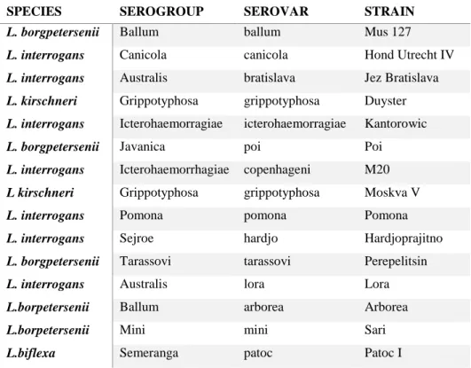
Leptospirosis is caused by an infection of spirochetal bacteria which belong to the genus Leptospira. They are Gram-negative, highly motile, elongated, helically coiled bacteria characterized by hook or question mark–shaped ends (Schuller et al., 2015). Taxonomy of the genus Leptospira is complex and leptospires are serologically classified by serovar (member of the genus Leptospira, which reacts with a specific monoclonal antiserum), serogroup (group of antigenically closely related leptospiral serovars) and strain (specific isolate of a defined leptospiral serovar) (Schuller et al., 2015). At present, there are over 250 pathogenic serovars (Hartmann et al., 2013a) which belong to 24 serogroups (Schuller et al., 2015).
Cats can be infected by feeding on animals harbouring leptospires or after exposure to infectious urine of cohabiting dogs (Hartmann et al., 2013a). Antibody prevalence was observed for cats with an outdoor lifestyle or those living in multicat households (Rodriguez et al., 2014). As observed in dogs, seasonality of leptospirosis was reported also for cats with a higher prevalence during most humid months (Rodriguez et al., 2014; Arbour et al., 2012). Serovars identified in cats include L. icterohaemorrhagiae, L. canicola, L. grippotyphosa, L. pomona, L. hardjo, L. autumnalis and L. ballum with a range of antileptospiral antibody prevalence between 0 and 35% reported (Hartmann et al., 2013a). In cats pathogenesis seems to be similar to that observed for dogs and humans, characterized by a systemic infection with invasion of many organs (kidneys, liver, spleen, central nervous system, eyes and genital tract) causing inflammation and tissue damage. Thanks to the adaptive humoral and cell-mediated immune response leptospires are removed from most organs except the kidneys, in which leptospires can persist with resultant chronic shedding (Hartmann et al., 2013a; Zuerner, 2015).
Clinical signs in infected cats seem to be rare, even if some have been reported manifesting in about 84 days in experimental studies or after few months in some reported cases after contact with possible animals harbouring leptospires or their urine (Arbour et al., 2012). Few studies have reported ascites, hepatomegaly (Agunloye and Nash, 1996) with severe centrilobular necrosis of the liver (Bryson and Ellis, 1976), vascular lesions in lung and brain with isolation of leptospires from thoracic fluid and
14
aqueous humour (Bryson and Ellis, 1976), ocular pathologies, particularly uveitis, and lameness (Arbour et al., 2012). Different studies reported that cats can shed leptospires in urine (Sprißler et al., 2019; Weis et al., 2017; Rodriguez et al., 2014), underlining their potential role as reservoirs or incidental hosts in transmission (Hartmann et al., 2013a). Chronic kidney disease in infected cats was also reported in some case reports (Arbour et al., 2012; Mason et al., 1972) with manifestation of polyuria and polydipsia (Arbour et al., 2012) and tubulointerstitial nephritis as histopathological findings (Arbour et al., 2012). However results from studies that evaluated serological evidence of exposure of cats to Leptospira spp., and kidney disease are conflicting (Shropshire et al., 2016: Rodriguez et al., 2014) and additional studies are needed to determine which is the real role of leptospirosis in the development of feline CKD.
Serological and molecular diagnosis can be respectively performed through microscopic agglutination test (MAT) or ELISA and PCR. Microscopic agglutination test is the most common diagnostic method used for antibody detection (Hartmann et al., 2013a) and it is based on determining the ability of serial dilutions of patient serum to agglutinate live leptospiral serovars in vitro. Positive MAT confirms the exposure to a serovar belonging to the corresponding serogroup (but not necessarily to the serovar tested) (Schuller et al., 2015). PCR targets the lipL32/hap1 gene or 23S rDNA, however a negative result on blood or urine does not rule out leptospirosis: leptospiraemia is transient (early stages of the disease), urinary shedding is delayed after acute infection and can be intermittent, moreover recent antibiotic treatment can cause negative results (Schuller et al., 2015). Other diagnostic methods are darkfield microscopy to identify entire leptospires in urine (poor sensitivity and specificity), and culture of biological samples (blood, urine, tissues) even if culturing leptospires is difficult and requiring up to six months of culture of fresh urine (Schuller et al., 2015).
Leishmania infantum
Feline leishmaniosis (FeL) is an emerging disease (Pennisi and Persichetti, 2018) determined by flagellated protozoan parasites of Leishmania genus (Pennisi et al., 2013a). In 1912 FeL was reported for the first time in Algeria (Soares et al., 2016) and since then it has been globally reported in endemic areas for canine leishmaniosis. Leishmania infantum is the most frequently detected species in both the New and Old
15
World but Leishmania amazonensis, Leishmania braziliensis, Leishmania mexicana, and Leishmania venezuelensis are also reported in the New World (Pennisi et al., 2013a; Pennisi et al., 2015; Soares et al., 2016; Pennisi and Persichetti, 2018) and infections by Leishmania tropica and Leishmania major were found in Turkey (Can et al., 2016; Paşa et al., 2015). Many studies investigated prevalence of anti-L. infantum antibodies or parasite DNA with ranges varying from 0 to 68.5% and from 0 and 60.7% respectively. This variability may be the consequence of different levels of endemicity in the investigated areas but, many factors may contribute such as the characteristics of the population under study or differences in diagnostic methodologies including the cut-off titres of antibody detection (Pennisi et al., 2015). Transmission of Leishmania spp. was not investigated in cats but it is believed to occur by sand fly bites as for other hosts. In fact, it is well known that sand flies feed on cats and they were found infected after feeding on cats with FeL (Pennisi et al., 2015; Maroli et al., 2013). Blood transfusion could also be a source of infection in cats as it is in dogs and humans therefore feline blood donors should be tested in L. infantum endemic areas in order to exclude subclinical infections (Pennisi et al., 2015; Pennisi and Persichetti, 2018). The cat immune response to L. infantum infection was scarcely investigated. In dogs progressive infection and development of lesions and clinical signs are linked to an impaired Th1 immune response but a pan-T cell exhaustion is observed early in some dogs before the onset of clinical disease (Boggiatto et al., 2010; Esch et al., 2013). Cats from endemic areas produce IFNγ and are therefore able to activate a cell-mediated adaptive immune response against the parasite that is variably associated with antibody or blood PCR positivity (Priolo et al., 2019). However at present we do not have data about correlation between progression of the infection and characteristics of the feline immune response.
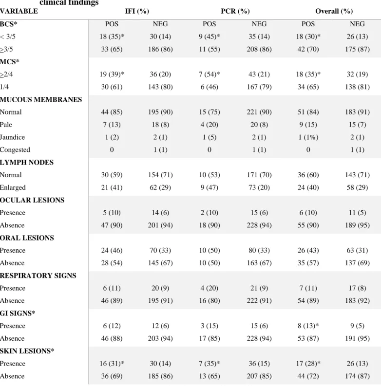
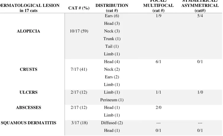
The most common clinical findings reported in FeL are skin or mucocutaneous lesions (ulcerative, crusty, nodular or scaly dermatitis, alopecia, poor coat condition), lymph node enlargment, ocular lesions (uveitis, nodular blepharitis and panophthalmitis), chronic gingivostomatis, hepatomegaly, spleen enlargment and non specific signs are also reported (weight loss, reduced appetite, dehydration, pale mucous membranes, fever, jaundice, polyuria/polydipsia, lethargy). Rare clinical manifestations include chronic nasal discharge and obstruptive upper respiratory tract
16
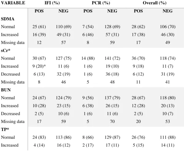
disease due to granulomatous rhinitis (Leal et al., 2018; Altuzarra et al., 2018). The more frequent clinicopathological abnormalities include mild to severe normocytic normochromic non-regenerative anemia, moderate to severe pancytopenia, lymphocytosis, hyperglobulinaemia and gammopathy, hypoalbuminemia, renal proteinuria, increased serum creatinine and increased serum alanine aminotransferase (ALT) (Pennisi et al., 2013a; Pennisi et al., 2015; Soares et al., 2016). Less information is available about renal involvement, even if in some clinical cases glomerular disease and chronic renal failure have been reported (Pennisi et al., 2013a). In a study conducted in 2004 four cats affected by FeL, followed up until their death or euthanasia, developed CKD progressively (Pennisi et al., 2004). In 2008 a case of Leishmania infection in a cat was reported with a positive PCR and isolation of Leishmania spp. from kidney (Caracappa et al., 2008). In 2010 histopathological lesions were investigated in 15 cats with leishmaniosis and one cat showed kidney moderate interstitial inflammatory infiltration composed predominantly of macrophages with some lymphocytes and plasma cells. Moderate interstitial fibrosis and tubular proteinuria were also present (Navarro et al., 2010). In 2016 in another study conducted in 14 followed up cats (four of them reported in the Pennisi et al., 2004 study) affected by FeL, 8 cats were affected by CKD with 3 cats in IRIS CKD stage 1, 4 cats in IRIS CKD stage 2 and one cat in IRIS CKD stage 4 (Pennisi et al., 2016). In another study diagnosis of FeL was performed by histopathology, immunohistochemistry and PCR and histopathology revealed a granulomatous nephritis with lymphoplasmacytic aggregates and macrophages containing some amastigotes (Puleio et al., 2011).
Diagnosis is performed by direct detection of Leishmania amastigotes in infected feline macrophages (rarely in circulating neutrophils) in smears from lymph nodes, bone marrow, skin, mucosal or eye lesions; histopathology with immunohistochemistry of lesions; molecular investigations of Leishmania DNA performed on EDTA-blood or other tissues (lymph node, bone marrow, skin) and non-invasive sampling (conjunctival or oral swabs), (Navarro et al., 2010; Pennisi and Persichetti, 2018; Pennisi et al., 2013a; Pennisi et al., 2015; Soares et al., 2016). Anti-Leishmania antibody detection is extensively used in cats, particularly IFI with a cut off established at 1:80 dilution (Pennisi et al., 2012), ELISA, direct agglutination test
17
(DAT) and WB techniques (Pennisi and Persichetti, 2018; Pennisi et al., 2013a; Pennisi et al., 2015; Soares et al., 2016) with WB offering the best sensitivity and specificity (Persichetti et al., 2017).
Feline immunodeficiency virus (FIV)
Feline immunodeficiency virus is an enveloped RNA virus, belonging to the family Retroviridae, subfamily Lentiviridae, group of viruses known to cause life-long infections with protracted incubation periods (Norris et al., 2007). Lentiviruses are complex retroviruses characterized by different genes, some of them responsible for their virulence and diversity: gag gene encodes the capsid protein p24 (important for diagnosis), pol gene encodes protease, integrase and reverse transcriptase proteins and enzymes determining the virulence of FIV, env gene encodes the viral glycoprotein (gp120) and the transmembrane protein (gp41) responsible for viral diversity among isolates. Five genetically distinct subtypes have been defined (A to E) and subtype A and B are the most frequently identified (Hosie et al., 2009).
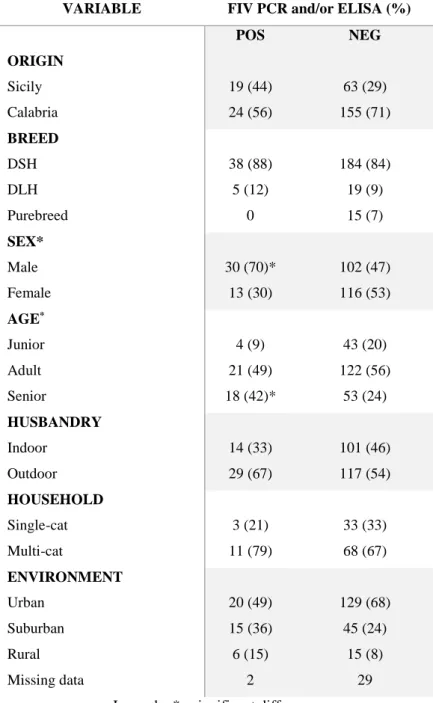
Biting is the principal route of FIV transmission between cats and pregnant queens in the acute phase of infection may transmit virus to their offspring during the prenatal and postnatal periods. A potential way for transmission could be intravenous, subcutaneous or intraperitoneal inoculation of blood products from infected cats (Norris et al., 2007).
Initially, viraemia with non specific signs, peripheral lymphadenopathy is present (duration weeks to months), followed by an asymptomatic phase (duration many years), while the final phase is charatcterized by viral replication with clinical disease, in part due to a CD4+ lymphopenia and immunodeficiency syndrome (Westman et al., 2019). Clinical signs frequently reported in infected cats include periodontitis, gingivitis, stomatitis, rhinitis, anemia, lymphadenopathy, myeloproliferative disorders, diarrhea, dermatitis, emaciation, central nervous system involvment, neuropathy and reproductive failure (Poli et al., 1993; Hosie et al., 2009). Laboratory findings frequently observed in FIV infected cats are anemia, cytopenia with neutropenia and lymphopenia, thrombocytopenia and hypergammaglobulinemia (Gleich and Hartmann, 2009; Collado et al., 2012). A significant relationship exists between CKD and FIV infection (White et al., 2010). Infected cats often present with
18
hypergammaglobulinemia that is believed to be due to chronic polyclonal B cell activation, with production of auto-antibodies and immune complexes (Poli et al., 1993; Pennisi et al., 1994). In FIV-infected cats azotemia, proteinuria, low USG and renal ultrasonographic abnormalities such as hyperechoic cortices and renomegaly were reported (Poli et al., 1993; Poli et al., 1995; Asproni et al., 2013; Baxter et al., 2012; Taffin et al., 2017). Histopathology revealed segmental glomerulosclerosis, glomerular capillary collapse, increased mesangial matrix, tubular dilatation and amyloid deposits with both medullary and glomerular location (Poli et al., 1993; Poli et al., 1995; Poli et al., 2012; Asproni et al., 2013) and FIV antigen (protein 24) was detected within tubular, glomerular, or interstitial cells (Poli et al., 1995). Recently, diagnosis of immune-complex glomerulonephritis was found associated with FIV infection (Rossi et al., 2019).
Diagnosis of FIV infection is routinely performed by rapid tests (ELISA or immunochromatographic techniques) that detect antibodies against viral structural proteins (the capsid protein p24 and a gp41 peptide). Western blot analysis is however considered the ‘gold standard’ for anti-FIV antibody detection. Indeed, the diagnostic specificity of ELISA and immunochromatography tests is below 100%, therefore any positive result in a low-prevalence population (young, indoor, pedigree cats) must therefore be confirmed for example, by Western blot. A positive result in a cat from a high-risk group (free-roaming, aged, entire male) is likely to be correct, because the frequency of true positives will exceed that of false positives in this group. Proviral DNA can be evidenced through EDTA-blood PCR, however its performance may be inferior to serological tests, with sensitivities and specificities ranging from 40 to 100%. In infected cats staging of the level of immune dysfunction is determined by counting CD4+ and CD8+ blood lymphocyte subpopulations even if these last two methods are not used routinely (Hosie et al., 2009).
Feline leukemia virus (FeLV)
Feline leukemia virus belongs to the genus Gammaretrovirus. There are three major subgroups of FeLV: A, B and C and FeLV-A is the most abundant subtype, responsible for transmission of the virus between animals. Subgroups B and C arise in subgroup A infected cats following the establishment of viremia (Willet and Hosie, 2013).
19
Horizontal and vertical transmission can both occur (Willis, 2000). Oronasal exposure to virus-containing secretions is the primary way of transmission and high levels of FeLV are present in the saliva of viraemic cats. Intimate contact between animals during grooming, sharing feeding bowls or fightning are the most likely routes of transmission and young age, high population density, poor hygiene are important risk factors (Lutz et al., 2009; Willet and Hosie, 2013). In addition to saliva and nasal secretions viraemic cats can shed the virus also in faeces and milk. In pregnant queens, viraemia usually leads to embryonic death, stillbirth or viraemic kittens, which will fade rapidly. In latently infected (cats with proviral DNA in bone marrow stem cells, as described later) queens, virus is usually not transmitted to the fetuses, but transmission can take place from individual mammary glands, where sequestered virus remains latent until the mammary gland develops during the last period of pregnancy (Lutz et al., 2009).
Stages of FeLV infection are influenced by feline immune response. The infection is characterized by an initial replication of the virus in the local lymphoid tissue in the oropharyngeal area. In immunocompetent cats viral replication may be avoided by an effective immune response without development of viremia (FeLV antigen, viral RNA or proviral DNA cannot be detected) and these cats have just an abortive infection. After local initial infection, FeLV disseminates through infected mononuclear cells. Cats have positive results on tests that detect free antigen in blood and they shed the virus. This viremia can last for weeks or months (transient viremia) but these “regressor” cats do not always eliminate the virus with time from the body, and proviral DNA is present in bone marrow stem cells as latent infection, however they do not shed the virus. In the regressive stage of infection all tests that detect FeLV antigen are negative, however provirus can be detected in the blood by sensitive PCR methods and in these cats the virus can potentially be reactivated at some time of their life. In other cats FeLV infection is not contained and they remain persistently viremic and infectious to other cats. These cats with progressive infection develop FeLV-associated diseases. It is noteworthy that a persistent atypical local viral replication (e.g., in mammary glands, bladder, eyes) is related to intermittent or low-grade production of antigen from a focal site of infection (Hartmann, 2012). The diseases associated with persistent infection are primarily disorders of haematopoiesis, such as
20
lymphoma (thymic, multicentric or alimentary) and leukaemias, immune suppression and anemia (Willis, 2000; Willet and Hosie, 2013). A dysregulation of the immune system leads to immunosuppression and infected cats can develop immune-mediated diseases caused by an overactive immune response. The most common abnormality seen is hypergammaglobulinemia which is caused by an excessive antibody response against the chronic persistent infection that is not neutralizing and may lead to antigen antibody complex formation. These immune complexes can deposit, usually in narrow capillary beds, leading to glomerulonephritis, polyarthritis, uveitis, and vasculitis (Hartmann, 2012). Glomerulonephritis is caused by an immune complex dense deposits located on the subepithelial and subendothelial sides of the glomerular basement membrane, as well as in the mesangial regions in most cases (Glick et al., 1978). Recently, diagnosis of immune-complex glomerulonephritis was found associated with FeLV infection (Rossi et al., 2019).
The first laboratory test used in the diagnosis of FeLV infection was an indirect immunofluorescent antibody (IFI) assay to detect viral antigen in blood cell smears, but this test has been replaced by commercially available rapid ELISA or immunochromatographic kits (Parry et al., 1989). These tests detect p27 viral capsid antigen that is a viral protein most abundant in the plasma of viraemic cats. As reported above, detection of regressive infection relies on real-time PCR commercially available for the quantification of FeLV proviral DNA. This technique is useful to identify regressive infection targeting proviral DNA integrated into the genome of cells within the bone marrow or lymphoid tissues and to confirm progressive infection by detecting viral RNA in blood or secretions (Willet and Hosie, 2013).
Feline coronavirus (FCoV)
Feline coronavirus is a large, spherical, enveloped, positive-sense single-stranded RNA virus that belongs to the family Coronaviridae of the order Nidovirales (Addie et al., 2009). There are two serotypes of FCoV recognized: type 1, which represents the vast majority of field strains found in naturally infected cats and type 2, which arise following recombination events between type 1 FCoV and canine coronavirus (CCoV). The two FCoV serotypes are distinguished primarily by the genetic and serological differences in their transmembrane spike (S) gene and protein,
21
respectively. The S protein is important as it is the part of the FCoV that binds to the host (feline) receptor, mediating host cell entry (Herrewegh et al., 1998; Addie et al., 2003; Tasker, 2018). Feline coronavirus infection is very common in cats with 40% infected cats reported (90% in multi-cat households), however natural infections are transient (~70%) and less frequently persistent infection occurs (~13%) (Tasker, 2018). The main way of transmission of FCoV is oro-fecal, in fact the virus replicates in the intestinal epithelium sometimes causing a mild diarrhea and is shed with faeces. After the first infection a proportion of FCoV-infected cats develop a severe immune-mediated disease called feline infectious peritonitis (FIP) as peritonitis is the most frequent consequence of the effusive form of the disease (Addie et al., 2009). Factors that contribute to development of FIP are: viral factors (mutation in the S gene), host factors (impaired immune response, breed, genetic, young age especially <2 years old), environmental factors (history of stress, overcrowded household) (Addie et al., 2009; Tasker, 2018). Strength of the T cell-mediated response influences the outcome of the infection and the severity of the clinical disease. Conversely in the presence of high levels of anti-FCoV antibodies monocytes and macrophages remain infected and a quiescent infection state develops (Addie et al., 2009). Activation of monocytes and macrophages leads to the pathologic features of FIP, including vasculitis, body cavity effusions, and fibrinous and granulomatous inflammatory lesions. Vasculitis observed in FIP is a phlebitis, mediated and dominated by activated virus-infected monocytes (Kipar and Meli, 2014). In those cats in which FCoV is able to replicate freely within the monocytes, infected monocytes attach to the walls of small and medium sized veins, releasing matrix metalloproteinase-9 (MMP-9) which destroys the collagen of the basal lamina of affected vessels with extravasation of the monocytes. Monocytes, differentiate into macrophages, and this allows plasma to leak out of the vessels (Kipar et al., 2005). Only veins are affected, most frequently the small- and medium-sized veins with involvment of leptomeninges, renal cortex, and eyes and, less frequently, veins in lungs and liver (Kipar and Meli, 2014).
Effusive FIP (wet form) is more severe and it is a fatal polyserositis affecting mainly the peritoneal cavity. In non-effusive FIP (dry form) pyogranulomatous lesions develop in different organs (especially eye, brain, kidneys, omentum, and liver), but actually FIP has a dynamic clinical spectrum and “dry” and “wet” lesions can be
22
concurrently found or at different times in a cat (Addie et al., 2009; Kipar et al., 2005). This explains why clinical signs related to FIP are variable. Affected cats can be interested by non-specific signs due to severe inflammation (fever refractory to antibiotics, lethargy, anorexia, and weight loss) (Addie et al., 2009). In the effusive form protein-rich fluid is found in the pleural space, peritoneal cavity, pericardial space, vaginal tunic of testicles and the subcapsular space of the kidneys, which can cause a possible clinical emergency; respiratory failure (pleural effusion), cardiac tamponade (pericardial effusion), or paralytic ileus (fibrinous peritonitis). The non-effusive form can be associated with progressive multifocal neurologic signs, signs of uveitis or chorioretinitis, hepatitis or gastro-intestinal signs (Lewis and O'Brien, 2010). Kidneys can be interested by renomegaly, ultrasound abnormalities as hyperechoic appearance, hypoechoic subcapsular rim, hyperechoic and hypoechoic areas in the renal medulla. Histologically a pyogranulomatous interstitial nephritis and immune-complex glomerulonephritis is evidenced (Lewis and O'Brien, 2010; Rossi et al., 2019).
Suspicion of FIP is often supported by clinicopathological abnormalities mild to moderate non regenerative anaemia, lymphopenia, increase in serum protein concentration (Addie et al., 2009), high albumin/globulin ratio with a cut-off value of 0.8 established (Hartmann, 2005), increase of liver enzymes, bilirubin, urea and creatinine) depending on the degree of organ damage (Addie et al., 2009) but diagnosis can be definetly confirmed by histopathology of suggestive lesions and the detection of FCoV in macrophages by immunofluorescent or immunohistochemistry staining of biopsies or smears from effusions or cerebrospinal fluid (Hartmann, 2005). Effusion fluid is generally yellow and sticky and typically positive in a Rivalta test. Serum antibody titres and RT-PCR may contribute to diagnostic information. Serum FCoV antibody tests available are ELISA, IFI test or rapid immunomigration tests. A positive FCoV antibody test indicates that there was contact with FCoV and that the cat has developed antibodies. Cats with FIP tend to have higher FCoV antibody titres than cats without FIP however low antibody titres do not rule out FIP, and a significant proportion of cats manifesting FIP are seronegative. Concerning PCR, FCoV shows a high rate of errors during replication and any mutations at the site of primer and/or probe binding can result in loss of PCR assay efficiency, and ultimately sensitivity
23
(Tasker, 2018). Both these factors compromise the ability of these diagnostic tests to provide a correct diagnosis.
1.3 PATHOGENESIS OF CHRONIC KIDNEY DISEASE
Development of CKD is characterized by an initiation phase and a progression phase. Indeed, primary renal disease initiates the damage to the kidneys with a variety of primary and secondary glomerulopathies, including immune complex glomerulonephritis, that act to initiate disease (Brown et al., 2016). Damage could be direct and or immune mediated and when injury is sustained and chronic follows the development of a chemotactic infiltration of inflammatory cells (neutrophils, platelets) that produce profibrotic cytokines (eg, transforming growth factor [TGF-β1]) which promote fibrogenesis by activation of matrix-producing cells (myofibroblasts, activated form of fibroblasts) (Reynolds and Lefebvre, 2013). Moreover, tubular epithelial cell cycle stops in response to toxic, obstructive, and ischemic injury with a lack of regeneration that may contribute to progression of disease and loss of nephrons (Jepson, 2016). In healthy cats, renal interstitium is composed of sparse cells (fibroblasts and dendritic cells) contained in an extracellular matrix composed of collagen, fibronectin, and glycoproteins. After an inciting injury, development of focal areas of inflammation and activation of mesenchymal cells begins. In CKD, fibrosis may represent a maladapted response of the kidney to injury with excessive fibrogenic response and expansion of extracellular matrix, which destroys the normal renal tissue (Jepson, 2016). Some pathophysiologic mechanisms can contribute to progression of CKD, aggravating the existing damage and reducing in some cases survival of cats. Vascular endothelial injury may lead to tissue hypoxia and ischemia, affecting renal parenchymal repair and contributing to renal damage and progression of CKD (Khan and Khan, 2015). In response to renal mass reduction, there are haemodynamic adataptions that lead to dilation of preglomerular afferent arterioles, increase in glomerular capillary pressure, and increased effective filtration pressure. Activation of the renin-angiotensin-aldosterone system contributes to kidney damage. Indeed, angiotensin II not only acts as a potent vasoconstrictor that contributes to the development of glomerular hypertension and hyperfiltration but also can modulate the
24
permeability of the glomerular filtration barrier, promoting proteinuria, and transcription and production of inflammatory and profibrogenic molecules. Aldosterone has also profibrotic effects contributing to the pathogenesis of CKD (Reynolds and Lefebvre, 2013; Jepson, 2016). Systemic hypertension may also affect renal function by inducing glomerular hypertension and proteinuria (Reynolds and Lefebvre, 2013). Proteinuria at the same time can contribute to kidney damage stimulating the inflammatory response. Even if in cats, the magnitude of proteinuria seems to be typically low compared to dogs, it has been significantly associated with the development of azotemia and reduced survival time. Finally, hyperphosphatemia can predispose to renal mineralization, with promotion of inflammation and fibrosis (Jepson, 2016). All these factors contribute to aggravate renal damage with development of subsequent clinical signs reported below.
25
1.4 CLINICAL AND CLINICOPATHOLOGICAL FINDINGS OF CHRONIC
KIDNEY DISEASE
Cats affected by CKD may have different complications that arise in advanced stages (Brown et al., 2016).
Polyuria and polydipsia are reported as the main clinical signs observed by the owners in the year that preceds CKD diagnosis (Bartlett et al., 2010). Polyuria may occur as a result of a decreased nephron mass and a consequent decrease of the urinary concentration ability. Polyuria is generally offset by polydipsia but dehydration may occur if water loss exceeds water intake (Bartges, 2012).
Urine specific gravity reflects the ability of tubules to concentrate and dilute urine to mantain fluid homeostasis. Healthy cats with the ability to concentrate urine have an USG ≥1.035. Some factors can minimally affect USG such as age, diet type, sex, fasting status, drinking avidity, refractometer type but generally older cats with USG <1.035 need further investigations (Rishniw and Bicalho, 2015). Some cats can retain their urine concentrating ability particularly in the early stages of CKD but with disease progression, USG usually gradually declines (Paepe and Daminet, 2013).
Decreased appetite is a common clinical finding of cats affected by CKD with a reported prevalence ranging from 21–92% (Freeman et al., 2016).
Cachexia and loss of muscle mass are common in companion animals with CKD (Freeman, 2012) and weight loss is also observed in cats affected by kidney disease with a prevalence range between 42 to 82%. Weight loss was related to a shorter survival time (Freeman et al., 2016). Multifactorial mechanisms are responsible for weight loss in CKD such as inflammation, malabsorption, increased energy requirements, and decreased appetite. Moreovercats with CKD and body weight ≤4 kg had a higher relative risk of death (Freeman et al., 2016).
Retention of uremic toxins (such as urea, creatinine, phosphates) is responsible for the uremic (Langston, 2003; McLeland et al., 2014) syndrome characterized by gastrointestinal manifestations such as reduced appetite and food intake, drooling and halitosis associated with, ulcerative stomatitis, vomiting, gastrointestinal hemorrhage, and diarrhea (Polzin, 2011). Gastric hyperacidity secondary to hypergastrinemia can contribute to some of the above manifestations. In fact, gastrin excretion is regulated
26
by kidneys therefore renal function decline is responsible for increase of gastrin levels and a consequent gastric hyperacidity (McLeland et al., 2014). Stomach histopathological lesions reported in cats affected by CKD include fibrosis and gastric mineralization, that can be a possible consequence of the dysregulation of the calcium-to-phosphorus ratio (McLeland et al., 2014).
Cats with CKD are affected by decreased production of erythropoietin (EPO) secondary to loss of functional renal mass. An altered/decreased iron metabolism can be seen as a consequence of chronic gastrointestinal hemorrhage and decreased intestinal absorption or intake (Gest et al., 2015; Javard et al., 2017). All these factors can be resposible for the development of anemia, and are poor prognostic indicators in terms of survival in animals affected by CKD. The anemia of CKD is usually non regenerative, normochromic normocytic, and its degree indicates the severity of loss of functional renal tissue (Elliot and Barber, 1998; Paepe and Daminet, 2013; Ettinger and Feldman, 2016). Cortical interstitial fibrosis is the renal lesion best correlated with the severity of anemia (Chakrabarti et al., 2013).
Progressive loss of functional nephrons leads to a decrease in the glomerular filtration rate that causes phosphorus retention, with hyperphosphatemia and an increased risk of development of secondary hyperparathyroidism. Hyperphosphatemia promotes parathyroid hormone (PTH) secretion that leads to phosphorus reabsorption in the renal tubules. However, as the glomerular filtration rate continues to decline, phosphorus retention becomes more severe with further secretion of PTH (Segev et al., 2016). At the same time calcitriol (active form of vitamin D) formed in the kidney, along with PTH is implicated in calcium homeostasis. In kidney disease there is a decrease in calcitriol concentration that decreases intestinal calcium absorption leading to hypocalcemia. The combination of low calcitriol and low calcium allows high concentration of PTH with an imbalance in the bone remodeling process and a consequent osteodystrophy (Segev et al., 2016; de Brito Galvao et al., 2013). Increases in plasma phosphate concentration can result in soft tissue mineralization especially in proton-secreting organs such as stomach or kidney where secretion of bicarbonates determines precipitation of calcium-phosphate crystals, however these mineralizations can also be seen in myocardium, lungs and liver (Chakrabarti et al., 2012; Ettinger and Feldman, 2016).
27
Total calcium (tCa) concentration includes hydrated free calcium ions (iCa), protein-bound calcium and a small portion of ionic complexes, such as calcium phosphate. Cats with CKD have increased risk of increased tCa. However, the biologically active iCa most accurately reflects true calcium status and measurement of iCa is necessary for an accurate assessment of calcium status in cats (van den Broek et al., 2017).
Chronic kidney disease can be responsible for increased blood pressure (BP) and increased blood pressure worsens the course of CKD. In cats increased BP can however be influenced by increased age, gender (with males and neutered cats having higher BP than females and intact animals), muscle condition score and skeletal muscle mass. Hypertension can be distinct in situational, idiopathic or secondary hypertension. Situational hypertension is caused by autonomic nervous system alterations that arise from the effects of excitement or anxiety on higher centers of the central nervous system. This can be minimized by measuring BP in a quiet area with the presence of the owner, away from other animals, before other procedures and only after the patients have been acclimated to their surroundings for 5-10 minutes. Idiopathic hypertension is suspected when there is a sustained increase in BP concurrent with normal complete blood count (CBC), serum biochemistry, and urinalysis results. Persistent, pathologically increased BP concurrent with a disease or condition known to cause hypertension is defined as secondary hypertension (Acierno et al., 2018). Chronic kidney disease, hyperthyroidism, primary hyperaldosteronism (PHA), hyperadrenocorticism (HAC) and phaeochromocytoma can be responsible for secondary hypertension (Taylor et al., 2017). Chronic kidney disease is the most common condition associated with feline hypertension. Azotaemia has been found in up to 74% of hypertensive cats, and conversely between 19% to 65% of cats with CKD have been found to be hypertensive (Taylor et al., 2017), moreover hypertension is associated with glomerulosclerosis and glomerular hypertension (Chakrabarti et al., 2013; Syme et al., 2006) and indirect blood pressure is correlated with the severity of CKD (Hori et al., 2018). Cats with CKD have to be monitored for hypertension because chronically sustained increases in BP cause injury to tissues with development of target organ damage (TOD). Eyes, brain, kidneys and myocardium are organs particularly vulnerable to injury. Indeed, hypertension has been associated with
28
proteinuria that can lead to a more rapid progression of renal disease, with ocular lesions (e.g. hypertensive retinopathy, choroidopathy, exudative retinal detachment, retinal hemorrhage, multifocal retinal edema, retinal vessel tortuosity, retinal perivascular edema, papilledema, vitreal hemorrhage, hyphema, secondary glaucoma, and retinal degeneration), with hypertensive encephalopathy (observed when systolic blood pressure exceeds 180 mm Hg) with lethargy, seizures, acute onset of altered mentation, altered behavior, disorientation, balance disturbances (e.g. vestibular signs, head tilt, and nystagmus), and focal neurologic defects because of stroke-associated ischemia. Hypertension appears to be a risk factor for ischemic myelopathy of the cranial cervical spinal cord, resulting in tetraparesis or tetraplegia with intact nociception in old cats. Cardiac abnormalities are common in hypertensive cats with cardiomegaly associated with left ventricular concentric hypertrophy (LVH). Epistaxis, aortic aneurysm and aortic dissection can be rare complications of hypertension (Acierno et al., 2018; Taylor et al., 2017).
Azotaemia is due to increased serum creatinine (sCr) and urea concentrations as a result of renal pathology (Paepe and Daminet, 2013). Serum creatinine is produced by muscle metabolism and is inversely related to glomerular filtration rate (GFR) (Sparkes et al., 2016; Yerramilli et al., 2016) with an exponential relationship with GFR, therefore early declines in GFR are characterized by small changes in creatinine (Sparkes et al., 2016). Significant changes in creatinine concentration occur when 60% to 70% of all nephrons are nonfunctional (Pressler, 2015). Plasma/serum creatinine values can be influenced by artifacts such as hemolysed samples with increased values or lipemic and icteric samples with lower estimates. Dehydration and muscle mass can also influence creatinine values, because depletion of body water leads to increases in creatinine concentration and muscle wasting leads to reduced creatinine values with understimation of kidney dysfunction if USG is not evaluated (Paepe and Daminet, 2013; Yerramilli et al., 2016). This aspect is of clinical relevance particularly in geriatric cats or during CKD progression where a tendence to muscle wasting is observed (Paepe and Daminet, 2013). Moreover some breeds tend to have higher levels of serum/plasma creatinine, such as Birman or Siberian cats and this should be considered when creatinine values are interpreted in these breeds (Paltrinieri et al., 2014; Reynolds et al., 2010).
29
Metabolic acidosis occurs commonly in cats with CKD as a consequence of retention of acids that are normally excreted through the kidneys (Bartges, 2012). Biochemical evidence of metabolic acidosis seems to occur with late stages of this disease syndrome (Elliot et al., 2003). Metabolic acidosis is characterized by an increased hydrogen ion concentration in blood that results in movement of hydrogen ions into cells in exchange for potassium ions that leave the cells and enter the circulation. Therefore potassium is excreted and this predispose to hypokalemia (Bartges, 2012).
The marked reduction in glomerular filtration rate of end-stage CKD tends to promote potassium retention and hyperkalemia (Polzin, 2011). In cats affected by CKD with hypokalemia, the muscle membrane becomes electrically hyperpolarized and refractory to stimulation for the action potentials. This explains manifestations of the kaliopenic polymyopathy/nephropathy syndrome characterized by a generalized appendicular muscle weakness, persistent ventroflexion of the neck, reluctance to walk and apparent muscular pain upon palpation. Moreover, in cats affected by hypokalemia reduction of renal function as well as anorexia are reported with an improvement of renal funtion when normokalemia is restored (Fettman, 1989; Ettinger and Feldman, 2016).
Proteinuria is a common laboratory finding in CKD, even if proteinuria in dogs seem to have a higher prevalence (90%) rather than proteinuria observed in cats (20%). This is compatible with published data, suggesting that the major disease process occurring in cats is chronic tubulointerstitial fibrosis rather than primary glomerular disease (Syme et al., 2006; Giraldi and Scarpa, 2018). However, a recent study found immune-complex glomerulonephritis in about half of examined tissue samples and this diagnosis was more frequently associated with FIV or FeLV infections, younger age and higher UPC values compared to non immune-complex renal disease (Rossi et al., 2019).
Physiologic or functional proteinuria is known and it can be distinguished from a pathologic proteinuria that is based on pre-renal, renal, or post-renal factors. Functional renal proteinuria is caused by heat, stress, seizure, venous congestion, fever and extreme muscle exercise and is generally of low grade and transient. Pre-renal proteinuria is caused by an overabundant filtered load of low molecular weight
30
proteins (hemoglobin, myoglobin, immunoglobulin light-chain monomers and dimers such as Bence Jones proteins from neoplastic plasma cells). Renal proteinuria is due to a defect in the glomerular filtration barrier, tubular reabsorption or interstitial damage. Post-renal proteinuria is due to protein that comes from any part of the urinary tract distal to the kidney (urinary tract infections, genital infections or inflammation) (Lees et al., 2005; Harley and Langston, 2012). Normally the majority of plasma albuminin is size excluded and charge excluded from the ultrafiltrate, but when glomerular damage occurs there is an increased passage of albumin into urine (Pressler, 2015). When albumin in the urine is in excess of 0.30 g/L the animal is affected by proteinuria, while albumin concentrations ≥0.01 and < 0.30 g/L are defined as microalbuminuria (Harley and Langston, 2012). Normally, proteins of molecular weight up to about 50 KD pass through the glomerular capillary wall and are reabsorbed by the proximal tubule but in CKD, changes in glomerular permeability can result in filtration of proteins with higher molecular weight and tubular damage reduces the reabsorption of low-molecular weight proteins. Proteinuria can also be affected by hemodynamic factors such as angiotensin II, local prostaglandins, endothelin, and other vasoactive mediators that can influence constriction or dilatation of afferent and efferent renal arterioles with development of glomerular hypertension (Harley and Langston, 2012). The level of proteinuria can be useful to evaluate the progression of CKD (Chakrabarti et al., 2012) and shorter survival times are associated with increased protein excretion (Syme et al., 2006) probably by invoking interstitial inflammation and fibrosis around renal tubules: filtered proteins may directly damage tubular cells or may activate complement and chemoattractants leading to the formation of inflammatory mediators which contribute to fibrosis (Harley and Langston, 2012).
Diagnostic methods for detection of proteinuria will be described below.
1.5 DIAGNOSIS AND STAGING OF CHRONIC KIDNEY DISEASE
Diagnosis of CKD in dogs and cats is based on evaluation of suggestive clinical signs, measurement of sCr, USG, proteinuria, and ultrasound evaluation of kidneys and urinary tract. Systolic blood pressure (SBP) measurement is required to substage
31
CKD in order to manage the risk for target organ damage related to hypertension. Moreover, as described later, novel biomarkers of CKD are currently being studied (Paepe and Daminet, 2013).
Diagnosis and staging of CKD in dogs and cats is at present based on internationally recognised guidelines established by a group of experts, the International Renal Interest Society (IRIS) (www.iris-kidney.com). Since 1998 IRIS objectives consisted in providing to practitioners guidelines for the diagnosis and treatment of renal disease in dogs and cats. Through the urine analyis and the measurement of creatinine, UPC, and blood pressure values, IRIS guidelines show how to confirm, stage and substage CKD. The staging system allows an accurate prognosis and more appropriate treatments to slow the progression of kideny damage, reduce the risk for complications and improve the clinical condition. Guidelines are regularly updated according to the most recent scientific evidence.
The staging system includes five stages including one stage for cats considered at risk for developing CKD and four stages (from stage 1 to stage 4) for grading the severity of renal damage in cats affected by CKD (table 1.a). Basically, staging is based on fasting blood creatinine, assessed on at least two occasions in the stable patient, but in non-azotemic individuals data from clinical examination and urine e analysis are also considered to exclude that they are “at risk” for CKD or are at “stage 1”.
32
Table 1.a IRIS staging system of CKD
STAGE BLOOD
CREATININE IN CATS (mg/dl)
COMMENTS
AT RISK <1.6 Increased risk of developing CKD in the future because of different factors (e.g. exposure to nephrotoxic drugs, breed, high prevalence of infectious disease in the area, or old age).
1 <1.6 Nonazotemic. Presence of other renal
abnormality (e.g. inadequate urinary concentrating ability without identifiable nonrenal cause, abnormal renal palpation or renal imaging findings, proteinuria of renal origin, abnormal renal biopsy results, increasing blood creatinine concentrations in samples collected serially)
2 1.6 – 2.8 Mild renal azotemia. Clinical signs usually mild or absent.
3 2.9 – 5.0 Moderate renal azotemia. Extrarenal signs may be present.
4 >5.0 Severe azotemia. Increasing risk of systemic clinical signs and uraemic crises
Modified from www.iris-kidney.com
As described previously, increased creatinine values are seen when a great portion of nephrons (70%) are no longer functional, moreover dehydration, muscle mass and breed can affect creatinine values. All these aspects can influence the correct staging of the disease as well as the choice of an adequate therapeutic plan. Therefore, different studies analyzed the role and utility of alternative biomarkers for CKD and the measurement of serum symmetric dimethylarginine (SDMA) has been recently proposed as a novel biomarker for CKD in dogs and cats (Hokamp and Nabity, 2016; Jepson et al., 2008; Braff et al. 2014; Hall et al., 2016; Hall et al., 2017, Peterson et al., 2018, Hall et al., 2014a). Symmetric dimethylarginine is produced in the nucleus of all nucleated cells after a post-translational modification and methylation of arginine residues of varius proteins and it is excreted by the kidney (Relford et al, 2016). According to recent studies, a SDMA value greater than 14 µg/dL shows a decrease in
33
GFR below 20% (Relford et al, 2016). This is because serum SDMA evaluation allows early detection of CKD in cats compared to serum creatinine measurement. Albeit increases of SDMA are found 17 months earlier than increases of creatinine in cats with CKD, SDMA higher sensitivity is associated with a lower specificity compared to creatinine (Hall et al., 2014a). The SDMA value correlates with plasma creatinine concentration (Jepson et al., 2008) and is inversely related to GFR (Hall et al., 2014a). Interestingly, it is not affected by lean body mass so it is a more sensitive marker for kidney disease in patients with muscle loss (Hall et al., 2014b; Relford et al, 2016). SDMA values may however be higher in kittens and in cats with kidney stones (Relford et al, 2016; Hall et al., 2017). In hyperthyroid cats, SDMA concentration together with the evaluation of USG were found useful to identify and predict hyperthyroid cats at risk to develop azotemia after treatment of hyperthyroidism (Peterson et al., 2018). Recently, SDMA values were found less frequently elevated than creatinine in apparently healthy Birman cats suggesting that the analysis of both creatinine and SDMA could be useful to prevent the over-diagnosis of CKD or errors in staging renal disease in this breed (Paltrinieri et al., 2018). Based on recent experimental and clinical data, SDMA measurement has been added to the IRIS staging system of CKD (table 1.b) (www.iris-kidney.com) as a persistent increase in SDMA above 14 µg/dL is considered suggestive of reduced renal function in adult cats. In the case of creatinine values <1.6 mg/dl, patients are now diagnosed with CKD at Stage 1. In the case of low body condition score (BCS) of patients diagnosed with CKD Stage 2, SDMA values ≥25 µg/dL are suggestive of more severe renal damage and treatment recommendations for CKD Stage 3 should be followed. Similarly, for Stage 3 patients with low BCS, SDMA values ≥45 µg/dl suggest following recommendations for IRIS CKD Stage 4 (www.iris-kidney.com).
34
Table 1.b IRIS staging system of CKD evaluating SDMA
AT RISK STAGE 1 STAGE 2 STAGE 3 STAGE 4 CREATININE VALUE (mg/dl) <1.6 <1.6 1.6 – 2.8 2.9 – 5.0 >5.0 SDMA VALUE (µg/dl) >14 ≥25 ≥45
COMMENTS In patients with low body condition score consider treatment
recommendations listed under IRIS CKD Stage 1 for this patient. In patients with low body condition score consider treatment recommendations
listed under IRIS CKD Stage 3 for this patient. In patients with low body condition score consider treatment recommendations listed under IRIS CKD Stage 4 for
this patient
Modified from www.iris-kidney.com
According to the IRIS guidelines, staging of cats with CKD is followed by substaging based on proteinuria and systolic hypertension detection. Concerning proteinuria, we have to consider that cats affected by CKD have lower levels of proteinuria than dogs (Harley and Langston, 2012). However, as mentioned before, shorter survival times have been associated with increased protein excretion in cats, underlining the importance of correct evaluation and monitoring of proteinuria in cats to avoid progression of the disease. Detection of proteinuria can be performed with different methods. The traditional reagent pad colorimetric method of “dipstick” devices is the first screening test used to detect albuminuria. However some artifacts can influence its performance, such as highly concentrated urine or pigmented urine that can give falsely elevated results. The sticks are designed for human urine which is rarely as concentrated as dog or cat urine. Moreover acidic urine can cause false negative and alkaline urine can cause false positive results (Harley and Langston, 2012). The sulfosalicylic acid (SSA) test can help to distinguish between a true and a false positive dipstick test. The sulfosalicylic acid test is a semi-quantitative test that can detect protein at >50 mg/dL. It is perfomed using equal parts urine supernatant to