Androgens are differentially associated with ovarian cancer
subtypes in the Ovarian Cancer Cohort Consortium
Jennifer Ose1,2, Elizabeth M. Poole3, Helena Schock1, Matti Lehtinen4, Alan A. Arslan5, Anne Zeleniuch-Jacquotte6, Kala Visvanathan7, Kathy Helzlsouer7, Julie E. Buring8,9, I-Min
Lee8,9, Anne Tjønneland10, Laure Dossus11, Antonia Trichopoulou12,13, Giovanna Masala14,
N. Charlotte Onland-Moret15, Elisabete Weiderpass16,17,18,19, Eric J. Duell20, Annika Idahl21, Ruth C. Travis22, Sabina Rinaldi23, Melissa A. Merritt24, Britton Trabert25, Nicolas
Wentzensen25, Shelley S. Tworoger3,9, Rudolf Kaaks1, and Renée T. Fortner1
1Division of Cancer Epidemiology, German Cancer Research Center, Heidelberg,
Baden-Württemberg, Germany 2Department of Population Sciences, Huntsman Cancer Institute,
University of Utah, Salt Lake City, Utah, USA 3Channing Division of Network Medicine,
Department of Medicine, Brigham and Women’s Hospital and Harvard Medical School, Boston,
MA, USA 4Karolinska Institutet, Department of Laboratory Medicine, Huddinge, Sweden
5Departments of Obstetrics and Gynecology, Population Health, and Environmental Medicine,
New York University School of Medicine; Perlmutter Cancer Center, New York University, New
York, New York 6Departments of Population Health, and Environmental Medicine, New York
University School of Medicine; Perlmutter Cancer Center, New York University, New York, New
York 7Department of Epidemiology, Johns Hopkins Bloomberg School of Public Health, Baltimore,
Maryland 8Division of Preventive Medicine, Department of Medicine, Brigham and Women’s
Hospital, Harvard Medical School, Boston, MA, USA 9Department of Epidemiology, Harvard T.H.
Chan School of Public Health, Boston, MA, USA 10Unit of Diet, Genes and Environment, Danish
Cancer Society Research Center, Copenhagen, Denmark 11INSERM, Centre for research in
Epidemiology and Population Health (CESP), U1018, Nutrition, Hormones and Women’s Health
team, Villejuif, France 12Hellenic Health Foundation, Athens, Greece 13WHO Collaborating Center
for Nutrition and Health, Unit of Nutritional Epidemiology and Nutrition in Public Health, Dept. of Hygiene, Epidemiology and Medical Statistics, School of Medicine, National and Kapodistrian
University of Athens, Greece 14Molecular and Nutritional Epidemiology Unit, Cancer Research
and Prevention Institute – ISPO, Florence – Italy 15Department of Epidemiology, Julius Center for
Health Sciences and Primary Care, University Medical Center Utrecht, Utrecht, The Netherlands
16Department of Community Medicine, Faculty of Health Sciences, University of Tromsø, The
Arctic University of Norway, Tromsø, Norway 17Department of Research, Cancer Registry of
Norway, Oslo, Norway 18Department of Medical Epidemiology and Biostatistics, Karolinska
Institute, Stockholm, Sweden 19Genetic Epidemiology Group, Folkhälsan Research Center,
Helsinki, Finland 20Unit of Nutrition and Cancer, Cancer Epidemiology Research Program,
Catalan Institute of Oncology (ICO-IDIBELL), Barcelona, Spain 21Department of Clinical
Corresponding author: Renée T. Fortner, Division of Cancer Epidemiology, German Cancer Research Center (DKFZ), Im
HHS Public Access
Author manuscript
Cancer Res
. Author manuscript; available in PMC 2018 January 15.Published in final edited form as:
Cancer Res. 2017 July 15; 77(14): 3951–3960. doi:10.1158/0008-5472.CAN-16-3322.
A
uthor Man
uscr
ipt
A
uthor Man
uscr
ipt
A
uthor Man
uscr
ipt
A
uthor Man
uscr
ipt
Sciences, Obstetrics and Gynecology, Umea University, Umea, Sweden 22Cancer Epidemiology
Unit, Nuffield Department of Population Health, University of Oxford, Oxford, UK 23Section of
Nutrition and Metabolism, International Agency for Research on Cancer (IARC), Lyon, France
24Department of Epidemiology and Biostatistics, School of Public Health, Imperial College
London, London, UK 25Division of Cancer Epidemiology and Genetics, National Cancer Institute,
National Institutes of Health, Department of Health and Human Services, Bethesda, MD, USA
Abstract
Invasive epithelial ovarian cancer (EOC) is the most lethal gynecologic malignancy. The etiology of EOC remains elusive; however, experimental and epidemiologic data suggest a role for hormone-related exposures in ovarian carcinogenesis and risk factor differences by histologic phenotypes and developmental pathways. Research on pre-diagnosis androgen concentrations and EOC risk has yielded inconclusive results, and analyses incorporating EOC subtypes are sparse. We conducted a pooled analysis of 7 nested case-control studies in the Ovarian Cancer Cohort Consortium to investigate the association between pre-diagnosis circulating androgens
(testosterone, free testosterone, androstenedione, dehydroepiandrosterone sulfate (DHEAS)), sex hormone binding globulin (SHBG), and EOC risk by tumor characteristics (i.e. histology, grade, and stage). The final study population included 1,331 EOC cases and 3,017 matched controls. Multivariable conditional logistic regression was used to assess risk associations in pooled individual data. Testosterone was positively associated with EOC risk (all subtypes combined, Odds Ratio (OR)log2=1.12 [95% Confidence Interval (CI) 1.02–1.24]); other endogenous
androgens and SHBG were not associated with overall risk. Higher concentrations of testosterone and androstenedione associated with an increased risk in endometrioid and mucinous tumors (e.g., testosterone, endometrioid tumors, ORlog2=1.40 [1.03–1.91]), but not serous or clear cell. An
inverse association was observed between androstenedione and high grade serous tumors (ORlog2=0.76 [0.60–0.96]). Our analyses provide further evidence for a role of hormone-related
pathways in EOC risk, with differences in associations between androgens and histologic subtypes of EOC.
Keywords
ovarian cancer; androgens; histologic subtypes; developmental pathways
Introduction
Reproductive history influences risk of ovarian cancer and it has been hypothesized that these associations are mediated by exposure to endogenous hormones, including androgens (1). Data from experimental studies link androgen-related signalling to ovarian cancer through increased cellular proliferation and reduced apoptotic rates (2–4). The relationship between androgens and epithelial ovarian cancer (EOC) risk has been examined in 7 nested case-control studies with the numbers of cases in these studies ranging from 31 to 1,052 (5– 10); these studies predominantly investigated EOC as a composite outcome. Emerging data show heterogeneity in risk factors by histologic subtypes (e.g., serous, endometrioid,
A
uthor Man
uscr
ipt
A
uthor Man
uscr
ipt
A
uthor Man
uscr
ipt
A
uthor Man
uscr
ipt
mucinous, clear cell) and by the hypothesized “dualistic pathway” of ovarian carcinogenesis (defined by differences in the genetic make-up and the morphological architecture of histologic phenotypes) (11–18). The relationship between androgens and EOC risk by disease subtype has been minimally explored. Analyses to date suggest heterogeneity by subtype (9, 10); however, individual studies evaluating EOC by subtype were either limited by small case numbers in subtype analyses (9), or restricted to women pregnant at the time of serum sampling (10).
We pooled and harmonized available data from 6 nested case-control studies within the Ovarian Cancer Cohort Consortium (OC3), plus the Finnish Maternity Cohort (FMC), to investigate the relationship of pre-diagnosis concentrations of androgens (e.g., testosterone, free testosterone, androstenedione, dehydroepiandrosterone-sulfate (DHEAS)) and sex-hormone binding globulin (SHBG) with EOC risk, overall and by subtype. Subtype analyses included analyses by histology, grade and stage, and by the hypothesized dualistic model of EOC development, i.e., type I vs. type II (19). Our study represents the largest investigation to date including individual-level data from 1,331 EOC cases and 3,017 matched controls, with 61 (clear cell) to 667 (serous) cases represented in the major histologic subtypes.
Methods
Study Population: Ovarian Cancer Cohort Consortium (OC3)
The OC3 has been described previously (12). For this investigation, eligible cohorts were required to have data on a defined set of a priori selected covariates (e.g., menopausal status at blood donation, oral contraceptive use at blood donation, parity) and pre-diagnosis measurements of testosterone, free testosterone, androstenedione or DHEAS. In addition to the OC3 cohorts, the FMC, a cohort of women pregnant at blood collection, contributed data to this investigation (for contributing cohorts see Supplementary Table S1). Available biomarker and questionnaire data from each cohort were centrally collated and harmonized at the Data Coordinating Center at the Brigham and Women’s Hospital.
Case characteristics
Eligible cases included women diagnosed with invasive EOC (International Classification of Disease Codes (ICD): ICD9 codes 183 and 158; ICD10 code C56) ascertained by self-report with medical record confirmation and/or linkage to cancer registries. Cases were
individually matched to two or three controls (free of cancer and alive at the time of diagnosis of the index case) on age, date, menopausal status and day or phase of menstrual cycle at blood collection in premenopausal women, with the exception of the FMC (matched on age and date at blood collection, parity at blood collection and at diagnosis/index date). Histomorphological data was complete, and the majority of cases had data on stage (82%); grade was available for 36% of the cases. We used histology and grade to classify tumors into type I ((48%, n=291); low-grade serous and endometrioid, all mucinous and clear cell) and type II ((52%, n=314); high-grade serous, high-grade endometrioid) (19). Serous and endometrioid cases missing grade data were excluded from these analyses; mucinous and clear cell tumors were included regardless of grade data availability, as these tumors are classified as type I independent of grade. In a sensitivity analysis, all mucinous and clear cell
A
uthor Man
uscr
ipt
A
uthor Man
uscr
ipt
A
uthor Man
uscr
ipt
A
uthor Man
uscr
ipt
cases missing grade were excluded from the type I subgroup (after exclusion, case n=77). The proportion of type I tumors was higher than expected; however, we observed the expected distribution (type I: 28% vs. type II: 72%) after excluding women from the FMC (all missing grade; younger at diagnosis and more frequently diagnosed with mucinous tumors than cases from the other cohorts).
Laboratory methods
In all studies, case-control sets were measured in the same batch and technicians performing the assays were blinded to case-control status and quality control samples. Information on sample type, laboratory assays, and intra- and inter-batch coefficients of variations for each cohort is summarized in Supplemental Table S2. Free testosterone was calculated based on measured concentrations of testosterone and SHBG, with albumin assumed to be a constant 40g/L, according to the mass law of action (20).
Statistical analyses
Hormone measurements were standardized across studies based on the cohort-specific mean concentrations in controls (see supplemental methods; Supplemental Table S3). Conditional logistic regression was used to estimate odds ratios (OR) and 95% confidence intervals (CI). ORs were estimated using log2-transformed biomarker concentrations and study-specific tertiles based on the distribution in controls. A continuous probit score, generating a rank for each participant in each cohort by hormone concentration, was used to test for trend across tertiles. We additionally evaluated associations in quintiles for EOC overall and the serous subtype. Multivariable models included: parity [never, ever, missing (2.8%)] and OC use [never, ever, missing (47%); excluding FMC 2.3% missing]. Additional adjustment for body mass index (BMI; kg/m2) among women with data available (n=747 cases), did not change the ORs (data not shown).
Statistical analyses were conducted using a two-stage approach: First, ORs were calculated within each cohort and pooled using DerSimonian and Laird random effects meta-analysis models to assess between-study heterogeneity (21). Second, ORs were calculated based on pooled individual participant data (22). ORs estimated from meta-analysis and the data pooling method were similar, and we observed no significant between-study heterogeneity. Therefore, presented results are based on the pooled analysis. The assumption of linearity was tested using restricted cubic splines; no significant deviations from linearity were observed. Statistical heterogeneity of associations across subtypes was assessed via a likelihood ratio test comparing a model allowing the association for the risk factor of interest to vary by subtype versus one assuming the same association across subtype using
polytomous conditional logistic regression (23).
We evaluated associations after stratification by menopausal status at blood collection (premenopausal vs. postmenopausal) and age at diagnosis (<55 vs. ≥55 years). Androgen concentrations are relatively stable in pregnancy (24), however, we excluded FMC members in sensitivity analyses given that all women were pregnant at the time they provided a blood sample. Finally, we conducted a sensitivity analysis after exclusion of women diagnosed
A
uthor Man
uscr
ipt
A
uthor Man
uscr
ipt
A
uthor Man
uscr
ipt
A
uthor Man
uscr
ipt
within two years after blood donation. A more detailed description of statistical procedures is available in the supplemental methods.
SAS Statistical Software, version 9.3 (SAS Institute, Cary NC, USA) was used for statistical analyses. P-values<0.05 were considered as statistically significant; all statistical tests and p-values were two-sided.
Results
In total 1,331 cases and 3,017 matched controls from 7 cohorts were included in this investigation (Table 1). Average age at blood collection ranged from 32 (FMC) to 61 years (CLUE II), and the majority of women were parous (89% cases, 94% controls) (Table 1). Average age at diagnosis ranged from 45 (FMC) to 67 (CLUE II) (Supplemental Table S4).
Androgens and overall EOC risk
A doubling of testosterone (i.e., 1-unit increase in log2-transformed testosterone) was
associated with a 12% increase in overall EOC risk (ORlog2=1.12; 95% Confidence Interval
(CI) [1.02–1.24)], and a 25% increase in risk comparing top to bottom tertile
(ORT3−T1=1.25 [1.06–1.48]; ptrend=0.03), Table 2). Free testosterone, androstenedione,
DHEAS and SHBG were not associated with overall risk of EOC. Results from analyses evaluating quintiles of androgen and SHBG concentrations were similar to those from models using tertiles (Supplemental Table S5); however, the OR comparing highest vs. lowest quintile of testosterone was not statistically significant (ORQ5−Q1=1.22 [0.99–1.52]). Histologic subtypes
The association between testosterone and EOC risk differed by histologic subtype
(phet=0.06). Higher concentrations of circulating testosterone were associated with increased
risk of endometrioid and mucinous tumors (e.g., endometrioid tumors: ORlog2=1.40 [1.03–
1.91]), but not with serous or clear cell tumors (e.g., serous tumors: ORlog2=0.96 [0.84–
1.11]). Free testosterone and androstenedione were associated with increased risk of mucinous tumors (e.g., androstenedione: ORlog2=1.33 [1.03–1.72], Table 2), but not with
any of the other histologic subtypes (e.g., androstenedione and endometrioid tumors: ORlog2=1.04 [0.76–1.43]). DHEAS and SHBG were not associated with any of the
examined histologic subtypes.
Tumor grade and developmental pathways
We observed significant heterogeneity in the association between androstenedione and low grade EOC and high grade serous disease; androstenedione was significantly inversely associated with high grade serous EOC (phet=0.02; all low grade cases: ORlog2=1.41 [0.86–
2.31]; high grade serous ORlog2=0.76 [0.60–0.96]) (Table 3). The association between
SHBG and EOC risk differed significantly by grade (phet=0.02); however, the individual
effect estimates were not statistically significant.
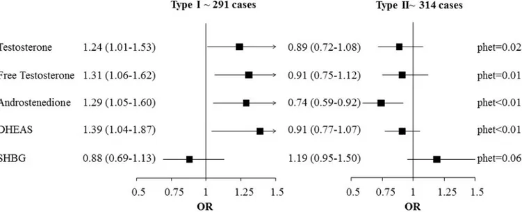
The association between androgens and EOC risk differed by developmental pathway (type I vs. type II tumors, phet, testosterone: 0.02; free testosterone: 0.01; androstenedione: <0.01;
A
uthor Man
uscr
ipt
A
uthor Man
uscr
ipt
A
uthor Man
uscr
ipt
A
uthor Man
uscr
ipt
DHEAS: <0.01) (Figure 1). Overall, higher concentrations of androgens were associated with increased risk of type I tumors, and reduced risk of type II tumors (e.g.,
androstenedione: type I: ORlog2=1.29 [1.05–1.60]; cases n=287; type II: ORlog2=0.74 [0.59–
0.92], cases n=307; phet<0.01). Significant heterogeneity for androstenedione (p<0.01) and
DHEAS (p=0.03) remained after exclusion of mucinous and clear cell cases missing data on grade from the type I subgroup (before exclusion, n=291 case-control sets; after exclusion, n=77 case-control sets). However, while of the same general magnitude, the effect estimates were no longer statistically significant (Supplemental Figure S1).
Sensitivity and Subgroup Analyses
We observed some evidence of heterogeneity for the androgens and SHBG and overall EOC by menopausal status at blood collection (androstenedione, phet=0.05; SHBG, phet=0.02) and
age at diagnosis (<55 years vs ≥55 years: androstenedione, phet=0.02; DHEAS, phet=0.05;
SHBG, phet=0.05). Both androstenedione and SHBG were positively associated with risk
only among women premenopausal at blood collection (androstenedione: premenopausal women, ORlog2=1.18 [1.03–1.35], postmenopausal women ORlog2=0.95 [0.82–1.12];
SHBG: premenopausal women, ORlog2=1.18 [1.00–1.39], postmenopausal women
ORlog2=0.89 [0.76–1.04]). No further significant heterogeneity was observed by menopausal
status at blood collection. Androstenedione was associated with increased risk of EOC among women diagnosed before age 55 years, but not among women diagnosed at age 55 or older (<55 at diagnosis, ORlog2=1.21 [1.05–1.40], ≥55 years at diagnosis, ORlog2=0.95
[0.82–1.10]). While the association between DHEAS and SHBG and EOC differed by age at diagnosis, the ORs were not statistically significant in either age at diagnosis subgroup (e.g., SHBG, <55 at diagnosis, ORlog2=1.16 [0.98–1.38], ≥55 years at diagnosis, ORlog2=0.92
[0.79–1.07]).
We observed no heterogeneity in analyses by stage at diagnosis. We observed an attenuation of the association between testosterone and EOC after excluding the FMC (n=576 cases, 43% of sample; after exclusion: ORlog2=1.06 [0.93 – 1.21]). Overall, ORs were similar for
the histologic subtypes after this exclusion, however, no longer statistically significant (e.g., testosterone and endometrioid tumors, before exclusion: n=164, ORlog2=1.40 [1.03 – 1.91];
after exclusion: n=73, ORlog2=1.39 [0.81 – 2.36]. The most substantial attenuation was for
the association between androstenedione and mucinous tumors (before exclusion: n=191 cases, ORlog2=1.33 [1.03 – 1.72]; after exclusion: n=49 cases, ORlog2=1.19 [0.74 – 1.92]).
Excluding women diagnosed within two years after blood donation did not meaningfully impact the results (data not shown).
Discussion
We investigated pre-diagnosis circulating concentrations of androgens and risk of EOC overall (n=1,331 cases) and by subtype (case range, n=61 clear cell to 667 serous), in a collaborative re-analysis of 7 nested case-control studies. The association between testosterone and risk of EOC differed by histologic subtype: endogenous androgens were predominantly associated with increased risk of endometrioid and mucinous tumors, while no significant associations were observed for serous or clear cell tumors, although some
A
uthor Man
uscr
ipt
A
uthor Man
uscr
ipt
A
uthor Man
uscr
ipt
A
uthor Man
uscr
ipt
androgens were inversely associated with high-grade serous and endometrioid (Type II) disease.
Ovarian cancer is comprised of four predominant histologic subtypes: serous, mucinous, endometrioid and clear cell. These histologic subtypes differ substantially by molecular alterations at diagnosis and presumed tissue of origin. The majority of serous tumors are high-grade neoplasms; this subtype represents the majority of invasive EOCs. Separate etiologic pathways are hypothesized for low- and high-grade serous EOC. It is hypothesized that a proportion of low-grade serous carcinomas develop from distal epithelium of the fallopian tube that implants on the ovarian surface epithelium (~ 80%), while high-grade serous tumors may arise from serous tubal intraepithelial carcinomas (STIC) within the fimbriated end of the fallopian tube (25, 26). Mucinous carcinomas are hypothesized to develop from the gastrointestinal mucosa or from transitional-type epithelium located at the tubal-peritoneal junction; borderline mucinous ovarian tumors are established precursors for this subtype (19). Both endometrioid and clear cell tumors have been proposed to arise from endometrial tissue, and have been associated with endometriosis and retrograde
menstruation (19, 27).
Beyond histologic subgroups, two hypothesized developmental pathways of tumorigenesis (type I and type II) have been defined using tumor molecular genetic characteristics (19, 25); in the absence of data on the tumor molecular profile, EOC is classified as type I or type II based on data on histology and grade. Type I tumors include low-grade serous, low-grade endometrioid, mucinous and malignant Brenner tumors (commonly present with KRAS, BRAF, PTEN, PIK3CA, CTNNB1, and ERBB2 mutations)—subtypes that have been hypothesized to develop in a step-wise manner from borderline tumors or endometriosis within or on the surface of the ovary, and are typically diagnosed at earlier disease stage (27). Type II tumors include high-grade serous, high-grade endometrioid, malignant mixed and undifferentiated tumors (typically present with TP53 mutations, but none of the mutations observed in type I disease) (19). These latter tumors comprise the majority of EOCs, are aggressive, and typically present at an advanced stage.
Prior epidemiologic data suggest risk factor differences by EOC subtype defined by histology (e.g. (12, 15–18)) and developmental pathway (11, 14). Consistent differences by histologic subtype of invasive EOC are observed for hormone-related risk factors including duration of OC use (lower risk of all histologic subtypes but mucinous; (12, 15)), older age at menopause (higher risk of all but mucinous; (12)), smoking (higher risk of mucinous, lower risk of clear cell; (12, 17)), parity (more strongly protective in non-serous subtypes; (12)), postmenopausal hormone therapy (HT) use (higher risk of serous and endometrioid subtypes only; (12, 18)), and adiposity (among non-HT users; higher risk of serous and endometrioid subtypes only); (16)). Data by the type I/II classification are sparse, but consistently show stronger associations between parity and type I, relative to type II, disease (11, 14). Three prospective studies evaluated circulating estrogens (10, 28) and/or androgens (9, 10) and invasive EOC risk by subtype. Higher concentrations of both estrogens and androgens were associated with increased risk of non-serous EOC subtypes (9, 10, 28), whereas higher concentrations of androstenedione had opposing effects on risk of type I (higher risk) and type II (lower risk) EOC (9).
A
uthor Man
uscr
ipt
A
uthor Man
uscr
ipt
A
uthor Man
uscr
ipt
A
uthor Man
uscr
ipt
In women, androgens are produced in the ovary, adrenal glands, and via peripheral conversion of androgen precursors (e.g., DHEA); in turn, androgens are the substrate for estrogen production by aromatase. DHEAS is a pre-androgen synthesized in the adrenal gland, and subsequently metabolized toward androstenedione and testosterone, or estradiol (29). Androstenedione, an intermediate between DHEA and DHEAS and testosterone, is produced in both the ovary (premenopausal women: 40%; postmenopausal women: 20– 30%) and the adrenal gland. In premenopausal women, approximately 25% of circulating testosterone originates in the ovary, 25% in the adrenal glands, and 50% is metabolized from precursors such as androstenedione in peripheral tissues (e.g., liver, adipose tissue) (29, 30); the proportion of testosterone of ovarian origin is higher in postmenopausal women (~50%) (29). These androgens are correlated with each other (e.g., r=0.54 between DHEAS and androstenedione to r=0.69 between DHEAS and testosterone; adjusted for menopausal status (6)) and weakly correlated with estradiol (e.g., estradiol and testosterone: premenopausal women: r=0.08 (31); postmenopausal women, r=0.23–0.38; (32, 33)) and body mass index (r=0.07–0.13; (31–33)).
Androgens may (1) directly influence ovarian carcinogenesis through androgen receptor (AR) signaling, or (2) impact risk through their role as estrogen precursors; associations with estrogens may be most evident in the context of progesterone insufficiency as observed in polycystic ovarian syndrome (PCOS). ARs and estrogen (ER) receptors are expressed in the normal ovary, including ovarian surface epithelial cells and cortical inclusion cysts, and the fallopian tube (34–36). In vivo data show that ovarian cancer preferentially develops in a hormonal milieu enriched with androgens (e.g., testosterone induces epithelial neoplasms in guinea pigs (37)) or estrogens (e.g., estrogen-induced tumor growth in high-grade serous ovarian cancers) (38, 39). The hyperandrogenic PCOS is characterized by functional ovarian hyperandrogenism, with an excess of testosterone produced in the ovarian thecal cells (40); up to 45% of cases additionally present with adrenal hyperandrogenism (41). Estimates of PCOS prevalence range from 5 to 15% (30); the syndrome has highest prevalence among reproductive-age women. PCOS-related androgen excess is observed in both pre- and postmenopausal women (42). Progesterone deficiency is a hallmark of PCOS, resulting in a higher ratio of estrogens to progesterone. PCOS (43, 44) and relatively high levels of estrogens unopposed by progesterone are associated with increased endometrial cancer risk (i.e., estrogen-alone HT (45), relatively high endogenous estrogens in postmenopausal women (33, 46)). These associations with endometrial cancer may be most relevant to the endometrioid or clear cell EOC, given endometrial tissue is a proposed tissue of origin for these subtypes. PCOS itself has not consistently been associated with ovarian cancer (43, 44, 47), though data by subtype are limited. Data to date suggest both estrogen-alone and estrogen plus progesterone HT are associated with increased risk of endometrioid EOC (18). In the current study, we evaluated three members of the androgen synthesis pathway— DHEAS, androstenedione, testosterone—and EOC risk by histology (i.e., accounting for hypothesized differences in cell of origin) and developmental pathway (i.e., “less” relative to “more” aggressive disease). We observed a significant positive association between
testosterone and risk of endometrioid ovarian cancer. There is limited in vitro evidence to support a role of androgens in the etiology of endometrioid EOC (34, 48). However, given the possible common tissue of origin, it is plausible that androgens impact risk similarly in
A
uthor Man
uscr
ipt
A
uthor Man
uscr
ipt
A
uthor Man
uscr
ipt
A
uthor Man
uscr
ipt
both endometrial cancer and endometrioid EOC. With respect to endometrial cancer, recent in vivo data have demonstrated that androgens induce epithelial proliferation in the mouse uterus (49), and epidemiologic data provide some support for an association between androgens and endometrial cancer risk (50). Together, this data on endometrial cancer provides indirect evidence supporting an association between androgens and endometrioid EOC. Androgens are an intermediate on the estrogen-synthesis pathway, and estrogen exposure unopposed by progesterone may be the underlying biological mechanism linking androgens to endometrioid EOC, particularly if in the context progesterone deficiency, as in PCOS and in postmenopausal women. Prior research has linked higher early pregnancy estradiol concentrations to a 2.5-fold increase in risk of endometrioid EOC (10), and postmenopausal HT use (12, 18) and adiposity (16) are associated with increased risk of this subtype. We adjusted for BMI in a sensitivity analysis, given (1) the association between PCOS and obesity and (2) adipose tissue is a key site of metabolism of androgens to estrogens in postmenopausal women. Adjustment for BMI did not impact the results. Data on history of PCOS were not available.
Higher concentrations of all investigated androgens, except DHEAS, were significantly associated with increased risk of mucinous tumors. Emerging data suggest the ovarian stroma proximal to mucinous EOC has higher concentrations of sex-steroid producing enzymes than distant stroma, providing support for a role for sex steroids in the development of mucinous disease (35). Androgens (directly, or after conversion to estrogens) may contribute to growth promotion in the early stages of mucinous disease; however, to our knowledge, the androgen responsiveness of mucinous tumors is not well characterized, and data on ER expression are limited (51, 52). The precise biological mechanisms underlying the observed associations between androgens and mucinous tumors remain an open question.
In line with two prior prospective studies (9, 10), both included in this analysis, we observed no association with pre-diagnosis androgen concentrations and increased risk of serous carcinomas. Recent data on estrogens and ovarian cancer are in line with our results on androgens, with no association observed between estrogens and risk of invasive serous tumors in the FMC (first-trimester estrogens) (10) or among postmenopausal women in the Women’s Health Initiative (28). We observed no associations with clear cell disease. However, sample size for this subtype was limited.
We observed significant heterogeneity in the strength of associations between androgens and risk of type I vs. type II tumors; higher androgen concentrations were associated with higher risk of type I, but lower risk of type II (predominantly high grade serous), tumors. These results are in agreement with the single prior study on endogenous androgens and EOC risk using the dualistic model classification (9); these data from the European Prospective Investigation into Cancer and Nutrition (EPIC) were included in the current analysis. There is indirect evidence for differences in hormone dependency in type I and type II tumors, based on the variation of ER expression between low-grade (ER expression: 58%) and high-grade serous carcinomas (ER expression: 27%) (53). However, the mechanisms linking androgen concentrations to lower risk of type II tumors in our study are unclear. While
A
uthor Man
uscr
ipt
A
uthor Man
uscr
ipt
A
uthor Man
uscr
ipt
A
uthor Man
uscr
ipt
chance and residual confounding may explain the results, future work should explicitly examine the impact of androgens on type II tumors.
Given the large sample size, our study was powered to investigate risk associations for less common tumors (e.g., mucinous tumors) and by developmental pathway (type I/type II). A general weakness of pooled analyses is the difference in data availability of covariates and differences in laboratory methods. In this investigation, data from each cohort were centrally compiled and harmonized and we addressed differences in absolute biomarker
concentrations (I) using study-specific tertiles and (II) standardizing hormone measurements using study-specific mean concentrations. Results were robust regardless of whether we calculated ORs from the pooling of individual data or from meta-analysis. For some of the investigated hormones the number of sets that could be used was reduced for subgroup analyses, which resulted in reduced power. In our primary analyses using the developmental pathway classification, we included all mucinous and clear cell tumors in the “type I” classification, as their classification is independent of grade. If there were systematic differences in the observed associations with type I disease in cases with and without grade data, this may result in a biased interpretation of the differences between type I and type II EOC. However, the associations observed in our primary analysis and in a sensitivity analysis restricted to women with complete data on grade were of similar magnitude. Many statistical tests are reported; therefore some significant observations may be due to chance. However, all statistical analyses were hypothesis driven. In line with the majority of other epidemiological studies, a single measurement of biomarkers was used to assess risk associations. This single measurement may not reflect long-term average concentrations and the storage time and conditions may impact the true value of the biochemical indicators. However, the stability of androgen measurements over time has been shown previously for a period over at least 2–3 years: (1) premenopausal women [ICC ranged from 0.58
(androstenedione) up to 0.81 (DHEAS), (54) and (2) postmenopausal women [ICC ranged from 0.66 (androstenedione) up to 0.92 (SHBG) (55).
The testosterone synthesis pathway (e.g., DHEAS, androstenedione, testosterone) may play an important role in the onset and progression of a subset of epithelial invasive ovarian carcinomas. Androgens may either have a direct impact on ovarian carcinogenesis, or act through increased synthesis of other steroid hormones (e.g., estrogens); this is an area for future epidemiologic research. While androgens were associated with increased risk of non-serous tumors, we observed an inverse association between androstenedione and high grade serous tumors. In addition to providing novel findings on hormone-related pathways in ovarian carcinogenesis, this study supports emerging data on the heterogeneity of epithelial invasive ovarian cancer and underscores the importance of examining etiologic differences for subtypes.
Supplementary Material
Refer to Web version on PubMed Central for supplementary material.
A
uthor Man
uscr
ipt
A
uthor Man
uscr
ipt
A
uthor Man
uscr
ipt
A
uthor Man
uscr
ipt
Acknowledgments
We thank the participants and staff of the Nurses’ Health Study and Nurses’ Health Study II for their valuable contributions as well as the following state cancer registries for their help: Alabama, Arizona, Arkansas, California, Colorado, Connecticut, Delaware, Florida, Georgia, Idaho, Illinois, Indiana, Iowa, Kentucky, Louisiana, Maine, Maryland, Massachusetts, Michigan, Nebraska, New Hampshire, New Jersey, New York, North Carolina, North Dakota, Ohio, Oklahoma, Oregon, Pennsylvania, Rhode Island, South Carolina, Tennessee, Texas, Virginia, Washington, and Wyoming.
We thank the participants and staff of CLUE II for their contributions as well as the Maryland Cancer Registry, Center for Cancer Surveillance and Control, Department of Health and Mental Hygiene, 201 W. Preston Street,
Room 400, Baltimore, MD 21201, http://phpa.dhmh.maryland.gov/cancer, 410-767-4055. We acknowledge the
State of Maryland, the Maryland Cigarette Restitution Fund, and the National Program of Cancer Registries of the Centers for Disease Control and Prevention for the funds that support the collection and availability of the cancer registry data.
Grant Support
The OC3 is supported by Department of Defense Ovarian Cancer Research Program Grant No.
W81XWH-12-1-0561. RT Fortner was supported by a Marie Curie International Incoming Fellowship of the European Commission’s Seventh Framework Programme (MC-IIF-623984).
The coordination of EPIC is financially supported by the European Commission (DG-SANCO) and the International Agency for Research on Cancer. The national cohorts are supported by Danish Cancer Society (Denmark); Ligue Contre le Cancer, Institut Gustave Roussy, Mutuelle Générale de l’Education Nationale, Institut National de la Santé et de la Recherche Médicale (INSERM) (France); German Cancer Aid, German Cancer Research Center (DKFZ), Federal Ministry of Education and Research (BMBF), Deutsche Krebshilfe, Deutsches Krebsforschungszentrum and Federal Ministry of Education and Research (Germany); the Hellenic Health Foundation (Greece); Associazione Italiana per la Ricerca sul Cancro-AIRC-Italy and National Research Council (Italy); Dutch Ministry of Public Health, Welfare and Sports (VWS), Netherlands Cancer Registry (NKR), LK Research Funds, Dutch Prevention Funds, Dutch ZON (Zorg Onderzoek Nederland), World Cancer Research Fund (WCRF), Statistics Netherlands (The Netherlands); ERC-2009-AdG 232997 and Nordforsk, Nordic Centre of Excellence programme on Food, Nutrition and Health (Norway); Health Research Fund (FIS), PI13/00061 to Granada; PI13/01162 to EPIC-Murcia), Regional Governments of Andalucía, Asturias, Basque Country, Murcia and Navarra, ISCIII RETIC (RD06/0020) (Spain); Swedish Cancer Society, Swedish Research Council and County Councils of Skåne and Västerbotten (Sweden); Cancer Research UK (14136 to EPIC-Norfolk; C570/A16491 and C8221/A19170 to Oxford), Medical Research Council (1000143 to Norfolk, MR/M012190/1 to EPIC-Oxford) (United Kingdom). For information on how to submit an application for gaining access to EPIC data and/or
bio-specimens, please follow the instructions at http://epic.iarc.fr/access/index.php
Support was provided by the following National Institutes of Health/NCI grants: R01 CA120061 (Finnish Maternity Cohort); UM1 CA186107, P01 CA87969, UM1 CA176726, and R01 CA67262 (Nurses’ Health Study, Nurses’ Health Study II); HL043851, HL080467, and HL099355 (Women’s Health Study).
CLUEII was supported by National Cancer Institute grant numbers: CA-97857, CA-86308; Grant sponsor: M. Jean Goutal (donation): Grant sponsors: The Woodrow Wilson Foundation/Johnson and Johnson, The Lloyds TSB Charitable Foundation for the Channel Islands.
The authors assume full responsibility for analyses and interpretation of these data.
References
1. Lukanova A, Kaaks R. Endogenous hormones and ovarian cancer: epidemiology and current hypotheses. Cancer Epidemiol Biomarkers Prev. 2005; 14:98–107. [PubMed: 15668482]
2. Risch HA. Hormonal etiology of epithelial ovarian cancer, with a hypothesis concerning the role of androgens and progesterone. J Natl Cancer Inst. 1998; 90:1774–86. [PubMed: 9839517]
3. Modugno F, Laskey RA, Smith AL, Andersen CL, Haluska P, Oesterreich S. Hormone response in ovarian cancer: time to reconsider as a clinical target? Endocrine-related cancer. 2012
4. Edmondson RJ, Monaghan JM, Davies BR. The human ovarian surface epithelium is an androgen responsive tissue. Br J Cancer. 2002; 86:879–85. [PubMed: 11953818]
A
uthor Man
uscr
ipt
A
uthor Man
uscr
ipt
A
uthor Man
uscr
ipt
A
uthor Man
uscr
ipt
5. Helzlsouer KJ, Alberg AJ, Gordon GB, Longcope C, Bush TL, Hoffman SC, et al. Serum gonadotropins and steroid hormones and the development of ovarian cancer. JAMA. 1995; 274:1926–30. [PubMed: 8568986]
6. Lukanova A, Lundin E, Akhmedkhanov A, Micheli A, Rinaldi S, Zeleniuch-Jacquotte A, et al. Circulating levels of sex steroid hormones and risk of ovarian cancer. International journal of cancer. 2003; 104:636–42. [PubMed: 12594820]
7. Tworoger SS, Lee IM, Buring JE, Hankinson SE. Plasma androgen concentrations and risk of incident ovarian cancer. Am J Epidemiol. 2008; 167:211–8. [PubMed: 17982156]
8. Rinaldi S, Dossus L, Lukanova A, Peeters PH, Allen NE, Key T, et al. Endogenous androgens and risk of epithelial ovarian cancer: results from the European Prospective Investigation into Cancer and Nutrition (EPIC). Cancer Epidemiol Biomarkers Prev. 2007; 16:23–9. [PubMed: 17220328] 9. Ose J, Fortner RT, Rinaldi S, Schock H, Overvad K, Tjonneland A, et al. Endogenous androgens and
risk of epithelial invasive ovarian cancer by tumor characteristics in the European Prospective Investigation into Cancer and Nutrition. International journal of cancer. 2015; 136:399–410. [PubMed: 24890047]
10. Schock H, Surcel HM, Zeleniuch-Jacquotte A, Grankvist K, Lakso HA, Fortner RT, et al. Early pregnancy sex steroids and maternal risk of epithelial ovarian cancer. Endocrine-related cancer. 2014; 21:831–44. [PubMed: 25270324]
11. Fortner RT, Ose J, Merritt MA, Schock H, Tjonneland A, Hansen L, et al. Reproductive and hormone-related risk factors for epithelial ovarian cancer by histologic pathways, invasiveness and histologic subtypes: Results from the EPIC cohort. International journal of cancer. 2015;
137:1196–208. [PubMed: 25656413]
12. Wentzensen N, Poole EM, Trabert B, White E, Arslan AA, Patel AV, et al. Ovarian Cancer Risk Factors by Histologic Subtype: An Analysis From the Ovarian Cancer Cohort Consortium. Journal of clinical oncology : official journal of the American Society of Clinical Oncology. 2016; 34:2888–98. [PubMed: 27325851]
13. Prat J. Ovarian carcinomas: five distinct diseases with different origins, genetic alterations, and clinicopathological features. Virchows Arch. 2012; 460:237–49. [PubMed: 22322322]
14. Merritt MA, De Pari M, Vitonis AF, Titus LJ, Cramer DW, Terry KL. Reproductive characteristics in relation to ovarian cancer risk by histologic pathways. Human Reproduction. 2013; 28:1406–17. [PubMed: 23315066]
15. Collaborative Group on Epidemiological Studies of Ovarian C. Beral V, Doll R, Hermon C, Peto R, Reeves G. Ovarian cancer and oral contraceptives: collaborative reanalysis of data from 45 epidemiological studies including 23,257 women with ovarian cancer and 87,303 controls. Lancet. 2008; 371:303–14. [PubMed: 18294997]
16. Collaborative Group on Epidemiological Studies of Ovarian C. Ovarian cancer and body size: individual participant meta-analysis including 25,157 women with ovarian cancer from 47 epidemiological studies. PLoS medicine. 2012; 9:e1001200. [PubMed: 22606070]
17. Collaborative Group on Epidemiological Studies of Ovarian C. Beral V, Gaitskell K, Hermon C, Moser K, Reeves G, et al. Ovarian cancer and smoking: individual participant meta-analysis including 28,114 women with ovarian cancer from 51 epidemiological studies. The Lancet Oncology. 2012; 13:946–56. [PubMed: 22863523]
18. Collaborative Group On Epidemiological Studies Of Ovarian Cancer. Menopausal hormone use and ovarian cancer risk: individual participant meta-analysis of 52 epidemiological studies. Lancet. 2015
19. Kurman RJ, Shih Ie M. Molecular pathogenesis and extraovarian origin of epithelial ovarian cancer–shifting the paradigm. Hum Pathol. 2011; 42:918–31. [PubMed: 21683865]
20. Sodergard R, Backstrom T, Shanbhag V, Carstensen H. Calculation of free and bound fractions of testosterone and estradiol-17 beta to human plasma proteins at body temperature. J Steroid Biochem. 1982; 16:801–10. [PubMed: 7202083]
21. DerSimonian R, Laird N. Meta-analysis in clinical trials. Control Clin Trials. 1986; 7:177–88. [PubMed: 3802833]
A
uthor Man
uscr
ipt
A
uthor Man
uscr
ipt
A
uthor Man
uscr
ipt
A
uthor Man
uscr
ipt
22. Smith-Warner SA, Spiegelman D, Ritz J, Albanes D, Beeson WL, Bernstein L, et al. Methods for pooling results of epidemiologic studies: the Pooling Project of Prospective Studies of Diet and Cancer. Am J Epidemiol. 2006; 163:1053–64. [PubMed: 16624970]
23. Wang M, Spiegelman D, Kuchiba A, Lochhead P, Kim S, Chan AT, et al. Statistical methods for studying disease subtype heterogeneity. Statistics in medicine. 2016; 35:782–800. [PubMed: 26619806]
24. Schock H, Zeleniuch-Jacquotte A, Lundin E, Grankvist K, Lakso HA, Idahl A, et al. Hormone concentrations throughout uncomplicated pregnancies: a longitudinal study. BMC Pregnancy Childbirth. 2016; 16:146. [PubMed: 27377060]
25. Nik NN, Vang R, Shih Ie M, Kurman RJ. Origin and pathogenesis of pelvic (ovarian, tubal, and primary peritoneal) serous carcinoma. Annu Rev Pathol. 2014; 9:27–45. [PubMed: 23937438] 26. Zeppernick F, Meinhold-Heerlein I, Shih Ie M. Precursors of ovarian cancer in the fallopian tube:
serous tubal intraepithelial carcinoma–an update. J Obstet Gynaecol Res. 2015; 41:6–11. [PubMed: 25330822]
27. Seidman, JD., Cho, KR., Ronnett, BM., Kurman, RJ. Surface Epithelial Tumors of the Ovary. In: Kurman, RJ.Hedrick Ellenson, L., Ronnett, BM., editors. Blaustein’s Pathology of the Female Genital Tract. 6th. USA: Springer; 2011.
28. Trabert B, Brinton LA, Anderson GL, Pfeiffer RM, Falk RT, Strickler HD, et al. Circulating Estrogens and Postmenopausal Ovarian Cancer Risk in the Women’s Health Initiative
Observational Study. Cancer Epidemiol Biomarkers Prev. 2016; 25:648–56. [PubMed: 26908437] 29. Yen, SS., Jaffe, RB., Barbieri, RL. Reproductive Endocrinology. 4th. Philadelphia: W.B. Saunders
Company; 1999.
30. Rosenfield RL, Ehrmann DA. The Pathogenesis of Polycystic Ovary Syndrome (PCOS): The Hypothesis of PCOS as Functional Ovarian Hyperandrogenism Revisited. Endocrine reviews. 2016; 37:467–520. [PubMed: 27459230]
31. Kaaks R, Berrino F, Key T, Rinaldi S, Dossus L, Biessy C, et al. Serum Sex Steroids in Premenopausal Women and Breast Cancer Risk Within the European Prospective Investigation into Cancer and Nutrition (EPIC). Journal of the National Cancer Institute. 2005; 97:755–65. [PubMed: 15900045]
32. James RE, Lukanova A, Dossus L, Becker S, Rinaldi S, Tjønneland A, et al. Postmenopausal serum sex steroids and risk of hormone receptor-positive and -negative breast cancer: a nested case-control study. Cancer Prevention Research. 2011; 4:1626–35. [PubMed: 21813404] 33. Lukanova A, Lundin E, Micheli A, Arslan A, Ferrari P, Rinaldi S, et al. Circulating levels of sex
steroid hormones and risk of endometrial cancer in postmenopausal women. International journal of cancer. 2004; 108:425–32. [PubMed: 14648710]
34. Gibson DA, Simitsidellis I, Collins F, Saunders PT. Evidence of androgen action in endometrial and ovarian cancers. Endocr Relat Cancer. 2014; 21:T203–18. [PubMed: 24623742]
35. Blanco LZ Jr, Kuhn E, Morrison JC, Bahadirli-Talbott A, Smith-Sehdev A, Kurman RJ. Steroid hormone synthesis by the ovarian stroma surrounding epithelial ovarian tumors: a potential mechanism in ovarian tumorigenesis. Modern pathology : an official journal of the United States and Canadian Academy of Pathology, Inc. 2017
36. Kyriakidis I, Papaioannidou P. Estrogen receptor beta and ovarian cancer: a key to pathogenesis and response to therapy. Archives of gynecology and obstetrics. 2016; 293:1161–8. [PubMed: 26861465]
37. Silva EG, Tornos C, Fritsche HA Jr, el-Naggar A, Gray K, Ordonez NG, et al. The induction of benign epithelial neoplasms of the ovaries of guinea pigs by testosterone stimulation: a potential animal model. Modern pathology : an official journal of the United States and Canadian Academy of Pathology, Inc. 1997; 10:879–83.
38. Silva EG, Tornos C, Deavers M, Kaisman K, Gray K, Gershenson D. Induction of epithelial neoplasms in the ovaries of guinea pigs by estrogenic stimulation. Gynecologic oncology. 1998; 71:240–6. [PubMed: 9826466]
39. Ciucci A, Zannoni GF, Buttarelli M, Lisi L, Travaglia D, Martinelli E, et al. Multiple direct and indirect mechanisms drive estrogen-induced tumor growth in high grade serous ovarian cancers. Oncotarget. 2016; 7:8155–71. [PubMed: 26797759]
A
uthor Man
uscr
ipt
A
uthor Man
uscr
ipt
A
uthor Man
uscr
ipt
A
uthor Man
uscr
ipt
40. Ehrmann DA. Polycystic ovary syndrome. The New England journal of medicine. 2005; 352:1223– 36. [PubMed: 15788499]
41. Luque-Ramirez M, Escobar-Morreale HF. Adrenal Hyperandrogenism and Polycystic Ovary Syndrome. Current pharmaceutical design. 2016; 22:5588–602. [PubMed: 27510480]
42. Markopoulos MC, Kassi E, Alexandraki KI, Mastorakos G, Kaltsas G. Hyperandrogenism after menopause. European journal of endocrinology. 2015; 172:R79–91. [PubMed: 25225480] 43. Gottschau M, Kjaer SK, Jensen A, Munk C, Mellemkjaer L. Risk of cancer among women with
polycystic ovary syndrome: a Danish cohort study. Gynecologic oncology. 2015; 136:99–103. [PubMed: 25451694]
44. Barry JA, Azizia MM, Hardiman PJ. Risk of endometrial, ovarian and breast cancer in women with polycystic ovary syndrome: a systematic review and meta-analysis. Human reproduction update. 2014; 20:748–58. [PubMed: 24688118]
45. Beral V, Bull D, Reeves G, Million Women Study C. Endometrial cancer and hormone-replacement therapy in the Million Women Study. Lancet. 2005; 365:1543–51. [PubMed: 15866308]
46. Allen NE, Key TJ, Dossus L, Rinaldi S, Cust A, Lukanova A, et al. Endogenous sex hormones and endometrial cancer risk in women in the European Prospective Investigation into Cancer and Nutrition (EPIC). Endocrine-related cancer. 2008; 15:485–97. [PubMed: 18509001]
47. Harris HR, Titus LJ, Cramer DW, Terry KL. Long and irregular menstrual cycles, polycystic ovary syndrome, and ovarian cancer risk in a population-based case-control study. International journal of cancer. 2017; 140:285–91. [PubMed: 27667654]
48. Maliqueo MA, Quezada S, Clementi M, Bacallao K, Anido M, Johnson C, et al. Potential action of androstenedione on the proliferation and apoptosis of stromal endometrial cells. Reprod Biol Endocrinol. 2004; 2:81. [PubMed: 15588330]
49. Simitsidellis I, Gibson DA, Cousins FL, Esnal-Zufiaurre A, Saunders PT. A Role for Androgens in Epithelial Proliferation and Formation of Glands in the Mouse Uterus. Endocrinology. 2016; 157:2116–28. [PubMed: 26963473]
50. Clendenen TV, Hertzmark K, Koenig KL, Lundin E, Rinaldi S, Johnson T, et al. Premenopausal Circulating Androgens and Risk of Endometrial Cancer: results of a Prospective Study. Horm Cancer. 2016; 7:178–87. [PubMed: 26925952]
51. Tkalia IG, Vorobyova LI, Svintsitsky VS, Nespryadko SV, Goncharuk IV, Lukyanova NY, et al. Clinical significance of hormonal receptor status of malignant ovarian tumors. Exp Oncol. 2014; 36:125–33. [PubMed: 24980769]
52. Sieh W, Kobel M, Longacre TA, Bowtell DD, deFazio A, Goodman MT, et al. Hormone-receptor expression and ovarian cancer survival: an Ovarian Tumor Tissue Analysis consortium study. The Lancet Oncology. 2013; 14:853–62. [PubMed: 23845225]
53. Escobar J, Klimowicz AC, Dean M, Chu P, Nation JG, Nelson GS, et al. Quantification of ER/PR expression in ovarian low-grade serous carcinoma. Gynecologic oncology. 2013; 128:371–6. [PubMed: 23103384]
54. Missmer SA, Spiegelman D, Bertone-Johnson ER, Barbieri RL, Pollak MN, Hankinson SE. Reproducibility of plasma steroid hormones, prolactin, and insulin-like growth factor levels among premenopausal women over a 2- to 3-year period. Cancer Epidemiol Biomarkers Prev. 2006; 15:972–8. [PubMed: 16702379]
55. Hankinson SE, Manson JE, Spiegelman D, Willett WC, Longcope C, Speizer FE. Reproducibility of plasma hormone levels in postmenopausal women over a 2–3-year period. Cancer Epidemiol Biomarkers Prev. 1995; 4:649–54. [PubMed: 8547832]
A
uthor Man
uscr
ipt
A
uthor Man
uscr
ipt
A
uthor Man
uscr
ipt
A
uthor Man
uscr
ipt
Figure 1. Odds ratios (95% CI) for EOC for doubling of androgen concentrations and EOC risk by the Type I and Type II classification: the Ovarian Cancer Cohort Consortium (OC3)
Results were derived from conditional logistic regression models, additionally adjusted for OC use (never/ever/missing) and parity (never/ever/missing). Pair-wise heterogeneity tests were performed, using the likelihood ratio test comparing models assuming (1) the same association between exposure and outcomes compared to (2) a model assuming different associations for each subtype. DHEAS=dehydroepiandrosterone sulfate; SHBG=sex hormone binding globulin
A
uthor Man
uscr
ipt
A
uthor Man
uscr
ipt
A
uthor Man
uscr
ipt
A
uthor Man
uscr
ipt
A
uthor Man
uscr
ipt
A
uthor Man
uscr
ipt
A
uthor Man
uscr
ipt
A
uthor Man
uscr
ipt
T ab le 1Case and control characteristics in pooled analysis of prospecti
v
e data on circulating androgens, SHBG and EOC risk: the Ov
arian Cancer Cohort
Consortium (OC3) Cohort
Refer
ence
No
Mean age at blood donation y
ears (SD) Nullipar ous% 1 Ev er OC use, % 1 P ostmenopausal, % 2 Mean BMI (SD) Clue II ≠ Case 46 60.8 (13.0) 19% 20% 85% 26.3 (5.8) ≠ Control 91 61.0 (12.9) 13% 13% 86% 25.4 (4.6) EPIC Ose et al. 2015 Case 451 55.9 (8.5) 18% 37% 77% 26.8 (4.9) Control 867 55.9 (8.6) 12% 45% 77% 26.3 (4.7) FMC Schock et al. 2014 Case 576 32.5 (4.8) 0% ≠≠ 0% ≠≠ Control 1,433 32.5 (4.7) 0% ≠≠ 0% ≠≠ NHS T w oroger et al. 2008 Case 117 57.7 (6.5) 8% 41% 79% 24.8 (4.8) Control 348 57.7 (6.5) 5% 47% 79% 24.7 (4.1) NHS II T w oroger et al. 2008 Case 15 46.1 (4.4) 20% 93% 20% 29.6 (9.8) Control 44 45.8 (4.3) 23% 86% 18% 25.9 (5.8) NYUWHS Lukano v a et al. 2003 Case 63 52.6 (8.6) 47% 29% 56% 24.5 (3.8) Control 112 52.0 (8.5) 38% 36% 54% 25.9 (4.3) WHS T w oroger et al. 2008 Case 63 55.7 (7.2) 25% 65% 75% 24.5 (3.9) Control 122 55.5 (7.0) 15% 71% 70% 25.1 (4.4) T otal Case 1,331 45.8 (13.7) 11% 40% 42% 26.2 (5.1) Control 3,017 44.8 (13.7) 6% 47% 39% 25.8 (4.6) 1 Among w
omen with data: parity 2.8% missing; OC use 47% missing (e
xcluding FMC: 2.3% missing)
2 At blood collection BMI = body mass inde
x; OC = oral contracepti
v
e; OC3 = Ov
arian Cancer Cohort Consortium; SHBG = se
x hormone binding glob
ulin; SD = standard de viation; CLUE = W ashington County , MD Study ‘Gi v
e us a clue to cancer and heart disease’; EPIC = European Prospecti
v
e In
v
estig
ation into Cancer and Nutrition; FMC = Finnish Maternity Cohort; NHS = Nurses’ Health Study;
NYUWHS = Ne
w Y
ork Uni
v
ersity W
omen’s Health Study; WHS = W
omen’s Health Study
.
≠ Data from Clue II ha
v
e not been published.
≠≠
Information on BMI and OC use w
A
uthor Man
uscr
ipt
A
uthor Man
uscr
ipt
A
uthor Man
uscr
ipt
A
uthor Man
uscr
ipt
T ab le 2Odds ratios (95% CI) for EOC o
v
erall and by histologic subtypes in tertiles and for doubling of androgen concentrations: the Ov
arian Cancer Cohort
Consortium (OC3) 1 In v asi v e EOC Ser ous Endometrioid Mucinous Clear Cell Sets OR (95%CI) ptr end Sets OR (95%CI) ptr end Sets OR (95%CI) ptr end Sets OR (95%CI) ptr end Sets OR (95%CI) ptr end T estoster one T1 398 ref 222 ref 35 ref 45 ref 15 ref T2 443 1.20 (1.02 – 1.41) 229 1.16 (0.92 – 1.46) 60 1.46 (0.88 – 2.42) 61 1.34 (0.86 – 2.08) 27 1.65 (0.73 – 3.73) T3 2 460 1.25 (1.06 – 1.48) 0.03 204 0.97 (0.76 – 1.24) 0.56 69 1.80 (1.08 – 3.01) 0.06 84 1.94 (1.25 – 3.02) 0.05 17 0.82 (0.34 – 2.00) 0.68 Doubling 3 1,301 1.12 (1.02 – 1.24) 0.02 655 0.96 (0.84 – 1.11) 0.61 164 1.40 (1.03 – 1.91) 0.03 190 1.29 (1.01 – 1.66) 0.04 59 1.12 (0.69 – 1.80) 0.65 phet 4 0.06 Fr ee T estoster one T1 286 ref 155 ref 25 ref 35 ref 11 ref T2 287 1.05 (0.85 – 1.28) 151 1.04 (0.79 – 1.38) 32 0.94 (0.48 – 1.86) 48 1.46 (0.85 – 2.52) 10 0.82 (0.27 – 2.48) T3 2 292 1.06 (0.87 – 1.31) 0.04 129 0.83 (0.62 – 1.10) 0.63 36 1.03 (0.53 – 1.99) 0.45 50 1.50 (0.88 – 2.54) 0.03 20 2.02 (0.67 – 6.12) 0.26 Doubling 3 865 1.10 (1.00 – 1.21) 0.05 435 0.97 (0.84 – 1.11) 0.61 93 1.11 (0.80 – 1.53) 0.53 133 1.33 (1.04 – 1.72) 0.03 41 1.32 (0.82 – 2.11) 0.26 phet 4 0.12 Andr ostenedione T1 450 ref 235 ref 46 ref 56 ref 21 ref T2 387 0.86 (0.73 – 1.02) 204 0.88 (0.70 – 1.12) 45 0.67 (0.4 – 1.13) 51 0.90 (0.57 – 1.42) 15 0.93 (0.41 – 2.10) T3 2 470 1.07 (0.90 – 1.28) 0.13 217 0.99 (0.77 – 1.28) 0.79 73 0.99 (0.59 – 1.67) 0.82 84 1.57 (1.01 – 2.42) 0.03 24 0.81 (0.37 – 1.77) 0.62 Doubling 3 1,307 1.08 (0.97 – 1.19) 0.16 656 0.97 (0.84 – 1.12) 0.69 164 1.04 (0.76 – 1.43) 0.82 191 1.33 (1.03 – 1.72) 0.03 60 1.07 (0.69 – 1.66) 0.77 phet 4 0.17
A
uthor Man
uscr
ipt
A
uthor Man
uscr
ipt
A
uthor Man
uscr
ipt
A
uthor Man
uscr
ipt
In v asi v e EOC Ser ous Endometrioid Mucinous Clear Cell Sets OR (95%CI) ptr end Sets OR (95%CI) ptr end Sets OR (95%CI) ptr end Sets OR (95%CI) ptr end Sets OR (95%CI) ptr end DHEAS T1 227 ref 128 ref 18 ref 8 ref 7 ref T2 245 1.08 (0.86 – 1.36) 127 1.06 (0.77 – 1.45) 23 0.81 (0.36 – 1.82) 16 1.04 (0.35 – 3.08) 12 3.36 (0.88 – 12.8) T3 2 219 0.95 (0.74 – 1.23) 0.87 111 0.83 (0.59 – 1.18) 0.45 28 1.02 (0.42 – 2.47) 0.85 20 1.53 (0.50 – 4.71) 0.29 14 3.50 (1.03 – 11.9) 0.10 Doubling 3 691 0.99 (0.89 – 1.10) 0.82 366 0.93 (0.81 – 1.08) 0.36 69 1.05 (0.72 – 1.53) 0.78 44 1.34 (0.81 – 2.23) 0.26 33 1.52 (0.87 – 2.65) 0.14 phet 4 0.22 SHBG T1 311 ref 147 ref 30 ref 56 ref 19 ref T2 250 0.82 (0.67 – 1.00) 141 0.89 (0.67 – 1.18) 27 0.98 (0.49 – 1.93) 28 0.61 (0.36 – 1.05) 12 0.75 (0.30 – 1.91) T3 2 325 1.09 (0.89 – 1.33) 0.56 157 1.14 (0.86 – 1.52) 0.39 37 1.49 (0.78 – 2.85) 0.44 51 0.96 (0.58 – 1.57) 0.79 12 0.75 (0.29 – 1.97) 0.50 Doubling 3 886 1.02 (0.91 – 1.14) 0.76 445 1.06 (0.91 – 1.25) 0.43 94 1.16 (0.80 – 1.67) 0.43 135 0.93 (0.68 – 1.28) 0.65 43 0.77 (0.46 – 1.30) 0.33 phet 4 0.431 Results were deri
v
ed from conditional logistic re
gression models, additionally adjusted for OC use (ne
v
er/e
v
er/missing) and parity (ne
v
er/e
v
er/missing);
2 The p v
alue for trend across tertiles is based on a continuous probit score (generating a rank for each person in each cohort by hormone le
v
el);
3 Linear trends for doubling of hormone concentrations were estimated on log2 scale; 4 P
air
-wise heterogeneity tests were performed, using the lik
elihood ratio test comparing models assuming (1) the same association between e
xposure and outcomes compared to (2) a model assuming
dif
ferent associations for each subtype.
DHEAS=deh
ydroepiandrosterone sulf
ate; SHBG=se
x hormone binding glob
A
uthor Man
uscr
ipt
A
uthor Man
uscr
ipt
A
uthor Man
uscr
ipt
A
uthor Man
uscr
ipt
Table 3Odds ratios (95% CI) for EOC for doubling of androgen concentrations and stratified by grade at diagnosis overall and for serous tumors: the Ovarian Cancer Cohort Consortium (OC3) 1
Sets OR (95%CI) Testosterone Low grade 55 1.28 (0.80 – 2.07) High grade All 407 0.94 (0.79 – 1.12) Serous 260 0.84 (0.67 – 1.04) phet2 0.25 phet3 0.12 Free Testosterone Low grade 38 1.34 (0.79 – 2.27) High grade All 277 0.95 (0.80 – 1.13) Serous 180 0.92 (0.74 – 1.13) phet2 0.24 phet3 0.19 Androstenedione Low grade 55 1.41 (0.86 – 2.31) High grade All 406 0.84 (0.69 – 1.01) Serous 259 0.76 (0.60 – 0.96) phet2 0.05 phet3 0.02 DHEAS Low grade 49 1.32 (0.89 – 1.97) High grade All 374 0.93 (0.81 – 1.07) Serous 234 0.91 (0.76 – 1.08) phet2 0.07 phet3 0.06 SHBG Low grade 38 0.59 (0.33 – 1.03) High grade
A
uthor Man
uscr
ipt
A
uthor Man
uscr
ipt
A
uthor Man
uscr
ipt
A
uthor Man
uscr
ipt
Sets OR (95%CI) All 286 1.12 (0.93 – 1.36) Serous 185 1.17 (0.92 – 1.49) phet2 0.02 phet3 0.02 1Results were derived from conditional logistic regression models, additionally adjusted for OC use (never/ever/missing) and parity (never/ever/ missing). Pair-wise heterogeneity tests were performed, using the likelihood ratio test comparing models assuming (1) the same association between exposure and outcomes compared to (2) a model assuming different associations for each subtype.
2
Comparing all high grade subtypes to low grade. 3