The Effects of Lung Protective Ventilation or
Hypercapnic Acidosis on Gas Exchange and
Lung Injury in Surfactant Deficient Rabbits
Helmut D. Hummler1*, Katharina Banke1, Marla R. Wolfson2, Giuseppe Buonocore3,
Michael Ebsen4, Wolfgang Bernhard5, Dimitrios Tsikas6, Hans Fuchs1
1 Division of Neonatology and Pediatric Critical Care, Department of Pediatrics, Children’s Hospital, Ulm University, 89070 Ulm, Germany, 2 Departments of Physiology, Medicine and Pediatrics,CENTRe: Collaborative for Environmental and Neonatal Therapeutics Research; Temple Lung Center; Center for Inflammation, Translational and Clinical Lung Research, Temple University School of Medicine, Philadelphia, Pennsylvania, United States of America, 3 Pediatric Neonatology Unit, Department of Molecular and Developmental Medicine, University Hospital of Siena, Siena, Italy, 4 Institute for Pathology, Medizinisches Versorgungszentrum, Staedtisches Krankenhaus Kiel, Germany, 5 Department of Neonatology, Children’s Hospital, University of Tuebingen, Tuebingen, Germany, 6 Centre of Pharmacology and Toxicology, Hannover Medical School, Hannover, Germany
Abstract
Background
Permissive hypercapnia has been shown to reduce lung injury in subjects with surfactant deficiency. Experimental studies suggest that hypercapnic acidosis by itself rather than decreased tidal volume may be a key protective factor.
Objectives
To study the differential effects of a lung protective ventilatory strategy or hypercapnic aci-dosis on gas exchange, hemodynamics and lung injury in an animal model of surfactant deficiency.
Methods
30 anesthetized, surfactant-depleted rabbits were mechanically ventilated (FiO2= 0.8,
PEEP = 7cmH2O) and randomized into three groups: Normoventilation-Normocapnia
(NN)-group: tidal volume (Vt) = 7.5 ml/kg, target PaCO2= 40 mmHg;
Normoventilation-Hypercap-nia (NH)-group: Vt = 7.5 ml/kg, target PaCO2= 80 mmHg by increasing FiCO2; and a
Hypo-ventilation-Hypercapnia (HH)-group: Vt = 4.5 ml/kg, target PaCO2= 80 mmHg. Plasma
lactate and interleukin (IL)-8 were measured every 2 h. Animals were sacrificed after 6 h to perform bronchoalveolar lavage (BAL), to measure lung wet-to-dry weight, lung tissue IL-8, and to obtain lung histology.
OPEN ACCESS
Citation: Hummler HD, Banke K, Wolfson MR, Buonocore G, Ebsen M, Bernhard W, et al. (2016) The Effects of Lung Protective Ventilation or Hypercapnic Acidosis on Gas Exchange and Lung Injury in Surfactant Deficient Rabbits. PLoS ONE 11 (2): e0147807. doi:10.1371/journal.pone.0147807 Editor: Edgardo Szyld, Icahn School of Medicine at Mount Sinai, ARGENTINA
Received: June 23, 2015 Accepted: January 8, 2016 Published: February 3, 2016
Copyright: © 2016 Hummler et al. This is an open access article distributed under the terms of the Creative Commons Attribution License, which permits unrestricted use, distribution, and reproduction in any medium, provided the original author and source are credited.
Data Availability Statement: All relevant data are within the paper and its Supporting Information file. Funding: Work performed in this study was supported in part by the Department of Defense/ Office of Naval Research N000141210597 and N000141210810 (MRW).
Competing Interests: The authors have declared that no competing interests exist.
Results
PaO2was significantly higher in the HH-group compared to the NN-group (p<0.05), with
val-ues of the NH-group between the HH- and NN-groups. Other markers of lung injury (wet-dry-weight, BAL-Protein, histology-score, plasma-IL-8 and lung tissue IL-8) resulted in sig-nificantly lower values for the HH-group compared to the NN-group and trends for the NH-group towards lower values compared to the NN-NH-group. Lactate was significantly lower in both hypercapnia groups compared to the NN-group.
Conclusion
Whereas hypercapnic acidosis may have some beneficial effects, a significant effect on lung injury and systemic inflammatory response is dependent upon a lower tidal volume rather than resultant arterial CO2tensions and pH alone.
Introduction
Neonates with respiratory distress syndrome (RDS), and children and adults with acute respi-ratory distress syndrome (ARDS) often require supplemental oxygen and mechanical ventila-tion because of decreased arterial PO2, which by itself may induce further ventilator induced lung injury (VILI) and result in significant mortality and morbidity [1–3]. Current approaches to reduce VILI take into account the concept of“volutrauma” emphasizing the mechanical stress by shear forces as a key factor inducing further injury when an often heterogeneously injured lung with protein-rich fluid filled alveoli is exposed to mechanical in- and deflation with gas [1,3,4]. Reducing tidal volume may attenuate mechanical disruption of the alveolar-capillary barrier, but may result in hypercapnic acidosis, which often is referred to as “permis-sive hypercapnia”. Hypercapnia has been shown to improve ventilation-perfusion matching resulting in improved PaO2[5], which may allow reduction of ventilator settings and thus con-tribute to lung protection. Clinical data clearly suggests that a low tidal volume ventilatory strategy is lung-protective and may improve outcome [1,6,7]. Utilization of this“permissive hypercapnia” strategy in patients was associated with improved outcome in several clinical studies in adults with ARDS [6–8], but not in studies in preterm infants with RDS, both termi-nated early [9,10]. The traditional concept of reducing mechanical stress induced by shear forces via low tidal volume ventilation as the key factor of“permissive hypercapnia” to reduce VILI has recently been challenged. Experimental data on lung-protective effects of hypercapnic acidosis without reduction in tidal volume in models of ischemia-reperfusion injury [11], ven-tilator induced lung injury [12,13] and surfactant deficiency [14] have challenged the concept that the decrease in tidal volume is the main factor for lung protection. Furthermore, reducing tidal volume may be associated with impaired oxygenation secondary to increased intrapul-monary shunt [15]. These studies have demonstrated that hypercapnic acidosis, induced by adding CO2into the inspiratory gas, attenuates lung injury compared to normocapnic control animals, although tidal volume was identical. However, there are no studies assessing the degree of lung injury in subjects with surfactant deficiency exposed to hypercapnia with a “nor-mal” tidal volume proven to be lung protective in clinical studies [6], as compared to a strategy with a similar degree of hypercapnia using very small tidal volumes. Therefore, we compared the effects of a lung protective ventilator strategy using a very small tidal volume resulting in permissive hypercapnia to that of a normal tidal volume with hypercapnia induced by
increasing FiCO2and a ventilator strategy of a normal tidal volume and normocapnia on gas exchange, lung injury, and hemodynamics in an animal model of surfactant deficiency (ARDS). We hypothesized that hypercapnia improves PaO2by attenuating the degree of lung injury during a hypoventilation strategy with very small tidal volumes only.
Materials and Methods
Animal preparation
All animals were cared for according to the current version of the German law on the protec-tion of animals and in strict accordance with the recommendaprotec-tions in the Guide for the Care and Use of Laboratory Animals of the National Institutes of Health. The experiments were approved by the governmental animal care committee (Regierungspraesidium Tuebingen, Per-mit No. 721). A peripheral i.v. catheter (24G) was inserted in an ear vein to induce anesthesia. Thirty adult New Zealand White Rabbits (weight: 3393±129 g; [mean ± SD]) were given 0.5 mg atropine i.v., and were anesthetized with ketamine (13.7±2.9 mg/kg) and xylazine (1.4±0.3 mg/kg) i.v. All subsequent procedures were performed under anesthesia, and all efforts were made to minimize suffering. After supine positioning, the animals were intubated with a 3.5 mm internal diameter cuffed endotracheal tube. A rectal temperature probe (Siemens Sirecust 302, Erlangen, Germany) was placed and a core temperature of 38.5–39.5°C was maintained using a heating mattress and an overhead warmer (Babytherm 8000, Dräger, Lübeck, Ger-many). The animals were placed on volume-controlled, positive pressure ventilation using a Stephanie infant ventilator (Fritz Stephan Medizintechnik GmbH, Gackenbach, Germany) with heated and humidified gas and the following settings: FiO2: 0.4, tidal volume: 7.5 ml/kg, PEEP: 3 cm H2O, inspiratory time: 0.5 s, ventilator rate: 40/min. The ventilator rate was adjusted to maintain a PaCO2within the target range of 35–45 mmHg. The inspiratory time was decreased to 0.4 s if a ventilator rate beyond 60 inflations/min was necessary to ensure that expiratory was always longer than inspiratory time,. Anesthesia was maintained by a continu-ous infusion of ketamine (67.6±11.0 mg/kg/h) and xylazine (6.8±1.1 mg/kg/h), and the dose was adjusted individually to maintain anesthesia deep enough to prevent spontaneous move-ments. Vecuronium (0.15 mg/kg i.v.) was given intermittently for muscle paralysis to prevent spontaneous respiratory activity as pilot experiments have shown that hypercapnic exposure may increase respiratory drive of the experimental animals, which would impair tidal volume targeting. Dextrose 2.5% with 135 mmol/l Na, 9 mmol/l K and 1 U Heparin/ml was adminis-tered at 5 ml/kg/h into a peripheral ear vein. A 3.5F arterial femoral line was inserted for con-tinuous blood pressure monitoring and sampling for blood gas analyses. It was concon-tinuously perfused with heparinized (1 U/ml) normal saline at a rate of 2 ml/h. A 4F thermodilution catheter was introduced via the right jugular vein. Its tip was successfully placed into the pul-monary artery in 16 out of the 30 animals to measure cardiac output by thermodilution with a Sat-2 Cardiac Output Monitor (Baxter, Santa Ana, CA). The pulmonary arterial and central venous lines were continuously perfused with heparinized normal saline (1 U/ml) at a rate of 2 ml/h. Esophageal pressure was measured using a fluid-filled 5F feeding tube with its tip placed into the distal esophagus [16]. This tube was perfused continuously with water (3 ml/h) to avoid bubble formation. Airway, esophageal, arterial, central venous and pulmonary arterial blood pressures were measured with Sorenson Transpac pressure transducers (Transpac 4, Abbott Critical Care Systems, North Chicago, IL) connected to amplifiers (Gould, Valley View, OH). All pressure transducers were calibrated using a water or mercury manometer. Immedi-ately before data acquisition, correct placement of the esophageal tube was checked by per-forming airway occlusions and by comparing airway and esophageal pressure [16]. A ratio of phasic esophageal and airway pressure changes of 1.0±0.1 was accepted. The airflow signal was
derived from the ventilator, calibrated by a mass flow meter (Gas Products Model 8270, Mathe-son, Montgomeryville, PA). All signals were digitized at a frequency of 100 Hz and recorded simultaneously using a computerized data acquisition system (DATAQ Instruments, Inc., Akron, OH).
Experimental protocol
After instrumentation FiO2was increased to 0.8, baseline measurements before lung lavage were recorded, and surfactant deficiency was induced by repeated lung lavages as described previously [17]. In brief, lungs were lavaged with 15 ml/kg warmed normal saline every 10 min to aim for a PaO2<200 mmHg for at least 15 min. Baseline measurements after lavage were recorded thereafter (time 0 h). Volume-controlled ventilation was continued with the settings described above, except for the positive end-expiratory pressure (PEEP) which was increased to 7 cmH2O, and animals were randomized into three groups using sealed envelopes: Group 1 was called the Normoventilation-Normocapnia (NN)-group: Vt was 7.5 ml/kg, and the ventila-tor rate was adjusted to maintain PaCO2within the target range of 35–45 mmHg. Group 2 was called the Normoventilation-Hypercapnia (NH)-group: Vt was 7.5 ml/kg, and a target PaCO2 of 75–85 mmHg was achieved by adding approximately 4% CO2into the inspiratory limb. Group 3 was called the Hypoventilation-Hypercapnia (HH)-group, the same target PaCO2of 75–85 mmHg was achieved by lowering the tidal volume to 4.5 ml/kg and the ventilator rate if necessary.
Hemodynamic and other support
Hemodynamic support was given by an intravenous volume infusion of 15 ml/kg normal saline given i.v. within 15 min, whenever the diastolic blood pressure dropped to<40 mmHg, and by continuous dopamine infusion in increments of 5μg/kg/min up to 30 μg/kg/min, whenever two volume infusions were unsuccessful. Whenever the base deficit exceeded 10 mmol/l, the difference between the actual base deficit and 5mmol/l was corrected with sodium bicarbonate 4.2% i.v. within 15 min using the formula: mEqual NaHCO3= (actual base deficit– 5 mmol/l) x 0.3 x kg. Animals surviving the 6 h observational period were sacrificed after 6 h with a thio-pental overdose (50 mg i.v.).
Physiological variables
Arterial, central venous, and pulmonary arterial blood pressure, and heart rate were obtained as average values from the last 5 min of each 30 min interval. Cardiac output was measured hourly and the average of three serial thermodilution measurements at each time point was cal-culated and corrected for weight. Peak inspiratory pressure, mean airway pressure, minute ven-tilation, tidal volume, respiratory rate and lung compliance were measured in 10 randomly selected breaths during the same 5 min period and was calculated using a computer program based on the equation of motion as described before [18].
Postmortem protocol
Immediately after death, the chest was opened, the lungs were inflated using a continuous air-way pressure of 20 cmH2O for 1 min, and then left at 10 cmH2O. A cannula was placed into the pulmonary artery, to perfuse the lung in situ with 100 ml Ringer’s lactate containing pro-caine (250 mg/l), heparin (20 U/ml), and CaCl (2,2 mmol/l) with a perfusion pressure of 30 cmH2O with the left atrium opened. Thereafter, the right lung was excised and its wet weight was measured immediately. Thereafter, the right lung was lavaged twice with 7.5 ml/kg normal
saline. Small samples (0.5 g) of dependent and non-dependent areas of the right lung were taken, weighed and homogenized in 4 ml normal saline to measure tissue concentrations of IL-8. The remainder of the right lung was dried in a drying oven to calculate the wet-to-dry weight ratio after daily weight measurements did not change for three days, taking into account the tissue lost for tissue samples. The left lung was perfused with a formaldehyde (3.6%)/glutar-aldehyde (0.5%) solution for 10 min. Finally the left lung was removed and submersed in the same solution maintaining an airway pressure of 10 cmH2O for at least 8 h.
Blood and tissue sampling, and BAL protocol
Arterial blood samples were drawn every 30 mins and immediately processed using an Omni 9 Analyzer (Roche Diagnostics, Mannheim) to measure blood gases, arterial oxygen saturation of hemoglobin as measured by CO-oximetry and lactate. Further, blood samples were drawn in Li-heparin tubes every 2h, centrifuged immediately to store plasma at–80°C for measurements of interleukin-8 (IL-8), and at 6h to measure total 15(S)-8-iso-prostaglandin F2α(15( S)-8-iso-PGF2α), advanced oxidation protein products (AOPP), total hydroperoxydes (TH) and non-protein bound iron (NPBI).
All effluent aliquots of the initial bronchoalveolar lavage (BAL) to induce lung injury were pooled, centrifuged (200xg for 10 min at 4°C) to remove cells. The supernatant was stored at– 80°C for further analysis of total protein and IL-8. The BAL fluid (BALF) obtained postmortem from the right lung was processed identically. Values were corrected for volume differences of the BAL volume recovered.
Biochemical analyses
Interleukin-8: Lung tissue (0.2–0.5 g) was washed twice with phosphate-buffered saline and homogenized in 5 ml of the buffer solution. The homogenate was centrifuged (5000xg for 5 min at 4°C) to obtain supernatant for measurement of IL-8 concentration. Lung tissue super-natant, plasma and BAL fluid supersuper-natant, was determined using a commercial rabbit-specific ELISA kit (OptEIA rabbit IL-8 set, PharMingen, San Diego, CA). The limit of detection was 3.1 pg/ml with inter-assay and intra-assay coefficient of variation of 8.4 and<5%, respectively.
Total (i.e. free and esterified) 15(S)-8-iso-PGF2αwas measured in 500-μL plasma samples by GC-MS/MS after saponification and immunoaffinity column chromatography extraction and derivatization as described elsewhere [19]. [3,3´,4,4´-2H4]15(S)-8-iso-PGF2α was used as the internal standard at a concentration of 100 pg/mL plasma.
Measurements of AOPP, TH and NPBI: Blood samples were immediately centrifuged at 5000 rpm and the supernatants were stored at−80°C after having added butylated hydroxytol-uene (BHT) to prevent oxidation during processing. NPBI was determined according to previ-ously described methods [20]. AOPP were measured as described by Witko-Sarsat et al. [21] using spectrophotometry on a microplate reader and expressed asμmol/l chloramine-T equiv-alents. TH production was measured with a d-ROMs Kit (Diacron s.r.l., Grosseto, Italy). This method makes it possible to estimate the total amount of hydroperoxide present in a blood sample by using a spectrophotometric procedure [22].
Total phospholipids were quantified from surfactant pellets of initial and final BALF. For this, cell-free BALF was centrifuged at 60,000xg for 60 min at 4°C. The pellets were re-suspended with 0.5 ml buffered Ringer’s solution with 1.5 mmol/l CaCl2and frozen at -80°C until analysis.
Lung histology
Fixed tissue samples were obtained from dependent and nondependent parts of the upper and lower lobes of the left lung in each animal. Samples were processed for light microscopy and
slides were stained with hematoxylin/eosin, and evaluated by a pathologist (M.E.) blinded to the animal’s group assignment by using a previously described score [23]. Briefly, variables scored were atelectasis, alveolar and interstitial inflammation, alveolar and interstitial edema, necrosis, and overdistension. Each variable was scored by using a 0–4 point scale with no injury = 0, injury in 25, 50, 75% and throughout the field scoring 1,2,3, and 4.
Statistical analysis
Differences of categorical variables between groups were analyzed by using Fisher’s exact test and of continuous variables by 2-tailedt- tests, Wilcoxon rank sum tests, ANOVA, ANOVA for repeated measures, Kruskal-Wallis ANOVA, Kruskal-Wallis ANOVA for repeated mea-sures where appropriate. Missing data points of repeated measurements secondary to early deaths were handled using a general linear model (Sigmastat V.2.03, Systat Software Inc., San Jose, CA). A p<0.05 was considered significant. The primary outcome measure was PaO2. A pre-study sample size calculation revealed that 9 animals/group would be necessary to detect a difference of 80 mmHg in PaO2between groups assuming anα = 0.05, and a β = 0.2, and a standard deviation in PaO2measurements of 51 mmHg, which was estimated from a previous study using the same animal model [24]. To compensate for potential data loss the sample size was increased to 10 animals/group. Values are presented as mean ± SD if normally distributed, or as median (range) unless otherwise stated.
Results
Of the 30 animals, 27 survived to the end of the experimental period. Two animals of the NN-, and one animal of the NH-group died prematurely due to cardiovascular failure.Fig 1shows gas exchange, pH and lactate levels over time. PaO2was significantly higher in the HH- vs. the NN-group. PaCO2was maintained close to the respective targets in all 3 groups. Arterial pH and lactate were lower in both hypercapneic groups as compared to the NN-group (p<0.05).
Mean arterial blood pressure, heart rate and cardiac output were not significantly different comparing the 3 groups (Fig 2). There was no significant difference in pulmonary arterial pres-sure (data not shown), but there was a trend towards a lower central venous prespres-sure in the HH-group as compared to the 2 normoventilation groups (NN and NH). Interventions accord-ing to the protocol to stabilize blood pressure were less likely to be necessary in the HH-group (Table 1). Moreover, less volume replacement was needed and the average dopamine dose was lower in HH-group as compared to the both normoventilation groups reaching statistical sig-nificance compared to the NH-group. Furthermore, less NaHCO3-replacement was necessary in the HH-group according as compared to the NN group.
Minute ventilation, tidal volume, peak inspiratory and mean airway pressure was lower in the HH-group as expected by the protocol (Table 2). There was no significant difference in lung compliance between groups (Table 2).
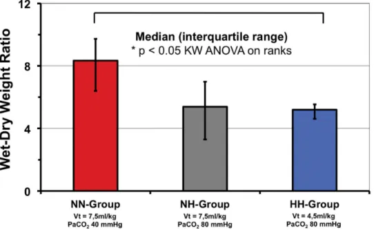
Wet-to-dry ratio of excised lungs tended to be lower in the NH group (p = 0.07) and was significantly lower in the HH-group as compared to the NN-group (p<0.05;Fig 3). Lung his-tology is shown inTable 3. Scores were significantly higher comparing lower lobes vs. upper lobes and for dependent vs. non-dependent specimen for most scores (data not shown). Scores for atelectasis, alveolar inflammation, alveolar hemorrhage, alveolar edema and the sum score were significantly lower for the HH-group as compared to the NN-group indicating less lung injury. In some scores evaluated the NH-group showed trends towards lower scores compared to the NN-group, none reaching statistical significance (Table 3).
Total protein concentration in BALF was lower in the NN-group compared with the HH-group only (Table 4). IL-8 in BALF was not significantly different between groups, whereas
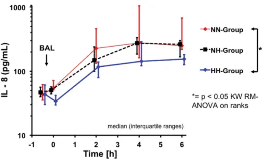
lung tissue IL-8 concentrations were significantly lower in the HH-group as compared to the NN-group in dependent lung tissue specimen (Table 4). Plasma Interleukin-8 was lower in the HH-group as compared to both normoventilation groups reaching statistical significance com-paring HH and NN-groups (Fig 4; p<0.05).
Phospholipid content in 60,000xg pellets of BALF was decreased at the end of the experi-ment compared with the initial values in all 3 groups, independent of ventilation strategy (Table 5).
There was a significant difference in 15(S)-8-iso-PGF2αlevels between NN- and HH-groups at baseline before lavage, but there was no difference in 15(S)-8-iso-PGF2αlevels between all experimental groups after 6h (Table 6). Whereas there were no differences in TH levels between all experimental groups at the end of the experiment, AOPP levels were significantly lower in the HH-group as compared to the normocapnia group and non-protein bound iron levels were significantly lower in both hypercapnia groups (Table 6).
Discussion
The aim of this study was to evaluate the effects of hypercapnic acidosis on the degree of lung injury in this animal model of surfactant deficiency. The design chosen for this study allows separating the protective effects by elevated PCO2levels from the lung protective effects of a small tidal volume. Although there seems to be some effect of hypercapnic acidosis on PaO2,
Fig 1. Gas exchange (PaO2[A], PaCO2[B]) and acid base characteristics (pH [C], lactate [D]) over experimental time. Time = 0 refers to time of
randomization. BAL = bronchoalveolar lavage. PaO2was significantly higher in the HH- vs. the NN-group. Arterial pH and lactate were lower in both
hypercapnia groups as compared to the NN-group;* = p< 0.05. doi:10.1371/journal.pone.0147807.g001
the hypothesis that hypercapnic acidosis improves PaO2was proven for the combined effect of a small tidal volume and hypercapnia only. Furthermore, PaO2was increased in the HH-group despite a lower peak and mean airway pressure as compared to both other groups. Lung injury was decreased in these animals as judged by wet-to-dry weight, BAL-Protein, histology. How-ever, we could not prove similar protective effects in the animals of the NH-group. Further-more, systemic IL-8 levels were significantly lower in the HH-group only, suggesting that systemic inflammation is reduced during low tidal volume use only. This finding suggests
Fig 2. Hemodynamic parameters over time: Arterial blood pressure [A], heart rate [B], central venous pressure [C] and cardiac index [D] over experimental time. Time = 0 refers to time of randomization. BAL = bronchoalveolar lavage. There were no significant differences between groups. doi:10.1371/journal.pone.0147807.g002
Table 1. Interventions to stabilize hemodynamics and pH.
Normoventilation-Normocapnia Normoventilation-Hypercapnia Hypoventilation-Hypercapnia
Dopamine dose [μg/kg/min]a 5.6 (0–15.9)‡ 15.1 (1.3–23.8) 1.9 (0–17.7)†
Volume replacement [ml/kg]b 48.3 (9.2–88.2)‡ 83.6 (8.2–122.0) 30.1 (8.7–83.9)†
NaHCO3[mEqu/kg]b 25 (0–45) 15 (0–32) 14 (0–22) *
a= averaged across time; b= cumulative amount given;
* = p < 0.05 (HH-group vs. NN-group);
†= p<0.05 (HH-group vs. NH-group); ‡= p< 0.05 (NN-group vs. NH-group).
more loss of alveolar and systemic compartmentalization in NN- and NH-groups which has been described in animals exposed to injurious mechanical ventilation [25].
Differences between our findings and those of previous studies may be related to several fac-tors. For example, the PEEP chosen in our study may have been sufficient to counteract the potential negative effects of decreased peak or mean airway pressure on pulmonary shunt and gas exchange. Arterial pO2increased in our model very early after randomization in both hypercapnia groups, and the mechanism involved may be related to improved ventilation-per-fusion matching secondary to pulmonary vasoconstriction and improved perventilation-per-fusion of non-dependent and better aerated lung areas [5]. Although data on PaO2, ventilator pressure, lung compliance, wet-to-dry weight, and BALF protein show trends towards protective effects in the NH-group as compared to the NN-group, a reduced tidal volume clearly was more protective than hypercapnia alone in our model. The lack of lung protection in our NH-group is in line with data from Rai et al. who did not detect a lung-protective effect of an elevated PaCO2(by increasing FiCO2) in rabbits with surfactant deficiency during both, injurious (tidal volume 12 ml/kg, no PEEP) or lung-protective ventilation (tidal volume 5 ml/kg, PEEP 12.5 cmH2O)
Table 2. Ventilation parameters and pulmonary mechanics.
Normoventilation-Normocapnia Normoventilation-Hypercapnia Hypoventilation-Hypercapnia
Tidal Volume [ml/kg] 7.4± 0.2 7.3± 0.3 4.5± 0.3 *
Ventilator Rate [breaths/min] 40.0± 8.2 40.8± 4.4 32.8± 8.5 *
Minute Ventilation [ml/kg/min] 314± 75 306± 37 158± 45 *
Peak Inspiratory Pressure [cmH2O] 25.7± 3.3 22.3± 3.3 18.2± 4.0 *
Mean Airway Pressure [cmH2O] 11.7± 1.4 10.8± 1.0 9.5± 1.2 *
Lung Compliance [ml/kg/cmH2O] 0.42± 0.09 0.51± 0.13 0.40± 0.12
Values are mean± SD;
* = p < 0.05 (HH-group vs. NN-group). doi:10.1371/journal.pone.0147807.t002
Fig 3. Wet-dry-weight ratio of excised lungs.* = p< 0.05. NN vs. NH groups: p = 0.07. doi:10.1371/journal.pone.0147807.g003
[26]. However, in contrast to Rai et al. [26] we aimed to minimize VILI by reducing tidal vol-ume and ventilator rate to very low settings to implement permissive hypercapnia and we were able to show a protective effect of hypercapnia induced with a small tidal volume.
Hypercapnic acidosis has been shown to suppress nuclear factor-kappa B activation and production of IL-8 in experimental settings [27,28], and the use of a lower tidal volume in patients undergoing mechanical ventilation during surgery resulted in lower serum IL-8 levels [29]. We found lower IL-8 values in the dependent lung tissue samples and a trend towards lower values in the BALF, which may reflect anti-inflammatory properties of lower tidal vol-ume and/or hypercapnic acidosis modulating lung injury. Our study design, however, does
Table 3. Lung histology scores.
Normoventilation-Normocapnia (NN-group) Normoventilation-Hypercapnia (NH-group) Hypoventilation-Hypercapnia (HH-group) Atelectasis 0.8 (0–1.3) 0.3 (0–1.3)a 0 (0–1.0) ** Alveolar inflammation 1.5 (1.3–2.0) 1.8 (0–2.5) 0.3 (0–2.5) *,b Interstitial inflammation 1.8 (1.3–2.5) 1.8 (0–2.5) 1.0 (0–2.5) Alveolar hemorrhage 0.8 (0.5–1.0) 0.8 (0–1.0) 0.1 (0–1.0) **,† Interstitial hemorrhage 0.9 (0.8–1.5) 0.8 (0–1.5) 0.4 (0–1.5)c Alveolar edema 0.8 (0.3–1.8) 0.3 (0–1.0) 0 (0–0.8) ** Interstitial edema 0.6 (0–0.8) 0.8 (0–1.3) 0.3 (0–0.8)† Necrosis 0 0 0 Overdistension 2.6 (2.3–3.0) 2.8 (2.5–3.0) 3.0 (2.8–3.0) * Sum-Score 9.9 (7.3–12.5) 9.3 (3.5–11.5) 4.8 (3.3–11.8) *
Values are median (minimum–maximum); * = p < 0.05 and
** = p<0.01 (HH-group vs. NN-group);
†= p<0.05 (HH-group vs. NH-group); a= p = 0.09 (NH-group vs. NN-group); b= p = 0.08 (HH-group vs. NH-group);
c= p = 0.09 (HH-group vs. NN-group); (ANOVA or Kruskal Wallis ANOVA on ranks where appropriate).
doi:10.1371/journal.pone.0147807.t003
Table 4. Protein and IL-8 in broncheal alveolar lavage fluid and lung tissue IL-8. Normoventilation-Normocapnia (NN-group) Normoventilation-Hypercapnia (NH-group) Hypoventilation-Hypercapnia (HH-group) BALF Protein [mg/kg] 24.3 (5.0–56.4) 16.1 (3.4–35.9) 7.2 (2.6–49.6) * BALF IL-8 [ng/kg] 29.8 (8.1–188.4) 18.3 (4.5–64.6) 14.9 (1.7–61.9)
Lung Tissue IL-8dependent lung [pg/g]
23.2 (6.4–100.7) 23.9 (2.0–55.8) 7.5 (3.9–19.9) * †
Lung Tissue IL-8non-dependent lung [pg/g]
17.7 (5.1–232.9) 42.3 (3.6–112.0) 12.6 (0.6–32.9)
Values are median (minimum–maximum); BALF IL-8 and lung tissue IL-8 were measured in right-sided lungs. Lung tissue IL-8 levels are normalized for tissue weight.
* = p < 0.05 (HH-group vs. NN-group);
†= p<0.05 (HH-group vs. NH-group).
not allow to clarify if the lower tidal volume or hypercapnic acidosis, or both, decreased inflammation.
Phospholipid content of BALF, as a surrogate parameter for total surfactant in the alveolar space at the end of the experiment was not different between groups. This suggests that type II pneumocytes only partially compensated for the loss of surfactant, in spite of the residual pools within their lamellar bodies, and that there was no difference in the effect of ventilation param-eters on surfactant homeostasis. As surfactant is partially lost along the airways during alveolar collapse and tidal volume affects surfactant turnover, the kinetics of surfactant secretion and re-uptake may nevertheless be different between the groups, which only can be addressed by isotope labeling [30]. Moreover, according to the Young-Laplace-equation alveolar collapse mostly occurs at low alveolar diameter during the end of the ventilation cycle, which is pre-vented by high PEEP. Therefore, the relatively high PEEP of 7 cmH2O may have prevented intrapulmonary shunt flow and substantially reduced the need of surfactant. This was previ-ously shown in neonatal rats, where an autogenous PEEP by high respiratory rate is associated with very low surfactant values [30,31]. Together, all this may have contributed to the good oxygenation of these surfactant-deficient animals, highlighting that PEEP and tidal volume are at least as important for lung protection as surfactant homeostasis.
Fig 4. Plasma interleukin-8 over time.* = p< 0.05, NN- vs. HH-groups. doi:10.1371/journal.pone.0147807.g004
Table 5. Broncheal alveolar lavage: Surfactant (phospholipid) content. Normoventilation-Normocapnia (NN-group) Normoventilation-Hypercapnia (NH-group) Hypoventilation-Hypercapnia (HH-group) Initial BAL [mg/ kg] 298 (121–506) 299 (231–467) 305 (249–829) Final BAL [mg/ kg] 49 (30–168) 58 (35–162) 47 (31–169)
Values are median (minimum–maximum). Surfactant phospholipid content was analyzed from BAL obtained from both lungs initially and from final BAL of the right lung. Values were corrected for BAL volume. No significant difference between groups was detected.
Markers on oxidative injury showed inconclusive results. Whereas we were able to docu-ment a lower NPBI in both hypercapnia groups and AOPP after 6h, the other markers and 15 (S)-8-iso-PGF2αlevels at 6h did not show differences between groups in our short-term model. Other authors have demonstrated that exposure to hypercapnia attenuated tyrosine nitration and histologic lung injury in rats exposed to 60% oxygen for 14d [32], suggesting that hyper-capnia may mediate hyperoxic lung damage. One might speculate that the high FiO2used in our study resulted in a significant hyperoxic stress, which may not be attenuated by hypercap-nia. However, the timeframe of our experiment was probably too short to assess a protective effect of hypercapnia on oxidative lung damage detectable by measuring F2-isoprostanes. Dur-ing hypoxic conditions exposure to 10% CO2was found to reduce total 8-isoprostane lung lev-els and reduced vascular remodeling and pulmonary hypertension [33].
Hemodynamic variables did not seem to be impaired by hypercapnic acidosis in our ani-mals. In fact, animals of the NH group needed more volume replacement and dopamine than the other groups, and animals ventilated with low tidal volume (HH-group) needed less hemo-dynamic support, suggesting that the lower tidal volume may be the main factor for reduced requirement of cardiovascular support. Other data suggests that beta-adrenergic activation may occur secondary to hypercapnic acidosis [34]. Lactate levels were lower in both hypercap-nia groups which may be related to decreased mitochondrial respiratory activity [35] and downregulation of the production of organic acids during acidosis [36] which protects acid-base balance in the setting of increased acid production [37].
In a previous experiment we have studied the effects of different tidal volumes with associ-ated different levels of permissive hypercapnia on lung injury and gas exchange using the same model and were able to show that a tidal volume below 4–5 ml/kg with a PaCO2of>80 mmHg did not increase lung protection further [38]. The major limitation of that study and of the present study is the short-term nature of both experiments showing beneficial effects within the 6 h study frame. Lung cell repair may be impaired in the setting of hypercapnic acidosis as shown in an short-term experimental study [39], but the relevance of these effects on long-term recovery from acute lung injury is unknown. Although we used an animal model with surfactant deficiency, the animals used do not have immature lungs and therefore the applica-bility of our findings to lung injury in preterm subjects may be limited. Another limitation is the fact that we did not use a 4thgroup with low tidal volume and normocapnia. However, a low tidal volume/normocapnia approach using a higher ventilator rate may have resulted in air trapping. It would be feasible using tracheal gas insufflation to wash out CO2. However,
Table 6. Markers of oxygen toxicity. 15(S)-8-iso-PGF2αlevels (measured at baseline and at the end of the experiment after 6h), Advanced Oxidation
Pro-tein Products (AOPP) and Total Hydroperoxydes (TH) and Non-proPro-tein Bound Iron (NPBI), measured at the end of the experiment (6h) in plasma samples. Normoventilation-Normocapnia Normoventilation-Hypercapnia Hypoventilation-Hypercapnia 15(S)-8-iso-PGF2α[pg/mL]
Baseline before lavage 59.8± 15.2 44.4± 18.9 46.1± 9.9 *
End of experiment 51.2± 19.3 60.5± 24.7 57.3± 24.1
AOPP [μg/dL] 16.5 (4.1–42.3) 10.9 (4.1–23.4) 6.4 (1.5–18.5) *
TH [UCARR] 108 (65–162) 144 (68–214) 82 (29–193)
NPBI [mmol/L] 30.1 (16.2–81.7)‡ 3.8 (0.6–9.9) 3.3 (1.9–6.0)
Values are mean± SD or median (range); * = p < 0.05 (HH-group vs. NN-group);
‡= p<0.05 (NN-group vs. NH- and HH-groups).
tracheal gas insufflation may cause airway epithelial injury as adequate humidification of bias flow into the trachea is difficult to achieve.
Currently it is unknown to what level permissive hypercapnia can be safely employed in preterm infants, children or adults with severe lung injury. Retrospective data in preterm infants suggests an association of an increased incidence of Bronchopulmonary Dysplasia in association with hypercapnia during the first week of life after adjustment for exposed baseline risk, severity of illness and mean airway pressure [40]. In most randomized clinical trials PCO2 values up to 55 mmHg were tolerated during the first days of life and neither proved relevant clinical benefits, nor were they associated with relevant side-effects when compared to control groups aiming for normocapnia [9,41,42]. In a recently published randomized trial necrotizing enterocolitis was more frequent in the permissive hypercapnia group [10]. Because experimen-tal evidence suggests that exposure to hypercapnic acidosis may cause clinically relevant changes in cerebral blood flow and cerebral tissue oxygenation [43] and adverse effects on cere-bral cortex enzyme activity and protein expression in the immature brain [44], follow-up stud-ies in preterm infants exposed to hypercapnic acidosis are needed to assess long-term effects on the developing brain of premature infants.
Conclusions
We conclude that in this model of surfactant deficiency hypercapnic acidosis combined with a low tidal volume improves arterial oxygenation and protects against inflammation and lung injury. However, these effects were not significant for hypercapnic acidosis with a normal tidal volume, showing that either low tidal volume alone or the combination of low tidal volume with permissive hypercapnia is essential for lung protection. More research is needed to study the interaction between tidal volume and CO2and their effects on hemodynamics and other systemic effects. Furthermore, clinical trials using different target ranges of tidal volume and PaCO2looking at side effects and including long-term follow-up of these patients need to be performed to assess whether this approach of protective ventilation with permissive hypercap-nia can be recommended for routine clinical use.
Supporting Information
S1 File. Arrive checklist. (PDF)
Acknowledgments
We thank Nelson Claure, University of Miami (Miami, FL) for his technical support. We also thank Roche Diagnostics (Mannheim, Germany) for kindly providing the AVL Omni 9 blood gas analyzer and Fritz Stephan Medizintechnik GmbH (Gackenbach, Germany) for providing the mechanical ventilator. We thank Maria-Theresia Suchy and Frank-Mathias Gutzki for their excellent laboratory and technical assistance in the GC-MS/MS analysis of 15(S)-8- iso-PGF2α.
Author Contributions
Conceived and designed the experiments: HH HF. Performed the experiments: HH KB MW GB ME WB DT HF. Analyzed the data: HH KB MW HF. Contributed reagents/materials/anal-ysis tools: MW GB ME WB DT. Wrote the paper: HH KB MW GB ME WB DT HF.
References
1. Clark RH, Gerstmann DR, Jobe AH, Moffitt ST, Slutsky AS, Yoder BA (2001) Lung injury in neonates: causes, strategies for prevention, and long-term consequences. J Pediatr 139: 478–486. PMID: 11598592
2. Ware LB, Matthay MA (2000) The acute respiratory distress syndrome. N Engl J Med 342: 1334–1349. PMID:10793167
3. Lee WL, Slutsky AS (2001) Ventilator-induced lung injury and recommendations for mechanical ventila-tion of patients with ARDS. Semin Respir Crit Care Med 22: 269–280. PMID:16088679
4. Dreyfuss D, Saumon G (1998) Ventilator-induced lung injury: lessons from experimental studies. Am J Respir Crit Care Med 157: 294–323. PMID:9445314
5. Yamamoto Y, Nakano H, Ide H, Ogasa T, Takahashi T, Osanai S, et al. (2001) Role of airway nitric oxide on the regulation of pulmonary circulation by carbon dioxide. J Appl Physiol 91: 1121–1130. PMID:11509507
6. Ventilation with lower tidal volumes as compared with traditional tidal volumes for acute lung injury and the acute respiratory distress syndrome. The Acute Respiratory Distress Syndrome Network. (2000) Ventilation with lower tidal volumes as compared with traditional tidal volumes for acute lung injury and the acute respiratory distress syndrome. The Acute Respiratory Distress Syndrome Network. N Engl J Med 342: 1301–1308. PMID:10793162
7. Hickling KG, Walsh J, Henderson S, Jackson R (1994) Low mortality rate in adult respiratory distress syndrome using low-volume, pressure-limited ventilation with permissive hypercapnia: a prospective study. Crit Care Med 22: 1568–1578. PMID:7924367
8. Amato MB, Barbas CS, Medeiros DM, Magaldi RB, Schettino GP, Lorenzi-Filho G, et al. (1998) Effect of a protective-ventilation strategy on mortality in the acute respiratory distress syndrome. N Engl J Med 338: 347–354. PMID:9449727
9. Carlo WA, Stark AR, Wright LL, Tyson JE, Papile L-A, Shankaran S, et al. (2002) Minimal ventilation to prevent bronchopulmonary dysplasia in extremely-low-birth-weight infants. J Pediatr 141: 370–374. PMID:12219057
10. Thome UH, Genzel-Boroviczeny O, Bohnhorst B, Schmid M, Fuchs H, Rohde O, et al. (2015) Permis-sive hypercapnia in extremely low birthweight infants (PHELBI): a randomised controlled multicentre trial. Lancet Respir Med 3: 534–543. doi:10.1016/S2213-2600(15)00204-0PMID:26088180 11. Laffey JG, Tanaka M, Engelberts D, Luo X, Yuan S, Transwell AK, et al. (2000) Therapeutic
hypercap-nia reduces pulmonary and systemic injury following in vivo lung reperfusion. Am J Respir Crit Care Med 162: 2287–2294. PMID:11112153
12. Broccard AF, Hotchkiss JR, Vannay C, Markert M, Sauty A, Feihl F, et al. (2001) Protective effects of hypercapnic acidosis on ventilator-induced lung injury. Am J Respir Crit Care Med 164: 802–806. PMID:11549536
13. Sinclair SE, Kregenow DA, Lamm WJE, Starr IR, Chi EY, Hlastala MP (2002) Hypercapnic acidosis is protective in an in vivo model of ventilator-induced lung injury. Am J Respir Crit Care Med 166: 403– 408. PMID:12153979
14. Strand M, Ikegami M, Jobe AH (2003) Effects of high PCO2on ventilated preterm lamb lungs. Pediatr
Res 53: 468–472. PMID:12595596
15. Feihl F, Eckert P, Brimioulle S, Jacobs O, Schaller MD, Mélot C, et al. (2000) Permissive hypercapnia impairs pulmonary gas exchange in the acute respiratory distress syndrome. Am J Respir Crit Care Med 162: 209–215. PMID:10903243
16. Coates AL, Stocks J (1991) Esophageal pressure manometry in human infants. Pediatr Pulmonol 11: 350–360. PMID:1758761
17. Lachmann B, Robertson B, Vogel J (1980) In vivo lung lavage as an experimental model of the respira-tory distress syndrome. Acta Anaesthesiol Scand 24: 231–236. PMID:7445941
18. Silva Neto G, Gerhardt T, Silberberg A, Gerhardt T, Claure N, Duara S, et al. (1992) Nonlinear pres-sure/volume relationship and measurements of lung mechanics in infants. Pediatr Pulmonol 12: 146– 152. PMID:1641270
19. Tsikas D, Schwedhelm E, Suchy MT, Niemann J, Gutzki FM, Erpenbeck VJ, et al. (2003) Divergence in urinary 8-iso-PGF(2alpha) (iPF(2alpha)-III, 15-F(2t)-IsoP) levels from gas chromatography-tandem mass spectrometry quantification after thin-layer chromatography and immunoaffinity column chroma-tography reveals heterogeneity of 8-iso-PGF(2alpha). Possible methodological, mechanistic and clini-cal implications. J Chromatogr B Analyt Technol Biomed Life Sci 794: 237–255. PMID:12954376 20. Paffetti P, Perrone S, Longini M, Ferrari A, Tanganelli D, Marzocchi B, et al. (2006) Non-protein-bound
iron detection in small samples of biological fluids and tissues. Biol Trace Elem Res 112: 221–232. PMID:17057261
21. Witko-Sarsat V, Friedlander M, Nguyen Khoa T, Capeillère-Blandin C, Nguyen AT, Canteloup S, et al. (1998) Advanced oxidation protein products as novel mediators of inflammation and monocyte activa-tion in chronic renal failure. J Immunol 161: 2524–2532. PMID:9725252
22. Buonocore G, Perrone S, Longini M, Terzuoli L, Bracci R (2000) Total hydroperoxide and advanced oxi-dation protein products in preterm hypoxic babies. Pediatr Res 47: 221–224. PMID:10674350 23. Hummler HD, Thome U, Schulze A, Schnabel R, Pohlandt F, Franz AR (2001) Spontaneous breathing
during partial liquid ventilation in animals with meconium aspiration. Pediatr Res 49: 572–580. PMID: 11264443
24. Thome UH, Schulze A, Schnabel R, Franz AR, Pohlandt F, Hummler HD (2001) Partial liquid ventilation in severely surfactant-depleted, spontaneously breathing rabbits supported by proportional assist venti-lation. Crit Care Med 29: 1175–1180. PMID:11395598
25. Haitsma JJ, Uhlig S, Göggel R, Verbrugge SJ, Lachmann U, Lachmann B(2000) Ventilator-induced lung injury leads to loss of alveolar and systemic compartmentalization of tumor necrosis factor-alpha. Intensive Care Med 26: 1515–1522. PMID:11126266
26. Rai S, Engelberts D, Laffey JG, Frevert C, Kajikawa O, Martin TR, et al. (2004) Therapeutic hypercap-nia is not protective in the in vivo surfactant-depleted rabbit lung. Pediatr Res 55: 42–49. PMID: 14561781
27. Takeshita K, Suzuki Y, Nishio K, Takeuchi O, Toda K, Kudo H, et al. (2003) Hypercapnic acidosis atten-uates endotoxin-induced nuclear factor-[kappa]B activation. Am J Respir Cell Mol Biol 29: 124–132. PMID:12600832
28. Contreras M, Ansari B, Curley G, Higgins BD, Hassett P, O'Toole D, et al. (2012) Hypercapnic acidosis attenuates ventilation-induced lung injury by a nuclear factor-κB-dependent mechanism. Crit Care Med 40: 2622–2630. doi:10.1097/CCM.0b013e318258f8b4PMID:22732277
29. Wolthuis EK, Choi G, Dessing MC, Bresser P, Lutter R, Dzoljic M, et al. (2008) Mechanical ventilation with lower tidal volumes and positive end-expiratory pressure prevents pulmonary inflammation in patients without preexisting lung injury. Anesthesiology 108: 46–54. PMID:18156881
30. Raith M, Schaal K, Koslowski R, Fehrenbach H, Poets CF, Schleicher E, et al. (2012) Effects of recom-binant human keratinocyte growth factor on surfactant, plasma, and liver phospholipid homeostasis in hyperoxic neonatal rats. J Appl Physiol 112: 1317–1328. doi:10.1152/japplphysiol.00887.2011PMID: 22323656
31. Bernhard W, Schmiedl A, Koster G, Orgeig S, Acevedo C, Poets CF, et al. (2007) Developmental changes in rat surfactant lipidomics in the context of species variability. Pediatr Pulmonol 42: 794–804. PMID:17659602
32. Masood A, Yi M, Lau M, Belcastro R, Shek S, Pan J, et al. (2009) Therapeutic effects of hypercapnia on chronic lung injury and vascular remodeling in neonatal rats. Am J Physiol Lung Cell Mol Physiol 297: L920–L930. doi:10.1152/ajplung.00139.2009PMID:19749000
33. Kantores C, McNamara PJ, Teixeira L, Engelberts D, Murthy P, Kavanagh BP, et al. (2006) Therapeutic hypercapnia prevents chronic hypoxia-induced pulmonary hypertension in the newborn rat. Am J Phy-siol Lung Cell Mol PhyPhy-siol 291: L912–L922. PMID:16829630
34. Steinhart CR, Permutt S, Gurtner GH, Traystman RJ (1983) beta-Adrenergic activity and cardiovascu-lar response to severe respiratory acidosis. Am J Physiol 244: H46–H54. PMID:6295189
35. Hillered L, Ernster L, Siesjö BK (1984) Influence of in vitro lactic acidosis and hypercapnia on respira-tory activity of isolated rat brain mitochondria. J Cereb Blood Flow Metab 4: 430–437. PMID:6470057 36. Hood VL, Schubert C, Keller U, Müller S (1988) Effect of systemic pH on pHi and lactic acid generation
in exhaustive forearm exercise. Am J Physiol 255: F479–F485. PMID:3414804
37. Hood VL, Tannen RL (1998) Protection of acid-base balance by pH regulation of acid production. N Engl J Med 339: 819–826. PMID:9738091
38. Fuchs H, Mendler MR, Scharnbeck D, Ebsen M, Hummler HD (2011) Very low tidal volume ventilation with associated hypercapnia—effects on lung injury in a model for acute respiratory distress syndrome. PLoS ONE 6: e23816. doi:10.1371/journal.pone.0023816PMID:21886825
39. Doerr CH, Gajic O, Berrios JC, Caples S, Abdel M, Lymp JF, et al. (2005) Hypercapnic acidosis impairs plasma membrane wound resealing in ventilator-injured lungs. Am J Respir Crit Care Med 171: 1371– 1377. PMID:15695495
40. Subramanian S, El-Mohandes A, Dhanireddy R, Koch MA (2011) Association of bronchopulmonary dysplasia and hypercarbia in ventilated infants with birth weights of 500–1,499 g. Matern Child Health J 15 Suppl 1: S17–S26.
41. Mariani G, Cifuentes J, Carlo WA (1999) Randomized trial of permissive hypercapnia in preterm infants. Pediatrics 104: 1082–1088. PMID:10545551
42. Thome UH, Carroll W, Wu T-J, Johnson RB, Roane C, Young D, et al. (2006) Outcome of extremely preterm infants randomized at birth to different PaCO2 targets during the first seven days of life. Biol Neonate 90: 218–225. PMID:16636534
43. Tomimatsu T, Pena JP, Longo LD (2006) Fetal hypercapnia and cerebral tissue oxygenation: studies in near-term sheep. Pediatr Res 60: 711–716. PMID:17065566
44. Fritz KI, Zubrow A, Mishra OP, Delivoria-Papadopoulos M (2005) Hypercapnia-induced modifications of neuronal function in the cerebral cortex of newborn piglets. Pediatr Res 57: 299–304. PMID: 15585683
![Fig 1. Gas exchange (PaO 2 [A], PaCO 2 [B]) and acid base characteristics (pH [C], lactate [D]) over experimental time](https://thumb-eu.123doks.com/thumbv2/123dokorg/4656212.42334/7.918.57.848.115.590/fig-gas-exchange-paco-characteristics-lactate-over-experimental.webp)
![Fig 2. Hemodynamic parameters over time: Arterial blood pressure [A], heart rate [B], central venous pressure [C] and cardiac index [D] over experimental time](https://thumb-eu.123doks.com/thumbv2/123dokorg/4656212.42334/8.918.58.848.119.591/hemodynamic-parameters-arterial-pressure-central-pressure-cardiac-experimental.webp)
![Table 4. Protein and IL-8 in broncheal alveolar lavage fluid and lung tissue IL-8. Normoventilation-Normocapnia (NN-group) Normoventilation-Hypercapnia(NH-group) Hypoventilation-Hypercapnia(HH-group) BALF Protein [mg/kg] 24.3 (5.0 –56.4) 16.1 (3.4 –35.9) 7](https://thumb-eu.123doks.com/thumbv2/123dokorg/4656212.42334/10.918.50.866.807.952/broncheal-normoventilation-normocapnia-normoventilation-hypercapnia-hypoventilation-hypercapnia-protein.webp)