Introduction. Vaccines and antiviral drugs are the most widely used methods of preventing or treating Influenza virus infection. The role of sea buckthorn (SBT) bud dry extract as a natural anti-viral drug against Influenza was investigated.
Methods. Influenza virus was cultured in the MDCK cell line, with or without SBT bud extract, and virus growth was assessed by HA and TCID50 virus titration in terms of cytopathic effect on cells. Several concentrations of extract were tested, the virus titer being measured on day 4 after infection.
Results. After infection, the virus titer in the control sample was calculated to be 2.5 TCID50/ml; treatment with SBT bud extract reduced the virus titer to 2.0 TCID50/ml at 50 µg/ml, while the HA titer was reduced from 1431 (control) to 178. Concentrations
lower than 50µg/ml displayed an inhibitory effect in the HA assay, but not in the TCID50 virus titration; however, observation of the viral cultures confirmed a slowdown of viral growth at all concentrations.
Discussion. Natural dietary supplements and phytotherapy are a growing market and offer new opportunities for the treatment of several diseases and disorders. These preliminary experiments are the first to show that SBT bud extract is able to reduce the growth of the Influenza A H1N1 virus in vitro at a concentration of 50 µg/ml. This discovery opens up the possibility of using SBT bud extract as a valid weapon against Influenza and, in addition, as the starting-point for the discovery of new drugs.
Originalarticle
Sea buckthorn bud extract displays activity
against cell-cultured Influenza virus
A. Torelli1, e. GiAnchecchi1, S. PiccirellA1, A. MAnenTi1, G. Piccini1, e. llorenTe PASTor1,
B. cAnovi2, e. MonToMoli1,3
1 visMederi Srl, Siena, italy; 2 Pool Pharma - Mi -Scientific Director, San Giuliano Milanese, italy; 3 University of Siena, italy
Key words
Sea buckthorn plant extract • Influenza prevention • Antivirals
Summary
Introduction
Influenza is caused by RNA viruses belonging to the family of Orthomyxoviridae. Three types of influenza viruses can be distinguished. Among these, influenza A (IAV) viruses mainly infect birds and mammals, where-as influenza B viruses almost only infect humans. In-fluenza A virus displays constant evolutionary changes, defined as “antigenic drift” [1] and “antigenic shift” [2], enabling new mutant strains to emerge and spread. Influ-enza viruses constitute a public health problem, as they can give rise to both epidemics and pandemics, causing high rates of morbidity and mortality [1]. Influenza vi-ruses are considered the leading cause of respiratory ill-ness in humans and are responsible for annual seasonal outbreaks that have a serious economic impact [3]. The average global burden of influenza may be in the order of 1 billion cases of flu, 3-5 million cases of severe ill-ness that need hospitalization and 250,000-500,000 deaths yearly [4]. The most effective means of fighting influenza is primary prevention through vaccination [5], which can prevent the disease after exposure or reduce the severity of symptoms. Antiviral drugs can also be used [6]. Indeed, in subjects who are not completely pro-tected by vaccination strategies, an important role has been played by antiviral treatments [7].
Currently, two main classes of antiviral drugs are avail-able; these are based on the ability to block viral M2
ion channels [8] and viral neuraminidase [9]. However, both classes of drugs have been associated with limited efficacy, adverse side effects [1] and the spread of drug resistance in circulating influenza viruses [10, 11]; this last problem has been associated with an uncontrolled administration of the drugs to humans and, sometimes, to farm animals [12]. Other classes of drugs can be used against influenza virus, some of these include: inhibitors of hemagglutinin, endosomes, lysosomes, proteases, polymerases, nucleoprotein (NP), nonstructural-1 (NS1) glycoprotein, RNA synthesis and caspase; however their efficacy in blocking Influenza virus and avoiding the development of antiviral resistances should be further investigated [13]. It is therefore necessary to develop new anti-influenza treatments that are able to inhibit the replication or cellular processes of viruses [1, 7].
Some studies have indicated that certain plant extracts are able to mimic the neuraminidase effect of anti-viral drugs. One of these plants is sea buckthorn (SBT) (Hippophae rhamnoides): commonly known as sea buckthorn, it belongs to the family Elaeagnaceae [14] and it is a shrubby to bushy plant that grows also at a high altitude and is widely distributed in Eurasia. In In-dia this plant grows predominantly in several high-alti-tude areas such as Sikkim, Himachal Pradesh, Kashmir, Jammu, and Uttar Pradesh but Hippophae is cultivated worldwide for its medicinal properties [15].
All parts of SBT contain large amounts of several active compounds [16] and these include: vitamins (folic acid, vitamin C, vitamin A, vitamin E, vitamin K, riboflavin), carotenoids (lycopene α, β, δ-carotene), phytosterols (amyrins, ergosterol, stigmasterol, lansterol), organic acids (malic and oxalic acids), polyunsaturated fatty ac-ids and essential amino acac-ids [17].
Several medicinal and therapeutic applications utilize the extracts obtained from the leaves of this plant, which display immunomodulatory [18] and anti-inflammato-ry [19] properties, as demonstrated by Padwad and col-leagues [16]. Furthermore, SBT leaves also exert anti-viral, anti-bacterial and anti-tumor effects [20, 21]. Pre-liminary results have also shown that SBT seed extracts have anti-bacterial activity (for example against Listeria
monocytogenes and Yersinia enterocolitica) [22, 23] and anti-viral activities, as reported by the group of Jain against Dengue virus [24].
SBT bud extracts are present in commercial dietary sup-plements, such as preparations of nutrient and vitamin products [25, 26], and are formulated into Influpirinvi-ral® (PoolPharma). However, no data are currently
avail-able on the putative antiviral activity of bud extracts when taken in the form of dietary supplements.
The aim of the present study was to evaluate the thera-peutic anti-viral potential of SBT bud extracts on Influ-enza A/H1N1 virus infection in Madin Darby Canine Kidney cells (MDCK). In 2009, this viral strain was re-sponsible for an Influenza pandemic that spread rapidly around the world [27]. Moreover, it currently circulates in the population, causing seasonal outbreaks, and its antigens are included in the available seasonal influenza vaccines [28].
Methods
Cells and Cell Cultures
Madin Darby Canine Kidney (MDCK) cells were pur-chased from Sigma-Aldrich (ECACC, Public Health England, Porton Down, United Kingdom) and cultured in Minimum Essential Medium Eagle (EMEM medium) supplemented with 2mM Glutamine, 10% Fetal Bovine Serum (FBS) EU Approved (Euroclone, Pero, Italy) and 100Ul/ml of penicillin-streptomycin. Sub-confluent cul-tures (70%-80%) were grown at 37°C in a humidified atmosphere containing 5% CO2, and subcultures were
performed every 3-4 days [29]. Except where indicated otherwise, all the above reagents were from Lonza (Ver-viers, Belgium).
SBT bud dry extract
SBT bud dry extract from PoolPharma S.r.l. (San Gi-uliano Milanese, Italy) was weighed on a precision scale and dissolved in sterile Dulbecco’s Phosphate-Buffered Saline (DPBS) at a final concentration of 1 mg/ml; the pH of the solution was measured (6.96). The dissolved extract was then sterile-filtered through a 0.22 µm filter.
Influenza A/California/7/2009 (H1N1) virus
Influenza A/California/7/2009 (H1N1) virus was ob-tained from the National Institute for Biological Stan-dards and Control (NIBSC) (Potters Bar, Hertfordshire, United Kingdom) and used according to the instructions provided by the supplier. The virus was propagated in MDCK cells [30], harvested and stored at -80°C. The propagated virus had a tissue culture infectious dose (TCID50) titer of 103.5.
Viral growth
Influenza A/California/7/2009 (H1N1) virus was propa-gated in MDCK cells cultured in UltraMDCK serum-free-medium (SFM) supplemented with 0.5 μg/ml of trypsin from bovine pancreas (TPCK) (Sigma-Aldrich, Saint Louis, MO, USA) and 100 UI/ml of penicillin-streptomycin. MDCK cells were seeded in a T25 cm2
tissue culture flask at a density of 1 x 10⁶ cells/ml.
Af-ter 24 hours (h), the cell medium was discarded and the cells were washed twice with sterile DPBS. After the DPBS had been discarded from the flask, the cells were treated with 500 µl of virus inoculum (5 ml of solution contained 50 µl of virus at 103.5 TCID50/ml (1:100
dilu-tion) and 2.5 µl of TPCK, and incubated for 1 h at 37°C in 5% CO2. After 1 h, the inoculum was removed, the
cells were washed with DPBS, and fresh UltraMDCK SFM, supplemented as previously described, was add-ed. The cells were incubated at 37°C in 5% CO2 and the
cytopathic effect was monitored every day until post-infection day 4. The culture medium was collected and analyzed for TCID50 and hemagglutination titer on the 4th day after infection.
Effect of SBT bud dry extract on MDCK cells
MDCK cells were seeded at a density of 6.5 x 10⁵ cells/
ml in 6-well plates in complete EMEM medium and were incubated for 24 h at 37°C in 5% CO2. SBT at
dif-ferent concentrations (1 μg/ml, 5 μg/ml, 10 μg/ml, 30 μg/ ml, 50 μg/ml, 75 μg/ml and 100 μg/ml) was then added to the medium in the wells. The cells were checked at 24 h, 48 h and 72 h by means of a light optical microscope to evaluate whether the extract had a cytotoxic effect on them. The experiment was repeated to confirm the pre-liminary results.
Effect of SBT on viral growth
The viral growth procedure was repeated in the condi-tions reported above. After infection, the inoculum was removed and the medium was replaced with fresh Ultra-MDCK SFM (supplemented with 100 UI/ml of penicil-lin-streptomycin and 0.5 g/ml of TPCK) containing dif-ferent concentrations of SBT: 2.5 μg/ml, 5 μg/ml, 7.5 μg/ ml, 10 μg/ml, 30 μg/ml, 50 μg/ml, 75 μg/ml and 100 μg/ ml. The infection grade was observed daily for cytopath-ic effect, and the culture medium was harvested on day 4 to be analyzed for virus content in terms of TCID50 and hemagglutination titer. In parallel, two control flasks were run: the first flask represented the cell control and
the culture medium was added with DPBS; the second flask represented the virus growth control and it was treated only with the live virus.
Virus titration by hemagglutination test
The ability of the influenza virus to agglutinate red blood cells from certain mammalian or avian species can be exploited to check for the presence and hemaggluti-nating activity of the virus in biological substrates (e.g. serum samples) [31, 32]. To evaluate the hemagglutinat-ing capability of the virus in previously infected cell tures, the hemagglutination test was used: 100 µl of cul-ture medium from the flask of interest was transferred to the 1st well of 12 of a 96-well V bottom plate then 50
μl of saline solution (0.9% NaCl) (Sigma-Aldrich, Saint Louis, MO, USA) was added from well 2 up to well 12; 2-fold serial dilutions of the culture medium (contained in the 1st well) in the saline solution (wells 2-12) were
performed from the 1st well up to the 12th well.
Then, 50 µl of turkey red blood cell (RBC) suspension (0.5% in saline solution) (Emozoo Snc, Casole d’Elsa, Italy) was added to each well and the plate was incu-bated for 45 minutes at room temperature (RT). After incubation, the plate was tilted to allow non-hemagglu-tinated RBCs to drip from the bottom of the wells and the result was read; the reciprocal of the highest dilution of the culture medium that was still able to cause ag-glutination indicated the titer of the virus in the culture medium. The experiment was repeated, this time with a 1:10 starting dilution of the culture media, and the virus titer was calculated in terms of hemagglutinating units (HAU) in 1 ml by applying the following formula: HAU in 1 ml=20x10[LOG(Dilution1)+LOG(Dilution2)]/2 .
Virus titration by TCID50
The “TCID50” titer is the viral dose that gives rise to a cytopathic effect in 50% of cells in the inoculated cul-ture. The virus titer was determined by means of the TCID50 assay, using the Spearman/Karber method [33] on treated MDCK cells, as reported by Lugovtsev [34]. Twelve plastic tubes (1.5 ml) were prepared and loaded with 900 μl of cell medium (EMEM), except for the 11th tube; 100 µl of supernatants from cultures was then
transferred into the first tube and serial 10-fold dilutions were performed from tube 1 to up to tube 10. The con-tents of the tubes were transferred into a 96-well cell culture plate: the content of the 1st tube was transferred
into the 1st column of the plate, the content of the 2nd tube
was transferred into the 2nd column, and so on up to the
10th tube. The 11th column was left empty. At the end
of the dilution steps, 100 μl of cell suspension (in com-plete EMEM medium, with 0.5% FBS and 0.5 μg/ml of TPCK, at a cell density of 5 x 10⁵ cells/ml) was added
to each well. The 12th column, containing only cell
me-dium and cell suspension, was used as a cell control. The plates were incubated for 5 days and the TCID50 titer was evaluated by checking the cytopathic effect in the cell mono-layer by means of a light microscope. The results were calculated by applying the Spearman/ Karber formula [33]: TCID50/100µl= X0-d/2+d(ΣXi/n),
where X0 is the positive logarithm of the highest
dilu-tion at which all wells are positive for cytopathic effect, d represents the dose distance in log, n is the number of repeats per dilution and ΣXi is the sum of all posi-tive wells, starting from X0. In the case of 10-fold
dilu-tion, d=log1010=1 and the formula can be simplified to
TCID50/100μl= X0-1/2+(ΣXi/n). To express the results
as TCID50/ml, one log was added.
Results
Evaluation of the cytotoxic effect of SBT on MDCK Cells
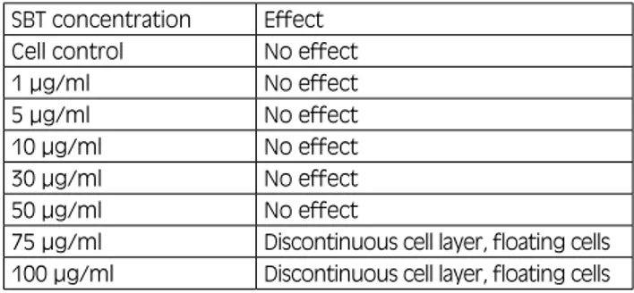
The possible cytotoxic effect of SBT on MDCK cells previously infected with Influenza A H1N1 virus was evaluated by means of direct observation of cells under a light optical microscope. SBT treatment of MDCK cells showed no cytotoxic effect up to a concentration of 50 µg/ml. At 75 μg/ml and 100 μg/ml, SBT had a cytotoxic effect on the cells: at 75 μg/ml, the cell monolayer had a discontinuous appearance and floating cells were pre-sent in the culture medium; this effect was more evident at a concentration of 100 μg/ml (Tab. I).
SBT effect on viral growth results Hemagglutination titer results
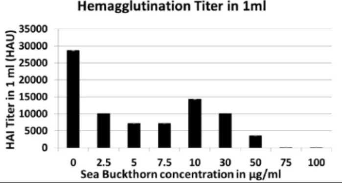
The ability of SBT bud extract to reduce Influenza H1N1 viral growth was evaluated on post-infection day 4. MDCK cell culture supernatants were harvested from the culture plates and assayed for hemagglutination ti-ter. Concentrations of SBT ranging from 2.5 μg/ml to 50 μg/ml markedly reduced the hemagglutination titer from 28621.6 HAU/ml (the value obtained from the viral growth control sample) (Fig. 1). The two highest concentrations of SBT used (75 μg/ml and 100 μg/ml) completely inhibited viral growth. However, they
dis-Tab. I. evaluation of the cytotoxic effect of SBT on mdCK Cells. This table shows the effect of different concentrations of SBT on mdCK cell cultures after 72 h of incubation. The concentrations of SBT test-ed were: 1 μg/ml, 5 μg/ml, 10 μg/ml, 30 μg/ml, 50 μg/ml, 75 μg/ml and 100μg/ml. No toxic effect was observed up to a concentration of 75 μg/ml. At 75 μg/ml and 100 μg/ml of SBT, the cells in the wells showed signs of toxicity: discontinuous cell layer and floating cells in the medium.
SBT concentration effect
Cell control No effect
1 µg/ml No effect
5 µg/ml No effect
10 µg/ml No effect
30 µg/ml No effect
50 µg/ml No effect
75 µg/ml discontinuous cell layer, floating cells 100 µg/ml discontinuous cell layer, floating cells
played a toxic effect on the cells; the results obtained at these high concentrations may therefore be affected cell toxicity, as was observed after staining the cell cultures with Trypan blue.
TCID50 virus titration results
Cell culture supernatants were harvested on day 4 after infection and treatment and assayed for TCID50 virus titer to assess the number of infectious (live) viral par-ticles released from the infected cells in the culture me-dium. The cytopathic effect was evaluated on day 5. The treatment of viral cultures with SBT at a concentration of 50 µg/ml markedly reduced the viral titer in terms of TCID50 (Fig. 2). At 75 µg/ml and 100 µg/ml, SBT was able to inhibit viral growth completely; however, this value could be affected by the toxicity that SBT showed on cells at such high concentrations. Direct observation of the viral cultures confirmed the results obtained from TCID50 and hemagglutination titration. The concentra-tion of SBT that proved to have the greatest impact on viral growth while exerting little negative effect on cells was 50 µg/ml. Figure 3 reports a series of representative pictures showing the effect of SBT at 0, 10, 30, 50, 75 and 100 µg/ml on day 4 on MDCK cells infected with Influenza A/H1N1 virus.
Discussion
In recent years, since the spread of resistant viral strain towards the available pharmacologic treatments and the occurrence of unpredictable pandemics, many investiga-tions have been carried out on new computer-designed molecules or available compounds in a search for new
Fig. 3. Cell viability of mdCK cells infected with Influenza h1N1 upon treatment with SBT. The figure shows a series of repre-sentative pictures from the light optical microscope (Trypan blue-stained; original magnification 100X) showing mdCK cell cul-tures infected with Influenza virus h1N1 A/California/7/2009 and treated with different concentrations of SBT: A, virus control; B, 10 µg/ml; C, 30 µg/ml; d, 50 µg/ml; e, 75 µg/ml; F, 100 µg/ml. Cells were stained with Trypan blue to observe dead cells. The virus control displayed a large number of dead cells (blue spots); 10 µg/ml and 30 µg/ml of SBT induced a reduction in the number of dead cells, and the cellular monolayer was more compact. pic-ture d represents the 50 µg/ml SBT concentration: no cytopathic effect and a limited number of dead cells. The 75 µg/ml and 100 µg/ml SBT concentrations showed a discontinuous cell layer with many dead cells in the culture medium.
Fig. 2. TCId50 virus titration results. The graph shows the results of the TCId50 titration assay performed on supernatants from vi-ral mdCK cultures treated with SBT. The values of the TCId50 titer in 1 ml are shown on the y axis; on the x axis the concentrations of SBT used to treat viral cultures are reported: 0 μg/ml (virus control), 2.5 μg/ml, 5 μg/ml, 7.5 μg/ml, 10 μg/ml, 30 μg/ml, 50 μg/ml, 75 μg/ml and 100 μg/ml. The virus control displayed a titer of 102.5 TCId50; the TCId50 titer was reduced when SBT was used at a concentration of 50 μg/ml. At 75 μg/ml and 100 μg/ml, SBT inhibited viral growth totally. These results represent a mean of two experiments (n = 2).
Fig. 1. hemagglutination Titration results on day 4 after SBT treat-ment. Supernatants of viral cultures treated with SBT were ana-lyzed by means of hemagglutination assay to determine whether the treatment was able to reduce the spread of Influenza virus. The values of the hemagglutination titer in 1 ml (volume assayed) are shown on the y axis; the x axis reports the concentrations of SBT (2.5 μg/ml, 5 μg/ml, 7.5 μg/ml, 10 μg/ml, 30 μg/ml, 50 μg/ml, 75 μg/ml and 100 μg/ml) used to treat viral cultures. No SBT was added to the virus control sample. The control sample displayed a titer of 28636; treatment with SBT dramatically decreased the hemagglutination titer. The most effective concentration of SBT was 50 μg/ml. At 75μg/ml and 100μg/ml, SBT totally inhibited viral growth, but in these cases the results could be affected by SBT toxicity. These results represent a mean of two experiments (n = 2).
anti-influenza drugs. In the present study, the antiviral activity of SBT bud extract (contained in Influpirinvi-ral®) against influenza H1N1 A/California/7/2009 was
evaluated in vitro on MDCK cells infected with this virus. A previous study conducted by Jain and col-leagues [24] demonstrated that SBT leaf extract at a concentration of 50 μg/ml exerted an antiviral activity against Dengue virus. We found that SBT bud extract had an antiviral activity on influenza virus H1N1, espe-cially at 50 μg/ml (the same concentration used by Jain’s group). Specifically, after 4 days the viral titer was eval-uated in terms of TCID50 titer and hemagglutination ti-ter upon treatment with SBT bud extract; both methods revealed that, at 50 μg/ml, the viral titer was reduced in comparison with the virus control. Direct observation of the virus cultures confirmed the antiviral activity at 50 μg/ml, although an antiviral effect was also visible at lower concentrations, starting from 2.5 μg/ml. When SBT was used at high concentrations (75 μg/ml and 100 μg/ml), viral growth was completely inhibited. Howev-er, the treatment had an adverse effect on cell cultures, and this could have affected the results obtained at these concentrations.
Conclusions
The data obtained from this preliminary study confirmed that SBT bud extract has an antiviral activity on influ-enza H1N1 A/California/7/2009 in vitro, supporting its potential use as an anti-influenza agent. Nevertheless, further investigations are needed in order to generate more data, to evaluate the preventive role of SBT treat-ment against Influenza infection, and to understand the specific mechanism of action of this extract both in vitro and in vivo.
References
[1] Loregian A, Mercorelli B, Nannetti G, et al. Antiviral strategies against influenza virus: towards new therapeutic approaches. Cell Mol Life Sci 2014;71:3659-83.
[2] Poovorawan Y, Pyungporn S, Prachayangprecha S, et al. Glob-al Glob-alert to avian influenza virus infection: from H5N1 to H7N9. Pathog Glob Health 2013;107:217-23.
[3] Killip MJ, Fodor E, Randall RE. Influenza virus activation of the interferon system. Virus Res 2015 Feb 9. doi: 10.1016. [4] http://www.who.int/mediacentre/factsheets/fs211/en/.
[5] Talbot HK, Griffin MR, Chen Q, et al. Effectiveness of seasonal vaccine in preventing confirmed influenza-associated hospi-talizations in community dwelling older adults. J Infect Dis 2011;203:500-8.
[6] Moscona A. Medical management of influenza infection. Annu Rev Med 2008;59:397-413.
[7] Krug RM. Influenza: An RNA-synthesizing machine. Nature 2014;516:338-9.
[8] Pinto LH, Lamb RA. The M2 proton channels of influenza A and B viruses. J Biol Chem 2006;281:8997-9000.
[9] Michiels B, Van Puyenbroeck K, Verhoeven V, et al. The value of neuraminidase inhibitors for the prevention and treatment of seasonal influenza: a systematic review of systematic reviews. PLoS ONE 2003;8:e60348.
[10] Deyde VM, Xu X, Bright RA, et al. Surveillance of resistance to adamantanes among influenza A(H3N2) and A(H1N1) virus-es isolated worldwide. J Infect Dis 2007;196:249-57.
[11] Bloom JD, Gong LI, Baltimore D. Permissive secondary muta-tions enable the evolution of influenza oseltamivir resistance. Science 2010;328:1272-5.
[12] Gasparini R, Amicizia D, Lai PL, et al. Compounds with anti-influenza activity: present and future of strategies for the op-timal treatment and management of influenza. Part I: Influ-enza life-cycle and currently available drugs. J Prev Med Hyg 2014;55:69-85.
[13] Gasparini R, Amicizia D, Lai PL, et al. Compounds with anti-influenza activity: present and future of strategies for the optimal treatment and management of influenza. Part II: Future compounds against influenza virus. J Prev Med Hyg 2014;55:109-29.
[14] Shipulina LD, Tolkachev ON, Krepkova LV, et al. Anti-viral anti-microbial and toxicological studies on Seabuckthorn (Hip-pophae rhamnoides). Seabuckthorn (Hippophae L.): A Multi-purpose Wonder Plant, vol. 2. Daya Publishing House 2005, New Delhi, India, pp. 471-83.
[15] Usha T, Diddha KS, Goyal AK et al. Molecular docking stud-ies of anti-cancerous candidates in Heppophae rhamnoides and Hippophae salicifolia. J Biomed Res 2014; 28:406-15. [16] Padwad Y, Ganju L, Jain M, et al. Effect of leaf extract of
Seabuckthorn on lipopolysaccharide induced inflamma-tory response in murine macrophages. Int Immunopharmacol 2006;6:46-52.
[17] Chauhan S, Varshneya C. The profile of bioactive compounds in Seabuckthorn: berries and seed oil. International Journal of Theoretical & Applied Sciences 2012;4:216-20.
[18] Geetha S, Singh V, Ram MS, et al. Immunomodulatory effects of seabuckthorn (Hippophae rhamnoides L.) against chro-mium (VI) induced immunosuppression. Mol Cell Biochem 2005;278:101-9.
[19] Ganju L, Padwad Y, Singh R, et al. Anti-inflammatory activity of Seabuckthorn (Hippophae rhamnoides) leaves. Int Immuno-pharmacol 2005;5:1675-84.
[20] Vermenichev SM. Experimental study of toxicity and anti-tumor activity of natural and synthetic compounds of pyrone series. Phenolic compounds and their physiological proper-ties. Proceedings of the 2nd All-Union Symposium on phenolic compounds held 17–21 May 1971 in Alma-Ata, ‘Nauka’ Ka-zakh, SSR (1973), pp. 210-4.
[21] Tsybikova DTs, Rasputina DB, Zalykeeva DN, et al. A study of leaves and the oil cake of Seabuchthorn. Biology, Chemistry and Pharmacology of Seabuckthorn, Nauka Sibirdiv, Novosi-birsk 1983, pp. 107-9.
[22] Negi PS, Chauhan AS, Sadia GA, et al. Antioxidant and anti-bacterial activities of various seabuckthorn (Hippophae rham-noides L.) seed extracts. Food Chemistry 2005;92:119-24. [23] Chauhan AS, Negi PS, Ramteke RS. Antioxidant and
antibacte-rial activities of aqueous extract of Seabuckthorn (Hippophae rhamnoides) seeds. Fitoterapia 2007;78:590-2.
[24] Jain M, Ganju L, Katiyal A, et al. Effect of Hippophae rhamnoi-des leaf extract against Dengue virus infection in human blood-derived macrophages. Phytomedicine 2008;15:793-9. [25] Xu MY, Sun XX, Tong WX. Medical research and
develop-ment of Seabuckthorn. Hippophae 1994;7:32-40.
[26] Beveridge S, Li T, Oomah BD, et al. Seabuckthorn prod-ucts: manufacture and composition. J Agric Food Chem 1999;47:3480-8.
[27] Kocik J, Kołodziej M, Joniec J, et al. Antiviral activity of novel oseltamivir derivatives against some influenza virus strains. Acta Biochim Pol 2014;61:509-13.
[28] Committee on Infectious Diseases. Recommendations for Pre-vention and Control of Influenza in Children, 2012-2013. Pedi-atrics 2012;130:780-92.
[29] Gaush CR, Hard WL, Smith TF. Characterization of an estab-lished line of canine kidney cells (MDCK). Proceedings of the Society for Experimental Biology and Medicine 1966-07-01. [30] Hamamoto I, Takaku H, Tashiro M, et al. High Yield
Pro-duction of Influenza Virus in Madin Darby Canine Kidney (MDCK) Cells with Stable Knockdown of IRF7. PLoS ONE 2013;8:e59892.
[31] Hirst GK. The quantitative determination of influenza virus and antibodies by means of red cell agglutination. J Exp Med 1942;75:49-64.
[32] Salk JE. A simplified procedure for titrating hemagglutinating capacity of influenza-virus and the corresponding antibody. J Immunol 1944;49:87-98.
[33] Spearman C. The Method of ‘Right and Wrong Cases’ (Con-stant Stimuli) without Gauss’ Formulae. Brit Jour of Psych, 2, 1908.
[34] Lugovtsev VY, Melnyk D, Weir JP. Heterogeneity of MDCK cell line and its applicability for Influenza virus research. PLoS ONE 2013;8:e75014.
n Received on April 28, 2015. Accepted on May 26, 2015.
n Conflicts of interests: Dr. Brenno Canovi is the Scientific Director of Pool Pharma, the company that distributes the Influpirinviral®,
the dietary supplement containing sea buckthorn bud extract. n Correspondence: Alessandro Torelli, VisMederi Srl, via
Fioren-tina 1, 53100 Siena, Italy - Tel. +39 0577 231254 - Fax +39 0577 43444 - E-mail: [email protected]