Effects of Hydrogen Sulfide-releasing
L
-DOPA Derivatives on
Glial Activation
POTENTIAL FOR TREATING PARKINSON DISEASE
*□SReceived for publication, February 17, 2010, and in revised form, March 26, 2010Published, JBC Papers in Press, April 5, 2010, DOI 10.1074/jbc.M110.115261 Moonhee Lee‡, Valerio Tazzari§, Daniela Giustarini¶, Ranieri Rossi¶, Anna Sparatore§, Piero Del Soldato储, Edith McGeer‡, and Patrick L. McGeer‡1
From the‡Kinsmen Laboratory of Neurological Research, University of British Columbia, Vancouver, British Columbia V6T 1Z3,
Canada, the§Dipartimento di Scienze Farmaceutiche Pietro Pratesi, Universita` degli Studi di Milano, Via Mangiagalli 25, 20133,
Milano, Italy, the¶Department of Evolutionary Biology, Laboratory of Pharmacology and Toxicology, University of Siena,
via A. Moro 4, I-53100, Siena, Italy, and储CTG Pharma, Viale Gran Sasso 17, 20131 Milano, Italy
The main lesion in Parkinson disease (PD) is loss of substantia nigra dopaminergic neurons. Levodopa (L-DOPA) is the most widely used therapy, but it does not arrest disease progression. Some possible contributing factors to the continuing neuronal loss are oxidative stress, including oxidation ofL-DOPA, and neurotoxins generated by locally activated microglia and astro-cytes. A possible method of reducing these factors is to produce
L-DOPA hybrid compounds that have antioxidant and
antiin-flammatory properties. Here we demonstrate the properties of four suchL-DOPA hybrids based on couplingL-DOPA to four different hydrogen sulfide-donating compounds. The donors themselves were shown to be capable of conversion by isolated mitochondria to H2S or equivalent SHⴚions. This capability was
confirmed by in vivo results, showing a large increase in intra-cerebral dopamine and glutathione after iv administration in rats. When human microglia, astrocytes, and SH-SY5Y neuro-blastoma cells were treated with these donating agents, they all accumulated H2S intracellularly as did their derivatives coupled
to L-DOPA. The donating agents and the L-DOPA hybrids
reduced the release of tumor necrosis factor-␣, interleukin-6, and nitric oxide from stimulated microglia, astrocytes as well as the THP-1 and U373 cell lines. They also demonstrated a neu-roprotective effect by reducing the toxicity of supernatants from these stimulated cells to SH-SY5Y cells.L-DOPA itself was
with-out effect in any of these assays. The H2S-releasing L-DOPA
hybrid molecules also inhibited MAO B activity. They may be useful for the treatment of PD because of their significant anti-inflammatory, antioxidant, and neuroprotective properties.
The pathogenesis of Parkinson disease (PD)2results primar-ily from loss of neurons, especially dopaminergic neurons of the
substantia nigra zona compacta. Levodopa (L-DOPA) therapy is the most widely used treatment because it helps to compensate for the deficiency of dopamine. WhereasL-DOPA treatment is very successful in the early stages of PD, it does not arrest dis-ease progression, and in the long term there are unwanted side effects, such as the development of dyskinesias (1–3). Many previous studies have investigated the reasons for such long term problems. One suggested mechanism is loss of neurons induced by oxidative stress, including the oxidation ofL-DOPA (4 – 6). Another is persisting neuroinflammation caused by acti-vated microglia and astrocytes (7–10). Hybrid molecules which combineL-DOPA with antioxidant and antiinflammatory moi-eties might therefore have therapeutic potential.
Hydrogen sulfide (H2S) has traditionally been regarded as nothing more than a noxious gas with an extremely unpleasant odor. But it is an essential body product. In solution it is a powerful reducing agent with endogenous antioxidant and neuroprotective properties. It is synthesized by the action of two enzymes, cystathione-synthase (CBS) and cystathione ␥-lyase (CGL). CBS is the main enzyme producing H2S in the brain (11, 12), whereas CGL is the main enzyme producing it in vascular tissue (13). Much attention has been given to this mol-ecule because it has multiple physiological and pathophysiolog-ical functions in body organs. A protective role for H2S in neu-rons against oxidative stress has been reported (14). It has also been demonstrated that H2S induces alterations in calcium channels [Ca2⫹]
i in astrocytes and microglia (15, 16) and enhances both NMDA receptor-mediated neurotransmission and long term potentiation (17).
Oxidative stress and neuronal cell death in PD is associated with microglial activation, which involves the release of inflammatory cytokines and free radicals (9, 10). Recently, pro-tective functions of H2S against LPS-induced inflammation in primary cultured and immortalized microglia have been reported (12, 18, 19). Such a protective effect has been demon-strated in vivo in the 6-OHDA rat model of Parkinson disease (20).
To date there are no disease-modifying drugs for the treat-ment of PD. As an approach to the developtreat-ment of next gener-ation agents, we have prepared hybrid compounds designed to *This research was supported by the Pacific Alzheimer Research Foundation.
□S The on-line version of this article (available at http://www.jbc.org) contains
supplemental Figs. S1–S11.
1To whom correspondence should be addressed: Kinsmen Laboratory of Neurological Research, University of British Columbia, 2255 Wesbrook Mall, Vancouver, BC V6T 1Z3, Canada. Tel.: 7377; Fax: 604-822-7086; E-mail: [email protected].
2The abbreviations used are: PD, Parkinson disease; DMEM, Dulbecco’s modified Eagle’s medium; PBS, phosphate-buffered saline; MTT, 3-(4,5-dimethylthiazol-2-yl)-2,5-diphenyltetrazolium bromide; IL, interleukin; TNF, tumor necrosis factor; MOPS, 4-morpholinepropanesulfonic acid;
OPT, o-phthalaldehyde; ANOVA, analysis of variance; CBS, cystathione -synthase; LPS, lipopolysaccharide; IFN, interferon; LDH, lactate dehydro-genase; MAO, monoamine oxidase.
at University of British Columbia, on May 28, 2010
www.jbc.org
combine the dopamine replacement properties of L-DOPA with the neuroprotective properties of H2S donors.
In this investigation, we synthesized four H2S-releasing moi-eties (ACS48, ACS50, ACS5, and ACS81) and examined whether they release H2S or equivalent SH⫺ions in both glia and neurons. The sulfurated moieties were chosen among those resembling the structures of known H2S-releasing com-pounds such as the dithiolethione ADT-OH (21) and diallyl-disulfide (22). We coupled these moieties toL-DOPA methyl ester through an amide linkage to create different lipophylic compounds potentially able to release H2S. The four hybrid molecules were designated ACS83, ACS84, ACS85, and ACS86. The structure of the four donors and the four hybrid molecules are shown in Fig. 1. We made a pilot in vivo test with ACS84 to confirm that these types of compounds can reach the brain.
We found that it reached the brain and that it produced an increase of intracerebral dopamine and glutathione. We tested the effects of all these compounds on prevention of neuronal cell death induced by stimulation of four types of cultured human glial cells: astrocytes, microglia, and the THP-1 and U373 cell lines. NaSH was used as the standard H2S donor for comparative purposes. We found that these hybrid molecules were able to release H2S or equivalent ions from all cell types tested, and from mitochondria isolated from U373 cells. EXPERIMENTAL PROCEDURES
Materials—All reagents were purchased from Sigma unless otherwise stated. The following substances were applied to the
cell cultures: bacterial LPS (from Escherichia coli 055:B5) and human recombinant interferon-␥ (INF␥) (from Bachem Cali-fornia, Torrance, CA). The following substances were used in the assays: diaphorase (EC 1.8.1.4, from Clostridium kluyveri, 5.8 units/mg solid), p-iodonitrotetrazolium violet (INT), nico-tinamide adenine dinucleotide (NAD⫹), and MTT (3-(4,5-di-methylthiazol-2-yl)-2,5-diphenyltetrazolium bromide).
Chemistry—Some of the sulfurated intermediates were already known and were prepared according to the literature. All novel compounds synthesized were characterized by melt-ing point (m.p.; Buchi apparatus),1H NMR spectra (Varian Mercuri 300VX spectrometer) and high-resolution mass tra (HRMS; APEX II ICR-FTMS Bruker Daltonics mass spec-trometer, ESI). Log P were calculated by ChemDraw Ultra 9.0 software.
ACS48 (4-(3-thioxo-3H-1,2-dithiol-4-yl)-benzoic acid) was obtained by hydrolysis of the corresponding methyl ester, which was synthesized as previously described by Adelaere (23). ACS50 ([2-methoxy-4-(3-thioxo-3H-1,2-dithiol-5-yl)-phenoxy]acetic acid, m.p. 198 –200 °C) was obtained by acidic hydrolysis of the corresponding methyl ester, which was synthesized as described by Lozac’h and Mollier (24). ACS5 (1,3-dithiole-2-thioxo-4-carboxylic acid) was synthesized as previously described by Dartigues et al. (25). ACS81 (3-(prop-2-en-1-yldisulfanyl)propanoic acid) was synthesized as follows: to a stirred solution of diallyl disulfide (2.4g; 13.6 mmol) in a mixture of ether (10 ml) and methanol (20 ml), under nitrogen FIGURE 1. The structure and synthetic scheme of H2S-releasing agents ACS 48, ACS 50, ACS 5, and ACS 81 and the H2S-releasingL-DOPA derivatives,
ACS83, ACS84, ACS85, and ACS86.
at University of British Columbia, on May 28, 2010
www.jbc.org
atmosphere at room temperature, was added a solution of 3-mercaptopropanoic acid (0.49 g, 4.6 mmol) in ether (5 ml), followed by a solution of 10MNaOH (0.46 ml). The reaction mixture was stirred at room temperature for 24 h and, after evaporation of the solvents under reduced pressure, the crude compound was taken up with ether and 1NHCl. After separa-tion of the organic phase and evaporasepara-tion of the ether, the res-idue was purified by column chromatography on silica gel, elut-ing with CH2Cl2/CH3COOC2H5(60:40). A colorless oil was obtained (520 mg; yield 63%).1H NMR (CDCl3):␦ ⫽ 5.91–5.77 (m, 1H); 5.24 –5.13 (m, 2H); 3.33 (d, J⫽ 7.30 Hz, 2H); 2.92 (t, J ⫽ 6.90 Hz, 2H); 2.80 (t, J⫽ 6.90 Hz, 2H). HMRS (ESI) m/z cal-culated for C6H10O2S2Na [M⫹Na]⫹: 201.00144; found: 201.00147.
Synthesis of Sulfurated Derivatives ofL-DOPA Methyl Ester
(ACS83, ACS84, ACS85, and ACS86)—L-DOPA methyl ester hydrochloride (200 mg, 0.8 mmol), 1-hydroxybenzotriazole (HOBt, 185 mg; 1.2 mmol) and 1-ethyl-3-(3-dimethylamino-propyl)carbodiimide hydrochloride (EDAC, 232 mg; 0.96 mmol) were added to a solution of the proper sulfurated com-pounds (ACS48, or ACS50, or ACS5, or ACS81; 0.8 mmol) in anhydrous N,N-dimethylformamide (DMF) (4 ml) and, after the addition of triethylamine (0.22 ml; 1.6 mmol), the solution was stirred at room temperature for 24 h under a nitrogen atmosphere. After evaporation of DMF, the residue was dis-solved in dichloromethane and the organic solution was washed sequentially with water, 1NHCl, and water and finally dried with anhydrous Na2SO4and evaporated to dryness. The crude compound was purified by flash chromatography (silica, CH2Cl2/MeOH, 98:2).
Methyl 3-(3,4-dihydroxyphenyl)-2-(4-(3-thioxo-3H-1,2-di-thiol-4-yl)benzamido)propanoate (ACS83)—Yield 35%. M.p. 160 –165 °C.1H NMR (DMSO-d
6):␦ ⫽ 9.21 (s, 1H); 8.81 (d, J ⫽ 7.63 Hz, 1H, collapses with D2O); 8.74 (s, 1H, collapses with D2O); 8.66 (s, 1H, collapses with D2O); 7.85 (d, J⫽ 8.21 Hz, 2H); 7.65 (d, J⫽ 8.21 Hz, 2H); 6.65 (d, J ⫽ 1.76 Hz, 1H); 6.59 (d, J ⫽ 8.21 Hz, 1H); 6.51 (dd, J⫽ 1.76, 8.21 Hz, 1H); 4.57–4.50 (m, 1H); 3.62 (s, 3H); 2.99 –2.85 (m, 2H). HMRS (ESI) m/z calcu-lated for C20H17NO5S3Na [M⫹Na]⫹: 470.01611; found: 470.01620. Calc. log P: 3.58.
Methyl 3-(3,4-dihydroxyphenyl)-2-(2-(2-methoxy-4-(3-thioxo-3H-1,2-dithiol-5-yl)phenoxy)acetoamido)propanoate, (ACS84)—Yield 53%. M.p. 153–155 °C.1H NMR (DMSO-d6): ␦ ⫽ 8.79 (s, 1H, collapses with D2O); 8.75 (s, 1H, collapses with D2O); 8.29 (d, J⫽ 7.91 Hz, 1H, collapses with D2O); 7.85 (s, 1H); 7.42–7.37 (m, 2H); 6.77 (d, J⫽ 8.21 Hz, 1H); 6.61 (d, J ⫽ 7.91 Hz, 1H); 6.56 (d, J⫽ 2.05 Hz, 1H); 6.41 (dd, J ⫽ 2.05, 7.91 Hz, 1H); 4.65– 4.54 (m, 2H); 4.51– 4.43 (m, 1H); 3.85 (s, 3H); 3.61 (s, 3H); 2.91–2.72 (m, 2H). HMRS (ESI) m/z calculated for C22H21NO7S3Na [M⫹Na]⫹: 530.03724; found: 530.03660. Calc. log P: 3.19.
Methyl 3-(3,4-dihydroxyphenyl)-2-(2-thioxo-1,3-dithiole-4-carboxamido)propanoate, (ACS85)—Yield 56%. M.p. 66 –70 °C. 1H NMR (DMSO-d
6):␦ ⫽ 9.24 (d, 1H, J ⫽ 7.92 Hz, collapses with D2O); 8.76 (s, 1H, collapses with D2O); 8.71 (s, 1H, col-lapses with D2O); 8.31 (s, 1H); 6.61– 6.58 (m, 2H); 6.46 (dd, J⫽ 1.76, 7.92 Hz, 1H); 4.47– 4.39 (m, 1H); 3.61 (s, 3H); 2.96 –2.76
(m, 2H). HMRS (ESI) m/z calculated for C14H13NO5S3Na [M⫹Na]⫹: 393.98481; found: 393.98524. Calc. log P: 1.68.
Methyl 2-(3-(allyldisulfanyl)propanamido)-3-(3,4-dihy-droxyphenyl)propanoate, (ACS86)—Yield 50%. Semisolid;1H NMR (DMSO-d6):␦ ⫽ 8.74 (s, 1H, collapses with D2O); 8.68 (s, 1H, collapses with D2O); 8.34 (d, J⫽ 7.92 Hz, 1H, collapses with D2O); 6.59 (d, J⫽ 7.91 Hz, 1H); 6.54 (d, J ⫽ 2.06 Hz, 1H); 6.41 (dd, J⫽ 2.06, 7.91 Hz, 1H,); 5.86–5.72 (m, 1H); 5.20–5.09 (m, 2H); 4.37– 4.30 (m, 1H); 3.56 (s, 3H); 3.35 (d, J⫽ 7.33 Hz, 2H); 2.82–2.64 (m, 4H); 2.49 –2.46 (m, 2H). HMRS (ESI) m/z calcu-lated for C16H21NO5S2Na [M⫹Na]⫹: 394.07534; found: 394.07469. Calc. log P: 2.32.
Animal Treatments and Sample Processing —Sprague-Daw-ley rats (350 – 400 g) were purchased from Charles River. Rats received administration of ACS84 dissolved in 500 l of PEG400 via the caudal vein. After 1 h, animals were anesthe-tized with pentobarbital (60 mg/kg), and blood was collected from the abdominal aorta in tubes containing 50 mg/ml K3EDTA and immediately processed. Plasma was obtained by centrifugation of blood at 10,000⫻ g for 15 s and immediately deproteinized by addition of 4 volumes of acetonitrile (ACN). The brain was then removed and cut longitudinally to obtain 2 equal halves. One half was homogenized in 5 volumes of 4% (w/v) trichloroacetic acid containing 1 mMK3EDTA for the analyses of glutathione and dopamine. The other half was homogenized in 5 volumes of a solution of 80% (v/v) acetoni-trile to measure ACS84 and its metabolites. All animal manip-ulations were made in accordance with the European Commu-nity guidelines for the use of laboratory animals. The experiments were authorized by the local ethical committee.
HPLC Analyses of ACS84 Metabolites—For the determina-tion of ACS84, ACS84-a, and ACS50, both brain and plasma samples were centrifuged to discard acetonitrile-denatured proteins (10,000⫻ g for 2 min). The clear supernatants were then diluted 1:1 with 0.05% trifluoroacetic acid, loaded onto an HPLC (Zorbax Eclipse XDB-C18 column, 4.6⫻ 150 mm, 5m, Agilent Technologies, Milan, Italy) and separated by the appli-cation of a trifluoroacetic acid/acetonitrile solution: 0 –9 min, 40% ACN in 0.05% (v/v) trifluoroacetic acid; 9 –10 min, 40 –90% ACN gradient. Analytes were detected at 435-nm wavelength. The identity of the peak was determined by analyz-ing plasma samples spiked with the respective authentic com-pounds. Calibration curves were generated in the 0.2–100M range by addition of ACS50, ACS84, or ACS84-a to rat plasma samples obtained from untreated animals.
HPLC Analyses of Dopamine and Glutathione for in Vivo Analysis—For the determination of GSH and dopamine, sam-ples were centrifuged to discard proteins (10,000⫻ g for 2 min). Dopamine was measured on the clear supernatant by UV-HPLC with fluorescence detection (excitation at 270 nm, emission at 320 nm, Zorbax Eclipse XDB-C18 column, 4.6⫻ 150 mm, 5m). Separation was carried out by the application of an isocratic run: 5% (v/v) methanol in 0.05% (v/v) trifluoroace-tic acid (26). Another aliquot of the supernatant (0.1 ml) was brought to pH⬃8.0 with 15l of 2MTris, and then 3l of 40 mMmonobromobimane (mBrB) were added for the analysis of GSH. After 10 min of incubation in the dark, samples were
at University of British Columbia, on May 28, 2010
www.jbc.org
acidified and analyzed by HPLC (Zorbax Eclipse XDB-C18 col-umn, 4.6⫻ 150 mm, 5m) as previously described (27).
Cell Culture and Experimental Protocols—The human monocyte THP-1 and astrocytoma U373 cell lines were obtained from the American Type Culture Collection (ATCC). The human neuroblastoma SH-SY5Y cell line was a gift from Dr. R. Ross, Fordham University, NY. These cells were grown in DMEM/F12 medium containing 10% fetal bovine serum (FBS), 100 international units/ml penicillin, and 100g/ml strepto-mycin (Invitrogen, Carlsbad, CA) under humidified 5% CO2 and 95% air. Human astroglial and microglial cells were isolated from surgically resected temporal lobe tissue. The procedures described previously for obtaining pure cultures of microglia and astrocytes were followed (28).
Experimental Protocol—Human astrocytes, U373 astrocy-toma cells and THP-1 cells (5⫻ 105cells), as well as human microglial cells (5⫻ 104cells) were seeded into 24-well plates in 1 ml of DMEM/F12 medium containing 5% fetal bovine serum. The CBS inhibitor hydroxylamine (1 mM) was added to inhibit endogenous production of H2S by CBS.L-DOPA, (⫺)-deprenyl or H2S-releasing moieties were then added at a concentration of 10M. Incubation of the mixtures was carried out for 2, 4, 8, or 12 h. The H2S-releasing moieties included NaSH, ACS 48, ACS50, ACS5, ACS81, and ADT-OH (21), as well as the hybrid molecules ACS83, ACS84, ACS85, and ACS86. Cells were washed with phosphate-buffered saline (PBS) twice and replated in 800l DMEM/F12 medium containing 5% FBS. The cells were then incubated at 37 °C for 2 days in the presence of inflammatory stimulants. For microglia and THP-1 cells, the stimulants were LPS at 1g/ml and IFN␥ at 333 units/ml. For astrocytes and U373 cells, the stimulant was IFN␥ alone at 150 units/ml. A companion set of cells was incubated in medium without inflammatory stimulants. After incubation, the super-natants (400l) were transferred to undifferentiated human neuroblastoma SH-SY5Y cells (2⫻ 105cells per well). The cells were incubated for a further 72 h, and MTT and LDH assays performed as described below.
SH-SY5Y Cell Viability Assays—The viability of SH-SY5Y cells following incubation with glial cell supernatants was eval-uated by the LDH release and MTT assays as previously described in detail (29). For the LDH assay, the amount of LDH released was expressed as a percentage of the value obtained in comparative wells where cells were 100% lysed by 1% Triton X-100. For the MTT assay, data are presented as a percentage of the value obtained from cells incubated in fresh medium only. Measurement of TNF␣ and IL-6 Release—Cytokine levels were measured in cell-free supernatants following 48 h of incu-bation of THP-1 cells, U373 cells, microglial cells, or astrocytes. The cell stimulation protocols were the same as used for mea-suring H2S generation. Quantitation was performed with ELISA detection kits (Peprotech, NJ) following protocols described by the manufacturer.
Measurements of Nitrite Release—Accumulation of NO2⫺as an indicator of NO synthesis was assayed by the standard Griess reaction. After stimulation of cells for 48 h, the media were centrifuged, and the cell-free supernatants mixed with an equal volume of Griess reagent (Sigma). Samples were incubated at room temperature for 15 min, and the fluorescence read at an
excitation of 380 nm and an emission of 540 nm using a plate reader.
Preparation of Mitochondria from U373 Cells—Preparation of functional mitochondria from human U373 cells was per-formed as described previously (30). Briefly, U373 cells were detached from a tissue culture flask using a cell scraper and were then transferred to 50 ml Falcon tubes for centrifugation (2,000 rpm for 10 min) at 4 °C. After their supernatants were discarded, cells were resuspended in 30 ml of ice-cold mito-chondria isolation buffer (10 mMTris-MOPS, 1 mMEGTA, and 0.2Msucrose, pH 7.4), and homogenized with a glass/Teflon potter homogenizer. The homogenates were transferred to 50 ml Falcon tubes for centrifugation (2,000 rpm for 10 min) at 4 °C. The supernatants were collected to perform high-speed centrifugation (30,000 rpm, 10 min) at 4 °C. After the superna-tants were discarded, the pellets were resuspended with 5 ml of ice-cold mitochondria isolation buffer and centrifuged again (30,000 rpm, 10 min). The pellets including mitochondria were resuspended very gently with 5 ml of PBS for H2S measure-ments in the presence of H2S releasers (ADT-OH, ACS48, ACS50, ACS5, and ACS81).
Measurement of H2S Levels—H2S levels were measured using a previously described method (21). To suppress endogenous production of H2S by CBS, all experiments were done with 1 mMof the specific CBS inhibitor hydroxylamine added to the solutions. Two sets of experiments were conducted: one in which the THP-1 cells and U373 cells were unstimulated and a second where they were stimulated with inflammatory media-tors for 48 h. For THP-1 cells, the stimulation was LPS at 1 g/ml and IFN␥ at 333 units/ml, and for U373 cells it was IFN␥ at 150 units/ml. The cells in each case were treated with 1 mM hydroxylamine plus NaSH, ADT-OH, ACS83, ACS84, ACS85, or ACS86 (10Meach) for 2, 4, 8, and 12 h. Following treat-ment, they were homogenized in 250l of ice-cold 100 mM potassium phosphate buffer (pH 7.4) containing trichloroacetic acid (10% w/v). Zinc acetate (1% w/v, 250l) was injected to trap the generated H2S. A solution of N,N-dimethyl-p-phenylenediamine sulfate (20M; 133 l) in 7.2MHCl and FeCl3(30M; 133l) in 1.2MHCl was added. Absorbance at 670 nm of the resulting mixture (300l) was determined after 10 min using a 96-well microplate reader (Bio-Rad). The H2S concentration of each sample was calculated against a calibra-tion curve of NaSH (1–250M) and results expressed asmol/g protein or nmol/ml.
GSH Level in Glial Cells in Vitro—The GSH level was assessed by the method of Hissin and Hilf (31). This assay detects reduced glutathione (GSH) by its reaction with o-phthalaldehyde (OPT) at pH 8.0. Cells (106) in 1.5-ml tubes were washed twice with PBS, and treated with 200l of 6.5% (w/v) trichloroacetic acid. The mixture was incubated on ice for 10 min and centrifuged (13,000 rpm, 1 min). The supernatant was discarded, and the pellets were resuspended in 200l of ice-cold 6.5% (w/v) trichloroacetic acid and centrifuged again (13,000 rpm, 2 min). Supernatants (7.5l) were transferred to 96-well plates containing 277.5l phosphate-EDTA buffer (pH 8.0) in 1MNaOH solution. Then 15l of OPT (1 mg/ml in methanol) was added. The reaction mixture was incubated in the dark at room temperature for 25 min. The fluorescence at
at University of British Columbia, on May 28, 2010
www.jbc.org
350 nm excitation/420 nm emission was measured in a multi-well plate reader. The concentration was calculated from a standard curve using a serial dilution of reduced GSH.
Activity of Monoamine Oxidase A and B—Activities of mono-amine oxidase A and B (MAO A and MAO B, respectively) after SH-SY5Y cells were exposed to glial-conditioned medium for 1 day were measured using a kit (Amplex Red monoamine oxi-dase kit, Molecular Probes Inc., Eugene, OR). Experiments were performed as described by the manufacturer.
Data Analysis—The significance of differences between data sets was analyzed by Student’s t test and one-way or two-way ANOVA. Multiple group comparisons were followed by a post-hoc Bonferroni test.
RESULTS
First, we evaluated the release rate in vivo of the H2S donor from ACS84, a typicalL-DOPA hybrid compound, adminis-tered intravenously (40 mg/kg) into rats. Table 1 demonstrates the formation of the main metabolites of ACS84. After 1 h, almost all of the ACS84 had disappeared from plasma with the concomitant appearance of both its demethylated derivative (ACS84-a) and of its dithiolethione moiety (ACS50). Measure-ments performed at earlier times from the treatMeasure-ments indicate that demethylation of ACS84 occurs rapidly, whereas the cleav-age of the amide bond between dithiolethione andL-DOPA occurs more slowly (data not shown). Low micromolar or sub-micromolar concentrations of ACS84 and its main metabolites were found in brain 1 h after administration.
Dopamine levels in brain were increased 2.2-fold by treat-ment with ACS84. Interestingly, GSH, a known antioxidant and neuroprotective agent (32) was increased 1.4-fold by this treat-ment. Although treatment with an equimolar dose ofL-DOPA also increased dopamine levels in brain, the increase was only by 1.6-fold, and there was no increase in GSH (Table 2). These data encouraged us to pursue in vitro experiments to determine whether H2S-releasingL-DOPA hybrid compounds can lead to accumulation of H2S in both glia and SH-SY5Y cells, and what effects this release might produce.
We commenced by examining the rate of cleavage of the H2S-releasing moieties (ADT-OH, ACS48, ACS50, ACS5,
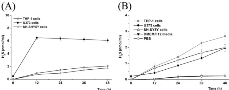
and ACS81) under extracellular conditions. We tested release from PBS, the DMEM/F12 culture medium, and human serum. We then compared this with the rate of uptake and cleavage intracellularly in THP-1, U373, and SH-SY-5Y cells (Fig. 2).
In PBS and the DMEM/F12 medium, H2S was released very slowly, reaching only 10% of the total amount available after 48 h (see Fig. 2B for ACS50 as a representative in DMEM/F12 medium and for ACS50 in PBS). Human serum caused a more rapid release with 50% of the potential being attained in 4.5–18 h. (ADT-OH, 4.5 h; ACS5,6 h; ACS 48, 6 h; ACS 50, 12 h; and ACS 81, 18 h) indicating the presence of weakly active cleaving enzymes in human serum. In contrast, NaSH, the model H2S donor, released its full potential immediately. It then showed a slow decay, declining by about 10% over 48 h (see supple-mental Fig. S1, B, D, and F), indicating minor decay of SH⫺ions. These data demonstrate that the H2S donating molecules are stable extracellularly, and, if administered in vivo, should be able to reach their target cells relatively intact.
Fig. 2 also indicates what happens when the H2S donors do reach their target cells. The data show intracellular levels of H2S in THP-1, SH-SY5Y, and U373 cells after being exposed to 10 Mof the H2S donors. H2S generated from the donor molecules slowly increased in the cytoplasm of THP-1 and SH-SY5Y cells over 48 h. Fig. 2 shows data from ACS50 as a representative of the S-donor compounds. All the H2S-donor compounds gave highly similar results. For U373 cells, the conversion was more robust than for THP-1 and SY-SY5Y cells with a maximum being reached by 12 h, after which there was a slow decline (Fig. 2A). The intracellular H2S levels reflect the net effect of at least three mechanisms: uptake of donors into the cells; intracellular cleavage of the molecules; and intracellular metabolism of the H2S generated. The other H2S-releasing compounds ACS48, ACS5, and ACS81 showed the same release kinetics in all cells tested (seesupplemental Fig. S1), as did the S-DOPA derivative ACS83 (seesupplemental Fig. S2).
These data establish that all the moieties are metabolized within each cell type to generate H2S. To explore a possible mechanism, we purified functional mitochondria from U373 TABLE 1
Levels of intact molecule and its metabolites in plasma and brain 1 h after iv rat administration of 40 mg/kg ACS 84
Plasma Brain
ACS84 ACS84-a ACS50 ACS84 ACS84-a ACS50
Mmean⫾ S.E. Mmean⫾ S.E.
1.1137⫾ 0.0867a 6.7267⫾ 0.8933 58.9333⫾ 3.6988 0.1230⫾ 0.0227 0.3497⫾ 0.0568 1.3227⫾ 0.2476 a
Values are mean⫾ S.E., n ⫽ 4.
TABLE 2
Dopamine,L-DOPA, and GSH levels in brain and/or plasma 1 h after iv rat administration of 40 mg/kg ACS84 or an equimolar dose ofL-DOPA Values are mean⫾ S.E., n ⫽ 4. One-way ANOVA test was carried out to evaluate the significance of differences.
Treatments Plasma Brain
Dopamine L-DOPA Dopamine L-DOPA GSH
M M M M M
ACS84 2.17⫾ 0.52a 0 13.2⫾ 1.3a 0.277⫾ 0.2775a 2402⫾ 39.3a
L-DOPA 29.2⫾ 3.8b 10.2⫾ 2.8b 9.60⫾ 0.08b 2.00⫾ 0.775b 1479⫾ 34
Vehicle 0 0 5.97⫾ 0.6 0 1670⫾ 60
ap⬍ 0.01 for ACS84 group compared with vehicle group. bp⬍ 0.01 forL-DOPA group compared with ACS84 group.
at University of British Columbia, on May 28, 2010
www.jbc.org
cells. All mitochondria contain molecules which have a high reducing potential such as NADH, FADH2, and cytochromes.
As shown in Fig. 3, all the donor moieties, but not ( ⫺)-depre-nyl, a classical inhibitor of monoamine oxidase B, were metab-olized to release H2S within 60 min by the mitochondria. Very similar results were obtained with the four different S-DOPA compounds (seesupplemental Fig. S3). The data indicate a potential mechanism by which H2S is generated intracellularly from the donor moieties.
Within cells, the most important reducing agent is gluta-thione (GSH). To determine whether the H2S donors were affecting GSH levels, we measured [GSH]iin SH-SY5Y cells exposed to the four different S-DOPAs (10Meach) for 8 h.
Equal concentrations of L-DOPA and NaSH were used as negative and positive controls, respectively. The results are shown in Fig. 4A. Treatment with NaSH and the S-DOPAs increased [GSH]iin SH-SY5Y cells under normal conditions by ⬃1.5-fold (p ⬍ 0.01). We then exposed the cells for 1 day to conditioned medium from stimulated THP-1 cells (LPS/IFN␥
for 2 days) or U373 cells (IFN␥ for 2 days). This treatment caused a huge decrease in [GSH]i(⬃90% decrease, p ⬍ 0.01).
NaSH and the four H2S-releasing S-DOPAs significantly atten-uated this decrease (p⬍ 0.01). Nevertheless, the values were still significantly lower than those obtained from control media. These data establish that H2S generated from the donor com-pounds is significantly converted into the antioxidant [GSH]i and that this [GSH]i is significantly depleted by exposure to supernatants from glial cells that have received inflammatory stimulation.
Fig. 4, B and C demonstrates the changes in MAO A and B in SH-SY5Y cells in the presence of NaSH,L-DOPA, and the four
H2S-releasing S-DOPAs. SH-SY5Y cells express both MAO A and MAO B, the latter accounting for 75% of the total MAO activity. Treatment with NaSH or four different S-DOPAs (10 Meach) for 8 h reduced the activity of MAO B, but not MAO
A. There was no effect ofL-DOPA. The decreases were about
85% (p⬍ 0.01) after exposure of SH-SY5Y cells to media from unstimulated THP-1 or U373 cells and about 65% after expo-sure to medium from THP-1 or U373 cells stimulated for 24 h (p⬍ 0.01). These data establish that all the S-DOPAs are selec-tive MAO B inhibitors.
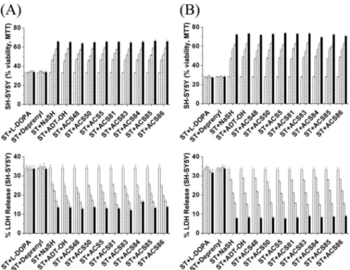
We next investigated the effects of 1, 3, 10, 30, and 50M
concentrations of the S-DOPA derivatives, as well as the H2S donors ADT-OH and NaSH on glial-mediated neurotoxicity. Fig. 5 shows the effect on SH-SY5Y viability of adding NaSH, ADT-OH,L-DOPA, (⫺)-deprenyl, ACS83, ACS84, ACS85, or ACS86 to LPS/IFN␥-activated THP-1 cells (5A) or IFN␥-acti-vated U373 cells (5B) at a standard concentration of 10M. It was found that the H2S-releasing agents ADT-OH and NaSH, but notL-DOPA or (⫺)-deprenyl, attenuated the neurotoxicity in an incubation time-dependent manner. This is shown both by the MTT assay (upper panels) and LDH assay (lower panels). The four S-DOPA derivatives ACS83, ACS84, ACS85, and ACS86 were comparably protective to NaSH and ADT-OH. Complete data showing the results at 1, 3, 10, 30, and 50M, and 2, 4, 8, and 12 h pretreatment are shown in supplemen-tal Figs. S4 and S5. Again, the results were highly similar for all FIGURE 2. Intracellular (A) and extracellular (B) H2S levels (nmol/ml) after treatment with 10MACS50 in the presence of 1 mMhydroxylamine. Values
are mean⫾ S.E., n ⫽ 4.
FIGURE 3. H2S levels (mol/g protein) generated by isolated functional
mitochondria in human U373 cells after treatment with 10MADT-OH, ACS48, ACS50, ACS5, ACS81, or (ⴚ)-deprenyl. Values are mean ⫾ S.E., n⫽ 4.
at University of British Columbia, on May 28, 2010
www.jbc.org
the SH-donors and all the S-DOPA derivatives in their concen-tration and incubation time dependence.
The 8-h time period shown in Fig. 5 demonstrated a robust effect of THP-1 and U373 cells, so it was deemed optimum for showing comparative effects using human microglia and astrocytes. It was found that the protective effect of pre-treatment with NaSH and the S-DOPAs (10M, 8 h) was comparable in human microglia and astrocytes to that found for THP-1 and U373 cells (Fig. 6A: microglia and 6B: astro-cytes). MTT data are shown in the upper panels and LDH release data in the lower panels. More detailed experimental data (3, 10, and 50M; 8 h of preincubation) showing com-parable effects at differing concentrations are shown in
supplemental Fig. S6.
Inflammatory stimulation of microglia or THP-1 cells causes them to release the inflammatory cytokines TNF␣ and IL-6, as well as to generate neurotoxic nitrite ions. Fig. 7 shows the effect of treatment with NaSH, ADT-OH, and the four S-DOPA compounds (10 M each, 8 h of preincubation) on THP-1
release of TNF␣ (Fig. 7A), IL-6 (Fig. 7C), and nitrite ions (Fig. 7E), and on human microglial release of TNF␣ (Fig. 7B), IL-6 (Fig. 7D) and nitrite ions (Fig. 7F). There was a substantially lower release of nitrite ions by stimulated microglia compared with THP-1 cells, even allowing for the 10-fold lower number of microglial cells that were seeded. This is perhaps related to the reported poor ability of human microglia to express iNOS. Nev-ertheless there were significant differences between the release of these materials in both types of cells: (1) between stimulated FIGURE 4. A, effect of direct treatment with NaSH,L-DOPA, ACS83, ACS84, ACS85, or ACS86 on GSH levels in SH-SY5Y cells. After SH-SY5Y cells were treated with NaSH,L-DOPA, ACS83, ACS84, ACS85, or ACS86 (10Meach) for 8 h and subsequently exposed to THP-1 or U373 conditioned medium (CM) for 1 day, they were extracted to measure GSH levels. B and C, effect of direct treatment with NaSH, ADT-OH (ADT),L-DOPA, ACS83, ACS84, ACS85, or ACS86 on activities of MAO A and MAO B in SH-SY5Y cells exposed to stimulated THP-1 or U373-conditioned medium (CM). After SH-SY5Y cells were treated with NaSH,L-DOPA, ACS83, ACS84, ACS85, or ACS86 (10Meach) for 8 h and subsequently exposed to THP-1 or U373 CM for 1 day, they were extracted to measure activities of MAO A (B) and MAO B (C). Notice that none of the compounds inhibited MAO A, but all of them significantly inhibited MAO B except forL-DOPA. Notice also that there was significantly less inhibition when the SH-SY5Y cells were exposed to CM. Values are mean⫾ S.E., n ⫽ 4. Two-way ANOVA was initially carried out to test the significance among the treatment groups. The significance of differences was then adjusted by applying the Bonferroni test for multiple comparisons. *, p⬍ 0.01 for NaSH or all the ACS-treated groups compared with untreated group; **, p⬍ 0.01 exposed to media from stimulated THP-1 or U373 compared with control media; and ***, p⬍ 0.01 exposed to stimulated THP-1 or U373 media treated with NaSH or all the ACS compounds compared with stimulated media only.
at University of British Columbia, on May 28, 2010
www.jbc.org
and unstimulated cells and (2) between stimulated cells that were untreated and stimulated cells that were treated with NaSH, ADT-OH, or any one of the S-DOPA com-pounds (p ⬍ 0.01). However, L-DOPA or (⫺)-deprenyl did not affect the release of any of these proinflammatory mediators (Fig. 7). Fig. 8 shows comparable data for IL-6 release from U373 cells (Fig. 8A) and cultured astrocytes (Fig. 8B). Cells were activated with IFN␥ as described under “Experimental Procedures” and were then treated similarly to the THP-1 and micro-glial cells as shown in Fig. 5. In both types of cells significant differences were found: (1) for the release of IL-6 from stimulated compared with unstimulated cells and (2) for stimulated cells treated with NaSH, ADT-OH, or the four S-DOPA compounds compared with stimu-lated cells that were untreated (p⬍ 0.01). Complete data showing a sim-ilar concentration dependence of the four donor moieties and the four S-DOPA derivatives on H2S release (3, 10, and 50M; 8 h pretreatment) are shown in supplemental Figs. S7 and S8. Supplemental Fig. S9
shows that the viability of THP-1, U373, microglia, and astrocytes was unaffected by any of the eight S-do-nating agents. Supplemental Fig. S10shows that the neuroprotective effects were additive when there was a combination of two S-DOPA derivatives (i.e. ACS 83⫹ 84, ACS 84⫹ 85, ACS 84 ⫹ 86) compared with the same derivatives alone. DISCUSSION
In the present study, we exam-ined the antioxidant and antiinflam-matory properties of four H2S donors (ACS48, ACS50, ACS5, and ACS81 as well as ADT-OH), and four L-DOPA hybrid compounds synthesized from the four donors (ACS83, ACS84, ACS85, and ACDS86). They all demonstrated therapeutic potential by being taken up by human microglia, and astro-cytes, as well as the humanTHP-1 U373 cell lines and then generating intracellular H2S. The H2S they gen-FIGURE 5. Effect on the viability of SH-SY5Y cells following treatment with supernatants from stimulated
THP-1 cells (A) or U373 cells (B) that had been exposed to 10Mof the neuroprotective moieties as described under “Experimental Procedures.” These were NaSH, the H2S-releasing dithiol-thione moiety ADT-OH, ACS48, ACS50, ACS5, ACS81, ACS83, ACS84, ACS85, or ACS86 pre-administered for (䡺, 0 h) (o, 2 h), (z, 4 h), (p, 8 h), and (f, 12 h).L-DOPA or (⫺)-deprenyl, which were without effect, served as the negative control and NaSH as the positive control. Notice that the SH donors were equally protective and were comparable to the the standard donor NaSH. Upper panels show MTT results whereas lower panels show LDH results. Values are mean⫾ S.E., n ⫽ 4. Two-way ANOVA was carried out to test the significance of differences. Multiple comparisons were followed with post-hoc Bonferroni tests. The time-dependent values were significantly different from control (p⬍ 0.01) for NaSH and each of the S-donating compounds.
FIGURE 6. Effect of treatment with NaSH, ADT-OH,L-DOPA, (ⴚ)-deprenyl, ACS48, ACS50, ACS5, ACS81, ACS83, ACS84, ACS85, or ACS86 (10Meach, 8 h of preincubation) on SH-SY5Y cell viability changes induced by activated human microglia (A) or activated astrocytes (B) as followed by MTT (upper panels) and LDH release (lower panels) assays. Values are mean⫾ S.E., n ⫽ 4. One-way ANOVA was carried out to test the significance of differences. Multiple comparisons were followed with post-hoc Bonferroni tests where appropriate. *, p⬍ 0.01 comparing the stimulated (ST) with the non-stimulated (NO-ST) group and **, p ⬍ 0.01 comparing stimulated (ST) group with the S-DOPA treatment groups.
at University of British Columbia, on May 28, 2010
www.jbc.org
erated acted in part to enhance levels of the classical antioxi-dant GSH. They also inhibited MAO B. Moreover, they acted as antiinflammatory compounds by ameliorating the neurotoxic effects of glial cell supernatants toward SH-SY5Y cells. Addi-tionally, they inhibited release of the proinflammatory media-tors TNF␣, IL-6, and nitrite ions (Figs. 7 and 8). The com-pounds were equipotent, butL-DOPA was without effect in all of these assays.
We have previously reported that depleting intracellular GSH by inhibiting its synthetic enzyme␥-glutamylcysteine syn-thase induces an inflammatory reaction in glial cells with
con-sequent neurotoxic effects (32). The compounds described here could help counteract the consequences of depressed GSH production. Our pilot in vivo data with ACS 84 showed that it reached the brain and was substantially metabolized as early as 1 h after iv administration to a rat. There was a more than a 2-fold increase in brain dopamine and a 1.4-fold increase in GSH. This dem-onstrates that a significant amount of the intact, and very lipophilic ACS84, crosses the blood brain bar-rier and is hydrolyzed in the brain into its two components L-DOPA and ACS50. Moreover the concom-itant inhibition of the dopamine-metabolizing enzyme MAO B, as had previously been shown for dithiolethiones (33), could contrib-ute to sustain the high concentra-tion of dopamine in brain. The increase of GSH presumably results from the H2S generated by metabo-lism of the donor moiety. While very preliminary, these data demon-strate that these L-DOPA hybrids may have therapeutic potential by enhancing dopamine and GSH levels.
Earlier studies have indicated that H2S is a reducing agent that can increase levels of intracellular gluta-thione (14) thereby inhibiting oxi-dative stress. It also decreases levels of peroxynitrite-derived nitrated proteins (34). Thus H2S might block oxidative stress-mediated cell death in PD by directly or indirectly detoxifying free radicals generated from oxidizedL-DOPA compounds. As far as neuroinflammation is concerned, it is well known that areas affected in PD, especially the substantia nigra, are characterized by the presence of activated micro-glia and activated astrocytes (4, 7–10, 35, 36). There is compa-rable glial activation in animal models of PD (37). Evidence that these reactive glial cells contribute to the neuronal degenera-tion comes from epidemiological studies where persons taking NSAIDs are reported to be relatively spared from PD (38, 39) especially if combined with coffee (40). Such protection is also reported for animal models of PD (37, 41).
The MPTP phenomena may be particularly revealing with respect to the potential consequences of inducing SN inflam-mation. Drug addicts who were originally exposed to MPTP developed a relentlessly progressive parkinsonian syndrome. FIGURE 7. Effect of treatment with NaSH, ADT-OH,L-DOPA, (ⴚ)-deprenyl, ACS48, ACS50, ACS5, ACS81,
ACS83, ACS84, ACS85, or ACS86 (10Meach, 8 h of preincubation) on the released levels of TNF␣ (A, B),
IL-6 (C, D) and nitrite ions (E, F) from THP-1 cells (A, C, E) or human microglia (B, D, F). Values are mean⫾ S.E., n⫽ 4. One-way ANOVA was carried out to test the significance of differences. Multiple comparisons were followed with post-hoc Bonferroni tests where appropriate. *, p⬍ 0.01 comparing the stimulated group with the non-stimulated group (NO-ST) and **, p⬍ 0.01 comparing the stimulated group (ST) with the S-DOPA treatment groups.
at University of British Columbia, on May 28, 2010
www.jbc.org
Postmortem studies showed persistent inflammation up to 17 years following their last exposure to the toxin (35). This human experience was duplicated in monkeys where inflam-mation of the SN was demonstrated up to 14 years after their last MPTP exposure (8). This suggests that inflammation of the SN, once initiated, may be self sustaining. If this is the case, antiinflammatory treatment may be essential to arresting pro-gression of PD.
Our study demonstrates that inflammatory stimulation of glial cells results in a 5–7-fold decrease in intracellular H2S (see
supplemental Fig. S11). This indicates an increase in intra-cellular consumption as reactive processes are induced. These experiments were performed in the presence of hydroxylamine to suppress endogenous H2S synthesis by CBS. But in a previous study (12) we showed that such inflammatory stimulation also sharply reduced the expression of CBS. Therefore inflamma-tory stimulation reduces intra-glial production of H2S while at the same time increasing consumption. The result is a dep-rivation of this endogenous anti-inflammatory and neuropro-tective agent.
There are studies demonstrating that serum levels of homo-cysteine, an intermediate of the trans-sulfuration pathway, were increased in PD patients receivingL-DOPA (42– 44). One of the main enzymes responsible for hyperhomocysteinemia is CBS, which is mainly expressed in astrocytes in brain (11, 12). So it could be hypothesized that endogenous production of H2S is reduced in PD brain, thus compounding the potential dam-age of oxidative stress associated withL-DOPA administration.
This provides additional rationale for designing methods of supplementing H2S availability in PD.
We have previously shown that SH-SY5Y cells express both MAO A and B (45). MAO B is primarily responsible for oxidative degradation of dopamine (46, 47). Treatment with NaSH and H2S-releasing moieties selectively inhibited MAO B (Fig. 4C). This could be one of the reasons why ACS84-treated rats have higher dopamine levels than vehicle or
L-DOPA injected rats (Table 2). However, we could not exclude
that dopamine is generated from degradation of ACS84 in rats. MAO B activity in SH-SY5Y cells was less inhibited in cells
exposed to the inflammatory effects of THP-1- or U373 cell-conditioned medium. This could be due to con-sumption of the generated H2S in stimulated cells, presumably through oxidative reactions.
In conclusion, our data demon-strate that H2S not only has anti-inflammatory activity against the glial toxicity which may be associ-ated with the pathogenesis of PD (36), but may also reduce the stress induced byL-DOPA oxidation (14). Furthermore, by inhibiting MAO B, H2S-releasingL-DOPA compounds could help restore the disease-depleted dopamine levels. The H2S-releasingL-DOPA derivatives (S-DOPAs) described here repre-sent compounds that can reach the brain and, in cells under stress, deliver ameliorating SH⫺ions in a time-dependent man-ner. They have properties that make them candidates for future treatment of PD. However the long term consequences of their use will require much further study.
REFERENCES
1. Fahn, S. (1991) Am. J. Clin. Nutr. 53, Suppl. 1, 380S–382S
2. Holloway, R. G., Shoulson, I., Fahn, S., Kieburtz, K., Lang, A., Marek, K., McDermott, M., Seibyl, J., Weiner, W., Musch, B., Kamp, C., Welsh, M., Shinaman, A., Pahwa, R., Barclay, L., Hubble, J., LeWitt, P., Miyasaki, J., Suchowersky, O., Stacy, M., Russell, D. S., Ford, B., Hammerstad, J., Riley, D., Standaert, D., Wooten, F., Factor, S., Jankovic, J., Atassi, F., Kurlan, R., Panisset, M., Rajput, A., Rodnitzky, R., Shults, C., Petsinger, G., Waters, C., Pfeiffer, R., Biglan, K., Borchert, L., Montgomery, A., Sutherland, L., Weeks, C., DeAngelis, M., Sime, E., Wood, S., Pantella, C., Harrigan, M., Fussell, B., Dillon, S., Alexander-Brown, B., Rainey, P., Tennis, M., Rost-Ruffner, E., Brown, D., Evans, S., Berry, D., Hall, J., Shirley, T., Dobson, J., Fontaine, D., Pfeiffer, B., Brocht, A., Bennett, S., Daigneault, S., Hodge-man, K., O’Connell, C., Ross, T., Richard, K., and Watts, A. (2004) Arch.
Neurol. 61,1044 –1053
3. Rascol, O., Brooks, D. J., Korczyn, A. D., De Deyn, P. P., Clarke, C. E., and Lang, A. E. (2000) N. Engl. J. Med. 342, 1484 –1491
4. Hald, A., and Lotharius, J. (2005) Exper. Neurol. 193, 279 –290
5. Kostrzewa, R. M., Kostrzewa, J. P., and Brus, R. (2002) Amino. Acids. 23, 57– 63
6. Jenner, P. (2008) Nat. Rev. Neurosci. 9, 665– 677
7. Hunot, S., and Hirsch, E. C. (2003) Ann. Neurol. 53, Suppl. 3, S49 –S60 8. McGeer, P. L., Schwab, C., Parent, A., and Doudet, D. (2003) Ann. Neurol.
54,599 – 604
9. McGeer, P. L., and McGeer, E. G. (2008) Mov. Disord. 23, 474 – 483 10. Tansey, M. G., McCoy, M. K., and Frank-Cannon, T. C. (2007) Exp.
Neu-rol. 208,1–25
11. Kamoun, P. (2004) Amino Acids 26, 243–254
12. Lee, M., Schwab, C., Yu, S., McGeer, E. G., and McGeer, P. L. (2009)
Neurobiol. Aging 30,1523–1534
13. Yang, G., Wu, L., Jiang, B., Yang, W., Qi, J., Cao, K., Meng, Q., Mustafa, A. K., Mu, W., Zhang, S., Snyder, S. H., and Wang, R. (2008) Science 322, 587–590
14. Kimura, Y., and Kimura, H. (2004) FASEB. J. 18, 1165–1167
15. Nagai, Y., Tsugane, M., Oka, J., and Kimura, H. (2004) FASEB. J. 18, 557–559
16. Lee, S. W., Hu, Y. S., Hu, L. F., Lu, Q., Dawe, G. S., Moore, P. K., Wong, P. T., and Bian, J. S. (2006) Glia 54, 116 –124
17. Eto, K., Ogasawara, M., Umemura, K., Nagai, Y., and Kimura. H. (2002) FIGURE 8. Effect of treatment with NaSH, ADT-OH,L-DOPA, (ⴚ)-Deprenyl, ACS48, ACS50, ACS5, ACS81,
ACS83, ACS84, ACS85, or ACS86 (10Meach, 8 h of preincubation, protocol 1) on release of IL-6 from U373 cells (A) or human astrocytes (B). Values are mean⫾ S.E., n ⫽ 4. One-way ANOVA was carried out to test the significance of differences. Multiple comparisons were followed with post-hoc Bonferroni tests where appropriate. *, p⬍ 0.01 comparing the unstimulated (NO-ST) group with the stimulated group. **, p ⬍ 0.01 comparing the stimulated (ST) group with the S-DOPA-treated groups.
at University of British Columbia, on May 28, 2010
www.jbc.org
J. Neurosci. 22,3386 –3391
18. Hu, L. F., Wong, P. T., Moore, P. K., and Bian, J. S. (2007) J. Neurochem. 100,1121–1128
19. Lee, M., Sparatore, A., Del Soldato, P., McGeer, E. G., and McGeer, P. L. (2010) Glia 58, 103–113
20. Hu, L. F., Lu, M., Tiong, C. X., Dawe, G. S., Hu, G., and Bian, J. S. (2010) Aging Cell, in press
21. Li, L., Rossoni, G., Sparatore, A., Lee, L. C., Del Soldato, P., and Moore, P. K. (2007) Free Radic. Biol. Med. 42, 706 –719
22. Benavides, G. A., Squadrito, G. L., Mills, R. W., Patel, H. D., Isbell, T. S., Patel, R. P., Darley-Usmar, V. M., Doeller, J. E., and Kraus, D. W. (2007) Proc. Natl. Acad. Sci. U.S.A. 104,17977–17982
23. Adelaere, B., and Guemas, J. P. (1989) Sulfur Letters 10, 31–36
24. Lozac’h, N., and Mollier, Y. (1950) Bulletin de la Societe Chimique de France1243–1244
25. Dartigues, B., Cambar, J., Trebaul, C., Brelivet, J., and Guglielmetti, R. (1980) Eur. J. Med. Chem. 15, 405– 412
26. Muzzi, C., Bertocci, E., Terzuoli, L., Porcelli, B., Ciari, I., Pagani, R., and Guerranti, R. (2008) Biomed. Pharmacother. 62, 253–258
27. Giustarini, D., Dalle-Donne, I., Milzani, A., and Rossi, R. (2009) FEBS J. 276,4946 – 4958
28. Klegeris, A., Giasson, B. I., Zhang, H., Maguire, J., Pelech, S., and McGeer. P. L. (2006) FASEB. J. 20, 2000 –2008
29. Klegeris, A., Walker, D. G., and McGeer, P. L. (1999) Neuropharmacology 38,1017–1025
30. Frezza, C., Cipolat, S., and Scorrano, L. (2007) Nature Protocols 2, 287–295 31. Hissin, P. J., and Hilf, R. (1976) Anal. Biochem. 74, 214 –226
32. Lee, M., Cho, T., Jantaratnotai, N., Wang, Y. T., McGeer, E. G., and Mc-Geer, P. L. (2010) FASEB J., in press
33. Drukarch, B., Flier, J., Jongenelen, C. A., Andringa, G., and Schoffelmeer, A. N. (2006) J. Neural Transm. 113, 593–598
34. Whiteman, M., Armstrong, J. S., Chu, S. H., Jia-Ling, S., Wong, B. S., Cheung, N. S., Halliwell, B., and Moore, P. K. (2004) J. Neurochem. 90, 765–768
35. Langston, J. W., Forno, L. S., Terrud, J., Reeves, A. G., Kaplan, J. A., and Karluk, D. (1999) Ann. Neurol. 46, 598 – 605
36. Whitton, P. S. (2007) Br. J. Pharmacol. 150, 963–976
37. Asanuma, M., and Miyazaki, I. (2008) Curr. Pharm. Des. 14, 1428 –1434 38. Chen, H., Jacobs, E., Schwarzschild, M. A., McCullough, M. L., Calle, E. E.,
Thun, M. J., and Ascherio, A. (2005) Ann. Neurol. 58, 963–967 39. Wahner, A. D., Bronstein, J. M., Bordelon, Y. M., and Ritz, B. (2007)
Neu-rology 69,1836 –1842
40. Powers, K. M., Kay, D. M., Factor, S. A., Zabetian, C. P., Higgins, D. S., Samii, A., Nutt, J. G., Griffith, A., Leis, B., Roberts, J. W., Martinez, E. D., Montimurro, J. S., Checkoway, H., and Payami, H. (2008) Mov. Disord. 23, 88 –95
41. Esposito, E., Di Matteo, V., Benigno, A., Pierucci, M., Cresscimanno, G., and Di Giovanni, G. (2007) Exp. Neurol. 205, 295–312
42. Postuma, R. B., and Lang, A. E. (2004) Neurology 63, 886 – 891 43. Łowicka, E., and Bełtowski, J. (2007) Pharmacol. Rep. 59, 4 –24 44. Zoccolella, S., dell’Aquila, C., Abruzzese, G., Antonini, A., Bonuccelli, U.,
Margherita-Canesi, M., Cristina, S., Marchese, R., Pacchetti, C., Zagaglia, R., Logroscino, G., Defazio, G., Lamberti, P., and Livrea, P. (2009) Mov. Disord. 24,1028 –1033
45. Klegeris, A., and McGeer, P. L. (2000) Exp Neurol. 166, 458 – 464 46. Damier, P., Kastner, A., Agid, Y., and Hirsch, E. C. (1996) Neurology 46,
1262–1269
47. Nagatsu, T., and Sawada, M. (2006) J Neural Transm Suppl. 71, 53– 65
at University of British Columbia, on May 28, 2010
www.jbc.org