Int. J. Mol. Sci. 2018, 19, x; doi: FOR PEER REVIEW www.mdpi.com/journal/ijms Article
1
Effect of Cyclic Stretch on Vascular Endothelial Cells
2
and Abdominal Aortic Aneurysm (AAA): Role in the
3
Inflammatory Response.
4
Martina Ramella1, Giulia Bertozzi1, Luca Fusaro1,2, Maria Talmon1, Marcello Manfredi3,5, Marta
5
Calvo Catoria1,2, Francesco Casella4, Carla M. Porta4, Renzo Boldorini1, Luigia G. Fresu1, Emilio
6
Marengo5, Francesca Boccafoschi1,2*
7
1 Department of Health Science, University of Piemonte Orientale (UPO), via Solaroli 17, Novara, Italy.
8
2 TissueGraft s.r.l. Spin-off of University of Piemonte Orientale (UPO), via Canobio 4/6, Novara, Italy.
9
3 ISALIT s.r.l. Spin-off of DISIT, University of Piemonte Orientale (UPO), Alessandria, Italy,
10
4 Vascular Surgery Unit, Ospedale Maggiore della Carità, Novara, Italy.
11
5 Department of Sciences and Technological Innovation, University of Piemonte Orientale (UPO),
12
Alessandria, Italy,
13
* Correspondence: Prof. Francesca Boccafoschi, via Solaroli 17, 28100 Novara, Italy. E-mail:
14
[email protected]; telephone number +39 0321-660556.
15
Received: date; Accepted: date; Published: date
16
Abstract: Abdominal aortic aneurysm (AAA) is a focal dilatation of the aorta, caused by both genetic
17
and environmental factors. Although vascular endothelium plays a key role in AAA progression,
18
the biological mechanisms underlying the mechanical stress involvement are only partially
19
understood. In this study, we developed an in vitro model to characterize the role of mechanical
20
stress as a potential trigger of endothelial deregulation in terms of inflammatory response bridging
21
between endothelial cells (ECs), inflammatory cells, and matrix remodeling. In AAA patients, data
22
revealed different degrees of calcification, inversely correlated with wall stretching and also with
23
inflammation and extracellular matrix degradation. In order to study the role of mechanical
24
stimulation, endothelial cell line (EA.hy926) has been cultured in healthy (10% strain) and
25
pathological (5% strain) dynamic conditions using a bioreactor. In presence of TNF-α, high levels of
26
MMP-9 expression and inflammation are obtained, while mechanical stimulation significantly
27
counteracts the TNF-α effects. Moreover, physiological deformation also plays a significant role in
28
the control of the oxidative stress. Overall our findings indicate that, due to wall calcification, in
29
AAA there is a significant change in terms of decreased wall stretching.
30
Keywords: cardiovascular diseases; abdominal aortic aneurysm; oxidative stress; inflammation;
31
calcification; cyclic stretch
32
33
1. Introduction
34
Abdominal aortic aneurysm (AAA) is a degenerative disease caused by permanent dilatation of
35
the aorta in the abdominal infrarenal tract. [1] AAA annual incidence is 0.4%-0.67% in western
36
population [2–4], while the prevalence is 4%-8% [5–7], and it is more common in men than in women.
37
Although the exact aetiology of AAA is unknown, there are several risk factors related to AAA
38
development such as male gender, age (≥ 50 years old), smoking habits, atherosclerosis and
39
hypertension, and some genetic factors. [8,9] Aneurysm can develop slowly, even silently and
40
asymptomatically until the rupture occurs, causing massive haemorrhage with an elevated risk of
41
death due to hypovolemic and haemorrhagic shock. [10] Extracellular matrix (ECM) degradation and
42
oxidative stress represent hallmarks of AAA progression. [11] Calcification is commonly found
within the aneurysm wall and leading to wall stiffening, and eventually to its rupture. [12] Moreover,
1
increased mechanical stresses due to turbulent flow within the wall contribute to AAA progression
2
and rupture. [13] The ability of a vascular wall to relax and passively contract depending on pressure
3
changes and blood flow is a physiological characteristic of large elastic arteries, and it is defined as
4
“compliance”. Bloodstream in the vascular compartment follows the laws of laminar flow; laminar
5
flow is altered by the reduction of flow velocity (blood stasis) as well as by the fluctuation of flow
6
(turbulence). The classical turbulent flow causes endothelial damage as it leads to the generation of
7
flows that are contrary to the direction of the circulatory current, also generating pockets of stasis.
8
[14] In arterial aneurysms, especially those with saccular morphology, there may be a slowing down
9
of flow until blood stasis.
10
Blood flow in the arterial aneurysm follows La Place's law, which explains how the parietal
11
tension (T) depends on the transmural pressure (Ptm), the wall thickness (d) and the radius of the
12
container (r) according to the equation:
13
T= (Ptm*r)/d.
14
If the volume increases, the parietal voltage increases. In fact, if the vessel expands, the increase
15
in the radius coupled with the decrease in the wall thickness increases the tension required to
16
counteract the transmural pressure. Blood vessels function as viscoelastic tubes and respond to a
17
transmural pressure gradient as a function of blood vessel wall composition. As vessel wall is
18
subjected to a transmural pressure gradient, a portion of the intraluminal energy is used to stretch
19
the fibers within the wall. The energy stored within the blood vessel fibers is later released back into
20
the system, upon closure of the aortic valve. As intraluminal pressure oscillates, the constant loading
21
and unloading of the fibers in the vessel wall result in a change in diameter of the blood vessel, which
22
is noted clinically as a palpable pulse. Although vascular ECs and vascular smooth muscle cells
23
(vSMCs) are exposed to both types of mechanical forces, shear stress resulting from blood flow is
24
sensed mainly by ECs [15], whereas both ECs and vSMCs are subjected to cyclic stretch resulting
25
from pulsatile pressure. In pathological remodeling, ECs can be influenced on structural as well as
26
functional aspects. Firstly, they can change morphology, acquiring a bigger size and an irregular
27
shape; they can also lose most of their regulatory roles. Endothelial layer can become more
28
permeable, allowing the transit of several substances and vSMC infiltration. Endothelium shows
29
inflammatory features, and it is characterized by the hyperexpression of proinflammatory cytokines
30
(IL-1, IL-6, TNF-α), proinflammatory chemokines (IL-8, MCP-1, and regulated upon activation,
31
normal T-Cell expressed- and secreted- RANTES) [16,17] and cell adhesion molecules (CAMs) such
32
as selectins and integrins, [18] with a significant production of reactive oxygen species (ROS), in
33
particular hydrogen peroxide (H2O2), superoxide (O2-), and hydroxyl radical (.OH). [19,20]
34
Deregulation of NADPH oxidase (NOX), xanthine oxidase (XO), superoxide dismutase (SOD),
35
thioredoxin (TRX), and catalase results in extreme ROS production. [21,22] Moreover, ROS regulate
36
ECM remodeling, acting directly on matrix metalloproteinases (MMPs) up-regulation, activating
37
nuclear factor kB (NF-kB) and activator protein (AP-1). [23–25] The aim of this work is to clarify the
38
role of vascular wall stretching in the maintenance of vascular physiology reproducing in vitro the
39
pathological dilatation (static and 5%), due to calcification, and physiological (10%) cyclic (1 Hz)
40
stretch of the vessel wall, in order to study the effects of mechanical stress on ECs functionality in
41
terms of inflammation, matrix remodeling, and oxidative stress production.
42
2. Results
43
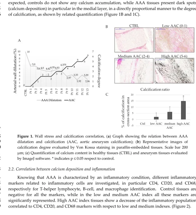
2.1. Relationship between wall stress and degree of calcification
44
Patient-specific AAA geometries are reconstructed, and structural analysis is performed to
45
calculate the wall stresses of the AAA models and their calcification. In figure 1A is shown how the
46
wall dilatation changes in relation to the amount of calcification. Retrospective analyses of literature
47
show that the aortic dilatation in healthy donors is 10% considering the ratio between systole and
48
diastole; this data is confirmed also by our measures on healthy controls. In fact, in presence of
aneurysm the dilatation is less than 10%, and it decreases when the calcification index increase
1
(Figure 1A). Control aortas and AAA sections are stained with Von Kossa to confirm the calcification
2
degrees obtained by the measurements with computed axial tomography (CAT) analysis. As
3
expected, controls do not show any calcium accumulation, while AAA tissues present dark spots
4
(calcium deposition) in particular in the medial layer, in a directly proportional manner to the degree
5
of calcification, as shown by related quantification (Figure 1B and 1C).
6
7
Figure 1. Wall stress and calcification correlation, (a) Graph showing the relation between AAA
8
dilatation and calcification (AAC, aortic aneurysm calcification); (b) Representative images of
9
calcification degree evaluated by Von Kossa staining in paraffin-embedded tissues. Scale bar 200
10
µ m; (c) Quantification of calcium content in healthy tissues (CTRL) and aneurysm tissues evaluated
11
by ImageJ software. * indicates p ≤ 0.05 respect to control.
12
2.2. Correlation between calcium deposition and inflammation
13
Knowing that AAA is characterized by an inflammatory condition, different inflammatory
14
markers related to inflammatory cells are investigated, in particular CD4, CD20, and CD68,
15
respectively for T-helper lymphocyte, B-cell, and macrophage identification. Control tissues are
16
negative for all the markers, while in the low and medium AAC index all these markers are
17
significantly represented. High AAC index tissues show a decrease of the inflammatory population
18
correlated to CD4, CD20, and CD68 markers with respect to low and medium indexes. (Figure 2).
1
Figure 2. Inflammatory cell infiltration in AAA. Representative immunohistochemistry for anti CD4,
2
CD8, CD68 staining. CD4+ is performed for T-helper lymphocytes, CD8+ for T-killer, and CD68 for
3
monocytes-macrophage. Healthy aorta, represented on the left, is negative for inflammatory cells
4
infiltration. Scale bar 200 µ m.
5
Other pro-inflammatory and calcification markers were investigated on tissue lysate, such as
6
MMP-9, IL-6 and osteopontin (Figure 3A and 3B). For all the considered markers, we obtain a
7
significant difference between patients and controls. Differences related to the degree of calcification
8
are appreciable: considering MMP-9, samples with medium index of calcification reach the higher
9
expression as observed by western blot assay as well as the higher proteolytic activity, as indicated
10
by zymography assay. The high calcification index indicates also the terminal phase of the
11
degradation, thus MMP-9 results decreased in terms of protein expression and proteolytic activity.
12
Considering IL-6 as an inflammatory marker, it decreases with the progression of calcium
13
accumulation confirming the previous data, particularly, it has the same trend as MMP-9. As
14
expected, patients with high calcification index have an increased expression of OPN, which has an
15
osteogenic activity (Figure 3B). No significant differences are observed between healthy donors and
16
patients in all AAC indices for MMP-2 activity (Figure 3A)
17
18
1
Figure 3. Matrix remodeling, inflammation and calcification in AAA tissues. (a) Gelatin zymography
2
performed on tissue lysates of patients with different degree of calcification and the respective
3
quantification of MMP-2 and MMP-9 activity; (b) Immunoblot on MMP-9, Il-6, and Osteopontin
4
(OPN) on tissue lysates of patients with different degree of calcification. The graph shows the relative
5
quantification. * statistically significant with respect to control p<0,05.
6
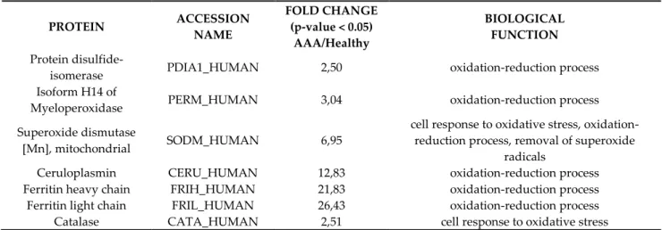
2.3. Oxidative stress proteins are overexpressed in AAA
7
The proteomic analysis of aortic abdominal aneurysms and control healthy vessel tissues was
8
performed with LC-MS in order to investigate the modulation of some proteins related to the
9
oxidative stress processes. Table 1 reports the identities, the modulation and the biological functions
10
of seven proteins resulted over expressed in the AAA respect with the healthy tissues. The
11
enrichment of all these proteins indicates that the oxidative stress pathway is strongly involved in
12
the AAA disease and that is particularly upregulated in the aortic abdominal vessel tissue.
13
Catalase (CATA) and Superoxide dismutase [Mn], mitochondrial (SODM) presented a fold
14
change of 2.51 and 6.95 respectively: these two proteins are involved in the cellular response to
15
oxidative stress pathway, as a result of the exposure to high levels of reactive oxygen species. SODM
16
is also involved in the removal of superoxide radicals and in oxidation-reduction processes.
17
Regarding the last biological function, we found that Protein disulfide-isomerase (PDIA1), Isoform
18
H14 of Myeloperoxidase (PERM), Ceruloplasmin (CERU), Ferritin heavy chain (FRIH), and Ferritin
19
light chain (FRIL) were all strongly upregulted (fold chnge of 2.50, 3.04, 12.83, 21.83 and 26.43) in the
20
AAA tissue.
21
22
Table 1. Proteomic analyses results.
1
PROTEIN ACCESSION NAME FOLD CHANGE (p-value < 0.05) AAA/Healthy BIOLOGICAL FUNCTION Proteindisulfide-isomerase PDIA1_HUMAN 2,50 oxidation-reduction process Isoform H14 of
Myeloperoxidase PERM_HUMAN 3,04 oxidation-reduction process Superoxide dismutase
[Mn], mitochondrial SODM_HUMAN 6,95
cell response to oxidative stress, oxidation-reduction process, removal of superoxide
radicals
Ceruloplasmin CERU_HUMAN 12,83 oxidation-reduction process Ferritin heavy chain FRIH_HUMAN 21,83 oxidation-reduction process Ferritin light chain FRIL_HUMAN 26,43 oxidation-reduction process Catalase CATA_HUMAN 2,51 cell response to oxidative stress
Table 1. Upregulated proteins in AAA vessel tissue after comparison to healthy tissues by proteomic
2
analysis. The upregulated proteins were selected using p value < 0.05.
3
A protein-protein interaction analysis (Figure 4) of oxidative stress-related proteins was
4
performed using STRING. The network analysis showed a high-connected network among these
5
proteins. Ferritin light chain and Ferritin heavy chain are co-expressed, showed evidence in
6
experimental and association in curated database. Also Superoxide dismutase, Catalase and Protein
7
disulfide-isomerase showed the same connections. The other linked proteins mainly showed
text-8
mining evidence.
9
10
Figure 4. STRING network analysis of oxidative stress-related proteins that are over expressed in
11
AAA vessel tissue respect with normal tissue. A proteome interactomic map was performed using
12
the STRING tool for obtaining cross correlation information. Homo sapiens was selected as a reference
13
organism. Different colored lines represent the existence of different types of evidence. A yellow line
14
indicates text-mining evidence; a purple line, experimental evidence, a cyan line indicates association
15
in curated database and black lines indicates co-expression data.
16
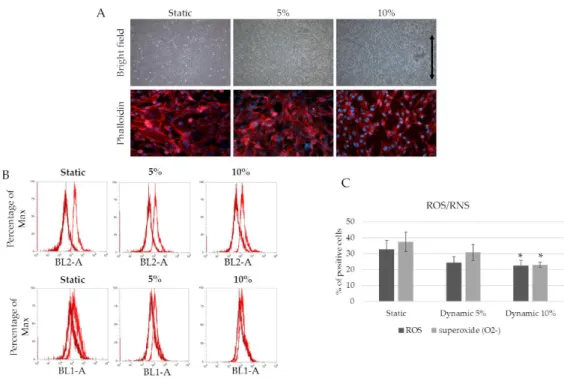
2.3. Mechanical stimulation drives ROS/Superoxide production in EA.hy926
17
Since mechanical stimulation seems to play a key role in driving the inflammatory response and
18
matrix remodeling, an in vitro dynamic model with physiological and pathological strain parameters,
19
culturing an endothelial cell line (EA.hy 926), was used. The effect of mechanical stimulation (5-10%
20
deformation, 1 Hz frequency) is evaluated in the presence and the absence of an inflammatory
21
stimulus (TNF-α 50 ng/ml) after three days of culture. Figure 5A shows cell morphology when 5%
22
and 10% mechanical strain is applied for 3 days. While no differences were detected in terms of cell
23
viability among the samples, 10% strain cultured cells display an elongated and oriented shape in the
strain direction, whereas random orientation is observed in the other samples (static and 5% strain).
1
In terms of ROS/RNS production, the difference between 10% strained cells (physiological) and 5%
2
strained or static cells (pathological) is confirmed, with lower ROS/RNS production in physiological
3
conditions (Figure 5B and 5C).
4
5
Figure 5. Mechanical strain drives ROS/RNS production. (a) EA.hy926 after 3 days of 5% and 10%
6
strain. Phalloidin was used to observe cell morphology; (b) FACS analyses for ROS and RNS
7
production. Dark red represents unstained cells, while light red represents the experimental samples.
8
BL2-A for superoxide detection; BL1-A for ROS detection. (c) graph of ROS/RNS production *
9
statistically significant with respect to static samples. p<0,05.
10
2.4. Strain affects CD62E expression and monocytes adhesion
11
As leukocytes adhesion on endothelium is promoted by CD62E expression on ECs membrane,
12
we evaluated the effect of mechanical stress on CD62E as well (Figure 6A). As expected, E-selectin is
13
upregulated in presence of TNF-α. Data show that the substrate deformation, both 5% and 10%
14
stretching at 1Hz, significantly and proportionally counteracts TNF-α effects on CD62E expression
15
(Figure 6A). In addition, data concerning monocytes adhesion nicely confirm these data. In fact, as
16
shown in Figure 6B, the mechanical deformation, at both 5% and 10% at 1Hz, significantly inhibits
17
the monocytes’ adhesion, also in presence of TNF-α.
1
Figure 6. Strain affect inflammation mediated by ECs (a) Representative immunofluorescence
2
staining for CD62E after mechanical (5 and 10%) and chemical (TNF-α 50ng/mL) stimulation. CD62E
3
is observed in red while DAPI is used for nuclear staining. Quantification of positive cells expressing
4
CD62E. Normalization of samples stimulated by TNF-α in relation to the respective control. Scale Bar
5
30 µm * p ≤ 0.05; (b) PBMCs adhesion on endothelial cells. PBMCs are observed in green.
6
Normalization of samples stimulated by TNF-α in relation to the respective control (static, 5% and
7
10%). Scale Bar 25 µm * p ≤ 0.05.
8
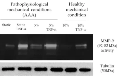
2.5. Strain affects MMP-9 expression and activity in ECs.
9
In static conditions, MMP-9 expression results strongly upregulated in presence of TNF-α.
10
Applying a 5% substrate deformation, no significant inhibition of TNF-α effects is shown, resulting
11
in an unmodified MMP-9 modulation (Figure 7), while no differences are found in absence of TNF-α
12
with respect to static conditions. When a 10% substrate deformation is applied, MMP-9 expression
13
results significantly downregulated in all conditions both in the absence and in the presence of
TNF-14
α.
15
16
Figure 7. Strain affect MMP-9 expression and activity (a) Representative zymography assay to detect
17
MMP-9 activity and expression after mechanical (5 and 10%) and chemical (TNF-α 50ng/mL)
18
stimulation. Tubulin is used for loading control.
19
3. Discussion
1
AAA is characterized by dramatic modifications of the medial layer, and it displays altered
2
mechanical behavior, inflammatory response, and matrix remodeling in the aortic wall. [26] The
3
calcification process has been acknowledged as a degenerative factor in inflammatory arterial
4
diseases. Calcium deposits show a reverse correlation with aortic dilatation and inflammatory cell
5
recruitment, as observed in our clinical data (results summarized in table 2). However, the specific
6
role of aortic dilatation in AAA progression is not completely elucidated. Taking together the results
7
on AAA patients, it is evident that the decrease in dilatation is related to the presence of vascular
8
calcifications in the medial layer. Moreover, the presence of calcification affects MMP-9-promoted
9
matrix remodeling. The severity of calcium deposition influences also IL-6-promoted inflammation,
10
because in patients with high AAC index, most of cells, including also the inflammatory ones, are
11
depleted and replaced by bone-like formation, resulting in a very low inflammatory infiltrate. Indeed,
12
the higher levels of inflammation and matrix remodeling are found in patients with medium
13
calcification index.
14
Inflammatory cell recruitment in AAA is sustained by the presence of inflammatory mediators
15
and by the increased expression of adhesion molecules able to interact with circulating inflammatory
16
cells. [27] Specifically, inflammatory cells infiltrate in the media and adventitia layers, inducing
17
oxidative stress and over expression of cytokines/chemokines and MMPs. The findings obtained by
18
proteomic analyses unveil also the involvement of oxidative stress in AAA patients, underlined by
19
the over expression of the proteins implied in oxidation-reduction process and in cell response to
20
oxidative stress. All these processes lead to elastic fiber breakdown, and depletion of vSMCs. As a
21
result, the aortic wall is weakened because of decreased thickness and reduced mechanical function.
22
The aortic wall cannot counteract the blood flow and pressure, and the aortic wall dilates to form
23
AAA. Among the inflammatory mediators, TNF-α plays a pivotal role in the initiation and
24
progression of vascular disorder by modulating the expression of molecules involved in vascular
25
tone, inflammation and remodeling, thus inducing endothelial dysfunction [28,29] and upregulating
26
the adhesion molecules, such as CD62E. An inflammatory condition, which leads to endothelial
27
dysfunction, contributes to the pathogenesis of vascular syndromes by predisposing vessels to plaque
28
rupture and intravascular thrombosis. [30] Due to endothelial dysfunctions, thus, ECs also contribute
29
to AAA progression. [31]
30
In our study, we have tested two different percentages of substrate deformation on endothelial
31
cell culture: 5% and 10% for 3 days at 1 Hz constant frequency, representing the resting heartbeat,
32
while the substrate deformation of 10% is selected to mimic the dilation of the aortic wall under
33
physiological conditions. [32] Our experimental settings are consistent with clinical findings on
34
healthy individuals and on AAA patients with medium calcification index. (table 2)
35
The results suggest that 10% stimulation controls inflammation and ROS/RNS production
36
compared to 5% dynamic and static samples; moreover, this experimental condition significantly
37
contrasts TNF-α-mediated inflammatory effects. Thus, these observations highlight the importance
38
of physiological vascular wall stretching as a powerful anti-inflammatory stimulus. Indeed, a
39
downregulation of MMP-9, CD62E, and a decrease of PBMC adhesion in 10% dynamic samples is
40
observed. In TNF-α-stimulated static samples, MMP-9, CD62E and PBMC adhesion increase,
41
demonstrating that these markers are closely related to the chronic inflammation. Overall, our
42
findings indicate the importance of physiological vascular wall stretching as a powerful
anti-43
inflammatory stimulus able to inhibit the pathological progression of AAA.
44
45
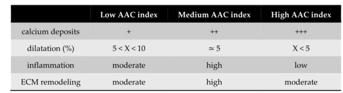
Table 2. Summary of obtained data.
1
Low AAC index Medium AAC index High AAC index
calcium deposits + ++ +++
dilatation (%) 5 < X < 10 ≃ 5 X < 5
inflammation moderate high low
ECM remodeling moderate high moderate
Table 2 shows the results obtained on AAA tissue in terms of calcium accumulation, dilatation,
2
inflammation value, and matrix remodeling.
3
4. Materials and Methods
4
4.1. Patients and healthy donors’ enrollment
5
Abdominal aortic aneurysm tissues were provided by the Vascular Surgery Unit, Hospital
6
Maggiore, Novara (Italy). AAA tissues were collected from 13 patients (100% male) subjected to open
7
surgical repair (OSR); demographical and clinical data are reported in Table 3. All data and samples
8
were collected from donors correctly informed for the use of excessive pathological material for
9
diagnostic and research purpose according to the local institute’s regulation and policies based on
10
Declaration of Helsinki (AVATAR, 1.0 – protocol 208/CE – CE 43/18). Peripheral blood from healthy
11
donors was collected from AVIS Novara in order to isolate PBMC’s (AVATAR, 1.0 – protocol 208/CE
12
– CE 43/18).
13
4.2. AAA patients: calcification modelling and wall dilatation
14
Calcificated regions are captured after the CAT exam. The amount of calcification was evaluated
15
through a score (aortic aneurysm calcification-AAC- from 0 to 8): AAC 0-1 are considered low
16
calcification index, AAC 2-3-4 are medium index, and AAC 5-6 high index [13]. Geometries of AAAs
17
are reconstructed, and images of the abdominal aorta are obtained from immediately distal to the
18
renal arteries to immediately proximal to the iliac bifurcation during doppler ultrasound
19
examination. Maximum AAA diameter, determined by B-MODE doppler ultrasound, is 72 mm. The
20
values of aneurysm dilatation are obtained using the following formula:
21
𝑨𝒐𝒓𝒕𝒊𝒄 𝒏𝒆𝒄𝒌 𝒔𝒚𝒔𝒕𝒐𝒍𝒆 − 𝒂𝒐𝒓𝒕𝒊𝒄 𝒏𝒆𝒄𝒌 𝒅𝒊𝒂𝒔𝒕𝒐𝒍𝒆 𝒂𝒐𝒓𝒕𝒊𝒄 𝒏𝒆𝒄𝒌 𝒅𝒊𝒂𝒔𝒕𝒐𝒍𝒆 : 𝒂𝒏𝒆𝒖𝒓𝒚𝒔𝒎 𝒔𝒚𝒔𝒕𝒐𝒍𝒆 − 𝒂𝒏𝒆𝒖𝒓𝒚𝒔𝒎 𝒅𝒊𝒂𝒔𝒕𝒐𝒍𝒆 𝒂𝒏𝒆𝒖𝒓𝒚𝒔𝒎 𝒅𝒊𝒂𝒔𝒕𝒐𝒍𝒆 = 𝟎. 𝟏: 𝒙22
Differences between systole and diastole of the aortic neck (healthy part) and the aneurysm are
23
normalized with their respective diastole. The proportion is obtained comparing the differences with
24
10% that is the physiological measured dilatation of the healthy aorta.
25
26
Table 3. Demographical and clinical feature of AAA patients.
1
Patients Age mean±S D Gende r DAAA mean±S D Hypercholesterolem ia Smoking Hypertension Ischemic cardiomyopath y Low AAC 72±4 Male 100% 5.6±1.4 100% 33% 100% 66% Medium AAC 75±6 Male 100% 5.4±1.3 50% 50% 88% 50% High AAC 71±12 Male 100% 5.3±0.6 100% 33% 100% 66%Table 3 shows demographical data (age, sex) and cardiovascular risk (DAAA aneurysm diameters,
2
hypercholesterolemia, smoking, hypertension, and ischemic cardiomyopathy). Age and DAAA are
3
represented as mean ± standard deviation. Patient data are divided by the grade of aortic calcification
4
index (AAC).
5
4.3. Histological analyses on human aortic samples
6
AAA and control samples were fixed in neutral buffered formalin for 24 hours, and 5 µ m-thick
7
sections were cut from paraffin-embedded tissues. Briefly, rehydrated sections were treated with a
8
1% aqueous silver nitrate solution (Sigma Aldrich, Italy). Silver is deposited replacing the calcium
9
reduced by the strong light, and thereby visualized as metallic silver. To counterstain the samples
10
was also used a 5% sodium thiosulfate solution and 0.1% nuclear fast red solution. Calcium deposits
11
and salts are detectable in black or brown-black, nuclei in red and cytoplasm in pink. All images were
12
acquired using Pannoramic MIDI 3DHISTECH and analyzed with Pannoramic Viewer software
13
(3DHISTECH, Hungary). For objective quantification of calcium content ImageJ software was used.
14
4.4. Proteomic analysis
15
Tissues obtained from AAA vessel and from healthy control vessel were lysed in RIPA buffer
16
(150 mM sodium chloride, 1% Triton X100, 0.5% sodium deoxycholate, 0.1% sodium dodecyl sulfate,
17
1 mM EDTA, 1 mM EGTA, 50 mM TRIS pH = 7.4) supplemented with protease inhibitors (0.2 mM
18
sodium othovanadate, 1 mM phenylmethyl sulfonyl fluoride and protease inhibitors cocktail, all
19
from Sigma, Italy). Proteins concentration was determined using the bicinchoninic acid assay (Pierce,
20
Rockford, IL, USA). Lysate proteins were digested using the following protocol: samples were
21
subjected to denaturation with TFE, to reduction with DTT 200 mM, alkylation with IAM 200 mM
22
and the complete protein trypsin digestion with 2 µg of Trypsin/Lys-C (Promega, Madison, WI,
23
USA). The peptide digests were desalted on the Discovery® DSC-18 solid phase extraction (SPE)
96-24
well Plate (25 mg/well) (Sigma-Aldrich Inc., St. Louis, MO, USA). Peptides were dried by Speed
25
Vacuum until the analysis.
26
LC–MS/MS analyses were performed on digests using a micro-LC Eksigent Technologies
27
(Dublin, USA) system with a stationary phase of a Halo Fused C18 column (0.5 × 100 mm, 2.7 µm;
28
Eksigent Technologies, Dublin, USA). The injection volume was 4.0 µL and the oven temperature
29
was set at 40 °C. The mobile phase was a mixture of 0.1% (v/v) formic acid in water (A) and 0.1%
30
(v/v) formic acid in acetonitrile (B), eluting at a flow-rate of 15.0 µL min− 1 at an increasing
31
concentration of solvent B from 2% to 40% in 30 min. LC system was interfaced with a 5600 +
32
TripleTOF system (AB Sciex, Concord, Canada) equipped with a DuoSpray Ion Source and CDS
33
(Calibrant Delivery System). The relative abundance of proteins was obtained using the label-free
34
quantification. Samples were subjected to data-dependent acquisition (DDA): the mass spectrometer
35
analysis was performed using a mass range of 100–1500 Da (TOF scan with an accumulation time of
36
0.25 s), followed by a MS/MS product ion scan from 200 to 1250 Da (accumulation time of 5.0 ms)
37
with the abundance threshold set at 30 cps (35 candidate ions can be monitored during every cycle).
38
The samples were, then, subjected to cyclic data independent analysis (DIA) of the mass spectra,
39
using a 25-Da window: the mass spectrometer was operated such that a 50-ms survey scan (TOF-MS)
was performed and subsequent MS/MS experiments were performed on all precursors.20,21 The MS
1
data were acquired with Analyst TF 1.7 (AB SCIEX, Concord, Canada). Three instrumental replicates
2
for each sample were subjected to the DIA analysis.
3
Protein identification was performed using Mascot v. 2.4 (Matrix Science Inc., Boston, USA), the
4
digestion enzyme selected was trypsin, with 2 missed cleavages and a search tolerance of 50 ppm
5
was specified for the peptide mass tolerance, and 0.1 Da for the MS/MS tolerance. The charges of the
6
peptides to search for were set to 2 +, 3 + and 4 +, and the search was set on monoisotopic mass. The
7
instrument was set to ESI-QUAD-TOF and the following modifications were specified for the search:
8
carbamidomethyl cysteines as fixed modification and oxidized methionine as variable modification.
9
The UniProt Swiss-Prot reviewed database containing human proteins (version 2015.07.07,
10
containing 42131 sequence entries) was used and a target-decoy database search was performed.
11
False Discovery Rate was fixed at 1%. The label-free quantification was carried out with PeakView
12
2.0 and MarkerView 1.2. (ABSCIEX, Concord, Canada). The up-regulated proteins were selected
13
using P value < 0.05 and fold change >1.5. The up-regulated proteins were analyzed by using STRING
14
software (http://string-db.org), which is a database of known and predicted protein-protein
15
interactions.
16
4.5. Dynamic cell culture
17
Human endothelial cells, EA.hy926 (ATCC® CRL-2922™) were cultured in high-glucose
18
Dulbecco’s modified Eagle’s medium (DMEM) enriched with 10% fetal bovine serum and penicillin
19
(100 U/mL), streptomycin (100 µ g/mL), and 2 mM glutamine mixture (all from Euroclone, Italy) at
20
37°C in humid 5% CO2 atmosphere. Rectangular silicone pieces (3cm x 1.5cm) were cut and sterilized
21
together with the culture chambers of the TC-3 bioreactor (Ebers Medical, Zaragoza, Spain). To
22
facilitate cell adhesion to the silicone substrate (both static controls and dynamic samples) a type I
23
collagen (50 µg/ml) coating was used. After 1 hour, the coating has been rinsed with sterile water to
24
remove the exceeding collagen. Finally, Ea.hy926 (2*104 /cm2) were seeded, and after 24 hours
25
(required for an optimal adhesion to the substrate), mechanical stimulation has been applied to the
26
cell culture. A stretching of 5 and 10% was maintained for 72 hours, 50ng/mL of TNF-α (Sigma
27
Aldrich, Italy) was added when required. Silicone controls were maintained under static conditions.
28
4.6. Phalloidin staining
29
Cells were fixed in formalin 4% and incubated with phalloidin TRITC (Sigma Aldrich, Italy) for
30
45’ at 37°C. DAPI (Sigma Aldrich, Italy) was used for nuclear staining. Samples were observed at
31
fluorescent microscope (DM2500 Leica, Germany).
32
4.7. Cellular ROS/Superoxide Detection Assay kit
33
Oxidative stress production was investigated through Cellular ROS/Superoxide detection assay
34
kit (ab139476, Abcam, Italy) following the manufacturer’s protocol. Briefly, detached cells (static
35
controls, 5%, and 10% of mechanical stimulation) were stained with oxidative stress reagent orange
36
and green, then flow cytometry analyses were performed (ATTUNE NxT Cytometer, Invitrogen) and
37
analysed by ATTUNE NxT flow cytometer software. Pyocyanin treated cells (400µ M) were used as
38
positive control.
39
4.8. Immunofluorescence
40
Immunofluorescence analyses were carried out on mechanically stimulated silicone cells and on
41
static controls. 1*104 EA.hy926/cm2 were seeded and maintained in culture according to experimental
42
protocols. After 3 days of stimulation, samples were fixed for 1 hour with 4% formalin. Samples were
43
blocked for 1 hour with a 5% goat solution and 0.3% TRITON X-100 in PBS 1X and, then, incubated
44
with the primary antibody (1:50 anti-E-selectin, Santa Cruz Biotechnology, USA) for 1 hour at room
45
temperature. E-selectin is detected by a secondary antibody TRITC-conjugated (Perkin-Elmer, Italy)
46
and observed at fluorescent microscope (DM2500 Leica, Germany). The images were acquired using
LAS software (Leica, Germany). Data are expressed as TNF-alpha-treated versus the respective
1
untreated samples ratio.
2
4.9. Leukocyte-endothelium adhesion assay
3
Peripheral blood mononuclear cells (PBMCs) were isolated with Histopaque® -1077 (Sigma
4
Aldrich, Italy) from peripheral blood obtained by healthy donors. PBMCs adhesion assay was
5
performed using Cell Biolabs’ CytoSelectTM Leukocyte-endothelium Adhesion Assay (Cell Biolabs
6
Inc, USA). After mechanical stimulations, PBMCs were labeled by the LeukoTrackerTM solution.
7
Labeled PBMCs were then incubated with static and dynamic cells in presence or not of TNF-α
8
(50ng/mL). After 1 h of incubation, nonadherent cells were removed by gently rinsing with PBS.
9
Adherent cells were counted in three separate fields using an inverted fluorescence microscope
10
(DM2500 Leica, Germany). Data are expressed as TNF-alpha-treated versus the respective untreated
11
samples ratio.
12
4.10. Western Blot
13
Culture cells and tissues were lysed in RIPA buffer supplemented with protease inhibitors.
14
Proteins concentration was determined using the bicinchoninic acid assay (Pierce, Rockford, IL,
15
USA). 50µg total proteins in sample buffer (62.5 mM Tris-HCl, pH 6.8, 20% glycerol, 5%
β-16
mercaptoethanol, 0.5% bromophenol blue) were resolved to SDS-PAGE and transferred to a
17
nitrocellulose membrane (Amersham Biosciences, Buckinghamshire, UK). Membranes were
18
incubated overnight with IL-6, OPN (Abcam, UK), MMP-9, Tubulin (Millipore, Italy) antibodies at
19
4°C. Proteins were revelated with secondary antibody-peroxidase conjugates (Perkin-Elmer, Italy).
20
Protein bands were visualized using ECL (Perkin-Elmer, Western lightning PLUS-ECL, Italy)
21
detection reagents in a chemosensitive visualizer (VersaDoc, BioRad, Italy). In order to check the
22
loaded proteins concentration, red ponceau (Sigma Aldrich, Italy) staining was considered.
23
4.11. Zymography assay
24
Non-reduced protein samples were resolved by SDS-PAGE gels containing gelatin (0.2%, Sigma
25
Aldrich, Italy). Briefly, after electrophoresis, gels were incubated with TRITON X-100 for 3 h at room
26
temperature, and then incubated in a solution of CaCl2 (1 mM) and NaCl (15 mM), pH 7.4 overnight
27
at 37°C. Subsequently, gels were fixed and then stained with Coomassie Blue. For objective
28
quantification ImageJ software was used.
29
4.12. Statistical analyses
30
All experiments were performed in triplicate. All data are expressed as mean values ± standard
31
deviation. Using Student's t-test, the p-value is calculated and the differences between variables with
32
a value of p <0.05 are considered statistically significant.
33
5. Conclusions
34
In conclusion, we found a negative correlation between calcium deposits and wall dilatation in
35
presence of AAA. A decreased wall stretching, due to the presence of calcification, affects
MMP-9-36
mediated matrix degradation and IL-6-mediated inflammation. As expected, in vitro model on
37
endothelial cell line shows that substrate deformation significantly regulates the inflammatory
38
response and matrix remodeling.
39
Author Contributions: Conceptualization: M.R., G.B., M.T., M.M., F.B.; Methodology: M.R., G.B., M.M., M.T.,
40
F.C., Validation: F.B., L.G.F.; Investigation, M.R., G.B., M.T., L.F., M.M., M.C., F.C., F.B.; Data Curation, M.R.,
41
G.B., M.T., F.C.; Writing – Original Draft Preparation: M.R., G.B.; Writing – Review & Editing: L.F., M.T., M.M.,
42
M.C., C.P., R.B., L.G.F., E.M., F.B.; Supervision: C.P., R.B., L.G.F., E.M., F.B.; Project Administration: R.B., L.G.F.,
43
F.B.; Funding Acquisition: R.B., L.G.F., E.M., F.B.
44
Funding: Part of this research was funded by TissueGraft s.r.l.
Acknowledgments: The authors would like to thank Dr. Carlino and Prof. Mascaro from AVIS Novara for blood
1
collection.
2
Conflicts of Interest: The authors declare no conflict of interest
3
Abbreviations
4
AAA Abdominal Aortic Aneurysm ECs Endothelial cells
TNF-α Tumor necrosis factor alpha MMP-9 Matrix metalloproteinase -9 ECM Extracellular matrix
PTM transmural pressure
vSMCs Vascular smooth muscle cells IL-1 Interleukin-1
IL-6 Interleukin-6 IL-8 Interleukin-8
MCP-1 Monocyte chemoattractant protein 1
RANTES Regulated on activation, normal T cell expressed and secreted CAMs Cell adhesion molecules
ROS Reactive oxygen species
NADPH Nicotinamide adenine dinucleotide phosphate NOX Nicotinamide adenine dinucleotide phosphate oxidase XO Xanthine oxidase
SOD Superoxide dismutase TRX Thioredoxin
MMPs Matrix metalloproteinases
NF-kB Nuclear factor kappa-light-chain-enhancer of activated B cells AP-1 Activator protein 1
OPN Osteopontin
CAT Computed axial tomography AAC Aortic aneurysm calcification RNS Reactive nitrogen species APOE Apoliprotein E
OSR Open surgical repair
DAAA Diameter of abdominal aortic aneurysm TFE Trifluoroethanol
DTT Dithiothreitol IAM Iodoacetamide
LC-MS/MS Liquid chromatography– tandem mass spectrometry CDS Calibrant Delivery System
DDA Data-dependent acquisition TOF Time of flight
DIA Data independent analysis
DMEM Dulbecco’s modified Eagle’s medium TRITC Tetramethylrhodamine
DAPI 4′,6-Diamidine-2′-phenylindole dihydrochloride RIPA Radioimmunoprecipitation buffer
EDTA Ethylenediamenetetraacetic acid
EGTA Ethyleneglycol-bis(2-aminoethylether)-N,N,N',N'-tetraacetic acid SDS-PAGE Sodium dodecyl sulfate polyacrylamide gel electrophoresis PBMCs Peripheral blood mononuclear cells
PBS Phosphate Buffered Saline
References
1
1. Moxon, J. V.; Parr, A.; Emeto, T.I.; Walker, P.; Norman, P.E.; Golledge, J. Diagnosis and Monitoring of
2
Abdominal Aortic Aneurysm: Current Status and Future Prospects. Curr. Probl. Cardiol. 2010, 35, 512–548,
3
doi:10.1016/j.cpcardiol.2010.08.004.
4
2. Forsdahl, S.H.; Singh, K.; Solberg, S.; Jacobsen, B.K. Risk factors for abdominal aortic aneurysms: a 7-year
5
prospective study: the tromsø study, 1994 2001. Circulation 2009, 119, 2202–2208,
6
doi:10.1161/CIRCULATIONAHA.108.817619.
7
3. Vardulaki, K.A.; Prevost, T.C.; Walker, N.M.; Day, N.E.; Wilmink, A.B.M.; Quick, C.R.G.; Ashton, H.A.;
8
Scott, R.A.P. Incidence among men of asymptomatic abdominal aortic aneurysms: Estimates from 500
9
screen detected cases. J. Med. Screen. 1999, 6, 50–54, doi:10.1136/jms.6.1.50.
10
4. Lederle, F.A.; Johnson, G.R.; Wilson, S.E.; Littooy, F.N.; Krupski, W.C.; Bandyk, D.; Acher, C.W.; Chute,
11
E.P.; Hye, R.J.; Gordon, I.L.; et al. Yield of repeated screening for abdominal aortic aneurysm after a 4-year
12
interval. Arch. Intern. Med. 2000, 160, 1117–1121, doi:10.1001/archinte.160.8.1117.
13
5. Scott, R.A.P.; Ashton, H.A.; Buxton, M.J.; Day, N.E.; Kim, L.G.; Marteau, T.M.; Thompson, S.G.; Walker,
14
N.M. The Multicentre Aneurysm Screening Study (MASS) into the effect of abdominal aortic aneurysm
15
screening on mortality in men: A randomised controlled trial. Lancet 2002, 360, 1531–1539,
16
doi:10.1016/S0140-6736(02)11522-4.
17
6. Lindholt, J.S.; Juul, S.; Fasting, H.; Henneberg, E.W. Screening for abdominal aortic aneurysms: Single
18
centre randomised controlled trial. Br. Med. J. 2005, 330, 750–752, doi:10.1136/bmj.38369.620162.82.
19
7. Ashton, H.A.; Gao, L.; Kim, L.G.; Druce, P.S.; Thompson, S.G.; Scott, R.A.P. Fifteen-year follow-up of a
20
randomized clinical trial of ultrasonographic screening for abdominal aortic aneurysms. Br. J. Surg. 2007,
21
94, 696–701, doi:10.1002/bjs.5780.
22
8. Nordon, I.M.; Hinchliffe, R.J.; Loftus, I.M.; Thompson, M.M. Pathophysiology and epidemiology of
23
abdominal aortic aneurysms. Nat. Rev. Cardiol. 2011, 8, 92–102, doi:10.1038/nrcardio.2010.180.
24
9. Rodella, L.F.; Rezzani, R.; Bonomini, F.; Peroni, M.; Cocchi, M.A.; Hirtler, L.; Bonardelli, S. Abdominal
25
aortic aneurysm and histological, clinical, radiological correlation. Acta Histochem. 2016, 118, 256–262,
26
doi:10.1016/j.acthis.2016.01.007.
27
10. Emeto, T.I.; Moxon, J. V; Biros, E.; Rush, C.M.; Clancy, P.; Woodward, L.; Moran, C.S.; Jose, R.J.; Nguyen,
28
T.; Walker, P.J.; et al. Urocortin 2 is associated with abdominal aortic aneurysm and mediates
anti-29
proliferative effects on vascular smooth muscle cells via corticotrophin releasing factor receptor 2. Clin Sci
30
2013, doi:10.1042/CS20130425.
31
11. Wassef, M.; Baxter, B.T.; Chisholm, R.L.; Dalman, R.L.; Fillinger, M.F.; Heinecke, J.; Humphrey, J.D.;
32
Kuivaniemi, H.; Parks, W.C.; Pearce, W.H.; et al. Pathogenesis of abdominal aortic aneurysms: A
33
multidisciplinary research program supported by the National Heart, Lung, and Blood Institute. J. Vasc.
34
Surg. 2001, 34, 730–738, doi:10.1067/mva.2001.116966.
35
12. Buijs, R.V.C.; Willems, T.P.; Tio, R.A.; Boersma, H.H.; Tielliu, I.F.J.; Slart, R.H.J.A.; Zeebregts, C.J.
36
Calcification as a risk factor for rupture of abdominal aortic aneurysm. Eur. J. Vasc. Endovasc. Surg. 2013,
37
46, 542–548, doi:10.1016/j.ejvs.2013.09.006.
38
13. Lederle, F.A.; Johnson, G.R.; Wilson, S.E.; Gordon, I.L.; Chute, E.P.; Littooy, F.N.; Krupski, W.C.; Bandyk,
39
D.; Barone, G.W.; Graham, L.M.; et al. Relationship of age, gender, race, and body size to infrarenal aortic
40
diameter. J. Vasc. Surg. 1997, 26, 595–601, doi:10.1016/S0741-5214(97)70057-0.
41
14. Chiu, J.-J.; Chien, S. Effects of dsisturbed flow on vascular endothelium: pathophysiological basis and
42
clinical perspectives. Physiol Rev 2011, 91, 327–387, doi:10.1152/physrev.00047.2009.
43
15. Davies, P.F. Flow-Mediated Endothelial Mechanotransduction. NIH Public Access 1995, 75, 519–560,
44
doi:10.1016/j.drudis.2011.09.009.
45
16. KOFLER, S.; NICKEL, T.; WEIS, M. Role of cytokines in cardiovascular diseases: a focus on endothelial
46
responses to inflammation. Clin. Sci. 2005, 108, 205–213, doi:10.1042/CS20040174.
47
17. Mackay, C.R. Chemokines: Immunology’s high impact factors. Nat. Immunol. 2001, 2, 95–101,
48
doi:10.1038/84298.
49
18. Golias, C.H.; Tsoutsi, E.; Matziridis, A.; Makridis, P.; Batistatou, A.; Charalabopoulos, K. Leukocyte and
50
endothelial cell adhesion molecules in inflammation focusing on inflammatory heart disease. In Vivo
51
(Brooklyn). 2007, 21, 757–770.
52
19. Tsutsui, H.; Kinugawa, S.; Matsushima, S. Oxidative stress and heart failure. Am. J. Physiol. Heart Circ.
53
Physiol. 2011, 301, H2181-90, doi:10.1152/ajpheart.00554.2011.
54
20. McCormick, M.L.; Gavrila, D.; Weintraub, N.L. Role of oxidative stress in the pathogenesis of abdominal
1
aortic aneurysms. Arter. Thromb Vasc Biol 2007, 27, 461–469, doi:10.1161/01.ATV.0000257552.94483.14.
2
21. Forstermann, U. Oxidative stress in vascular disease: causes, defense mechanisms and potential therapies.
3
Nat Clin Pr. Cardiovasc Med 2008, 5, 338–349, doi:10.1038/ncpcardio1211.
4
22. Forstermann, U. Nitric oxide and oxidative stress in vascular disease. Pflugers Arch 2010, 459, 923–939,
5
doi:10.1007/s00424-010-0808-2.
6
23. Parodi, F.E.; Mao, D.; Ennis, T.L.; Bartoli, M.A.; Thompson, R.W. Suppression of experimental abdominal
7
aortic aneurysms in mice by treatment with pyrrolidine dithiocarbamate, an antioxidant inhibitor of
8
nuclear factor-κB. J. Vasc. Surg. 2005, 41, 479–489, doi:10.1016/j.jvs.2004.12.030.
9
24. Costanzo, A.; Moretti, F.; Burgio, V.L.; Bravi, C.; Guido, F.; Levrero, M.; Puri, P.L. Endothelial activation by
10
angiotensin II through NFκB and p38 pathways: Involvement of NFκB-inducible kinase (NIK), free oxygen
11
radicals, and selective inhibition by aspirin. J. Cell. Physiol. 2003, 195, 402–410, doi:10.1002/jcp.10191.
12
25. Li, Q.; Engelhardt, J.F. Interleukin-1beta induction of NFkappaB is partially regulated by H2O2-mediated
13
activation of NFkappaB-inducing kinase. J Biol Chem 2006, 281, 1495–1505, doi:M511153200
14
[pii]\r10.1074/jbc.M511153200.
15
26. Ramella, M.; Bernardi, P.; Fusaro, L.; Manfredi, M.; Casella, F.; Porta, C.M.; Nicolai, L.; Galeazzi, E.; Boldorini,
16
R.; Settembrini, A,M.; Settembrini, P.; Marengo, E.; Cannas, M.; Boccafoschi, F. Relevance of inflammation
17
and matrix remodeling in abdominal aortic aneurysm (AAA) and popliteal artery aneurysm (PAA)
18
progression. Am J Transl Res 2018, 10(10), 3265-3275.
19
27. Wang, Q.; Ren, J.; Morgan, S.; Liu, Z.; Dou, C.; Liu, B. Monocyte Chemoattractant Protein-1 (MCP-1)
20
regulates macrophage cytotoxicity in abdominal aortic aneurysm. PLoS One 2014, 9,
21
doi:10.1371/journal.pone.0092053.
22
28. Palmieri, D.; Perego, P.; Palombo, D. Estrogen receptor activation protects against TNF-α-induced
23
endothelial dysfunction. Angiology 2014, 65, 17–21, doi:10.1177/0003319713477909.
24
29. Zhang, H.; Park, Y.; Wu, J.; Chen, X.; Lee, S.; Yang, J.; Dellsperger, K.C.; Zhang, C. Role of TNF-alpha in
25
vascular dysfunction. Clin Sci 2009, 116, 219–230, doi:CS20080196 [pii]\r10.1042/CS20080196.
26
30. Hadi, H.A.R.; Carr, C.S.; Al Suwaidi, J. Endothelial Dysfunction: Cardiovascular Risk Factors, Therapy,
27
and Outcom. Vasc Health Risk Manag 2005, 1(3), 183–198.
28
31. Ramella, M.; Boccafoschi, F.; Bellofatto, K.; Follenzi, A.; Fusaro, L.; Boldorini, R.; Casella, F.; Porta, C.;
29
Settembrini, P.; Cannas, M. Endothelial MMP-9 drives the inflammatory response in abdominal aortic
30
aneurysm (AAA). Am. J. Transl. Res. 2017, 9(12), 5485-5495.
31
32. Golledge, J.; Muller, J.; Daugherty, A.; Norman, P. Abdominal aortic aneurysm: Pathogenesis and
32
implications for management. Arterioscler. Thromb. Vasc. Biol. 2006, 26, 2605–2613.
33
© 2018 by the authors. Submitted for possible open access publication under the terms
34
and conditions of the Creative Commons Attribution (CC BY) license
35
(http://creativecommons.org/licenses/by/4.0/).