implants have proved to be a very reliable means for dental rehabili-tation, not only in healthy edentulous patients but also in patients with autogenous grafting sides after tumor surgery. In light of these results, we rehabilitated the grafted mandible with dental implants.
The process of demineralization and resorption after fibula bone grafting is marginal and takes place within the first year after aug-mentation. The fibula provides adequate bone for the insertion of implants, both in quantity and in quality. The amount of grafted bone is available; implant stability depends on bone density, a crucial factor for successful osseointegration.17The fibula bone structure has a cortical thickness superior to that of the iliac crest, and fixtures placed in a microvascular fibula bone graft show very good primary stability and osseointegration because of the high bone-implant in-terface. In our case, dental implants were stable after the operation and after the loading period, but severe resorption was seen during the follow-up. As none of the implants were lost and still stable in this time, the patient is using his overdenture prosthesis without any problem. Gbara and Darwich18concluded that the complication rate associated to the loss of implants was 3.4% in free flap rehabilitated with dental implants. Bone resorption was seen around the dental implants in the follow-up period (Fig. 7). Schwartz-Arad et al19 observed an average resorption of 0.78 to 1.22 mm after 8 years. Resorption of the bone around the implants may be explained by the excess forces transmitted to the neck of the implants being much more because of the high cortical content of the fibula graft. For the reported case, the resorption rate was higher than the literature, but stabilization of the implants was sufficient for occlusal support so prostheses were used in the same way.
CONCLUSIONS
Eosinophilic granuloma lesions could involve large areas of the maxillofacial bones, and so it is very crucial to diagnose the lesions in early stages. Misdiagnosis and delayed treatment may cause ex-tensive lesions and pathologic fractures. Free fibula flap with dental implants is a safe and reliable method for comprehensive functional and aesthetic mandibular defect reconstruction.
REFERENCES
1. Regezi A. Oral pathology clinical pathologic correlations. 2003;303Y305 2. Ardekian L, Peled M, Rosen D, et al. Clinical and radiographic
features of eosinophilic granuloma in the jaws. Oral Surg Oral Med Oral Pathol Oral Radiol Endod 1999;87:238Y242
3. Hartman KS, Colonel L. Histiocytosis X: a review of 114 cases with oral involvement. Oral Surg Oral Med Oral Pathol 1980;49:38
4. Peled M, El-Naaj IA, Lipin Y, et al. The use of free fibular flap for functional mandibular reconstruction. J Oral Maxillofac Surg 2005;63:220
5. Pontual M, Silveira M. Eosinophilic granuloma in the jaws.
Oral Surg Oral Med Oral Pathol Oral Radiol Endod 2007;104:e47Ye51 6. Putters TF, Visscher JGAM, Visscher JGAM, et al. Intralesional
infiltration of corticosteroids in the treatment of localised Langerhans’ cell histiocytosis of the mandible Report of known cases and three new cases. Int J Oral Maxillofac Surg 2005;34:571Y575
7. Uckan S, Gurol M, Durmus E. Recurrent multifocal Langerhans cell eosinophilic granuloma of the jaws: report of a case. J Oral Maxillofac Surg 1996;54:906Y909
8. Piatelli A, Paolantonio M. Eosinophilic granuloma of the mandible involving the periodontal tissues. A case report. J Periodontol 1995;66:731Y736
9. Saunders JGC, Eveson JW, Addy M, et al. Langerhans cell histiocytosis presenting as bilateral eosinophilic granulomata in the molar region of the mandible. J Clin Periodontol 1998;25:340Y342 10. Pringle GA, Daley TD, Veinot LA, et al. Langerhans’ cell
histiocytosis in association with periapical granulomas and cysts. Oral Surg Oral Med Oral Pathol 1992;74:186
11. Uckan S, Gurol M, Durmus E. Recurrent multifocal Langerhans cell eosinophilic granuloma of the jaws: report of a case. J Oral Maxillofac Surg 1996;54:906Y909
12. Marti K, Skouteris C. Large preauricular swelling in a 75-year-old woman. J Oral Maxillofac Surg 2004;62:730Y735
13. Arceci RJ, Brenner MK, Pritchard J. Controversies and new approaches to treatment of Langerhans cell histiocytosis. Hematol Oncol Clin North Am 1998;12:339Y350
14. Kessler P, Wiltfang J, Schultze-Mosgau S, et al. Langerhans cell granulomatosis: a case report of polyostotic manifestation in the jaw. Int J Oral Maxillofac Surg 2001;30:359Y361
15. Wong G, Pharoah M. Eosinofilic granuloma of the mandibular condyle. J Oral Maxillofac Surg 1997;55:870Y878
16. Yeung R, Samman N. Stereomodel-assisted fibula flap harvest and mandibular reconstruction. J Oral Maxillofac Surg 2007;65:1128Y1134 17. Sumi Y, Hasegawa T. Interface analysis of titanium implants
in a human vascularized fibula bone graft. J Oral Maxillofac Surg 2001;59:213Y216
18. Gbara A, Darwich K. Long-term results of jaw reconstruction with microsurgical fibula grafts and dental implants. J Oral Maxillofac Surg 2007;65:1005Y1009
19. Schwartz-Arad D, Yaniv Y, Levin L, et al. A radiographic evaluation of cervical bone loss associated with immediate and delayed implants placed for fixed restorations on edentulous jaws. J Periodontol 2004;75:652
Direct Access to a Frontal Sinus
Osteoma and Reconstruction of
the Orbital Roof Displaced by
the Lesion by Titanium Mesh
Matteo Nicolotti, MD,* Fabrizio Grivetto, MD,* Matteo Brucoli, MD,* Arnaldo Benech, MD, PhD*Þþ
Abstract: Osteomas are the most common benign tumors of the paranasal sinuses. They are usually localized in the frontal sinus and less often in the other paranasal sinuses. In this article, we report the surgical treatment of an unknown frontal sinus osteoma discovered after an acute exophthalmos. We have chosen an external approach to obtain a radical excision of the tumor, but we prefer a direct frontal incision following a horizontal wrinkle to the classic bicoronal flap to avoid an unsightly scar because of patient’s hair loss. We discuss the surgical approach, the reconstruction of the roof of the orbit in-volved, and patient’s satisfaction.
Key Words: Frontal sinus, osteoma, exophthalmos, frontal sinusitis
From the *A.O.U. Maggiore della Carita`, and †School of Specialization in Oral and Maxillofacial Surgery, University of East Piedmont; and ‡Maxillofacial Surgery Department, A.O.U. Maggiore della Carita`, Novara, Italy.
Received February 12, 2012.
Accepted for publication March 31, 2012.
Address correspondence and reprint requests to Matteo Nicolotti, MD, A.O.U. Maggiore della Carita`, University of East Piedmont BA. Avogadro,[ C.so Mazzini 18, 28100 Novara, Italy; E-mail: [email protected]
The authors report no conflicts of interest. Copyright* 2012 by Mutaz B. Habal, MD ISSN: 1049-2275
DOI: 10.1097/SCS.0b013e3182587a26
Brief Clinical Studies The Journal of Craniofacial Surgery
&
Volume 23, Number 4, July 2012e364
* 2012 Mutaz B. Habal, MDO
steomas are the most common benign tumors of the paranasal sinuses. They are usually localized in the frontal sinus and less often in the other paranasal sinuses.1,2Osteomas are commonly an incidental finding in 1% of plain sinus radiographs and 3% of computed tomographic (CT) scans.3,4Generally, conservative treat-ment with periodical radiographs or CT scans is recommended for asymptomatic osteomas.1Y3In this article, we report the surgical treatment of an unknown frontal sinus osteoma discovered after an acute exophthalmos.
MATERIALS AND METHODS
This work has been approved by the ‘‘Maxillofacial School Com-mittee,’’ the official institution’s review board of the Maxillofacial Department of the University of East Piedmont ‘‘A. Avogadro.’’ The patient, a 62-year-old white man, has come to our observation be-cause of an increasing headache affecting the left frontal orbital re-gion associated to an acute exophthalmos of the left eye. Computed tomographic scan of paranasal sinus showed an inhomogeneous bony neoplasm, which displaced caudally the orbital roof invading the intraorbital space. It occupied also the major part of the endo-sinusal volume generating a frontal sinusitis and an orbital cellulitis (Figs. 1 and 2).
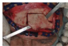
The patient, initially treated by intravenous antibiotic and corti-costeroid therapy, underwent surgical treatment. We have chosen an external approach to obtain a radical excision of the tumor, which had involved the posterior wall of the sinus. The classic bicoronal flap would provide a very evident scar because of the patient’s hair loss. Under general anesthesia, we performed a direct frontal incision following a horizontal wrinkle about 3 cm above the eyebrow line and a subperiosteal flap up to the superior orbital rim. According to CT scan, a polygonal area of frontal bone was cut with a Lindmann burr and then removed with a small chisel. The osteoma was easily separated from the surrounding bony walls by the same chisel and then removed by a clamp.
The orbital roof was partially removed with the tumor, and then we restored it by a preplated titanium mesh 0.8 mm thick, fixed to the bony flap of the anterior wall of the frontal sinus by 2 screws of 1.5 mm. The continuity of the orbital roof has been perfectly reconstructed (Figs. 3 and 4).
The bony fragment, previously removed, was set back in place and fixed with 3 preplated titanium plates (Fig. 5). The flap was sutured by layers and the skin by intradermic 3-0 nylon suture. The surgical procedure lasted 45 minutes.
RESULTS
The histological examination of the specimen confirmed the radio-logic diagnosis of osteoma with prevalent amounts of compact bone. Postoperative CT scan showed the correct reconstruction of the ante-rior wall of the frontal sinus and of the orbital roof (Fig. 6).
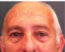
Hertel exophthalmometry performed at admission was 24 mm on the left eye and 17 mm on the right one. The exophthalmos regressed after 5 days of drug therapy, and 15 days after surgery, the new value was 18 mm on the left eye. Also, the Hess-Lancaster scheme has improved after regression of exophthalmos. A month later, the frontal scar was almost invisible (Fig. 7). Recovery of skin sensitivity has been slow. After 8 months, skin was still numb at the frontoparietal junction. The patient, who was previously informed about this com-plication, has confirmed that he absolutely prefers a mild discomfort rather than an unsightly scar.
DISCUSSION
Exclusion criteria for the unique endonasal endoscopic treatment are as follows: frontal osteomas with a lateral localization respect to the sagittal plane passing through the lamina papyracea, intracranial extension, complete involvement of the posterior and anterior wall of the frontal sinus, and anteroposterior sinus pavement smaller than 1 cm.5The classic external approach is a hemicoronal or bicoronal flap with an incision starting from the tragus placed 2 cm posterior to the hairline. This surgical incision often gives rise to an unsightly
FIGURE 1. Computed tomographic slide showing endosinusal bony mass.
FIGURE 2. Clinical evidence of left eye exophthalmos.
FIGURE 3. Removal of the osteoma.
FIGURE 4. Frontal sinus wall bony flap.
FIGURE 5. Reconstruction of the orbital roof by a preplated titanium mesh 0.8 mm thick.
FIGURE 6. Postoperative CT scan.
The Journal of Craniofacial Surgery
&
Volume 23, Number 4, July 2012 Brief Clinical Studies* 2012 Mutaz B. Habal, MD
e365
scar, especially in patients with hair loss. When indicated, we use the horizontal frontal approach also in the osteosynthesis of the frac-tures of the anterior wall of the frontal sinus, especially if the patient already has traumatic cutaneous wound.
The loss of sensitivity due to the cut of the branches of the supraorbitalis and supratrochlearis nerves is the main discomfort reported by patients in the postoperative course. In our experience, patients do not completely recover skin sensitivity a year after sur-gery. However, they declare to prefer an incomplete but satisfactory sensitivity recovery rather than a coronal scar. We prefer a direct horizontal incision in the wrinkles of the forehead to reduce opera-tory time, to avoid anesthetic scars at the expense of a minimal loss of sensitivity.
REFERENCES
1. Seiden AM, el Hefny YI. Endoscopic trephination for the removal of frontal sinus osteoma. Otolaryngol Head Neck Surg
1995;112:607Y611
2. Goldenberg D, Gilboa M, Danino J, et al. A large ethmoido-orbital osteoma presenting with epiphora in an 11-year-old boy.
J Pediatr Ophthalmol Strabismus 2000;37:238Y240
3. Huang HM, Liu CM, Lin KN, et al. Giant ethmoid osteoma with orbital extension, a nasoendoscopic approach using an intranasal drill. Laryngoscope 2001;111:430Y432
4. Brunori A, de Santis S, Bruni P, et al. Life threatening intracranial complications of frontal sinus osteomas: report of two cases. Acta Neurochir (Wien) 1996;138:1426Y1430
5. Castelnuovo P, Valentini V, Giovannetti F, et al. Osteomas of the maxillofacial district: endoscopic surgery versus open surgery. J Craniofac Surg 2008;19:1446Y1452
Magnetic Resonance Imaging in
Isolated Sagittal Synostosis
Michael Engel, MD, DDS, Juergen Hoffmann, MD, DDS, Joachim Mu¨hling, MD, DDS,
Gregor Castrillo´n-Oberndorfer, MD, DDS,
Robin Seeberger, MD, DDS, Christian Freudlsperger, MD, DDS Abstract: Isolated fusion of the sagittal suture is the most prevalent form of craniosynostosis. Although the typical clinical appearance
usually points the way to the right diagnosis, computed tomographic (CT) scans are still recommended as necessary tools for both the diagnosis of scaphocephaly and the preoperative planning. Because CT scans are accompanied by the biological effects of ionizing ra-diation, some authors have already postulated the use of magnetic resonance imaging (MRI) especially because MRI seems to be valuable for detecting intracranial anomalies compared with CT scans. Hence, we investigated the preoperative MRIs of 42 children with isolated sagittal synostosis to evaluate the frequency of brain anomalies and their therapeutic consequences.
In our study, 10 patients (23.8%) showed pathologic MRI find-ings such as ventricular dilatation and hypoplastic corpus callosum, whereas 32 patients (76.2%) had an unremarkable MRI except a pathognomonic secondary deformation of the brain caused by the abnormally shaped skull, which was present in all patients. Seven patients showed clinically significant symptoms including papille-dema or psychomotoric developmental delay; however, the clinical appearance was not predictive for pathologic MRI findings and vice versa.
As the detection of brain anomalies had no influence on the surgical procedure or led to any additive therapy in our patients, we conclude that evaluation of possible pathologic brain findings does not legitimate the general use of MRI in clinically normal children with isolated sagittal synostosis.
Key Words: Sagittal synostosis, craniosynostosis, MRI, papilledema
S
caphocephaly is the morphologic consequence of the premature sagittal suture synostosis. Isolated fusion of the sagittal suture is the most prevalent form of craniosynostosis, occurring with a fre-quency of 1 case per 2000 to 4000 live births, and shows a typical male predominance. Typical clinical hallmark is the long narrow head, widest in the temporal regions and narrowing toward the top of the head, with associated ridging over the fused sagittal suture.1Y6Patients with isolated sagittal synostosis are at risk for increased intracranial pressure (ICP), which can be identified clinically as papilledema.7Furthermore, studies have shown an increased risk for neurodevelopment delays in patients with scaphocephaly; however, the exact reasons for this association are still unclear.8Y11
The treatment is complex and requires surgical intervention be-fore the age of 1 year for correction. The objectives of this treatment are to induce normal brain development, to prevent increased ICP, and to achieve an acceptable cranial morphology.9,12
Although this characteristic clinical appearance should point the way to the right diagnosis, computed tomographic (CT) scans are still recommended as necessary tools for both the diagnosis of scaphocephaly and the preoperative planning.12Y15 Besides these well-known reasons, some authors further justified the use of CT scans with its capacity to discover structural brain abnormalities.12 However, with the recent concerns of radiation exposure in early childhood, it is increasingly clear that these radiographic investiga-tions should be used judiciously, and alternative imaging methods should be discussed.14
To avoid radiographic examination in childhood, magnetic res-onance imaging (MRI) can be a helpful tool in the preoperative diagnostic and imaging of patients with isolated sagittal synostosis. Magnetic resonance imaging opens to researchers a window to the underlying brain, leading to gross observations that the overall shape of the brain may be dysmorphic in craniosynostosis.16,17Magnetic resonance imaging is an excellent technique for the diagnosis of
From the Department of Oral and Maxillofacial Surgery, University Hospital Heidelberg, Heidelberg, Germany.
Received December 25, 2011.
Accepted for publication March 3, 2012.
Address correspondence and reprint requests to Michael Engel, MD, DDS, Department of Oral and Maxillofacial Surgery, University Hospital Heidelberg, Im Neuenheimer Feld 400, 69120 Heidelberg, Germany; E-mail: [email protected]
The authors report no conflicts of interest. Copyright* 2012 by Mutaz B. Habal, MD ISSN: 1049-2275
DOI: 10.1097/SCS.0b013e3182543258
FIGURE 7. Clinical evidence of a minimal frontal scar after 2 months from surgery.
Brief Clinical Studies The Journal of Craniofacial Surgery