Original Paper
Ann Nutr Metab 2008;53:86–90 DOI: 10.1159/000164691Plasma Antioxidants and Asymptomatic
Carotid Atherosclerotic Disease
Graziano Riccioni
a
Tonino Bucciarelli
c
Nicolantonio D’Orazio
d
Nicola Palumbo
b
Emanuela di Ilio
c
Francesco Corradi
c
Alfonso Pennelli
c
Lydia A. Bazzano
e
a Cardiology Unit and b Analysis Laboratory, ‘San Camillo de Lellis’ Hospital, Manfredonia , and c Clinical Biochemistry Laboratory and d Human Nutrition, Department of Biomedical Science,
University ‘G. D’Annunzio’, Chieti , Italy; e Department of Epidemiology, Tulane University School of
Public Health and Tropical Medicine, New Orleans, La. , USA
protein cholesterol, triglycerides and C-reactive protein)
were not significantly associated with carotid
atherosclero-sis. Conclusions: Low plasma concentrations of antioxidant
vitamins (A, E,
 -carotene) and lycopene were associated
with early carotid atherosclerotic lesions as measured by
CIMT. Regular intake of foods rich in lycopene and
antioxi-dant vitamins may slow the progression of atherosclerosis.
Copyright © 2008 S. Karger AG, Basel
Introduction
Cardiovascular diseases, specifically coronary heart
disease and cerebrovascular diseases, are the most
com-mon causes of morbidity and mortality worldwide [1] .
Moreover, cerebrovascular disease is the most important
cause of long-term disability in Western societies.
Oxida-tive stress, particularly from the oxidation of low-density
lipoprotein cholesterol in the artery wall, induces an
in-flammatory reaction [2] which stimulates recruitment of
monocytes and their differentiation into macrophages.
Macrophages accumulate lipids intracellularly, forming
foam cells, which results in increased thickness of the
ar-terial wall [3–5] . Antioxidants, which may inhibit lipid
peroxidation, could play an important protective role [6,
7] against the formation of simple and complex
Key Words
Atherosclerosis
ⴢ Carotid arteries ⴢ Antioxidants ⴢ Lycopene ⴢ
Vitamin A
ⴢ Vitamin E ⴢ  -Carotene
Abstract
Background: Atherosclerosis remains clinically mute for a
long time and frequently manifests itself with an acute
car-diovascular event. The possibility of detecting this disease in
a subclinical phase and reducing or reversing its progression
is an issue of relevance. Published studies on the association
between antioxidant vitamins and carotenoids and carotid
intima-media thickness (CIMT) have been inconclusive.
Methods: We enrolled 220 consecutive, asymptomatic
par-ticipants. After carotid ultrasound investigation, a medical
history was taken, a physical examination was performed
and venous blood samples were collected. Venous blood
samples were analyzed for concentrations of antioxidant
tamins and carotenoids. Results: Low concentrations of
vi-tamin A (p ! 0.01), vivi-tamin E (p ! 0.001), lycopene (p ! 0.01)
and
 -carotene (p ! 0.001) were significantly associated with
carotid atherosclerosis (CIMT 6 0.8 mm). In addition,
margin-ally higher body mass index, plasma haemoglobin and
high-density lipoprotein cholesterol were also associated with
ca-rotid atherosclerosis, while other laboratory parameters
considered in this study (total cholesterol, low-density
Received: January 10, 2008 Accepted after revision: July 10, 2008 Published online: October 21, 2008
sclerotic lesions, which progressively protrude into the
arterial lumen, causing stenosis or occlusion [4, 8] . In
particular, increased carotid intima-media thickness
(CIMT) represents an early phase of the atherosclerotic
process [9–11] and is widely used as a marker of
subclin-ical atherosclerosis which correlates with established
cor-onary heart disease [12, 13] .
Few studies have examined the relationship between
plasma antioxidant concentrations and CIMT, and those
that have report conflicting results. The aim of this study
was to assess the relationship between asymptomatic
el-evated CIMT and plasma levels of antioxidants (vitamin
A, vitamin E,
 -carotene and lycopene).
Patients and Methods
Subjects
We enrolled 220 consecutive, asymptomatic participants (109
males, 111 females) who presented to our institute (Cardiology
Unit of ‘San Camillo de Lellis’ Hospital, Manfredonia, Italy)
be-tween July and December 2007. The participants ranged in age
from 45 to 65 years and underwent carotid ultrasound
investiga-tion of the extracranial carotid arteries.
Inclusion and Exclusion Criteria and Definitions of Clinical
Parameters
Participants were identified as asymptomatic if they had not
experienced a transient ischaemic attack, amaurosis fugax or
stroke. Participants were excluded if they had symptomatic
ca-rotid artery disease that necessitated revascularization therapy,
current infectious or inflammatory disease, recent operations or
endovascular interventions, bilateral carotid occlusion,
monolat-eral/bilateral stent implantation or monolatmonolat-eral/bilateral
endoar-terectomy.
Participants were classified as current smokers if they
an-swered ‘yes’ to the question ‘have you smoked cigarettes, cigars,
or a pipe within the past 30 days?’. Body weight was measured
using a balance scale. During the height and weight
measure-ments, the subjects wore light clothing and no shoes. Body mass
index was computed as the ratio of weight in kilograms to the
square of height in meters. Arterial hypertension was diagnosed
in subjects with blood pressure values 1 140/90 mm Hg measured
repeatedly (at least twice) and was assumed to be present in
pa-tients taking antihypertensive drugs [14] . Diabetes mellitus was
defined according to the clinical practice recommendations 2002
criteria of the Expert Committee of the American Diabetes
As-sociation [15] . Hyperlipidaemia was defined as an elevation of
low-density lipoprotein cholesterol values above 130 mg/dl and
was assumed to be present in all patients taking lipid-lowering
therapies [16] .
The diagnosis of peripheral artery disease was performed
ac-cording to the American College of Cardiology/American Heart
Association practice guidelines [17] . Stroke was defined as a
neu-rological deficit evaluated after 24 h by a neurologist or internist
according to the American Heart Association/American Stroke
Association Council guidelines [18] . History of myocardial
in-farction was assessed according to the consensus document of the
Joint European Society of Cardiology/American College of
Car-diology Committee for the redefinition of myocardial infarction
[19] .
The study was performed in accordance with the Helsinki
Declaration of 1975, as revised in 1983, and approved by the
eth-ical committee of San Camillo de Lellis Hospital. All patients had
to provide written informed consent.
Carotid Ultrasound Investigation
Carotid ultrasound investigations were performed by means
of a colour-coded Acuson Sequoia C512 (Siemens Medical
Solu-tions USA Inc.) carotid duplex machine with a 7.5-MHz linear
transducer. The investigation included longitudinal and
trans-verse examinations of the carotid arteries. Diameters of both the
left and right carotid arteries were measured and calculated at the
site of maximal stenosis in the extra-cranial common carotid
ar-teries according to the European Carotid Surgery Trial method
[20] . The CIMT measurements were performed 10 mm proximal
to the carotid bulb or 20 mm proximal to the flow divider. CIMT
was measured between the leading edge of the first echogenic line
(lumen-intima interface) and the second echogenic line (upper
layer of the adventitia) in the far (deeper) artery wall. All
measure-ments were performed on frozen, enlarged images ( ! 2) at the end
of a heart cycle (end diastole), with the transducer in the
medio-lateral direction [21] . Measurements were performed in both
common carotid arteries, and the larger of the two values was
used in data analysis. Offline analysis of the CIMT was performed
using video images based on the Atherosclerosis Risk in
Commu-nities study protocol [22] . Carotid atherosclerosis was defined as
a CIMT between 0.8 and 1.2 mm, while carotid plaque was
de-fined as focal echo structures encroaching into the vessel lumen
where the CIMT was 1 1.2 mm. These cut-offs were chosen
be-cause they were used in previous randomized clinical trials [23,
24] .
Clinical and Laboratory Data
After carotid ultrasound investigation, medical history
(hy-pertension, diabetes mellitus, family history of atherosclerosis,
myocardial infarction, hyperlipidaemia, angina pectoris,
periph-eral artery disease, history of prior cerebral accident) and data
from a physical examination [age, gender, body mass index, blood
pressure, cardiac rate, smoking habits (smoker, ex-smoker or
non-smoker)] were collected.
Venous blood samples were obtained at the baseline visit.
Blood analyses included haemoglobin, total cholesterol,
high-density lipoprotein cholesterol, low-high-density lipoprotein
choles-terol, triglycerides and C-reactive protein (measured at the
Anal-ysis Laboratory of San Camillo de Lellis Hospital) and plasma
concentrations of vitamin A, vitamin E, lycopene and
 -carotene
(measured at the Clinical Biochemistry Laboratory, Chieti, Italy).
All investigators and laboratory personnel were blinded to the
subjects’ status. Antecubital venous blood samples from all
sub-jects
were handled identically and blindly through all stages of the
blood collection, storage, retrieval and analytic processes.
Blood Sample Collection, Storage and Preparation
Blood samples were collected in polypropylene tubes
contain-ing EDTA 1 m
M. Samples were stored in an ice box prior to
cen-trifugation at 3,000 g for 10 min at 4
°
C. Then, 200-
l aliquots of
plasma were transferred into foil-wrapped polypropylene tubes.
Plasma samples were either used for immediate extraction or
stored in the dark at –80
°
C until analysis was performed. Sample
preparation was performed as described by Lee et al. [25] .
HPLC System and Conditions for Antioxidant Measurements
Vitamin A, vitamin E, lycopene and
 -carotene
concentra-tions were determined by HPLC. Two Waters 515 HPLC pumps
(Whatman, Clifton, N.J., USA) equipped with a Waters
auto-in-jector (model 717 plus auto-sampler) and a Waters 996
photodi-ode array detector were used as the HPLC system. Data
acquisi-tion and processing were performed using the chromatography
software Empower-Pro (Waters). Analysis was performed by
iso-cratic elution. The flow rate was 1.5 ml/min. The mobile phases
used were as follows: (A) methanol/ n -butanol/water (89.5/5/5.5
v/v/v) premixed and vacuum filtered through a 0.45-
m
polypropylene membrane filter (Whatman) before use; (B) methanol/ n
-butanol/water (76/19.5/4.5 v/v/v) premixed and vacuum filtered
before use as for A. The elution isocratic profile was 0–7 min 100%
A, 7–15 min 100% B and 15–20 min 100% A for column
re-equil-ibration. Auto-injections of 20
l were performed at 5
°
C. The
analytical column used was a replaceable Partisphere 5 C-18
car-tridge with an inner diameter of 110 ! 4.7 mm and particle size
of 5
m (Whatman, Waters Corporation, Milford, Mass., USA)
protected by a guard cartridge (C-18, 5
m) system and
main-tained at 45
°
C. The photodiode array wavelength ranged from
270 to 460 nm, and the chromatograms were extracted at 340 nm
for vitamin A, 288 nm for vitamin E and vitamin E-acetate (used
as internal standard) and 441 nm for lycopene and
 -carotene.
The run time was 20 min.
Statistical Analysis
Study participants were grouped into two categories
accord-ing to CIMT, i.e. ! 0.8 and 6 0.8 mm. For each baseline
character-istic, the mean value or corresponding percentage of study
par-ticipants was calculated according to the CIMT category.
The statistical significance of differences was examined using
Student’s t test (continuous variables) and the
2test (categorical
variables).
A two-sided p value ! 0.05 was considered statistically
signifi-cant. Data were analyzed using SPSS statistical software (version
15.0 for Windows, SPSS Inc., Chicago, Ill., USA).
Results
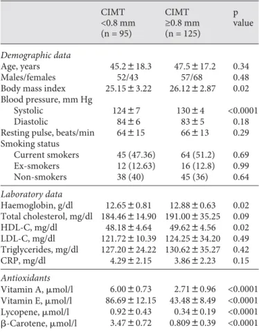
Means ( 8 SD) or percentages of patients for various
demographic characteristics and established
cardiovas-cular disease risk factors are summarized in table 1
ac-cording to CIMT category. Of the 220 participants who
took part in the study, 125 subjects (56.8%) had carotid
atherosclerosis, of whom 52 (41.6%) had disease localized
to the common carotid arteries, 43 (34.4%) had disease
localized to the internal carotid arteries and 30 (24%) had
disease localized to the external carotid arteries.
A significant association was identified between low
concentrations of vitamin A (p ! 0.01), vitamin E (p !
0.001), lycopene (p ! 0.01) and
 -carotene (p ! 0.001) and
evidence of carotid atherosclerosis as measured by CIMT.
Marginally higher body mass index, plasma
haemoglo-bin and high-density lipoprotein cholesterol were also
as-sociated with carotid atherosclerosis, while other
labora-tory parameters considered in the study (total
cholester-ol, low-density lipoprotein cholestercholester-ol, triglycerides and
C-reactive protein) were not significantly associated with
carotid atherosclerosis.
Discussion
The primary finding of our study is that low plasma
levels of antioxidant vitamins and oxygenated
carot-enoids (vitamin A, vitamin E,
 -carotene and lycopene)
Table 1.
Characteristics of the study population
CIMT <0.8 mm (n = 95) CIMT ≥0.8 mm (n = 125) p value Demographic data Age, years 45.2818.3 47.5817.2 0.34 Males/females 52/43 57/68 0.48Body mass index 25.1583.22 26.1282.87 0.02 Blood pressure, mm Hg
Systolic 12487 13084 <0.0001
Diastolic 8486 8385 0.18
Resting pulse, beats/min 64815 66813 0.29 Smoking status Current smokers 45 (47.36) 64 (51.2) 0.69 Ex-smokers 12 (12.63) 16 (12.8) 0.99 Non-smokers 38 (40) 45 (36) 0.64 Laboratory data Haemoglobin, g/dl 12.6580.81 12.8880.63 0.02 Total cholesterol, mg/dl 184.46814.90 191.00835.25 0.09 HDL-C, mg/dl 48.1884.64 49.6284.56 0.02 LDL-C, mg/dl 121.72810.39 124.25834.20 0.49 Triglycerides, mg/dl 127.20824.22 130.62835.27 0.42 CRP, mg/dl 4.2982.15 3.8682.23 0.15 Antioxidants Vitamin A, mol/l 6.0080.73 2.7180.96 <0.0001 Vitamin E, mol/l 86.69812.15 43.4888.49 <0.0001 Lycopene, mol/l 0.9280.43 0.3480.19 <0.0001 -Carotene, mol/l 3.4780.72 0.80980.39 <0.0001 Values in parentheses represent percentages. HDL-C = High-density lipoprotein cholesterol; LDL-C = low-High-density lipoprotein cholesterol; CRP = C-reactive protein.
are associated with carotid atherosclerosis. Other
epide-miological studies have evaluated the relationship
be-tween antioxidant vitamins and evidence of subclinical
atherosclerosis; however, the results have been
inconclu-sive.
For example, in a cross-sectional study of 392 men and
women (age 45–65 years), D’Odorico et al. [26] found no
association between vitamin A plasma concentrations
and the presence of carotid/femoral atherosclerosis.
Sim-ilarly, Iannuzzi et al. [27] examined 310 women for early
carotid atherosclerosis and found no association between
intake or plasma concentrations of vitamin A and the
presence of carotid plaques. In prospective cohort
stud-ies, Dwyer et al. [28] and McQuillan et al. [29] found no
association between CIMT atherosclerosis or
progres-sion and vitamin A plasma concentrations. In a nested
case-control study, Iribarren et al. [30] also failed to find
an association between plasma vitamin A (retinol)
con-centration and CIMT. In contrast, a case-control study
conducted by Polidori et al. [31] found a significant
asso-ciation between lower plasma vitamin A (retinol)
concen-trations and carotid and iliofemoral atherosclerosis.
The results of published studies regarding vitamin E
and carotid atherosclerosis are also inconclusive.
Ian-nuzzi et al. [27] , Polidori et al. [31] and McQuillan et al.
[29] found an inverse association between CIMT and
plasma vitamin E concentration, whereas D’Odorico et
al. [26] , Dwyer et al. [28] and Giannetti et al. [32] found
no association between vitamin E plasma concentration
and the presence of carotid/femoral atherosclerosis. Even
for the well-studied relationship between
 -carotene and
carotid atherosclerosis, the results of published studies
are unclear and conflicting. Many studies have found
that the consumption of
 -carotene is inversely related to
the risk of coronary heart disease [33, 34] and that the risk
of carotid and femoral atherosclerosis decreased with
in-creasing plasma
 -carotene concentrations [26] ,
suggest-ing a protective role for
 -carotene in early atherogenesis.
However, several well-conducted studies have found no
association between
 -carotene plasma concentrations
and CIMT or peripheral vascular disease [29, 30, 32] . In
contrast, the results of published studies of the
associa-tion between lycopene and carotid atherosclerosis are
generally concordant and show a significant inverse
rela-tionship between serum concentrations of lycopene and
CIMT, supporting the hypothesis that plasma lycopene
may decrease the risk of atherosclerosis and play an
im-portant role in the early stage of atherogenesis [29, 32,
35] .
One important limitation of this study is the
cross-sectional nature of our findings. Because antioxidant
val-ues, lipid profiles, anthropometrics and questionnaires
were collected concurrently with the ultrasound
assess-ment of carotid arteries, temporality cannot be inferred
from these findings. Further studies which examine this
important relationship are warranted.
Conclusions
In summary, we found a strong inverse relationship
between antioxidant plasma levels and the presence of
carotid atherosclerosis. Atherosclerosis remains
clinical-ly mute for a long time and frequentclinical-ly manifests itself
with an acute cardiovascular event; therefore, the
possi-bility of detecting the disease in a subclinical phase and
reducing or reversing its progression is an issue of
rele-vance. The evaluation of CIMT in asymptomatic patients
with CUS allows us to identify atherosclerotic disease in
its early phases, to evaluate disease progression and to
monitor the effects of interventions. In particular,
regu-lar intake of rich foods rich in lycopene and other
anti-oxidant vitamins may slow the progression of
rotic processes and modify the early stages of
atheroscle-rosis, with a consequent reduction in cardiovascular
events.
References
1 Ezzati M, Vander Hoorn S, Lawes CM, Leach R, James WP, Lopez AD, Rodgers A, Murray CJ: Rethinking the ‘diseases of affluence’ paradigm: global patterns of nutritional risks in relation to economic development.PLoS Med 2005; 2:e133.
2 Holvoet P, Mertens A, Verhamme P, Bo-gaerts K, Beyens G, Verhaeghe R, Collen D, Muls E, Van de Werf F: Circulating oxidized LDL is a useful marker for identifying pa-tients with coronary artery disease.
Arterio-scl Thromb Vasc Biol 2001; 21: 844–848.
3 Libby P, Algra A, Aikawa M, Schonbeck U: Cholesterol and atherosclerosis. Biochim
Biophys Acta 2000; 1529: 299–309.
4 Lusis AJ: Atherosclerosis. Nature 2000; 407:
233–241.
5 Riccioni G, De Santis A, Cerasa V, Menna V, Di Ilio C, Ballone E, D’Orazio N: Atheroscle-rotic plaque formation and risk factors. Int J
Immunopathol Pharmacol 2003; 16: 25–31.
6 Diaz MN, Frei B, Vita JA, Keaney FR Jr: An-tioxidants and atherosclerotic heart disease.
7 Riccioni G, Bucciarelli T, Mancini B, Di Ilio C, Capra V, D’Orazio N: The role of the anti-oxidant vitamin supplementation in the pre-vention of cardiovascular diseases. Expert
Opin Investig Drugs 2007; 16: 25–32.
8 Steinberg D, Parthasarathy S, Carew TE, Khoo JC, Witzum JL: Beyond cholesterol: modifications of low-density lipoprotein that increase its atherogenicity. N Engl J Med
1989; 320: 915–924.
9 Zureik M, Ducimetiere P, Touboul PJ, Cour-bon D, Bonithon-Kopp C, Berr C, Magne C: Common carotid intima-media thickness predicts occurrence of carotid atherosclerot-ic plaques: longitudinal results from the Ag-ing Vascular Study (EVA) study. Arterioscler
Thromb Vasc Biol 2000; 20: 1622–1629.
10 Heiss G, Sharrett AR, Barnes R, Chambless LE, Szklo M, Alzola C: Carotid atherosclero-sis measured by B-mode ultrasound in popu-lations: associations with cardiovascular risk factors in the ARIC Study. Am J
Epide-miol 1991; 134: 250–256.
11 Salonen R, Salonen JT: Determinants of ca-rotid intima-media thickness: a population-based ultrasonography study in eastern
Finnish men. J Intern Med 1991; 229: 225–
231.
12 Davis PH, Dawson JD, Riley WA, Lauer MN: Carotid intimal-medial thickness is related to cardiovascular risk factors measured from childhood through middle age: the
Musca-tine study. Circulation 2001; 104: 2815–2819.
13 Salonen JT, Salonen R: Ultrasonographically assessed carotid morphology and the risk of coronary heart disease. Arterioscler Thromb
1991; 11: 1245–1249.
14 Joint National Committee: The Seventh Re-port of the Joint National Committee on Pre-vention, Detection, Evaluation, and Treat-ment of High Blood Pressure: the JNC 7
report. JAMA 2003; 289: 2560–2572.
15 Expert Committee on the Diagnosis and Classification of Diabetes Mellitus: Ameri-can Diabetes Association: clinical practice recommendations 2002. Diabetes Care 2002; 25(suppl 1):S1–S147.
16 National Cholesterol Education Program (NCEP) Expert Panel on Detection, Evalua-tion, and Treatment of High Blood Choles-terol in Adults (Adult Treatment Panel III): Third Report of the National Cholesterol Ed-ucation Program (NCEP) Expert Panel on Detection, Evaluation, and Treatment of High Blood Cholesterol in Adults (Adult Treatment Panel III) final report.
Circula-tion 2002; 106: 3143–3421.
17 ACC/AHA 2005 Practice Guidelines for the management of patients with peripheral ar-terial disease (lower extremity, renal, mesen-teric, and abdominal aortic): a collaborative report from the American Association for Vascular Surgery/Society for Vascular Sur-gery, Society for Cardiovascular Angiogra-phy and Interventions, Society for Vascular Medicine and Biology, Society of
Interven-tional Radiology, and the ACC/AHA Task Force on Practice Guidelines (Writing Com-mittee to Develop Guidelines for the Man-agement of Patients With Peripheral Arterial Disease): endorsed by the American Associ-ation of Cardiovascular and Pulmonary Re-habilitation; National Heart, Lung, and Blood Institute; Society for Vascular Nurs-ing; TransAtlantic Inter-Society Consensus; and Vascular Disease Foundation.
Circula-tion 2006; 113:e463–e654.
18 Primary prevention of ischemic stroke: a guideline from the American Heart Associa-tion/American Stroke Association Stroke Council: cosponsored by the Atherosclerotic Peripheral Vascular Disease Interdisciplin-ary Working Group; Cardiovascular Nurs-ing Council; Clinical Cardiology Council; Nutrition, Physical Activity, and Metabo-lism Council; and the Quality of Care and Outcomes Research Interdisciplinary Work-ing Group: the American Academy of Neu-rology affirms the value of this guideline.
Stroke 2006; 37: 1583–1633.
19 Alpert JS, Thygese K, Antman E, Bassand GP: Myocardial infarction redefined – a con-sensus document of the Joint European So-ciety of Cardiology/American College of Cardiology Committee for the redefinition of myocardial infarction. J Am Coll Cardiol
2000; 39: 959–969.
20 European Carotid Surgery Trialists’ Collab-orative Group: MRC European Carotid Sur-gery Trial: interim results for symptomatic patients with severe (70–99%) stenosis or
with mild (0–29%) stenosis. Lancet 1991; 337:
1235–1243.
21 Kanters SD, Algra A, van Leeuwen MS, Ban-ga JD: Reproducibility of in vivo carotid in-tima-media thickness measurements. Stroke
1997; 28: 665–671.
22 Howard G, Sharrett AR, Heiss G, Evans GW, Chambless LE, Riley WA, Burke GL: Carot-id artery intimal-medial thickness distribu-tion in general populadistribu-tions as evaluated by
B-mode ultrasound. Stroke 1993; 24: 1297–
1304.
23 Crouse JR III, Byngton RP, Bond MG, Espe-land MA, Craven TE, Sprinkle JW, McGov-ern ME, Furberg CD: Pravastatin, lipids and atherosclerosis in the carotid arteries
(PLAC-II). Am J Cardiol 1995; 75: 455–459.
24 Mercuri M, Bond MG, Sirtori CR, Veglia F, Crepaldi G, Feruglio FS, Descovich G, Ricci G, Rubba P, Mancini M, Gallus G, Bianchi G, D’Alò G, Ventura A: Pravastatin reduces intima-media thickness progression in an asymptomatic hypercholesterolemic Medi-terranean population: the Carotid Athero-sclerosis Italian Ultrasound Study. Am J Med
1996; 101: 627–634.
25 Lee BL, Chua SC, Ong HY, Ong CN: High-performance liquid chromatographic meth-od for routine determination of vitamins A
and E and  -carotene. J Chromatogr 1999;
581: 41–47.
26 D’Odorico A, Martines D, Kiechl S, Egger G, Ozer F, Bonvicini P, Sturinolo GC, Nacca-rato R, Willeit J: High plasma levels of alpha- and beta-carotene are associated with a low-er risk of athlow-eroscllow-erosis. Results from the
Bruneck study. Atherosclerosis 2000; 153:
231–239.
27 Iannuzzi A, Celentano E, Panico S, Galasso R, Covetti G, Sacchetti L, Zarrilli F, De Mi-chele M, Rubba P: Dietary and circulating antioxidant vitamins in relation to carotid plaques in middle-aged women. Am J Clin
Nutr 2002; 3: 582–587.
28 Dwyer JH, Paul-Labrador MJ, Fan J, Shircore AM, Bairez Merz CN, Dwyer KM: Progres-sion of carotid intima-media thickness and plasma antioxidants: the Los Angeles Ath-erosclerosis Study. Arterioscler Thromb
Vasc Biol 2004; 24: 313–318.
29 McQuillan BM, Hung J, Beilby JP, Nidorf M, Thompson PL: Antioxidant vitamins and the risk of carotid atherosclerosis. The Perth Carotid Ultrasound Assessment study
( CUDAS). J Am Coll Cardiol 2001; 38: 1788–
1794.
30 Iribarren C, Folsom AR, Jacobs DR Jr, Gross MD, Belcher JD, Eckfeldt JH: Association of serum vitamin levels, LDL susceptibility to oxidation, and autoantibodies against MDA-LDL with carotid atherosclerosis. A case-control study. The ARIC Study Investi-gators. Atherosclerosis Risk in
Communi-ties. Arterioscler Thromb Vasc Biol 1997; 7:
1171–1177.
31 Polidori MC, Praticò D, Parente B, Mariani E, Cecchetti R, Yao Y, Sies H, Cao P, Mecocci P, Sthal W: Elevated lipid peroxidation bio-markers and low antioxidant status in ath-erosclerosis patients with increased carotid or iliofemoral intima media thickness. J
In-vest Med 2007; 55: 163–167.
32 Giannetti J, Perinelli R, Petrucci R, Lazzerini G, De caterina M, Bellomo G, De Caterina R: Inverse association between carotid intima-media thickness and the antioxidant lyco-pene in atherosclerosis. Am Heart J 2002;
143: 467–474.
33 Bolton-Smith C, Woodward M, Tunstall-Pe-doe H: The Scottish Heart Health Study. Di-etary intake by food frequency question-naire and odds ratios for coronary heart disease risk. II. The antioxidant vitamins
and fibre. Eur J Clin Nutr 1992; 46: 85–93.
34 Rimm EB, Stampfer MJ, Ascherio A, Giovan-nucci E, Colditz GA, Willett WC: Vitamin E consumption and the risk of coronary heart
disease in men. N Engl J Med 1993; 328: 1450–
1456.
35 Rissanen TH, Voutilainen S, Nyyssonen K, Salonen R, kaplan GA, Salonen JT: Serum ly-copene concentrations and carotid athero-sclerosis: the Kuopio Ischaemic Heart Dis-ease Risk Factor Study. Am J Clin Nutr 2003;
77: 13–18.