UNIVERSITA’ DEGLI STUDI DEL PIEMONTE ORIENTALE
“Amedeo Avogadro”
PhD Program
Biotechnology for Human Health
Cycle XXVI
HIGH-THROUGHPUT ANTIBODY VALIDATION
PLATFORMS
SUPERVISOR:
PhD STUDENT:
TABLE OF CONTENTS
1. ABSTRACT 4
2. INTRODUCTION 6
2.1 THE ANTIBODY MOLECULE 6
2.2 THE IMPORTANCE OF ANTIBODIES SPECIFICITY IN RESEARCH 7 2.3 IN VIVO AND IN VITRO ANTIBODY PRODUCTION TECHNOLOGIES 10
2.3.1 Phage display technology 12
2.3.2 Yeast display technology 15
2.3.3 Differences between phage and yeast display 18
2.4 ANTIBODY VALIDATION 20
2.5 PROTEIN MICROARRAY TECHNOLOGY 22
2.5.1 Protein array applications 24
2.5.2 Protein array improvements 26
2.6 PROTEIN PRODUCTION WITHOUT CELL BOUNDERIES 27
2.7 IN SITU PROTEIN ARRAY 29
2.7.1 Protein in situ array (PISA) 30
2.7.2 Nucleic acid programmable protein array (NAPPA) 31
2.7.3 Multiple spotting technique (MIST) 31
2.7.4 TUS-TER array 32
2.7.5 DNA array to protein array (DAPA) 32
2.7.6 In situ puromicyn-capture from mRNA array 33
3. AIM OF THE PROJECT 37
4. MATERIALS AND METHODOLOGIES 39
5. RESULTS 45
5.1 CHARACTERIZATION AND VALIDATION OF RABBIT POLYCLONAL ANTIBODIES 45 5.1.1 Western blot and ELISA polyclonal antibodies validation 48
5.1.2 Protein microarray optimization 51
5.1.3 Protein array validation of yeast polyclonal supernatants 53
5.1.4 Protein array validation of purified polyclonal scFv-Fc 56
5.1.5 Comparison with commercial polyclonal antibody: CDK2A 58
5.2 PROTEIN ARRAY ON DEMAND ASSESSMENT 61
5.2.1 Protein array on demand template construction 62
5.2.2 Protein expression and Ter binding 64
5.2.3 Protein array on demand 67
5.3 SET UP OF A NEW PROTEIN ARRAY ON DEMAND PROTOCOL 69
5.3.1 Array slide and DNA template set up 69
5.3.2 Protein array on demand improvements 72
5.3.3 New protein array on demand 75
6. DISCUSSION 78
6.1 POLYCLONAL ANTIBODIES VALIDATION 78
6.2 PROTEIN ARRAY ON DEMAND 80
6.3 NEW PROTEIN ARRAY ON DEMAND SET UP 81
1. ABSTRACT
Antibodies are molecules able to specifically bind a particular antigen. Thanks to this capability they are extremely useful in many application. Indeed, antibodies are utilized in different routinary experiments, such as: Western blot, Enzyme-linked immunosorbent assay (ELISA), Immunohystochemistry, Immonofluorescence, Flow-cytometry, Immunoprecipitation, etc. Obviously, to carry out a good experiment a good antibody is needed. Indeed, many commercial available antibody aren’t efficient and leads to wrong results. This problem derives from a wrong antibody selection, production and validation. This project is aimed at setting up a selection, production and validation platform for polyclonal antibodies. In particular, the polyclonal antibodies were selected, in form of scFv, with a phage/yeast display combination, produced in yeast cells as minibody with a CH2-CH3 of rabbit IgG, validated with high-throughput technologies. A list of 78 proteins
based on proteins structure and solubilization was drafted. Of these proteins only the first seven were used for scFv selection with two rounds of phage display and two additional rounds of yeast display. The selected scFvs were cloned in a yeast expression vector that allows the production of scFv fused to a rabbit IgG constant part. The collected yeast supernatants, containing the polyclonal, were sent us for the validation step. The polyclonal antibodies validation consists of three different analysis: Western blot, ELISA and protein microarray. From this validation arose that five polyclonal antibodies were highly specific and sensitive to the target protein, revealing that the entire platform is efficient. Furthermore, one of them was compared to the commercial ones using ELISA and protein microarray technologies. Only with protein microarray it could be possible observed that the commercial polyclonal presented cross-reactivity to other proteins. Instead, the yeast polyclonal resulted specific only to the target protein. This result confirmed that the selection with phage/yeast display combination is extremely specific avoiding the selection of sticky or unspecific antibodies. Moreover, protein microarray resulted a very useful and sensitive antibody validation technology. The second aim of this project concerned the use of a new technology to perform high-throughput antibody validation. Indeed, protein microarray is a successful tool, but present some drawbacks, such as production and purification of all the tested proteins and possible degradation of them once immobilized.
technology consists of: cDNA immobilization on the slide; expression of the proteins of interest by in vitro transcription and translation system; capture the nascent proteins; reveal the proteins. Different types of in situ protein array have been set up, but the one used in this project is called protein array on demand. The peculiarity of this array is the presence of the capture agent directly on the template DNA. In this way only the DNA has to be printed on the slide. First, a DNA plasmid suitable for this system was constructed and subsequently tested with four different proteins available in our laboratory. Subsequently, a protein array on demand was performed, following the published method. The analysis showed that protein array on demand was less efficient and reproducible. For this reason a new protein array on demand was set up. The array slide, template and IVTT system were analyzed and tested to perform the best protein array on demand conditions. After different experiments the array on demand was performed using: CodeLink slide; biotinylated PCR DNA mixed with streptavidin; IVTT based on rabbit reticulocytes lysate. Since that the array was performed with an anti-tag antibody, an additional array was performed using an anti-GFP antibody direct against the eGFP. Even if the signal wasn’t high it could be affirmed that this protein array on demand was functional and better than the previous ones. In conclusion, a new protein array on demand was set up and can be potentially used for antibody validation.
2. INTRODUCTION
2.1 THE ANTIBODY MOLECULE
Antibodies, also called immunoglobulins, are molecules produced by B cells of the immune system. The main activity of an antibody is to disrupt the pathogen presents in the organism through two different mechanisms: opsonization and neutralization. These mechanisms involves the antibody binding to a particular antigen in a strong manner. The main type of immunoglobulin used in research is the G (IgG) and it is a protein composed of two polypeptide chains link together by covalent binding [1] (Fig. 1). In every polypeptide an heavy and a light chain is present. The heavy chain is composed by three constant domains called CH1, CH2 and CH3 and a variable domain called VH. The CH1 and CH2 domains
are linked together by covalent binding, forming a flexible hinge region. On the other hand, the light chain is composed only by two domains, one variable (VL) and one constant (CL).
The antibody’s part that confers the antigen specificity is the variable region (VH and VL), in
particular three loops, called complementarity determining region (CDRs) 1,2 and 3, are responsible of the antibody diversity [2]. Finally, the constant region is responsible for glycosylation which is important for the recognition of effector molecules, such as immunoglobulin receptors or C1 complement complex.
Figure 1. IgG structure and other antibody format. Structure of a full-length IgG and the possible
smaller formats used in research: Fragment variable (Fv); single-chain fragment variable (scFv); Fragment-antigen-binding (Fab); minibody.
The scientific interest in antibodies arising from their ability to specifically recognize a determined target. The production of these molecules is not simple due to the higher molecular weight and post-translational modifications in the constant part. For this reason other antibody formats have been constructed maintaining the antibody specificity region (Fig. 1) [3]. The most used in research are:
1. Fragment-antigen-binding (Fab), composed by CH1 and VH link with random and flexible
peptide linker to VL and CL (50kDa);
2. single-chain fragment variable (scFv) molecules, formed by VH and VL link with a linker
peptide of 15aa (25-30kDa);
3. fragment variable (Fv), only VH and VL are linked without a linker between them
(25kDa);
4. minibody, formed by scFv-CH3 protein that self-assembled into a bivalent dimer
(80kDa).;
5. ScFv-Fc, bivalent molecule composed by two scFvs fused to CH2-CH3 IgG region
(120kDa).
2.2 THE IMPORTANCE OF ANTIBODIES SPECIFICITY IN RESEARCH
The Human Genome Project, in 2000, has revealed a great number of genes that were previously unknown and subsequently shifted the attention from the genome to the proteome. The major approach to explore the whole human proteome is the use of affinity reagents, among which antibodies are the most widely used. Antibodies are able to recognize specifically a selected antigen. It is possible to produce antibody directed to a particular epitope (monoclonal) or different epitopes of the same antigen (polyclonal). Furthermore, antibodies can be easily marked with many kind of molecules such as: enzymes, fluorophores, biomolecules and drugs. All these features have lead to the use of antibodies in various research fields. Indeed, they are necessary for many standard analysis such as:
Western blot (WB), where antibodies allow the recognition of denaturated proteins present in a lysate or protein mix;
Enzyme-linked immunosorbent assay (ELISA), in which antibodies reveal the presence of full-lengh proteins on plastic plates;
Immunoprecipitation (IP), where antibodies are not only used to detect the protein presence, but also to precipitated protein complexes;
Flow-cytometry (FC), in which marked antibodies allow proteins recognition on cells or different marked antibodies can allow the separation of different cells;
Immunohistochemistry (IHC), where tissues are stained with antibodies allowing to study tissue structure and cells distribution;
Immunofluorescence (IF), in which tissues are depicted with antibodies conjugated with a fluorophore allowing us to compare not only protein distribution in different cells types but also cancerous cells with normal ones.
Antibodies are also employed in many new techniques. For example, they are very important in protein microarray technology, where they are used to detect the expression of proteins in different samples or cell lysates [4; 5; 6]. Recently, they have been used to address in specific tissue nanoparticles carrying drugs or other molecules [7; 8; 9]. Furthermore, antibodies are also utilized for the isolation of specific cells, such as stem cells [10; 11]. Obviously, all these analysis are based on the antibody specificity. Indeed, antibodies that recognize the wrong molecules or cross-react against other antigens will cause an incorrect result. These problems occur in the case of non-specific validation. Moreover, some analysis can’t be carried out because an antibody, against the specific antigen, is not available. For this reason, another technology has been introduced for proteome analysis: mass spectrometry (MS)[12]. The main ability of MS is the analysis of proteins without affinity reagents use. On the other hand, MS is not high-throughput and can’t be used in different applications, such as immunofluorescence and immunohystochemistry, for this reason technologies using antibodies are preferred. In the last decade, many articles have reported the inefficiency of different commercial antibodies [13; 14; 15]. This is the result of a validation tested only with few experiments. Indeed, many antibodies can have diverse sensibility in different analysis. To overcome this problem the Human Protein Organization (HUPO) have started a Human Antibody Initiative (HAI) that have the aim to [16] :
1. create a catalogue of validated antibodies produced by companies or academic research laboratories;
2. create a protein atlas that collect the differences of protein expression in normal and disease tissue.
products are collected. The website provides data about all antibodies recognizing a certain protein and also the different methods used for each analysis (WB, ELISA, IP,IHC, FC, IF). The second portal is the protein atlas (www.proteinatlas.org) that collects millions of high-resolution images showing the spatial distribution of proteins in 44 different normal human tissues and 20 different cancer types, as well as 46 different human cell lines. The data is combined with validation performed for each antibody, including WB and IM analysis and, for a large fraction, a protein array assay and immunofluorescent based confocal microscopy (Fig. 2). For every protein searched information about expression profiles, protein classes and chromosome location are supplied. At the moment, 20329 gene products are collected in that portal.
Figure 2. The human protein atlas [17]. Example of the data gives by the protein atlas database.
These two database are very useful for antibody research and utilization, even if many target remain without a specific antibody. For this purpose high-throughput antibody production, selection and validation is needed.
2.3 IN VIVO AND IN VITRO ANTIBODIES PRODUCTION TECHNOLOGIES
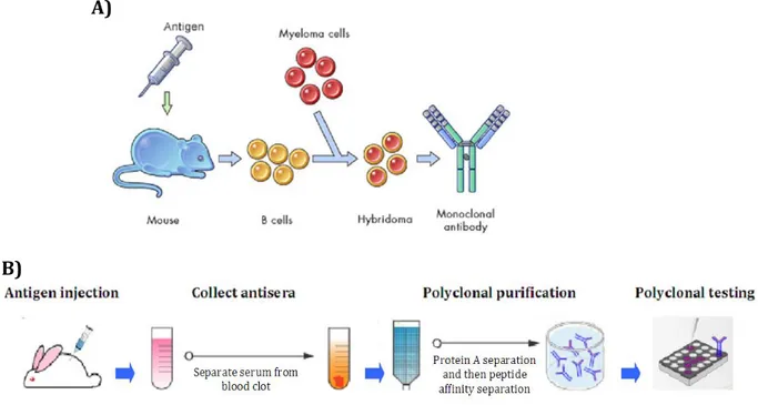
Antibodies are very powerful molecules in research, therapy and diagnosis. For this reason, it has been necessary to develop efficient methodologies to produce them. In 1975, Kohelr and Milstein have sewn up the “hybridoma method” (Fig. 3), based on B lymphocytes and tumoral cells fusion [18]. In particular, this method consists of the injection of a putative antigen and adjuvants in animals (mouse, rat or rabbit) to induce an immune rensponse that lead to antibodies production by B cells. When the animal sera is positive for antibodies against the antigen, the lymphocytes are collected and fused to tumoral cells to allow the formation of hybridomas. These cells are able to produce antibodies, such as B lymphocytes, and can reproduce itself in vitro for a long period, as a tumor cell line.
Figure 3. In vivo antibody production technology. A) The “hybridoma method” involves the use
of animals (mouse, rat or rabbit). The hybridoma is a cell derived from the fusion of animal B cells, stimulated for antibody production through antigen injection, and tumor cells (myeloma). This cell type is able to produce antibody for long time in vitro allowing the antibody selection. B) Polyclonal antibodies production: animals (normally rabbit) are injected with the antigen supplemented with adjuvant and their blood is collected and the sera separated. Subsequently, the antisera can be purified with protein A, for IgGs separation, and peptide affinity column, for antigen specific IgGs separation. The polyclonals set can be subsequently tested.
A)
adjuvants, and after 7-14 days the animal sera is collected and used directly as polyclonals antibodies or successively purified [19]. These methods are the well known methodologies for antibodies production, but they still to be very laborious, expensive and time-consuming. For these reasons in vitro methods have been developed. These technologies don’t involve the antibodies production by animals, but utilize antibody libraries. The library consists in a collection of antibodies cDNA that can be screen to select the one of interest. There are different types of antibody libraries differing each other in how they are made [20]:
1. naïve; 2. immune; 3. semi-synthetic; 4. synthetic.
The naïve library derives from Ig of human donors or animals that are not immunized to a particular antigen. The light and heavy chains are amplified by polymerase chain reaction (PCR) and collected [21]. For this type of library the diversity is directly dependent to the number of the donors. On the opposite side the immune antibody libraries consist in the RNA amplification of immunoglobulins derived from immunized individuals or animals [20]. The semi-synthetic and synthetic libraries are based on random oligonucleotides synthesis to construct CDRs regions. In the semi-synthetic only a part of the antibody, normally the CDR3, is synthetic [22]. These last types of library are used to increase the library diversity. All these libraries contain at least from 107 to 1011 different antibody
clones that can be used in all the in vitro antibody production technologies. In general, these methods are called “display technologies” and allow the linkage between genotype and phenotype. In this way the cDNA of the selected antibody is immediately avaliable for further applications. In vitro technologies, differently to the hybridoma method, allow a faster and easier selection and a major specificity of the antibodies selected has been shown [23]. Furthermore, in vitro antibody production allows to select antibodies against toxins and compounds that can’t be injected in the animals. In the last decade, different display technologies have been developed, most used are: phage display and yeast display [24; 25; 26]. These two techniques are based on the display of the antibody on the cell surface, fusing it with a cell surface protein. In case of phage display the glycoproteins III or VIII are used; for yeast display mannoproteins are the used ones. The corresponding antibody cDNA is present inside the yeast cell or phage particle.
2.3.1 PHAGE DISPLAY TECHNOLOGY
Phage display is a technology that allows the selection of molecules through the exposition of peptides, proteins or antibodies on phage particles. The idea to use filamentous phages as an expression vector was introduced by Smith in 1985 [27]. Filamentous phages considered to be a good expression vector because [28]:
1. their genome can be modified, with insertion, without causing packaging problems; 2. their genome can be isolated;
3. they can tolerate stringent conditions necessary for selection steps;
4. coat proteins can be fused to other molecules, without loosing their infective role; 5. they can be accumulated in large amount in bacterial cells thanks to their non-lytic
propagation.
The main phage utilized for phage display is the filamentous phage M13 [29](Fig. 4).
Figure 4. Filamentous phage M13 structure. The M13 genome is a single stranded DNA of 6407
bases. The DNA molecule is covered by the major coat protein gpVIII of which 2700 copies are present. On the opposite sides of the phage particle there are minor coat proteins: gpIII, gpVI, gpVII and gpIX. The proteins III and VI are located at one side and at the other there are gpVII and gpIX. The minor coat proteins are present in 5 copies each. A single phage particle is 1000nm in length and 5nm in diameter.
It is composed by a capsid that holds a single stranded DNA (ssDNA) encoding for capsid, infection and cell cycle proteins. The capsid is formed by five proteins: gpIII, gpVI, gpVII, gpVIII and gpIX. The gpVIII, called major coat protein, is present in 2700 copies for each phage particle and covers the entire ssDNA molecule. The other proteins are called minor coat proteins because only five copies each are present in a single phage. They are situated at the double-ended of a phage particle: gpIII and gpVI at one side; gpVII and gpIX at the
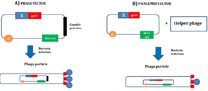
for proteins exposition. A cDNA library can be directly inserted in the phage genome fused to gpIII or gpVIII gene [28] (Fig. 5). This allows the formation of phages containing each: 1. a DNA molecule containing the cDNA of a protein fused to phage protein; 2. the corresponding protein fused to all the copies of the phage protein. This method is potential only with peptides or small proteins because when big proteins, such as antibodies, are fused to gpIII or gpVIII the phage packaging or infection is compromised. To overcome this problem a new method was introduced. This new method uses a different vector called phagemid for protein display [30](Fig. 5). The phagemid is a plasmid containing:
1. phage replication origin; 2. packaging signal;
3. gpIII or gpVIII gene fused to the cDNA library.
The phagemid is transformed in the bacteria cell that will be infected by a helper phage containing wild-type phage genome. The resulting phages will have internalized the phagemid and will present only some copies of gpIII or gpVIII fused to the displayed protein [24].
Figure 5. Phage display vectors. Phage display technology can be carry out with two different
strategies. A)Phage vector: the cDNA library (blue) is directly inserted in the phage genome, fused to the gpIII gene (red). The resulting phages will present as genome the phage vector containing the cDNA of a protein X fused to the gpIII gene; as phenotype, the same protein X (blue) fused to all the gpIII copies (red). B) The phagemid vector is a plasmid containing the gpIII gene (red), the packaging signal (yellow) and the M13 ori (green). The cDNA library is inserted in this plasmid fused to the gpIII gene. To infect the bacteria a wild-type phage, called helper phage, is needed. Since that also a wild-type gpIII gene is present during the infection, only some gpIII copies (red) of the nascent phages, containing the phagemid vector, will be fused to the X protein (blue).
When a library is expressed with phage display it has to be selected to find the cDNA fragments of interest. The selection stage is called biopanning and consists of three steps (Fig. 6)[28]:
1. target immobilization: the antigen is bound on a solid support;
2. phage binding: exposure of the phage presenting antibody to the antigen; 3. removing unbound phages: washes to eliminate aspecific phages;
4. phage elution: the phage positive for antigen binding are eluted using free antigen or a competitor.
Normally, these steps are repeated three times with more stringent washing conditions to eliminate the major amount of unspecific antibodies.
Figure 6. Biopanning of a scFv phage library. To select the phage presenting the antibody (scFv)
of interest is necessary to perform rounds of selection. The selection consist in: 1. target immobilization; 2. phage binding; 3. removing of the unbound phages; 4. elution. This allow the enrichment of the phages carrying the scFv recognizing the target.
Phage display technology is a potential tool that can be used for different applications, but the main remains protein-protein interactions, in particular antibody-antigen. In antibody phage display a Fab or scFv can be expressed. Antibody libraries, containing 1011 clones,
selected against the specific antigen. It has been also demonstrated that with phage display specific antibodies against protein, haptens and complex antigens, as carcinoma cells, can be isolated [31]. Furthermore, monoclonal antibodies optimized with phage display technology are now used for clinical purpose, such as Motavizumab and Palivizumab, humanized antibody direct against respiratory sinchythial virus (RSV) [32]. Its has been reported that approximately 30% of all human antibodies now in clinical trial are derived from phage display [33]. This confirms the great specificity of phage display derived antibodies.
2.3.2 YEAST DISPLAY TECHNOLOGY
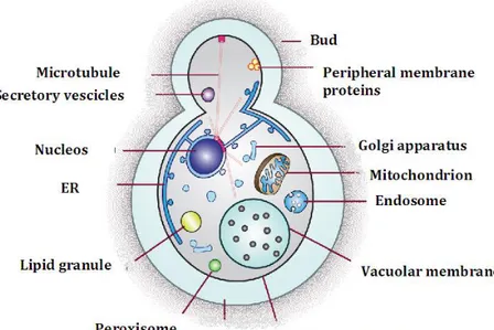
Another display technology that utilized organism as display system is the yeast display. Yeasts are unicellular eukaryotic organisms that have a generally-regarded-as-safe status (GRAS). The yeast cell is characterized by all the typical eukaryotic compartments such as: mitochondria, nucleus, endoplasmic reticulum, lisosomes, vesicles and Golgi apparatus (Fig. 7).
Figure 7. Yeast cell. Structure of a yeats cell. In the citosol are present: nucleos, endoplasmic
reticulum, lipid granules, microtubules, Golgi apparatus, vacuolar membrane, vesicles and mitochondria. Al this compartments are covered by two layer: plasmamembrane and the cell wall.
All these elements form the cytoplasm of a yeast cell that is cover by a plasma membrane. The plasma membrane is in turn covered by a thick layer called cell wall and the space between them is called periplasmic space. The cell wall is 200nm thick and is principally composed by mannoproteins and β-linked glucans [34]. The proteins normally choose for yeast display are mannoproteins. Two types of them are present in yeast:
1. Extractable with SDS (associated with glucans through non-covalent binding);
2. Extractable only with glucans digestion (associated with glucans through covalent binding).
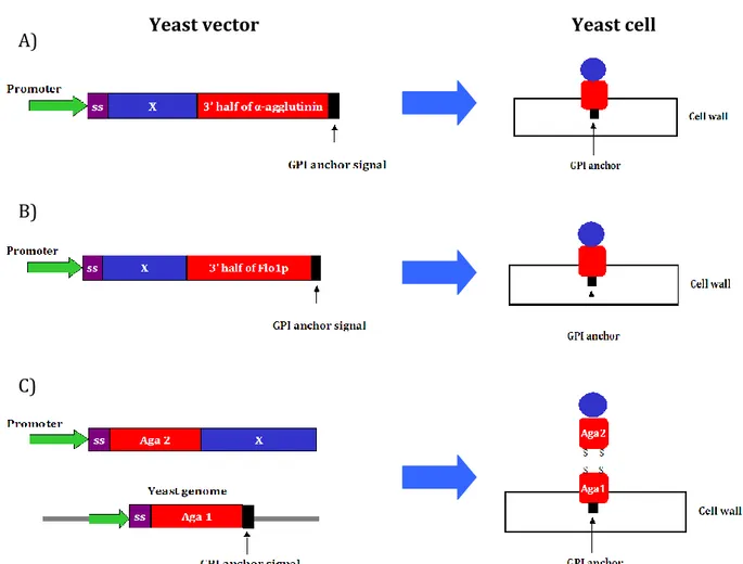
Only the second type of mannoproteins is used for yeast display, in particular the most used are: α-agglutinin, a-agglutinin and flocculins [35]. All these proteins have a glycosylphosphatidylinositol (GPI) anchor that is important for cell-surface protein expression. In yeast display the cDNA library cloned in yeast plasmid contains [36]: an inducible promoter, a secretion signal and the cDNA library fused to the mannoprotein. If the display is carried out with α-agglutinin or flocculin 1p only the C-term half part, correspondent to the 3’ half of the gene, is present in the plasmid, because in this part is present the GPI anchor signal that is essential for the cell wall attachment (Fig. 8 A,B). When the display is carried out with the a-agglutinin the Aga2 gene is fused the library instead Aga1 gene is present in the yeast genome (Fig. 8 C). The Aga1 and Aga2 domains are linked together through covalent binding.
Figure 8. Yeast display vectors. Different proteins are used for yeast display selection. The most
utilized are: a-agglutinin, α-agglutinin and flocculin. In case of α-agglutinin (A) and flocculin (B) the library is fused to the 3’ half part of the corresponded gene, allowing the expression of the GPI anchor the C-terminal part of the mannoproteins and the displayed protein. The a-agglutinin protein is composed by two domains translate by two different gene Aga1 and Aga2. in this case the library is fused to the Aga2 gene and the Aga1 is present in the yeast genome (C).
The plasmid is present in multiple copies in the yeast cell and it has been demonstrated that almost 105-106 copies of mannoprotein are presenting the target gene [35]. After the
library expression a selection step is necessary. In yeast display the selection is achieved with magnetic bead and flow-cytometric sorting. The magnetic beads are used to decrease the size of the library, that is too much for flow-cytometric sorting. The employment of high-speed flow-cytometry allows the detection of the amount of protein expressed by yeast and the affinity to the target using a two-color labeling scheme. The resulting graph shows (Fig. 9) [37]:
1. yeast displaying protein with less affinity to the target (black line); 2. yeast displaying protein with high affinity to the target (green line);
Yeast vector Yeast cell
A)
B)
3. yeast displaying no express of proteins (blue line);
4. yeast displaying proteins with no binding activity to the target (yellow line).
Figure 9. Yeast displaying scFv selection with flow-cytometry cell sorting [37].
Flow-cytometric analysis of yeast displaying scFv. The double-color detection allows to distinguish yeast for antigen binding and scFv expression on the yeast surface through anti-tag antibody (c-myc).
Since its introduction 10 years ago [38], yeast display has been used to express a variety of proteins for improved affinity, specificity, expression, stability, and catalytic activity. More recently, it has been widely used for antibody selection and engineering [37; 39]. It has been demonstrated that the distribution of affinity of antibodies selected by yeast display is similar to the antibodies isolated with phage display. Furthermore, yeast display is suitable for antibody affinity maturation [40].
2.3.3 DIFFERENCES BETWEEN PHAGE AND YEATS ANTIBODY DISPLAY
Either phage and yeast display are powerful tools for high specific antibody selection and maturation. Phage display was introduced a decade before yeast display and many paper have been published using these technologies [41] (Fig. 11).
Figure 11. Phage and yeast display publications [41]. Number of publications regarding phage
and yeast display by their discovery to 2008.
The main differences between phage and yeast display are:
1. type of organism: phage is a prokaryotic and doesn’t allow the expression of complex proteins or post-translational modifications. Yeast is an eukaryotic organism.
2. safety: phage is a virus, and because of this not a safe organism; yeast has a GRAS.
3. library dimension: phage allows the use of largest libraries till 1011 clones; yeast uses
smaller library. In yeast display additional rounds of magnetic-cell sorting are needed for the selection of largest library.
4. number of protein copies express by a single cell: using a phage vector is possible to have a maximum of 2700 copies displayed on a single phage, but it is true only for small proteins. With yeast display till 105-106 protein copies can be present on a single cell.
Recently, to exploit the advantage of the two technologies, researchers try to couple them. In 2011, Patel et al. have introduced a cross-species display system in which phage and yeast display are used in parallel to antibody library selection [42]. In this paper an adapter-direct display platform have been used. In this platform two different vectors are used: display vector and helper vector. The display vector avoids the anchoring sequence and can be used to display proteins on different species in combination with the helper vector that is species-specific. Using this technology Patel and coworkers have selected antibodies with a Kd in the nanomolar order. Furthermore they observed that the first round of selection with phage panning allows the enrichment of the library to 106; this
phage and yeast display, was published in 2012 by Ferrara et al. [43]. In this paper the researchers have selected antibodies against the antigen 85 of tuberculosis bacteria through two round of phage selection followed by one or two round of yeast display selection using FACS analysis. The passage between phage and yeast vector has been achieved by homologous recombination. In conclusion the combination of the two display method seems to improve the potential of both technologies.
2.4 ANTIBODY VALIDATION
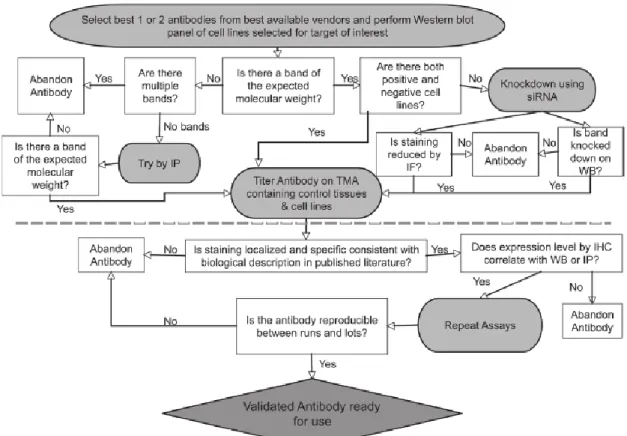
The great use of antibodies in research has brought to the development of high-throughput production, selection and validation methods. If the antibody production is well characterized, the validation has been underrated. Many articles take a lot of attention in the antibodies production and selection and don’t perform a real antibody validation. Indeed, as often as not the validation is carried out with Western blot and ELISA test only. For this reason many antibodies can be not functional in immunohistochemistry or show cross-reactivity if used in more sensible technique such as protein microarray. To validate antibodies there isn’t a standard protocol because they have to be choosen depending on the experiment that has to be performed. From this point of view, Rimm’ s laboratory has developed an algorithm for antibody validation for immunohistochemistry/quantitative immunofluorescence that can be used also for other application [15](Fig. 12).
Figure 12. The Rimm’s algorithm. The part upper the dot line concern the antibody validation cell
lines for antibody specificity testing. The lower part consists in further validation with tissue microarray for localization of the antibody target and to test different antibody lots.
This algorithm starts from the validation in Western blot of different cell lysates that express the target protein. This part allows to test the specificity of the antibody using techniques such as IP or short interfering RNA (siRNA). Subsequently the antibody will be used for in immunohistochemistry test to verify their reproducibility and the capability to localize the target in every tissue expressing it. Tissue microarray and protein microarray are emerging as important tool for antibody validation [44; 45]. Indeed, in 2012 Sjöberg at al. have shown the validation of monoclonal, polyclonal and scFv antibodies against protein containing SH2 domain using high-throughput protein microarray [46]. This technology is very useful because is sensitive and allows to test an antibody on thousand of characters in a single experiment.
2.5 PROTEIN MICROARRAY TECHNOLOGY
The great interest arisen by proteome studies have led to development and improvements of different highly innovative technologies. In particular, there was a need to developed a method that increase the number of characters analyzed in one experiment, with less consumption of materials and solutions. One of the major tool that has been set up for proteomic studies is protein microarray. This technology allows the immobilization of thousands of proteins on a solid surface, in a miniaturized format, and their analysis, in parallel, in a single experiment. Every kind of proteins can be immobilized on a microarray slide: peptides, full-length proteins, antibodies, receptors, enzymes or aptamers. As solid support, slides made of glass, polymer or plastic are used. This support is subsequently treated to allow the protein immobilization through different types of binding (Fig. 13):
Non-covalent (hydrophobic or entrapment: nitrocellulose, polystyrene; positive charges: poly-lysine);
Covalent (active groups such as: aldehyde, epoxy, esters, etc.);
Figure 13. Proteins immobilization on different microarray slides. The proteins can be
immobilized in different way on a solid surface. Non-covalent binding: proteins are capture through hydrophobic groups or entrap in a matrix (e.g. nitrocellulose). Covalent binding: proteins are immobilized through active groups such as epoxy, ester or aldhehyde that are able to form covalent bounds with all kind of proteins. Biomolecular interactions: the proteins are fused to tags or biomolecules such as biotin that permit the proteins capture by known groups or other molecule such as streptavidin.
The deposition of proteins on the slide is called “printing” or “spotting” and can be achieve through particular instruments called array printer. These instruments utilized pins to permit the deposition of nanolitre of protein. The pins are divided in two type:
1. contact pins: place nanolitre of sample directly on the slide surface;
2. non-contact pins: deposit drops of sample using capillary or ink jet technology.
Once the proteins are spotted they can be directly analyzed treating the slide with the desired solutions. The mainly detection method use with protein microarray is fluorescence, that is sensitive, safe and can be read with charge coupled device (CCD) camera or laser scanners. Although, other label methods are used, such as chemiluminescence and radioactivity. In recent years, also label-free methods have been used such as: mass spectrometry, surface plasmon resonance imaging (SPRI) and surface-enhanced laser desorption (SELDI)[47]. Protein microarray technology allows also the quantification of the signals using positive and negative controls and a calibration scale.
2.5.1 PROTEIN MICROARRAY APPLICATIONS
Protein microarrays are subdivided in three main groups : 1. analytical;
2. functional; 3. reverse-phase.
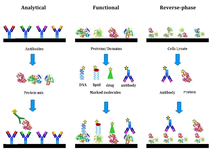
The analytical arrays are used to profile protein affinity and expression in different cells. The most common analytical array is antibody array, where this type of protein is immobilized on the slide and a proteins mixture is the probe (Fig. 14). Functional arrays allow the detection of proteins function and interactions. Normally full-length or domains are spotted and analyzed with proteins or other molecules such as: DNA, RNA, lipids, carbohydrates and drugs (Fig. 14). Finally, reverse-phase array are used to detect altered proteins or to highlight proteome differences between two cell types or healthy and diseased cells. Indeed, this kind of array consists in the printing of a cell lysate that can be characterized using a specific antibody or proteins (Fig. 14).
Figure 14. Protein arrays subtypes. Protein microarrays are divided in three subgroups:
analitical, functional and reverse-phase. Analytical arrays are normally used in protein expression analysis using antibodies as capture molecules. Functional arrays allow the analysis of protein function, interaction (DNA, lipid, drug, protein, RNA) or antibody validation and consist in the immobilization of full-length protein or domain. Reverse-phase array are characterized by immobilization of cells lysate instead of single molecules and are used in proteome analysis.
It is immediately clear that protein microarray can be a powerful tool for many different applications. First it has been widely used for protein interaction studies that allow to understand signaling pathways [48] and to discover binding proteins [49]. Protein microarray are also important for clinical purpose because allows biomarkers and autoantigens discovery [50; 51], where healthy and diseased sera are used to screen proteins/peptides library. Furthermore, host-pathogens interface studies that lead to vaccine development can be carry out [52]. Another important fields in which protein microarray are widely used is antibody validation. Many times commercial antibodies seems to be less specific for their target and cross-reactivity can also be detect. These problems lead to a wrong or less accurate validation. Protein microarrays are potential tool for this purpose because provide multiplex analysis of different antigens revealing specificity and selectivity of the antibody. In 2008, A. Lueking and coworkers have shown
the characterization of a monoclonal antibody against the variant 6 of CD44 molecule [53]. Sjöberg R. et al. have used protein microarray to validate selectivity and specificity of affinity reagents direct against SH2-domain[46]. In this study monoclonal antibodies, single
chain fragment variable and polyclonal antibodies have been utilized as affinity reagents demonstrating that all these type of antibody format are feasible for protein microarray validation. Furthermore, they have shown that with protein microarray is possible to validate antibodies against proteins with 89% of similarity determine the exclusion of some aspecific monoclonals. Finally, it is possible to assure that protein microarray can be one of the major tool for antibody validation.
2.5.2 PROTEIN MICROARRAY IMPROVEMENTS
As described before, to perform a protein microarray is necessary to produce and purify all the proteins that have to be printed on the slide. This become an issue when you think to produce thousands of proteins in the same time. The development of protein libraries has allowed a more easily search of proteins cDNA but their production and purification remain a time-consuming issue. Furthermore, protein microarrays are not stable for long period so it is necessary to proceed with the experiment immediately after the spotting. Many groups of researchers have try to overcome these two problems link to protein microarray technology. The first innovation for protein microarray improvement was the use of cell-free protein expression systems, that are able to transcribed a DNA template and translate the correspondent mRNA in a protein [54]. The combination of this two tools has brought to a new technology called “in situ protein array” [55]. This new type of protein microarray consists in the immobilization of DNA instead of proteins eliminating the stability issue. Once the DNA is spotted it can be translate in proteins directly just when is needed, erasing the protein production and purification processes. Even if standard protein array continue to be widely used, many groups took interest in this “in situ protein array” developing different strategy to use it.
2.6 PROTEIN PRODUCTION WITHOUT CELL BOUNDERIES
Protein production is one of the key steps in biotechnology and functional proteomics. Conventional protein expression systems rely on prokariotic cells as bacteria, where the most used is Escherichia coli (E. coli) or eukaryotic cells as yeast, insect and mammalian cell systems [56; 57; 58]. The prokaryotic expression systems are easily adaptable to high-throughput expression methodologies, but are less efficient in the expression of complex mammalian proteins, in particular those which require post-translational modification. At the opposite site, systems based on eukaryotic organisms are capable of expressing post-translationally modified proteins but are difficult to be integrated into high-throughput methodologies. In order to find a new method that allows an easier, cost-effective and high-throughput application, cell-free based systems were developed [59]. In 1961 Niremberg and Matthaei have demonstrated the possibility to translate in vitro proteins with an E. coli extract using RNA or synthetic nucleotide as a template [59]. Subsequently, Zubay et al. has shown the coupled transcription and translation of a β-galactosidase chain using an E. coli extract and a virus DNA [60]. Compared to cell-based methods, cell-free expression systems are considerably faster since they don’t require cell transfection, cell culture and extensive protein purification procedures (Fig. 16A). Moreover, they don’t involve cell lysis steps that could denaturate proteins. In addition, cell-free systems allow the production of toxic, insoluble or membrane proteins, that are difficult to produce using cell-based methods. Finally, they can be used in a microlitre scale, making them ideal for high-throughput applications [61; 62].
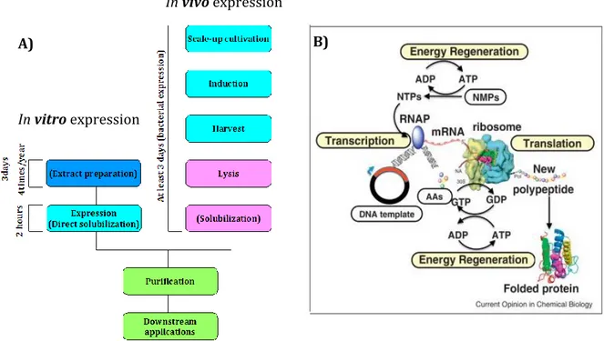
Figure 16. Protein production systems. A) Protein expression with in vivo or in vitro methods. In
case of in vivo (bacteria) cells have to be cultivated, induced and collected; subsequently a cell lysis has to be carried out and in some cases also protein solubilization is needed. All these passages takes 3 days. When an in vitro expression system is used the protein production takes only 2 hours and the extract necessary for the expression has to be prepared only 4 times /year. B) Schematic representation of the passages occurring during an IVTT reaction [63]: DNA template has to be transcribed in mRNA with all the transcription complex, RNA polymerase and energy sources; the mRNA is translate in protein using amminoacids, tRNAs, ribosome and energy sources.
The most recent in vitro transcription and translation (IVTT) systems are composed of crude cell lysate supplemented of [64] (Fig. 16B):
1. energy sources (ATP, ADP); 2. RNA polymerase (T7, SP6 or T3) ; 3. amino acids;
4. tRNA; 5. enzymes; 6. NTPs ;
7. salts and ions.
In vitro expression
In vivo expression
Initially, only E. coli lysate was used, because it is easier to grow in a large scale and it is also less expensive. Successively, IVTT systems based on eukaryotic extracts, as rabbit reticulocytes and wheat germ, were generated [65]. These systems seem to be more stable than the E. coli one and more suitable for eukaryotic protein production. Recently, also insect and human cells lysates have been developed, showing an improvement in terms of protein quantity [66; 67]. The major advantage of cell-free systems is their flexibility that has allowed significant improvements in protein yields, folding and post-translational modifications through the addiction of chaperons or specific enzymes (glycosylation, phosphorilation, etc.) to the cells extract [68]. Thanks to all these improvements, IVTT systems are increasingly used in protein array technology, protein structural studies, large scale analysis of proteins, screening of antibody mutants, IVEC technology and display technologies such as ribosome display, mRNA display and in vitro compartimentalization [55; 62; 69; 70; 71].
2.7 IN SITU PROTEIN ARRAY
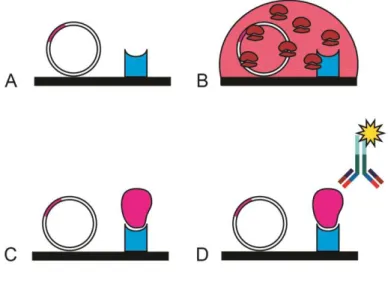
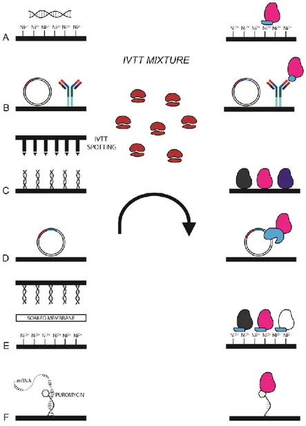
The great innovation of protein production in a cell-free mode and the possibility to transcribe and translate a DNA template in a single step has allowed the generation of high-throughput technologies, such as in situ protein arrays. These particular microarrays permit to build the protein array whenever is needed, starting from a DNA template. An in situ protein array consists of four different steps (Fig. 17): 1) immobilizing a DNA template on a solid support (Fig. 17A); 2) treating the support with an IVTT mixture (Fig. 17B); 3) capturing the nascent protein on the solid support (Fig. 17C); 4) revealing the protein of interest (Fig. 17D). In this way the standard protein array drawbacks of protein stability and long period of storage are overcome. The first in situ protein array which has been developed is the protein in situ array (PISA), also known as DiscernArray, by He and Taussing in 2001[55]. Afterwards, many other in situ protein arrays have been developed: Nucleic acid programmable protein array (NAPPA) [72], multiple spotting technique (MIST) [73], TUS-TER array [74], DNA array to protein array (DAPA)[75], in situ puromycin-capture from mRNA array [76].
Figure 17. In situ protein microarray technology. Schematic resume of the main steps to
perform an in situ protein microarray. A) printing of a DNA template and a capture agent; B) production of the proteins treating the slide with an IVTT mixture; C) capture of proteins from the capture agent; D) revealing of the protein of interest.
2.7.1 PROTEIN IN SITU ARRAY (PISA)
The PISA method (Fig. 18A) permits the generation of protein arrays in one step starting from PCR DNA fragments. In particular, these PCR fragments contain all the DNA sequence necessary for IVTT (T7 promoter, translation initiation site, poly(A)28 tail, transcription
and translation terminators) and the protein cDNA fused with a double (His)6-tag
sequence, which allows a stronger affinity binding to nickel ions (Ni2+) than standard
(His)6-tag. In this way, the generation of the DNA template doesn’t involve cloning
passages, but different PCR cycles. As cell-free system a commercial kit based on rabbit reticulocytes lysate is currently used (T7 TnT Quick for PCR fragments).The template is added to the IVTT mixture and deposited on a Ni-NTA-coated microtiter plate or Ni-NTA magnetic agarose beads. After the reaction, the nascent protein is bound to the solid support thanks to the double (His)6-tag and can be subsequently analyzed. The authors tested the system using single-chain fragment variable (scFv) and experienced that in 25μl of reaction 120ng of protein was produced, of which 50% was bound on the microtiter [77].
2.7.2 NUCLEIC ACID PROGRAMMABLE PROTEIN ARRAY (NAPPA)
The Nucleic acid programmable protein array was designed by La Bear and colleagues in 2004 [72] (Fig. 18B). In this method a plasmid DNA is cross-linked to psoralen-biotin conjugate through UV light and subsequently printed with avidin on a glass slide covered with aminopropyltriethoxysilane (APTES). The DNA template encodes the protein/peptide of interest, fused at the C-terminal to glutathione S-trasferase (GST)-tag. The presence of the GST-tag allows the capture of the nascent protein by a monoclonal anti-GST antibody spotted on the slide concurrently with the plasmid DNA. After the deposition of DNA and antibody, the slide is treated with IVTT system based on rabbit reticulocytes lysate and the protein, produced and captured, can be immediately analyzed. Instead of PISA, this method permits a high-density format thanks to the type of support used. Indeed it has been shown that 512 spots, spaced 900μm from each other, can be printed in a single slide and all the proteins are produced using only 100μl of IVTT reaction. Furthermore, they have demonstrated that 675pg of protein can be produced and capture in each spot. In 2008, the same group of researchers have improved the NAPPA technology by the use of an high quality super-coiled DNA and adding bovin serum albumin (BSA) to the printing solution [78]. Finally, in recent years, they have shown that an IVTT system based on HeLa cells led up to a major quantity of protein produced by printed DNA [79].
2.7.3 MULTIPLE SPOTTING TECHNIQUE (MIST)
The multiple spotting technique is an high-throughput approach where a microarray slide is spotted a first time to allow the immobilization of proteins and a second time to permit the deposition of another compound, exactly in correspondence of the previous one [80]. Agenendt et al. have applied this method to build a new type of in situ protein array in which the first printing allows the spotting of DNA templates, plasmid or PCR fragments, and the second involves the deposition of the IVTT mixture [81] (Fig. 18C). After the expression, the proteins are immobilized on Nickel Chelate-coated or APTES slide and subsequently analyzed. The importance of this new approach is the limitation of protein diffusion and cross-talking, due to the deposition of the IVTT for every single spot. Furthermore, Angenendt et al. have demonstrated that only 35fg of unpurified PCR DNA are sufficient for the detection of full-length green fluorescent protein (GFP) in subnanolitre volume [81]. Moreover, the MIST allows to perform high-density protein
microarray with up to 13,000 spots in one slide. By now the MIST technology has not been used for particular studies, but its functionality is well characterized.
2.7.4 TUS-TER ARRAY
The most recent in situ protein array developed is the one called TUS-TER or Protein array on demand [74] (Fig. 18D). This method is based on the interaction between the E. coli protein TUS and a double stranded DNA sequence of 23bp called Ter. In vivo the binding of this protein to Ter allows the termination of the DNA replication. The peculiarities of this binding is that even if it is not covalent it is very strong (Kd= 3.4 x10⁻¹³) and it occurs only
when the protein encounter this particular DNA sequence [82]. Chatterjee et al. have exploited the capability of this protein fusing it to cDNA of target proteins that can be immobilized from the Ter sequence [74]. In particular a plasmid DNA is used as template, because contains transcription and translation sequence and target protein-TUS cDNA, but also as capture agent because it contains the Ter sequence. After the DNA printing on a nitrocellulose-coated slide, is only necessary to treat it with IVTT mixture to obtain the protein immobilization on the slide. The great advantage of this method is that there is no need for an additional molecule to capture the nascent protein, but only the presence of a plasmid DNA onto the microarray surface. To validate the system the Authors have also shown that the nascent proteins captured directly from the Ter site are present in the correspondent spot, therefore protein diffusion is avoided.
2.7.5 DNA ARRAY TO PROTEIN ARRAY (DAPA)
The DAPA methodology consists of the printing of a protein microarray using a DNA array as a mould [83] (Fig. 18E). In particular PCR fragments encoding for different proteins, fused to a tag, are spotted onto epoxysilane slide and subsequently the array is placed face to face with a second slide able to capture tagged proteins. Between the two slides a permeable membrane soaked with the IVTT mixture is present in order to allow the protein production. Afterwards, the proteins are produced starting from the DNA array, diffused through the membrane and are captured by the second slide. Using GFP as a control He and colleagues have evaluated that from 0,1ng of DNA template up to 30fmol of
reuse the same DNA array to produce many protein arrays. It has been demonstrated that a single DNA array can be reused almost 20 times without significant variation [84]. Finally, it has been demonstrated that scFvs can be spotted on a slide using DAPA and they are also functional. These results are promising for the utilization of DAPA technology in proteome studies.
2.7.6 IN SITU PUROMICYN-CAPTURE FROM mRNA ARRAY
This type of microarray is the only one using RNA as a template. It is based on the assumption that when a ribosome encounters a region of double-stranded RNA or DNA-RNA hybrid it is not immediately released but it stalls on the mDNA-RNA. Tao and Zhu have hypothesized that this ribosome stalling can be long enough to provide the incorporation of a puromycin-grafted oligo immobilized on a solid phase, therefore the nascent polypeptide is directly captured by the puromycin [76] (Fig. 18F). This strategy consists of the immobilization, on a streptavidin-coated slide, of biotinylated puromycin-oligo and mRNA, which anneals at a 3’ end to an RNA bumper oligo, so as to form a double-stranded RNA. After printing, the slide is treated with an in vitro translation mixture based on rabbit reticulocytes lysate and after the synthesis, the nascent peptide is captured by the puromycin-oligo. Finally, the slide is treated with a solution containing RNAse that degrades the mRNA, in order to have on the slide only the newly synthesized protein. After the assessment of the technology, the Authors have evaluated that, using this approach, 0,8 fmol/spot of protein are captured on the slide surface and this results are comparable with those of traditional protein microarray.
Figure 18. Schematic representation of all in situ protein microarrays. A) PISA: protein in situ
array; B) NAPPA: nucleic acid programmable protein array; C) MIST: multiple spotting technique; D) TUS-TER array; E) DAPA: DNA array to protein array; F) in situ puromycin-capture from mRNA array.
2.7.7 IN SITU PROTEIN ARRAY APPLICATIONS
The main application of in situ protein microarrays is the individuation of protein-protein interactions. All the types of in situ protein array, previously described, can be used for interaction studies [55; 72; 74; 75]. In particular the NAPPA methodology is a great tool for this type of application due to the possibility of producing at same time query and target protein. La Bear et al. have demonstrated this capability using the Cdk inbhitor p16 as a query protein and Cdk4 and Cdk6 as positive targets, whereas Cdk2 has been used as negative control [72]. Furthermore, new protein-protein interactions were discovered
arrays which have been used for other kind of applications are NAPPA and PISA methods. The NAPPA was used in immunogenicity and immunoprofiling studies. In 2009, Montor et al., have directed a genome-wide study on Pseudomonas aeruginosa outer membrane proteins immunogenicity. According to this method, the reactivity of patients sera, positive or negative to cystic fibrosis, infected with P. aeruginosa, to bacteria outer membrane proteins was analyzed [86]. In this study the Authors identified a total of 48 antigens, 12 of which were detected in 10 patients and were considered as promising candidate for in-depth studies. Other similar studies were performed on different organisms, like Ornithodoros moubata [87], Plasmodium falciparum [88]and Coxiella burnetii [89], confirming that NAPPA is an important tool for immunogenicity studies and vaccine development. Moreover, NAPPA can also be considered suitable for immunoprofiling, since two different studies, in which this technology was used to analyze possible autoantibody biomarkers for early breast cancer and juvenile arthritis, were published [90; 91]. The usage of this novel methodology demonstrated that is possible to identify autoantibody biomarkers that is important not only for the diagnosis and the follow-up of the disease, but also to determine disease stages. In the matter of PISA method has been published the possibility to perform screening studies, functional assay and identification of mutants [92].
2.7.8 IMPROVEMENTS AND FUTURE PROSPECTIVES
In situ protein arrays have demonstrated to be a great tool for proteome studies and they have overcame the main drawbacks related to standard protein arrays. Under this aspect, many improvements regarding protein production, sensitivity and cross-talk were done in recent years. In 2010 a group of researchers have demonstrated that combining E. coli with wheat germ IVTT lysate, a major quantity of functional protein can be produced and captured on a glass slide, compared to rabbit reticulocytes lysate, that is the most IVTT used in in situ array technology [93]. Furthermore, based on NAPPA technology, these Authors have developed an autofluorescent microarray where the target protein is fused to a GFP, instead of GST-tag, and the nascent protein is captured by anti-GFP monoclonal antibody. With this approach, the protein is revealed immediately after the production, without the need of other treatments. Another important improvement of in situ protein arrays regards protein diffusion during the IVTT reaction that can cause cross-talking
between different spots. In a standard NAPPA slide the distance between two spot is 625μm that means approximately 2000 proteins analyzed on a single slide. This spot to spot space is the minimum distance necessary to avoid cross-talking events using NAPPA technology. To increase the density of NAPPA slide, without the presence of protein diffusion, a new type of slide was set up [94]. This slide is made of silicon and is composed of semispherical nanowells of 250μm in diameter, 75μm in depth and distanced each other 375μm. These nanowells are produced with photolithography and can be subsequently functionalized with different chemical groups. Moreover, every nanowell can contain 5nl of IVTT reaction mix indicating a minimal consume of cell-free IVTT systems that are expensive tools. The last challenge of in situ protein arrays is the use of label-based methods that are limiting and less sensitive, instead of label-free methods. For this reason, many groups have tried to combine this new type of arrays with label-free methods. Two different groups have demonstrated the possibility of combining NAPPA or MIST to matrix-assisted laser desorption/ionization (MALDI)[95; 96]. Furthermore, in 2012, an on-chip microfluidic protein microarray based on NAPPA technology was developed [97]. This method consists in a generator element where a dsDNA encoding for His6 tagged proteins and a detector element composed by a Cu(II)-NTA group are immobilized. When the microfluidic chip is treated with the IVTT mixture, the protein is produced from the DNA template and directly absorbed by the detector element and can be immediately used in surface plasmon resonance imaging (SPRI). Finally, the possibility to combine NAPPA methodology with other label-free methods like: nanogravimetry, atomic force microscope and anodic porous alumina, has been reported (APA)[98]. Thanks to all these improvements, in situ protein arrays are increasingly becoming a new technology able to analyze thousands of elements in almost one day and suitable for many other purposes.
3. AIM OF THE PROJECT
As described in the introduction, antibodies have become an important tool not only in scientific research, but also in proteome discovery, diagnosis and therapy. For this purpose, several improvements have been done in antibody production, selection and validation [23], but many proteins still remain unknown and without antibodies able to recognize and identify them. Moreover, many commercial antibodies present cross-reactivity or less specificity against their target [10; 11]. This leads to a not specific antibodies validation. It is became clear that an antibody has to be validated in relation with the experimental needs, but it can results laborious and cost-time expensive. For this reason the aim of this project is to develop new high-throughput technologies for antibodies validation. My laboratory has take part in a National Institute of Health (NIH) project, coordinated by Andrew Bradbury (National Laboratories of Los Alamos), that has the aim to produce, with high-throughput technologies, specific polyclonal antibodies against hundreds different proteins. In particular the project consists of (Fig. 19):
1. production of the target proteins;
2. selection of polyclonal antibodies through a phage/yeast display combination [43]; 3. validation of the polyclonal antibodies with Western blot, ELISA and protein
microarray;
4. set up of a new platform for high-throughput antibodies validation: protein array on demand.
Figure 19. Experimental scheme of the project. The project consists of four different parts: 1.
Protein production (antigens); 2. Selection of polyclonal antibodies against the putative antigens; 3. Validation of antibodies with WB, ELISA and protein microarray; 4. High-throughput antibodies validation using “protein array on demand”.
The first part of the project (proteins production and antibodies selection) has been carried out in collaboration with Los Alamos Laboratories. After the scFvs selection, through phage/yeast display combination, and scFv-Fc rabbit polyclonals production in yeast cells, the polyclonals were sent us for validation with WB, ELISA and standard protein array. Subsequently, since protein microarray has some drawbacks, such as intensive protein production and immobilized proteins functionality, we want to set up an high-throughput protein array suitable for antibody validation. In particular, an in situ protein array called “protein array on demand” has been chosen for this purpose.
5. MATERIALS AND METHODS
Materials. As microarray slides FAST (Whatman) and Codelink® activated (Surmodics)
were used. In ELISA, Western blot and protein microarray analysis the subsequent antibodies were employed: anti-SV5 (in house made), anti-mouse IgG horseradish peroxidase (HRP) conjugated (Dako), anti-mouse IgG alkalin phosphatase (AP) conjugated (Sigma), anti-mouse IgG Cyanine3 (Cy3) conjugated (Listarfish), anti-rabbit IgG HRP conjugated (Sigma), anti-rabbit IgG Cyanine5 (Cy5) conjugated (Listarfish) and polyclonal rabbit anti-CDK2A (full-length protein) (Santa Cruz). To reveal biotinylated proteins streptavidin HRP conjugated (BIOSPA), streptavidin AP conjugated (Sigma) and streptavidin phycoerythrin (PE) conjugated (Life Technologies) were used. As AP and HRP developer the nitro blue tetrazolium chloride/5-bromo-4-chloro-3-indolyl-phosphate (NBT/BCIP) (Roche) and 3,3’,5,5’-Tetramethylbenzidine (TMB) (Sigma) were used. All PCR reactions were performed with GoTaq Flexi (Promega). In vitro transcription and translation kit used are: T7 TnT quick coupled transcription and translation system (Promega), T7 TnT quick for PCR DNA coupled transcription and translation system (Promega), TnT coupled Wheat Germ extract System (Promega) and 1-Step Human coupled in vitro expression kit (Pierce). Bacteria strains used: DH5α and NM522. Additional compounds: streptavidin (Biospa), bovine serum albumin (BSA) (Sigma), triton-X 100 (Sigma), tween-20 (Sigma), ampicillin (Sigma), 2xTY media (Sigma), select agar (Sigma), agarose (Sigma), TEMED and APS (Sigma), Page ruler Plus Protein marker (Fermentas), Quant-it™ PicoGreen® (Life Technologies).
Buffers and solutions. Codelink print buffer 6x (300mM sodium phosphate, pH 8.5),
Codelink saturation buffer (10mM Tris-Hcl, 50mM ethanolamine, pH 9), phosphate buffered saline (PBS) (NaCl 0.137M, Na2HPO4 4,3mM, KCl 2,7mM, KH2PO4 1,5mM), TE
buffer (Tris 10mM, EDTA 1mM pH 7.4), sample buffer 2x (Tris-HCl pH6,8 125mM, SDS 4%, glycerol 20%, β-mercaptoethanol 10%, blue bromophenol 0,004%), coomassie staining (acetic acid 10%, methanol 45%, coomassie blue R-250 0,1%), coomassie destaining (acetic acid 10%, methanol 10%).
Western blot antibodies validation. Biotinylated proteins were denaturated with SB 2x
and loaded on a polyacrylamide gel for SDS-PAGE. After gel transfer, the nitrocellulose membrane was saturated in 4% milk in PBS plus tween-20 0,01% (PBST) for 1 hour at room temperature. The nitrocellulose was treated with polyclonal supernatants at 1:5 dilution in 2% milk in PBST and subsequently with anti-rabbit IgG-AP at 1:5000 in the same buffer. The membrane has been developed with NBT BCIP.
Sanger sequencing. PCR fragments were quantified by agarose gel and a sequencing
reaction was carried out with: primer at 3,2μM concentration, 1μl of Big Dye Terminator (Applied Biosystems) mix and distillated water till 10μl final volume. The amount of DNA used depends on its length, according with the Big Dye datasheet. Susequently, the sequences reaction was purification on columns containing sephadex resin (Princeton Separation). After resin hydratation with 800μl of water, two centrifugation at 3600 rpm for 2’ were carried out, to completely eliminate the water. The sequencing reaction was loaded on the resin and another centrifugation was performed, on order to eluate the purified reaction. This was mixed with formammide, denaturated for 2’ at 96°C and analyzed with ABI 3130xl Genetic Analyzer.
Antibodies validation with ELISA. Yeast supernatants, produced by National Laboratories
of Los Alamos, containing rabbit polyclonal antibodies were validated using ELISA test. The corresponding proteins were coated on 96 well plate, at 10ng/μl in PBS and stored at 4°C overnight. The saturation was carried out with 2% milk in PBS and supernatants were used at 1:5 dilution in saturation buffer. The antibodies were revealed with anti-rabbit IgG-HRP at 1:5000. To verify the antigen coating a streptavidin-HRP at 1:200 were used.
Antibodies validation with protein microarray. All the biotinylated proteins, sent us from
the Los Alamos National Laboratories, were brought to 50ng/μl concentration and put in a 384 well plate for microarray printing. The proteins were print on FAST slide, at 15°C and 50% humidity using the Biodeassay Calligrapher (BioRad). When printing finished, the slide was stored overnight at room temperature in a less humidity chamber. The day after, it was saturated with 4% milk in PBST at room temperature, with shacking, for 1 hour. Subsequently, the slide was treated with polyclonal supernatants at 1:5 dilution in 2% milk PBST for 1hour at room temperature. As secondary antibody an anti-rabbit IgG-Cy5, at
![Figure 9. Yeast displaying scFv selection with flow-cytometry cell sorting [37]](https://thumb-eu.123doks.com/thumbv2/123dokorg/4814537.50047/18.892.275.589.199.570/figure-yeast-displaying-scfv-selection-flow-cytometry-sorting.webp)