www.PRSGlobalOpen.com
1
L
ichen sclerosus (LS) is a chronicinflamma-tory disease of uncertain etiology. Initially, it was described at the end of the 19th century by Hallopeau and Darier as a variant of lichen planus; currently, it is considered as a separate entity.
Typical lesions involve sclerotic patches, with cen-tral atrophy, generally located in the genital area. Extragenital manifestations are characterized by polygonal whitish papules or plaques, erosions, or lichenification (unique or in combination).1
Hyper-keratosis, vacuolization of the basal cells, and ho-mogenization of dermal collagen with a band-like chronic dermal infiltrate of inflammatory cells is evi-denced by light microscopy.1,2
Women are more commonly affected than men, with a bimodal peak of incidence in the
prepubertal and postmenopausal age. There is a strong association with human leukocyte antigen class II DQ7 and autoimmune disorders, such as vitiligo, alopecia areata, thyroid disease, and perni-cious anemia. There is still controversy regarding the role of infectious diseases (human papillo-mavirus, Borrelia burgdorferi). Histopathologically, LS is characterized by a band of inflammation as a zone of homogeneous paucicellular superficial dermal edema and sclerosis, with variable vacuo-lar interface degeneration and epidermal atrophy or lichenification.2 Direct immunofluorescence
shows fibrin deposit in the superficial dermis and at the dermal epidermal junction. LS often exhib-its a prominent inflammatory infiltrate composed mainly of lymphocytes and plasma cells. The acral form of LS involving the palmoplantar region is rarely reported.3–5
Data from recent case reports show that the most common approach for LS is treatment with immu-nosuppressant agents (topical and systemic). Sur-gical procedure is described in only 1 case report (split-skin graft by Sonnex TS in 1985).6
Disclosure: The authors have no financial interest
to declare in relation to the content of this article. The
Article Processing Charge was paid for by the
Depart-ment of Dermatology, A.O. Spedali Civili, University
of Brescia, Brescia, Italy.
Nested Graft for Acral Lichen Sclerosus of the
Feet: A Surgical Treatment for an Inflammatory
Disease
From the Department of Dermatology, A.O. Spedali Civili, University of Brescia, Brescia, Italy.
Received for publication August 15, 2015; accepted December 8, 2015.
Copyright © 2016 The Authors. Published by Wolters Kluwer Health, Inc. on behalf of The American Society of Plastic Surgeons. All rights reserved. This is an open-access article distributed under the terms of the Creative Commons Attribution-Non Commercial-No Derivatives License 4.0 (CCBY-NC-ND), where it is permissible to download and share the work provided it is properly cited. The work cannot be changed in any way or used commercially.
DOI: 10.1097/GOX.0000000000000595 Giulio Gualdi, MD Paola Monari, MD Laura Pelizzari, MD Daniele Cammalleri, MD Piergiacomo Calzavara-Pinton, MD
Gualdi et al.
Summary: The “nested graft” is an innovative and well-defined surgical technique used for chronic wound healing that induces the de-senescence of fibroblasts in the wound bed. We report a case of a 76-year-old man af-fected by plantar chronic wounds because of acral lichen sclerosus and atrophicus localized at both feet and treated for many years successfully with immunosuppressive agents. For cardiological dysfunction, systemic therapy was reduced to low dosage of steroids with an increase of ulcer-ations (5 × 2 cm). So we decided to perform the nested graft on the plan-tar region. After the surgical procedure, all the grafted ulcers healed, and at a 4-month follow-up, no signs of lichen sclerosus were present. (Plast
Reconstr Surg Glob Open 2016;4:e633; doi: 10.1097/GOX.0000000000000595; Published online 7 March 2016.)
Reconstructive
2
PRS Global Open • 2016
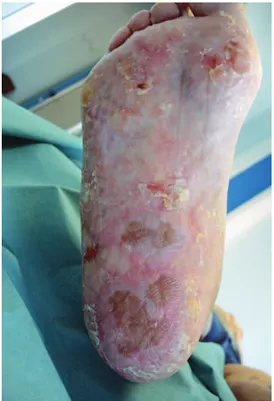
Herein, we report a case of a 76-year-old man affected for 15 years by chronic plantar ulcers be-cause of acral LS and atrophicus (Figs. 1 and 2)7,8
No genital or oral lesions were present, but a severe nail dystrophy had led to a complete absence of the nail plate.
A biopsy taken from a plantar lesion demonstrat-ed epidermal acanthosis with upper hyperkeratosis and compact ortho-parakeratosis. The granular layer
was only present focally. There were scattered spon-giotic phenomenon with lymphocyte exocytosis and balloon degeneration of basal keratinocytes with aci-dophilic bodies.9
The patient had been successfully treated for many years with immunosuppressive agents (sys-temic prednisone, cyclosporin A, and azathioprine) and topical application of tacrolimus ointment 0.1% leading to a complete wound healing. Because of metabolic dysfunction (heart failure, diabetes, and increasing of transaminase and creatinine), systemic therapy was reduced to low dosage of steroids with subsequent reactivation of the disease and compari-son of plantar ulcerations reappearance (5 × 2 cm).
Therefore, we decided to perform surgery using a graft-derived technique. Reverdin, in 1869,10
de-scribed the skin graft for regenerative purpose for the first time. The skin graft needs a well-granulating wound bed tissue, which chronic wounds often lack. So skin graft is not indicated in many affected patients.
We recently described an innovative surgical tech-nique used for chronic venous ulcers called “nested graft”: the aim of the nested graft is not to cover the wound bed, as with the traditional full-thickness skin graft, but to repopulate the chronic ulcer bed with healthy cells. We also demonstrated that the nested graft is able to induce wound healing through the de-senescence in wound bed fibroblasts.11,12 The
donor site is prepared using povidone-iodine, and a local anesthesia is achieved using 1% lidocaine.
Fig. 1. Right foot before surgery.
3
Gualdi et al.
•
Nested Graft for Acral Lichen Sclerosus of the Feet
Full-thickness explants, without hypodermal fat, are taken from the donor site using a 6-mm punch bi-opsy; the donor site is immediately repaired with a simple suture. The graft is then deposited in a Petri dish containing physiological saline. The receiving site (ulcer bed) is prepared with soft curettage, and then full-thickness circular fragments of the ulcer are removed by using a 5-mm punch biopsy. For re-moving the explants from the donor site, a slightly larger punch (6 mm) is used. Due to the physiologi-cal contraction of the transfer tissue, the dimension-al match results perfectly.
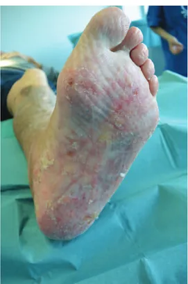
For the first time, we performed the nested graft on an inflammatory skin ulcer because of LSA. Our patient tolerated the surgical procedure well lead-ing to complete wound heallead-ing in 4 weeks, and at the 4-month follow-up, no signs of the LSA were present. The islands of grafted skin maintained their status and were not affected by the underlying inflammatory disease.13 On the contrary, the grafted
skin “healed” the surrounding skin suffering from
LS. This could be explained by the proliferation of grafted cells through the skin inducing a microen-vironment remodeling process that influenced the epidermal atrophy and lichenoid infiltrate at dermo-epidermal junction.12 At the follow-up visit, a slow
pigmentation grafted island was evident; the pig-mentation was because of the presence of resident melanocytes in the donor skin (Figs. 3 and 4). In conclusion, our procedure could well be an innova-tive surgical treatment for a chronic inflammatory disease.
Giulio Gualdi, MD
Department of Dermatology A.O. Spedali Civili, University of Brescia Brescia, Italy E-mail: [email protected]
REFERENCES
1. Caspary P, Carpena AB, de Almeida H Jr. Plantar involvement in lichen sclerosus. Int J Dermatol. 2009;48:662–663.
2. Keith PJ, Wolz MM, Peters MS. Eosinophils in lichen scle-rosus et atrophicus. J Cutan Pathol. 2015;42:693–698. 3. Powell JJ, Wojnarowska F. Lichen sclerosus. Lancet.
1999;353:1777–1783.
4. Wolff K, Goldsmith LA, Katz SI, et al. Fitzpatrick Dermatology
in General Medicine. 7th ed. New York, NY: Mc Graw Hill;
2012.
5. Murphy R. Lichen sclerosus. Dermatol Clin. 2010;28: 707–715.
6. Sonnex TS, Eady RA, Sparrow GP, Mayou B. Ulcerative lichen planus associated with webbing of the toes. J R Soc
Med. 1986;79:363–365.
7. Steff M, Toulemonde A, Croue A, et al. [Acral lichen sclerosus et atrophicus]. Ann Dermatol Venereol. 2008;135: 201–204.
8. Viana Fde O, Cavaleiro LH, Unger DA, et al. Acral lichen sclerosus et athrophicus—case report. Ann Bras Dermatol. 2011;86(4 supp1):S82–S84.
9. Fung MA, LeBoit PE. Light microscopic criteria for the diagnosis of early vulvar lichen sclerosus: a comparison with lichen planus. Am J Surg Pathol. 1998;22:473–478. 10. Ehrenfried A, Reverdin and Other Methods of
Skin-Grafting. Historical Boston Med Surg J. 1909;161:911–917. 11. Gualdi G, Monari P, Farisoglio C, et al. Nested graft in
chronic wounds: a new solution for an old problem. Int
Wound J. 2011;8:127–131.
12. Gualdi G, Crotti S, Monari P, et al. The nested graft acts by inducing process of de-senescence of the fibroblasts in chronic venous ulcers. Int Wound J. 2015;8:127–131. 13. Isedeh P, Al Issa A, Lim HW, et al. Uncommon responses
of segmental vitiligo to melanocyte-keratinocyte trans-plantation procedure. J Cutan Med Surg. 2015;19:177–181.